
Elimination of the Femoral Neck in Measuring Femoral Version Allows for Less Variance in Interobserver Reliability
Abstract
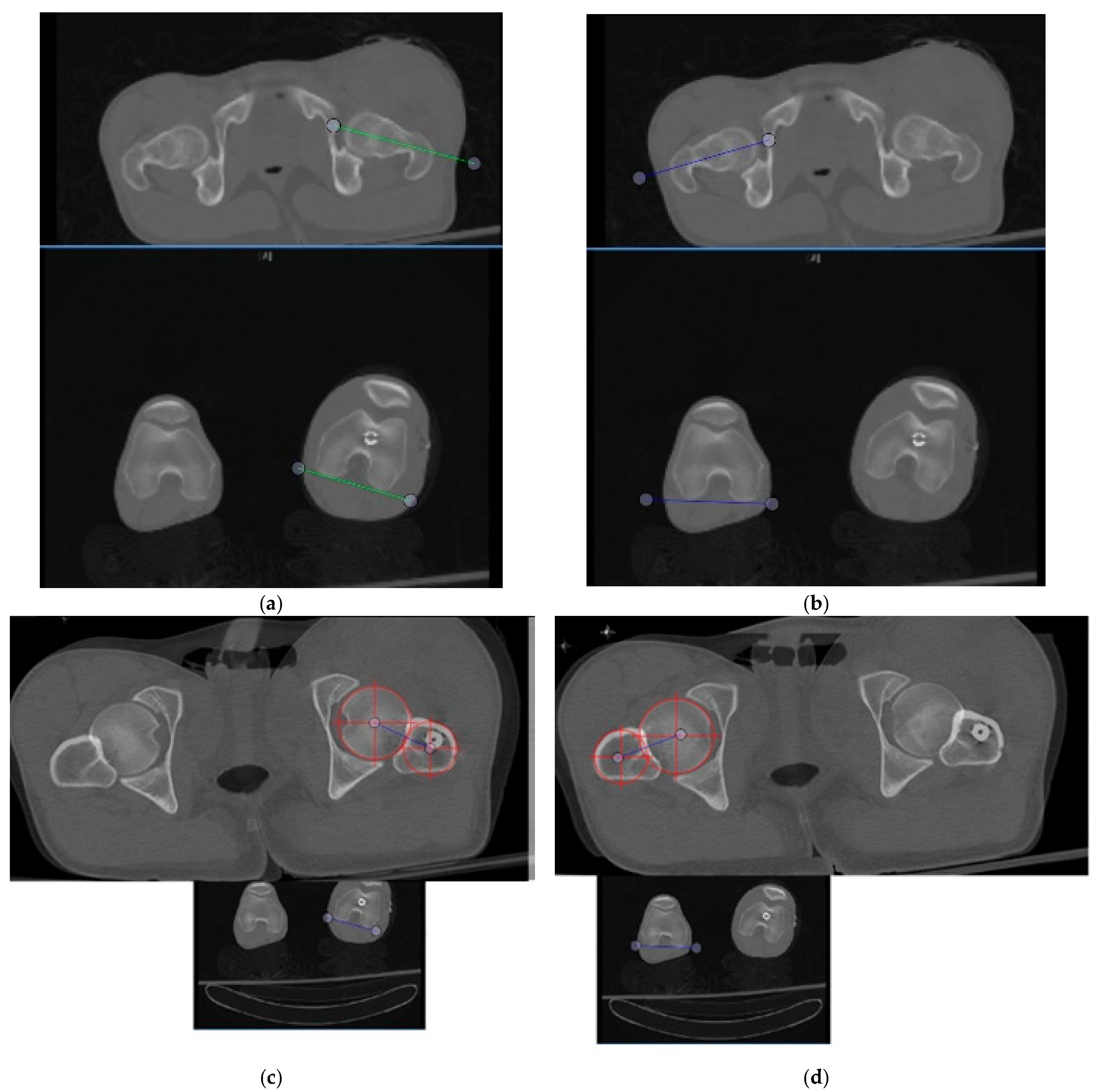
:1. Introduction
2. Materials and Methods
3. Results
4. Discussion
5. Conclusions
Author Contributions
Funding
Institutional Review Board Statement
Informed Consent Statement
Data Availability Statement
Conflicts of Interest
References
- Kingsley, P.C.; Olmsted, K.L. A Study to Determine the Angle of Anteversion of the Neck of the Femur. J. Bone Jt. Surg. Am. 1948, 30, 745–751. [Google Scholar] [CrossRef]
- Billing, L. Roentgen Examination of the Proximal Femur End in Children and Adolescents: A Standardized Technique Also Suitable for Determination of the Collum-, Anteversion-, and Epiphyseal Angles: A Study of Slipped Epiphysis and Coxa Plana. Acta Radiol. 1954, 110, 1–80. [Google Scholar]
- Murphy, S.B.; Simon, S.R.; Kijewski, P.K.; Wilkinson, R.H.; Griscom, N.T. Femoral Anteversion. J. Bone Jt. Surg. Ser. A 1987, 69, 1169–1176. [Google Scholar] [CrossRef]
- Botser, I.B.; Ozoude, G.C.; Martin, D.E.; Siddiqi, A.J.; Kuppuswami, S.; Domb, B.G. Femoral Anteversion in the Hip: Comparison of Measurement by Computed Tomography, Magnetic Resonance Imaging, and Physical Examination. Arthroscopy 2012, 28, 619–627. [Google Scholar] [CrossRef] [PubMed]
- Dickschas, J.; Harrer, J.; Reuter, B.; Schwitulla, J.; Strecker, W. Torsional Osteotomies of the Femur. J. Orthop. Res. 2015, 33, 318–324. [Google Scholar] [CrossRef] [PubMed]
- Koerner, J.D.; Patel, N.M.; Yoon, R.S.; Sirkin, M.S.; Reilly, M.C.; Liporace, F.A. Femoral Version of the General Population. J. Orthop. Trauma 2013, 27, 308–311. [Google Scholar] [CrossRef] [PubMed]
- Kuo, T.Y.; Skedros, J.G.; Bloebaum, R.D. Measurement of Femoral Anteversion by Biplane Radiography and Computed Tomography Imaging: Comparison with an Anatomic Reference. Investig. Radiol. 2003, 38, 221–229. [Google Scholar] [CrossRef] [PubMed] [Green Version]
- Schneider, B.; Laubenberger, J.; Jemlich, S.; Groene, K.; Weber, H.M.; Langer, M. Measurement of Femoral Antetorsion and Tibial Torsion by Magnetic Resonance Imaging. Br. J. Radiol. 1997, 70, 575–579. [Google Scholar] [CrossRef] [PubMed]
- Strecker, W.; Keppler, P.; Gebhard, F.; Kinzl, L. Length and Torsion of the Lower Limb. J. Bone Jt. Surg. Ser. B 1997, 79, 1019–1023. [Google Scholar] [CrossRef]
- Tomczak, R.J.; Guenther, K.R.; Rieber, A.; Mergo, P.; Ros, P.R.; Brambs, H.J. MR Imaging Measurement of the Femoral Antetorsional Angle as a New Technique: Comparison with CT in Children and Adults. Am. J. Roentgenol. 1997, 168, 791–794. [Google Scholar] [CrossRef] [PubMed]
- Yoshioka, Y.; Cooke, T.D.V. Femoral Anteversion: Assessment Based on Function Axes. J. Orthop. Res. 1987, 5, 86–91. [Google Scholar] [CrossRef] [PubMed]
- Hartel, M.J.; Petersik, A.; Schmidt, A.; Kendoff, D.; Nüchtern, J.; Rueger, J.M.; Lehmann, W.; Grossterlinden, L.G. Determination of Femoral Neck Angle and Torsion Angle Utilizing a Novel Three-Dimensional Modeling and Analytical Technology Based on CT Datasets. PLoS ONE 2016, 11, e0149480. [Google Scholar] [CrossRef] [PubMed]
- Hinterwimmer, S.; Rosenstiel, N.; Lenich, A.; Waldt, S.; Imhoff, A.B. Femorale Osteotomien Bei Patellofemoraler Instabilität. Unfallchirurg 2012, 115, 410–416. [Google Scholar] [CrossRef] [PubMed]
- Waidelich, H.-A.; Strecker, W.; Schneider, E. Computertomographische Torsionswinkel- Und Längenmessung an Der Unteren Extremität. In RöFo-Fortschritte auf dem Gebiet der Röntgenstrahlen und der Bildgebenden Verfahren; Georg Thieme Verlag Stuttgart: New York, NY, USA, 1992; Volume 157, pp. 245–251. [Google Scholar] [CrossRef]
- Hernandez, R.J.; Tachdjian, M.O.; Poznansk, A.K.; Dias, L.S. CT Determination of Femoral Torsion. Am. J. Roentgenol. 1981, 137, 97–101. [Google Scholar] [CrossRef] [PubMed] [Green Version]
- Jarrett, D.Y.; Oliveira, A.M.; Zou, K.H.; Snyder, B.D.; Kleinman, P.K. Axial Oblique CT to Assess Femoral Anteversion. Am. J. Roentgenol. 2010, 194, 1230–1233. [Google Scholar] [CrossRef] [PubMed]
- Vaidya, R.; Dimovski, R.; Cizmic, Z.; Vaidya, A.; Gheraibeh, P.; Hudson, I. Use of Inherent Anteversion of an Intramedullary Nail to Avoid Malrotation in Comminuted Femur Fractures: A Prospective Case—Control Study. J. Orthop. Trauma 2018, 32, 623–628. [Google Scholar] [CrossRef] [PubMed]
- Enderlein, G. Fleiss, J.L.: The Design and Analysis of Clinical Experiments. Wiley, New York-Chichester-Brislane-Toronto-Singapore 1986, 432 S., £38.35. Biometrical J. 2007, 30, 304. [Google Scholar] [CrossRef]
- Kaiser, P.; Attal, R.; Kammerer, M.; Thauerer, M.; Hamberger, L.; Mayr, R.; Schmoelz, W. Significant Differences in Femoral Torsion Values Depending on the CT Measurement Technique. Arch. Orthop. Trauma Surg. 2016, 136, 1259–1264. [Google Scholar] [CrossRef] [Green Version]
- Jaarsma, R.L.; Pakvis, D.F.M.; Verdonschot, N.; Biert, J.; van Kampen, A. Rotational Malalignment after Intramedullary Nailing of Femoral Fractures. J. Orthop. Trauma 2004, 18, 403–409. [Google Scholar] [CrossRef] [PubMed]


| Measurement Technique | Mean Femoral Version * | Range of Version | ICC |
|---|---|---|---|
| Method 1 | 9.50 ± 4.82° | −20° to 37° | 0.960 (95% CI 0.909–0.982) |
| Method 2 | 18.73 ± 2.69° | −23° to 52° | 0.993 (95% CI 0.987–0.996) |
Publisher’s Note: MDPI stays neutral with regard to jurisdictional claims in published maps and institutional affiliations. |
© 2021 by the authors. Licensee MDPI, Basel, Switzerland. This article is an open access article distributed under the terms and conditions of the Creative Commons Attribution (CC BY) license (https://creativecommons.org/licenses/by/4.0/).
Share and Cite
Dimovski, R.; Teitge, R.; Bolz, N.; Schafer, P.; Bobba, V.; Vaidya, R. Elimination of the Femoral Neck in Measuring Femoral Version Allows for Less Variance in Interobserver Reliability. Medicina 2021, 57, 1363. https://0-doi-org.brum.beds.ac.uk/10.3390/medicina57121363
Dimovski R, Teitge R, Bolz N, Schafer P, Bobba V, Vaidya R. Elimination of the Femoral Neck in Measuring Femoral Version Allows for Less Variance in Interobserver Reliability. Medicina. 2021; 57(12):1363. https://0-doi-org.brum.beds.ac.uk/10.3390/medicina57121363
Chicago/Turabian StyleDimovski, Radomir, Robert Teitge, Nicholas Bolz, Patrick Schafer, Vamsy Bobba, and Rahul Vaidya. 2021. "Elimination of the Femoral Neck in Measuring Femoral Version Allows for Less Variance in Interobserver Reliability" Medicina 57, no. 12: 1363. https://0-doi-org.brum.beds.ac.uk/10.3390/medicina57121363