
Loading Effect of Chitosan Derivative Nanoparticles on Different Antigens and Their Immunomodulatory Activity on Dendritic Cells

 ,
,
Abstract
:
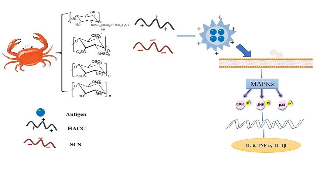
1. Introduction
2. Results
2.1. Characterization of Chitosan Derivatives
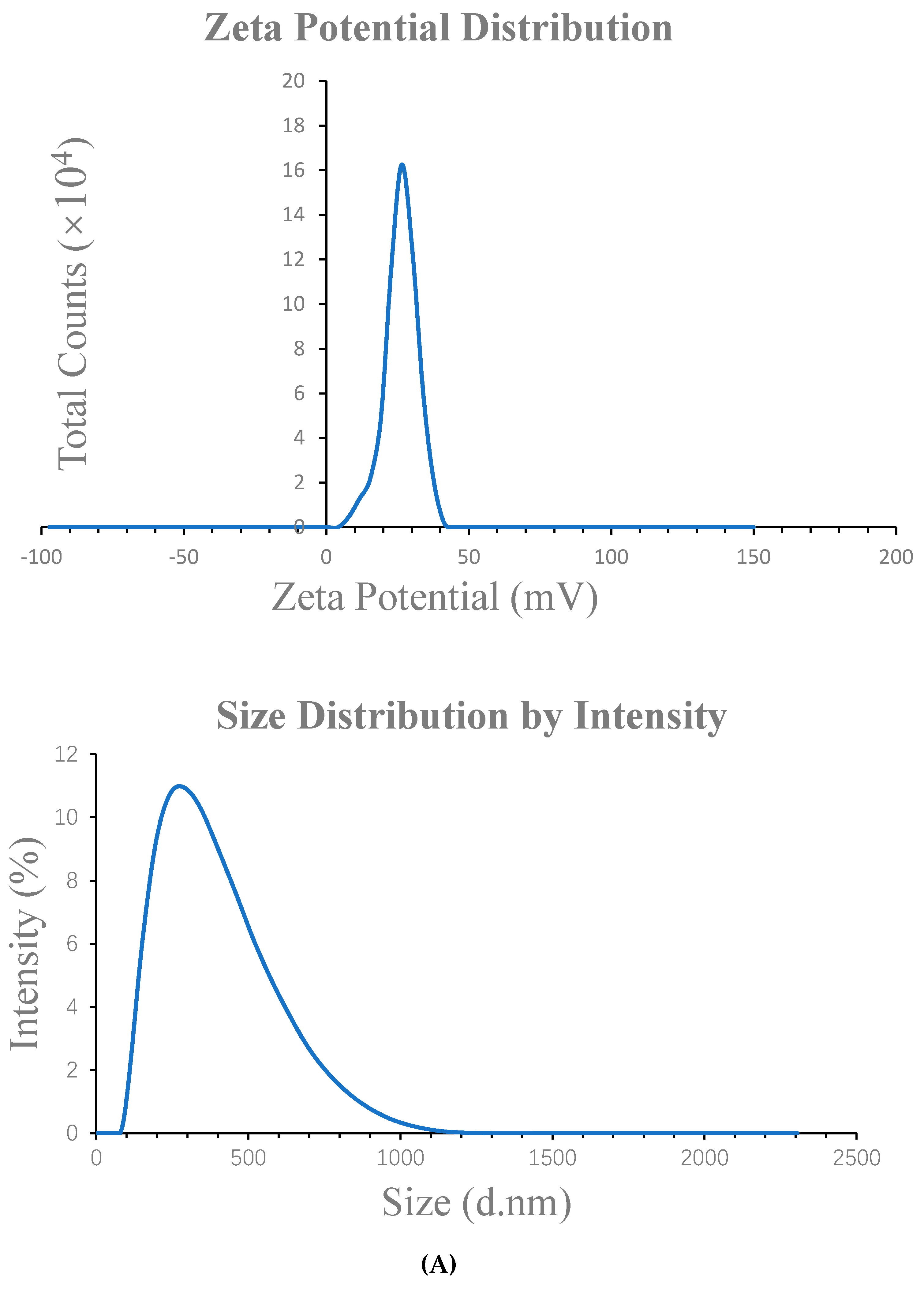
2.2. Characterization of the NPs
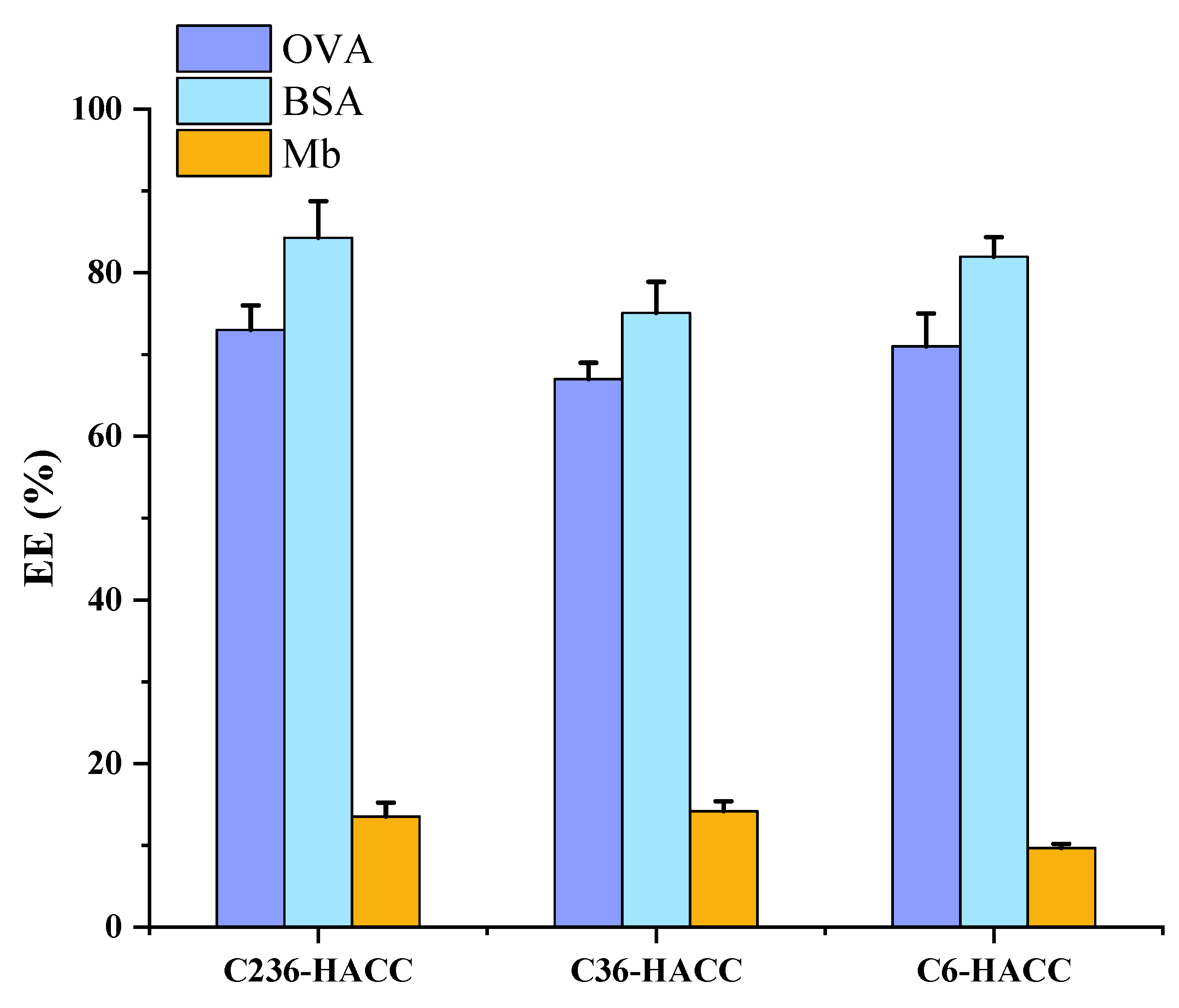
2.3. Encapsulation Efficiency of NPs
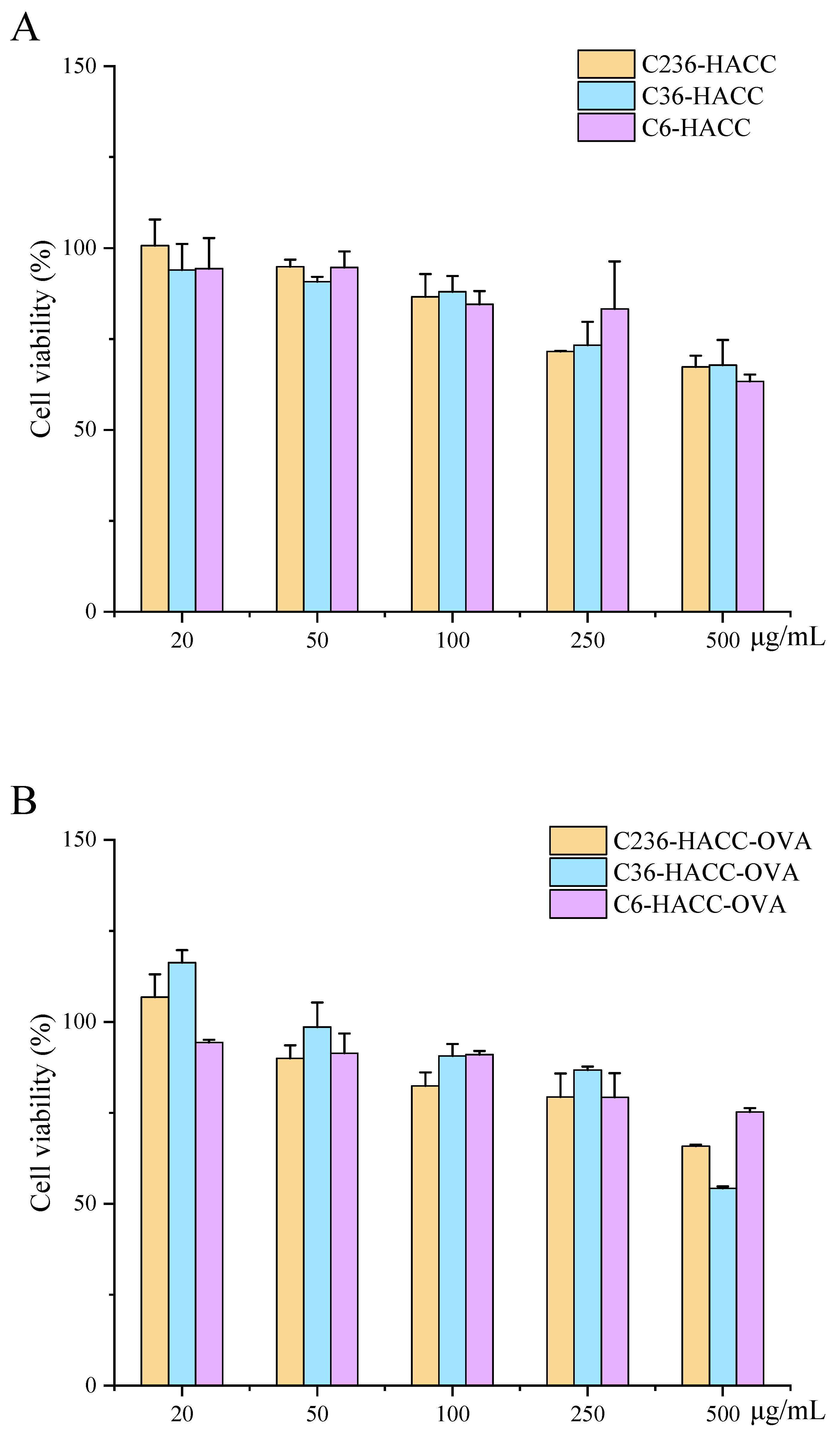
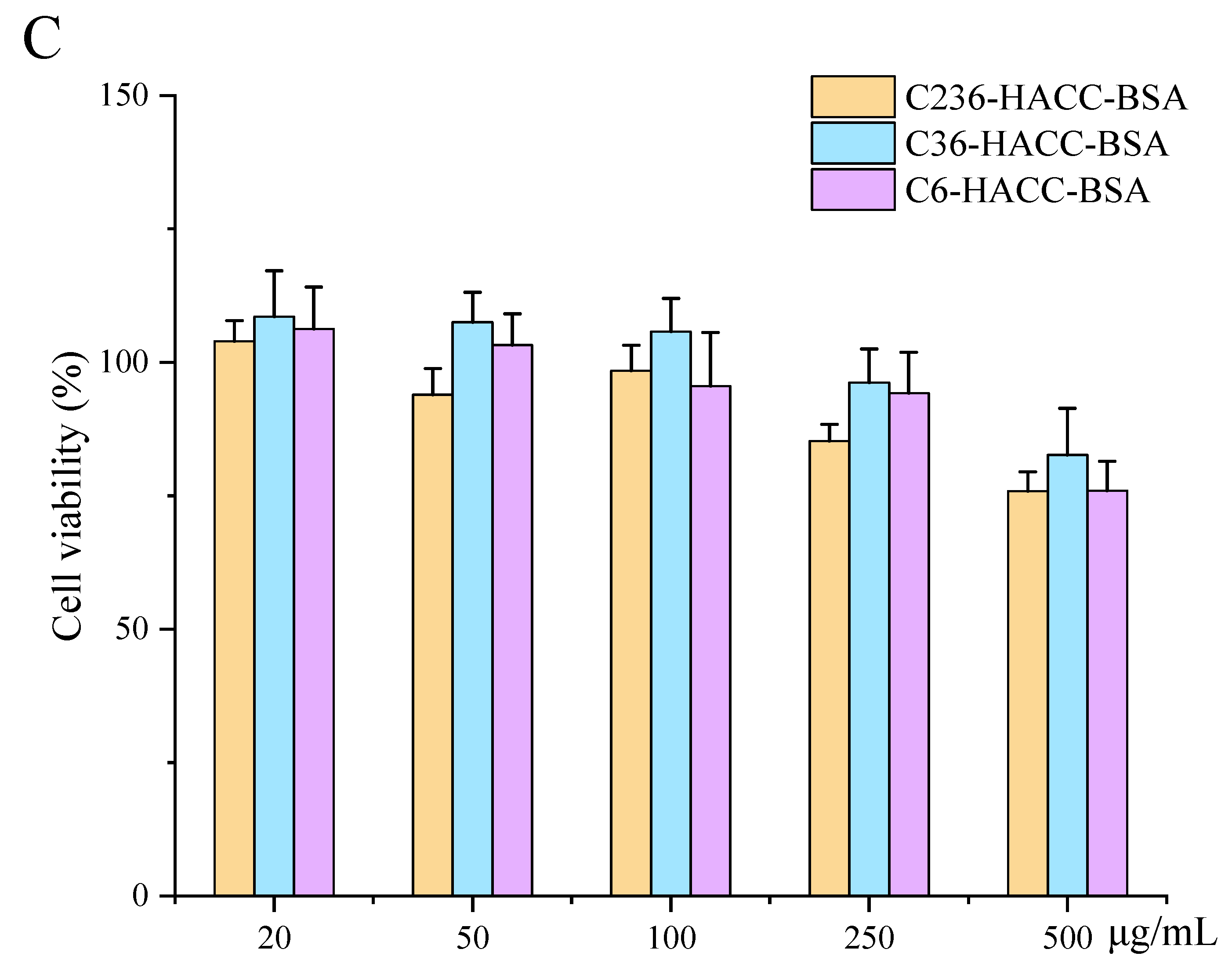
2.4. Cytotoxicity of NPs
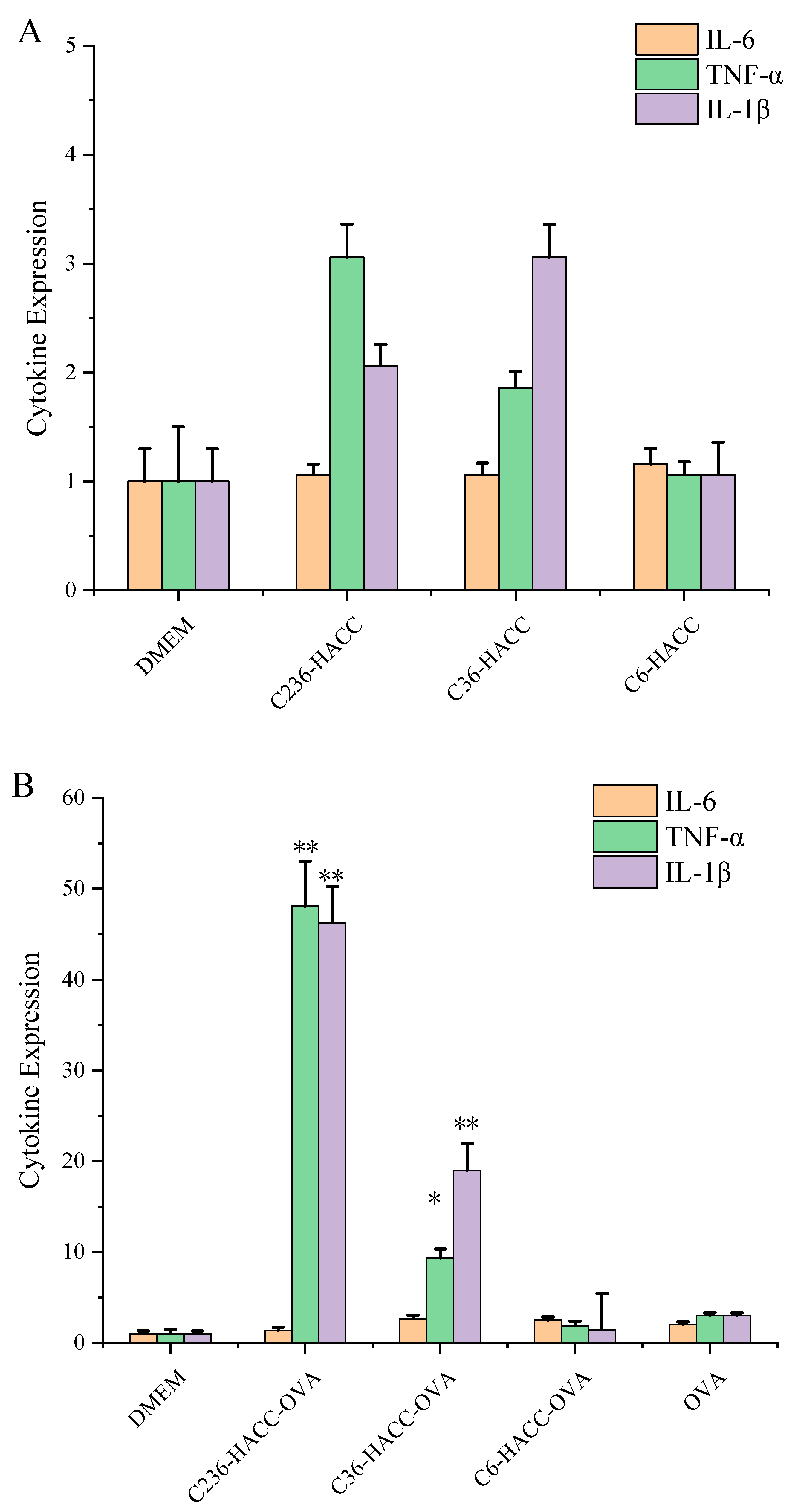
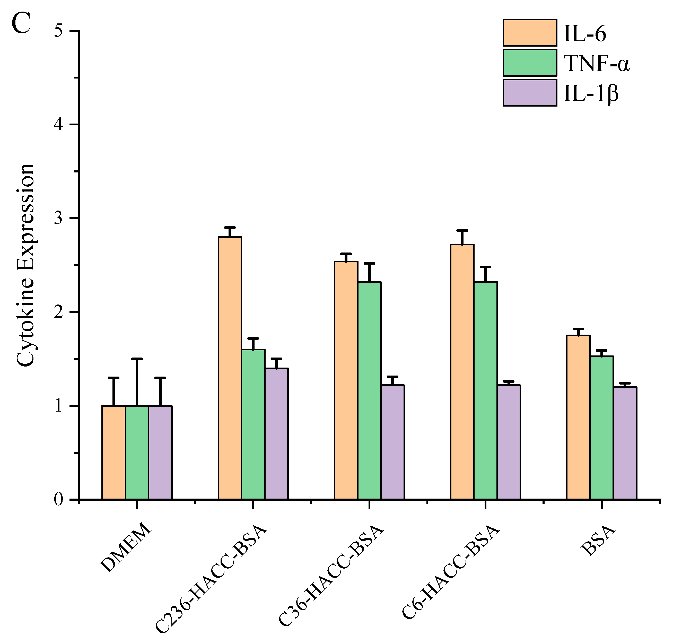
2.5. RNA Expression of NPs on DCS Cells
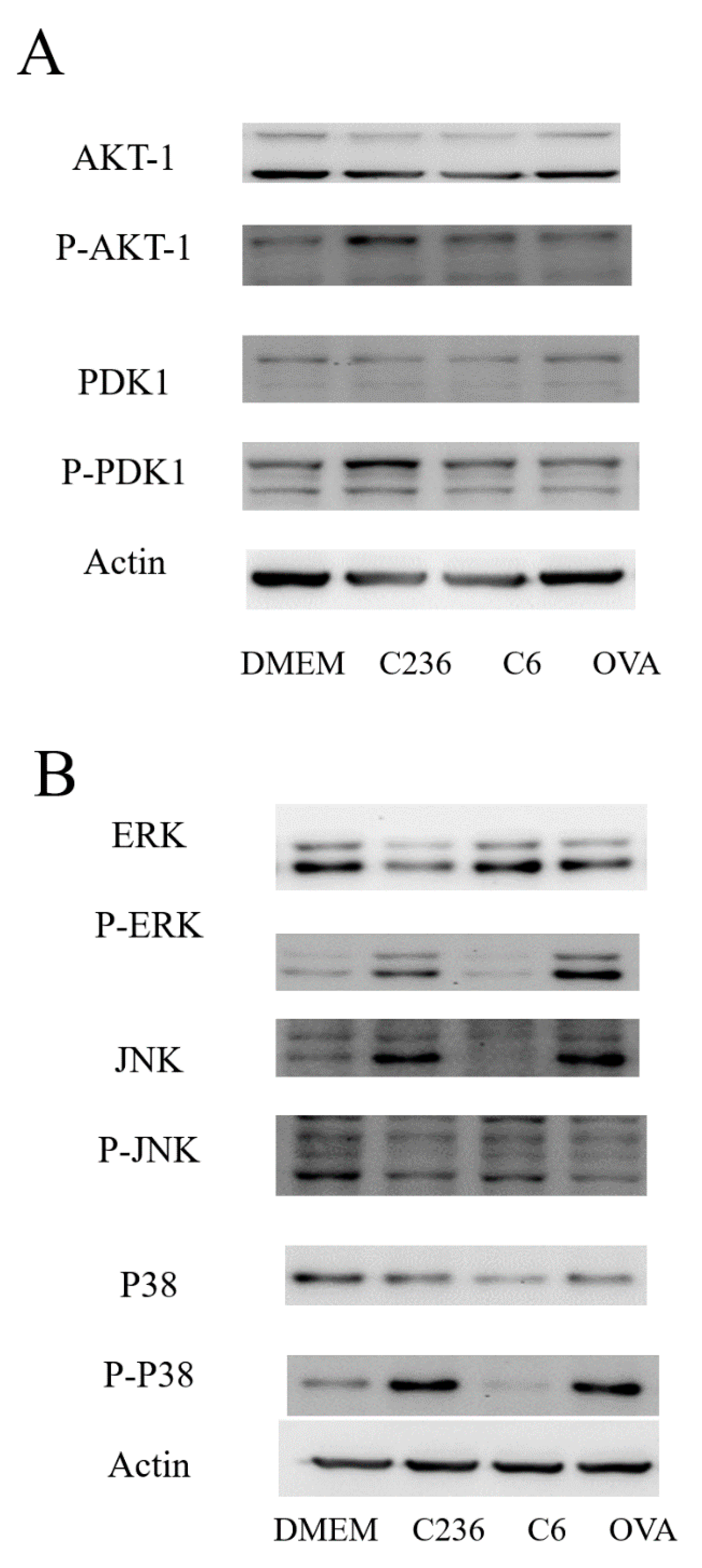
2.6. The Protein Phosphorylation Level in Cell Pathway
2.7. The Determination of NPs Endotoxin
2.8. The Release of NPs
3. Discussion
4. Materials and Methods
4.1. Materials
4.2. Preparation of Hydroxypropyl Trimethyl Ammonium Chloride Chitosan (HACC) and Chitosan Sulfate (SCS)
4.3. Preparation of Chitosan Derivative NPs Loaded with Different Antigens
4.4. Characterization of Different Chitosan Derivatives
4.5. Characterization of Chitosan Derivative NPs Loaded with Different Antigens
4.6. Encapsulation Effect of NPs
4.7. Cell Culture
4.8. In Vitro Experiment
4.8.1. Cytotoxicity of NPs
4.8.2. RNA Expression Analysis
4.8.3. Determination of Protein Phosphorylation Level in Cell Pathway
4.9. The Endotoxin of NPs
4.10. The Antigen Release of NPs
5. Conclusions
Author Contributions
Funding
Institutional Review Board Statement
Informed Consent Statement
Acknowledgments
Conflicts of Interest
References
- Hogenesch, H. Mechanism of immunopotentiation and safety of aluminum adjuvants. Front. Immunol. 2012, 3, 406. [Google Scholar] [CrossRef] [PubMed] [Green Version]
- Aguilar, J.C.; Rodriguez, E.G. Vaccine adjuvants revisited. Vaccine 2007, 25, 3752–3762. [Google Scholar] [CrossRef] [PubMed]
- Moran, H.B.T.; Turley, J.L.; Andersson, M.; Lavelle, E.C. Immunomodulatory properties of chitosan polymers. Biomaterials 2018, 184, 1–9. [Google Scholar] [CrossRef] [PubMed]
- Li, X.; Xing, R.; Xu, C.; Liu, S.; Qin, Y.; Li, K.; Yu, H.; Li, P. Immunostimulatory effect of chitosan and quaternary chitosan: A review of potential vaccine adjuvants. Carbohydr. Polym. 2021, 264, 118050. [Google Scholar] [CrossRef] [PubMed]
- Yang, Y.; Xing, R.; Liu, S.; Qin, Y.; Li, K.; Yu, H.; Li, P. Immunostimulatory effects of sulfated chitosans on RAW 264.7 mouse macrophages via the activation of PI3K/Akt signaling pathway. Int. J. Biol. Macromol. 2018, 108, 1310–1321. [Google Scholar] [PubMed]
- Zhang, G.; Jia, P.; Liu, H.; Hu, T.; Du, Y. Conjugation of chitosan oligosaccharides enhances immune response to porcine circovirus vaccine by activating macrophages. Immunobiology 2018, 223, 663–670. [Google Scholar] [CrossRef] [PubMed]
- Carroll, E.C.; Jin, L.; Mori, A.; Munoz-Wolf, N.; Oleszycka, E.; Moran, H.B.T.; Mansouri, S.; McEntee, C.P.; Lambe, E.; Agger, E.M.; et al. The Vaccine Adjuvant Chitosan Promotes Cellular Immunity via DNA Sensor cGAS-STING-Dependent Induction of Type I Interferons. Immunity 2016, 44, 597–608. [Google Scholar] [CrossRef] [Green Version]
- Mori, A.; Oleszycka, E.; Sharp, F.A.; Coleman, M.; Ozasa, Y.; Singh, M.; O’Hagan, D.T.; Tajber, L.; Corrigan, O.I.; McNeela, E.A.; et al. The vaccine adjuvant alum inhibits IL-12 by promoting PI3 kinase signaling while chitosan does not inhibit IL-12 and enhances Th1 and Th17 responses. Eur. J. Immunol. 2012, 42, 2709–2719. [Google Scholar] [CrossRef]
- Liu, Y.-J. Dendritic Cell Subsets and Lineages, and Their Functions in Innate and Adaptive Immunity. Cell Press 2001, 106, 259–262. [Google Scholar] [CrossRef] [Green Version]
- Zhao, K.; Sun, Y.; Chen, G.; Rong, G.; Kang, H.; Jin, Z.; Wang, X. Biological evaluation of N-2-hydroxypropyl trimethyl ammonium chloride chitosan as a carrier for the delivery of live Newcastle disease vaccine. Carbohydr. Polym. 2016, 149, 28–39. [Google Scholar] [CrossRef]
- Xu, Y.; Mao, H.; Yang, C.; Du, H.; Wang, H.; Tu, J. Effects of chitosan nanoparticle supplementation on growth performance, humoral immunity, gut microbiota and immune responses after lipopolysaccharide challenge in weaned pigs. J. Anim. Physiol. Anim. Nutr. 2020, 104, 597–605. [Google Scholar] [CrossRef]
- Schijns, V.E.; Lavelle, E.C. Trends in vaccine adjuvants. Expert Rev. Vaccines 2011, 10, 539–550. [Google Scholar] [CrossRef]
- Smith, D.M.; Simon, J.K.; Baker, J.R., Jr. Applications of nanotechnology for immunology. Nat. Rev. Immunol. 2013, 13, 592–605. [Google Scholar] [CrossRef]
- Lebre, F.; Bento, D.; Ribeiro, J.; Colaco, M.; Borchard, G.; de Lima, M.C.P.; Borges, O. Association of chitosan and aluminium as a new adjuvant strategy for improved vaccination. Int. J. Pharm. 2017, 527, 103–114. [Google Scholar] [CrossRef]
- Dai, C.; Kang, H.; Yang, W.; Sun, J.; Liu, C.; Cheng, G.; Rong, G.; Wang, X.; Wang, X.; Jin, Z.; et al. O-2′-hydroxypropyltrimethyl ammonium chloride chitosan nanoparticles for the delivery of live Newcastle disease vaccine. Carbohydr. Polym. 2015, 130, 280–289. [Google Scholar] [CrossRef]
- Li, X.; Kong, X.; Shi, S.; Zheng, X.; Guo, G.; Wei, Y.; Qian, Z. Preparation of alginate coated chitosan microparticles for vaccine delivery. BMC Biotechnol. 2008, 8, 89. [Google Scholar] [CrossRef] [PubMed] [Green Version]
- Bento, D.; Jesus, S.; Lebre, F.; Goncalves, T.; Borges, O. Chitosan Plus Compound 48/80: Formulation and Preliminary Evaluation as a Hepatitis B Vaccine Adjuvant. Pharmaceutics 2019, 11, 72. [Google Scholar] [CrossRef] [PubMed] [Green Version]
- Xu, C.; Xing, R.; Liu, S.; Qin, Y.; Li, K.; Yu, H.; Li, P. Immunostimulatory effect of N-2-hydroxypropyltrimethyl ammonium chloride chitosan-sulfate chitosan complex nanoparticles on dendritic cells. Carbohydr. Polym. 2021, 251, 117098. [Google Scholar] [CrossRef]
- Horne, D.S. Casein structure, self-assembly and gelation. Curr. Opin. Colloid Interface Sci. 2002, 7, 456–461. [Google Scholar] [CrossRef]
- Boonyo, W.; Junginger, H.E.; Waranuch, N.; Polnok, A.; Pitaksuteepong, T. Chitosan and trimethyl chitosan chloride (TMC) as adjuvants for inducing immune responses to ovalbumin in mice following nasal administration. J. Control. Release 2007, 121, 168–175. [Google Scholar] [CrossRef] [PubMed]
- Liu, L.; Cao, F.; Liu, X.; Wang, H.; Zhang, C.; Sun, H.; Wang, C.; Leng, X.; Song, C.; Kong, D.; et al. Hyaluronic Acid-Modified Cationic Lipid-PLGA Hybrid Nanoparticles as a Nanovaccine Induce Robust Humoral and Cellular Immune Responses. ACS Appl. Mater. Interfaces 2016, 8, 11969–11979. [Google Scholar] [CrossRef] [PubMed]
- Dan, H.C.; Cooper, M.J.; Cogswell, P.C.; Duncan, J.A.; Ting, J.P.; Baldwin, A.S. Akt-dependent regulation of NF-{kappa}B is controlled by mTOR and Raptor in association with IKK. Genes. Dev. 2008, 22, 1490–1500. [Google Scholar] [CrossRef] [Green Version]
- Li, S.; Xiong, Q.; Lai, X.; Li, X.; Wan, M.; Zhang, J.; Yan, Y.; Cao, M.; Lu, L.; Guan, J.; et al. Molecular Modification of Polysaccharides and Resulting Bioactivities. Compr. Rev. Food Sci. Food Saf. 2016, 15, 237–250. [Google Scholar] [CrossRef] [Green Version]
- Wang, J.; Hu, Y.; Wang, D.; Liu, J.; Zhang, J.; Abula, S.; Zhao, B.; Ruan, S. Sulfated modification can enhance the immune-enhancing activity of lycium barbarum polysaccharides. Cell Immunol. 2010, 263, 219–223. [Google Scholar] [CrossRef]
- Xing, R.; He, X.; Liu, S.; Yu, H.; Qin, Y.; Chen, X.; Li, K.; Li, R.; Li, P. Antidiabetic Activity of Differently Regioselective Chitosan Sulfates in Alloxan-Induced Diabetic Rats. Mar. Drugs 2015, 13, 3072–3090. [Google Scholar] [CrossRef] [PubMed] [Green Version]


| Sample | Antigen | Zeta Potential Average (mV) | Size Average (nm) | Polydispersity Index |
|---|---|---|---|---|
| C236-HACC | OVA | 25.40 ± 1.80 | 248.50 ± 21.56 | 0.173 ± 0.050 |
| BSA | 38.40 ± 2.11 | 163.20 ± 19.87 | 0.187 ± 0.009 | |
| Mb | 38.40 ± 0.99 | 213.40 ± 15.43 | 0.079 ± 0.013 | |
| C36-HACC | OVA | 30.50 ± 0.81 | 210.80 ± 12.76 | 0.143 ± 0.061 |
| BSA | 31.30 ± 2.41 | 193.30 ± 17.54 | 0.204 ± 0.018 | |
| Mb | 16.00 ± 0.21 | 255.00 ± 15.24 | 0.336 ± 0.016 | |
| C6-HACC | OVA | 28.33 ± 3.67 | 250.80 ± 6.71 | 0.188 ± 0.001 |
| BSA | 32.90 ± 2.40 | 135.70 ± 31.81 | 0.191 ± 0.054 | |
| Mb | 32.80 ± 1.98 | 217.30 ± 29.10 | 0.153 ± 0.003 |
| Control | C236-HACC-OVA NPs | C6-HACC-OVA NPs | LPS |
|---|---|---|---|
| 0.0096 | 0.10 ± 0.009 | 0.29 ± 0.011 | 0.63 ± 0.021 |
| Primer | Forward Primer Sequences | Reverse Primer Sequences |
|---|---|---|
| IL-6 | TGGGACTGATGCTGGTGACA | ACAGGTCTGTTGGGAGTGGT |
| IL-1β | GCAGAGCACAAGCCTGTCTTCC | ACCTGTCTTGGCCGAGGACTAAG |
| TNF-α | GCGACGTGGAACTGGCAGAAG | GCCACAAGCAGGAATGAGAAGAGG |
| GAPDH | ACTCACGGCAAATTCAACGGCA | GACTCCACGACATACTCAGCAC |
Publisher’s Note: MDPI stays neutral with regard to jurisdictional claims in published maps and institutional affiliations. |
© 2021 by the authors. Licensee MDPI, Basel, Switzerland. This article is an open access article distributed under the terms and conditions of the Creative Commons Attribution (CC BY) license (https://creativecommons.org/licenses/by/4.0/).
Share and Cite
Xu, C.; Xing, R.; Liu, S.; Qin, Y.; Li, K.; Yu, H.; Li, P. Loading Effect of Chitosan Derivative Nanoparticles on Different Antigens and Their Immunomodulatory Activity on Dendritic Cells. Mar. Drugs 2021, 19, 536. https://0-doi-org.brum.beds.ac.uk/10.3390/md19100536
Xu C, Xing R, Liu S, Qin Y, Li K, Yu H, Li P. Loading Effect of Chitosan Derivative Nanoparticles on Different Antigens and Their Immunomodulatory Activity on Dendritic Cells. Marine Drugs. 2021; 19(10):536. https://0-doi-org.brum.beds.ac.uk/10.3390/md19100536
Chicago/Turabian StyleXu, Chaojie, Ronge Xing, Song Liu, Yukun Qin, Kecheng Li, Huahua Yu, and Pengcheng Li. 2021. "Loading Effect of Chitosan Derivative Nanoparticles on Different Antigens and Their Immunomodulatory Activity on Dendritic Cells" Marine Drugs 19, no. 10: 536. https://0-doi-org.brum.beds.ac.uk/10.3390/md19100536