Article

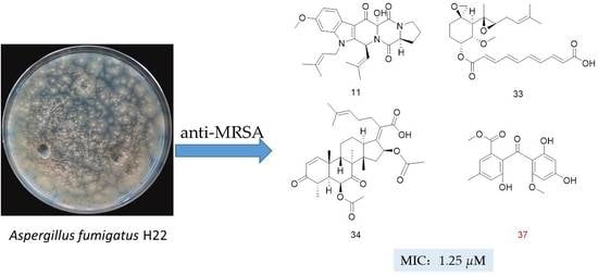
Discovery of Anti-MRSA Secondary Metabolites from a Marine-Derived Fungus Aspergillus fumigatus

1
Key Laboratory of Structure-Based Drug Design & Discovery of Education, College of Traditional Chinese Materia Medica, Shenyang Pharmaceutical University, Shenyang 110016, China
2
State Key Laboratory of Mycology, Institute of Microbiology, Chinese Academy of Sciences, Beijing 100101, China
*
Authors to whom correspondence should be addressed.
Submission received: 11 April 2022 / Revised: 23 April 2022 / Accepted: 25 April 2022 / Published: 28 April 2022
(This article belongs to the Special Issue Bioactive Secondary Metabolites of Marine Fungi)

Abstract

:
Methicillin-resistant Staphylococcus aureus (MRSA), a WHO high-priority pathogen that can cause great harm to living beings, is a primary cause of death from antibiotic-resistant infections. In the present study, six new compounds, including fumindoline A–C (13), 12β, 13β-hydroxy-asperfumigatin (4), 2-epi-tryptoquivaline F (17) and penibenzophenone E (37), and thirty-nine known ones were isolated from the marine-derived fungus Aspergillus fumigatus H22. The structures and the absolute configurations of the new compounds were unambiguously assigned by spectroscopic data, mass spectrometry (MS), electronic circular dichroism (ECD) spectroscopic analyses, quantum NMR and ECD calculations, and chemical derivatizations. Bioactivity screening indicated that nearly half of the compounds exhibit antibacterial activity, especially compounds 8 and 11, and 3338 showed excellent antimicrobial activities against MRSA, with minimum inhibitory concentration (MIC) values ranging from 1.25 to 2.5 μM. In addition, compound 8 showed moderate inhibitory activity against Mycobacterium bovis (MIC: 25 μM), compound 10 showed moderate inhibitory activity against Candida albicans (MIC: 50 μM), and compound 13 showed strong inhibitory activity against the hatching of a Caenorhabditis elegans egg (IC50: 2.5 μM).

Graphical Abstract

1. Introduction

Methicillin-resistant Staphylococcus aureus (MRSA) is recognized as one of the most common bacteria in both community and hospital-acquired infections, causing significant morbidity and mortality [1]. Compared to non-resistant Staphylococcus aureus infections, the mortality rate of MRSA infections increases by 64% [2]. Vancomycin is a last-resort treatment for MRSA infections. However, strains that are less susceptible to vancomycin are emerging in clinics [3,4]. As a result, new antibiotics to treat MRSA infections are desperately needed. In 2017, the development of new antibiotics for the treatment of MRSA infections is listed as a high urgency level by the WHO (World Health Organization) [5].
The marine environment is one of the most complex atmospheres on the earth, due to the huge variations in predation, temperature, pressure, light, and nutrient circumstances, etc. [6]. The organisms that thrive in marine environments could produce extremely diverse and complicated functional secondary metabolites that differ from those observed in terrestrial environments [6,7,8]. In recent decades, an increasing number of bioactive marine natural products (MNPs) have piqued the interest of chemists and pharmacologists for their medicinal values [9,10], such as the earliest marine sponge-derived anticancer drug cytarabine (Cytosar-U®), the marine sponge-derived antiviral drug vidarabine (Arasena A®), the mollusk-derived ziconotide (Prialt®) for the treatment of neuropathic pain, the famous sponge-derived anticancer drugs trabectedin (Yondelis®) and eribulin mesylate (Halaven®), and the marine cyanobacterium-derived anticancer drug disitamab vedotin (Aidixi™) and tisotumab vedotintftv (TIVDAK™), etc. [11,12,13,14,15,16,17].
Marine fungi have been shown to produce a variety of secondary metabolites with a variety of structures and bioactivities [18], including antibacterial, antiviral, anticancer, and anti-inflammatory characteristics, and have already provided a number of promising leads against MRSA [19,20]. Pestalone is a well-known anti-MRSA compound that was discovered by Fenical and colleagues, after co-culturing a fungus of the genus Pestalotia with a unicellular marine bacteria (strain CNJ-328) [21,22].
In our search for new anti-MRSA agents from marine-derived fungi, the EtOAc extract of the fungus Aspergillus fumigatus H22 was found to show strong anti-MRSA activity by in vitro anti-MRSA assay. A chemical investigation on its extract led to the identification of 45 secondary metabolites (Figure 1), including six new novel compounds, including fumindoline A–C (13), 12β,13β-hydroxy-asperfumigatin (4), 2-epi-tryptoquivaline F (17), and penibenzophenone E (37). The isolation and structure characterization of the new compounds, as well as the antibacterial activity of all the compounds, are described in this work.

2. Results

Structure Elucidation of the Isolated Compounds

Fumindoline A (1) was obtained as a chartreuse powder and had the molecular formula of C21H23N3O4, based on HRESIMS data (Figure S8), corresponding to 12 indices of hydrogen deficiency. This molecular formula was corroborated by 1H NMR and 13C NMR spectroscopic data. The 1H NMR data (Table 1) showed characteristic signals for a 1,2,4-trisubstituted benzene ring (δ 8.21 (d, J = 8.7 Hz), 6.91 (dd, J = 8.7 and 2.3 Hz), and 7.05 (d, J = 2.3 Hz)), two singlet olefinic protons (δH 8.59 (s) and 6.79 (s)), three singlet methyl groups (δH 2.08 (s), 2.17 (s), and 3.89 (s)), and two exchangeable (δ 8.42 (br. s) and 11.85 (br. s)). The 13C NMR and HSQC data of 1 revealed the presence of twenty-one carbon resonances, including three methyls (δC 20.6, 27.3, and 55.4), three methylenes (δC 24.9, 31.2, and 38.3), five sp2 methines (δC 94.8, 110.2, 111.1, 118.5, and 123.1), and ten nonprotonated carbons (eight sp2 carbons at δC 115.0, 128.9, 135.1, 138.7, 142.4, 142.8, 158.3, and 160.7; one amide carbonyl carbon at δC 164.7, and one carboxyl carbon at δC 174.2).
The planar structure of 1 was defined by the 2D NMR spectra, particularly the 1H–1H COSY and HMBC data (Figures S5 and S7). The HMBC correlations from H-9 to C-8, C-11, C-12, and C-13, from H-10 to C-8, and C-11, and from H-12 to C-8, C-10, C-11, and C-13, together with the 1H–1H COSY correlations of H-9/H-10/H-12, which indicated a 1,2,4-trisubstituted benzene. The 1H–1H COSY correlations of NH-15/H2-16/H2-17/H2-18, as well as the HMBC correlations from H2-16 to C-14, C17, and C18, from H2-17 to C-16, C-18, and C-19, from H2-18 to C-16, C-17, and C-19, led to the identification of the γ-aminobutyric acid residue. The HMBC correlations from H-6 to C-2, C-7, C-8, and C-14, H-9 to C-7, and H2-16 to C-14, as well as the chemical shifts of C-2 (δ 135.1), C-3 (δ 142.4), C-5 (δ 158.3), C-6 (δ 111.1), and C-14 (δ 164.7), supported a 2-pyridinecarboxylic acid moiety that was connected with a γ-aminobutyric acid moiety through C-14 and linked with a 1,2,4-trisubstituted benzene moiety through C-7, and completed the assignment of the moiety. The HMBC correlations from H-20 to C-2, C-3, and C-21, together with the 1H–1H COSY correlations of H-20/H3-22/H3-23, suggested that the isobutenyl group was located at C-3 of the 2-pyridinecarboxylic acid moiety. The key HMBC correlations from H3-24 to C-11 indicated that the methoxy group was located at C-11. Furthermore, these data accounted for 11 of the 12 degrees of unsaturation, implying the presence of an additional cycle, attributed to the NH bridging between C-2 and C-13 to establish the indole-pyridinecarboxylic acid skeleton (Figure 2). Therefore, the 2D structure of 1 was determined as shown below.
Fumindoline B (2) was obtained as a chartreuse powder. Its molecular formula, C22H23N3O4, with 14 degrees of unsaturation, was established on the basis of the HRESIMS data (Figure S15). The UV spectrum showed absorptions at 282 nm and 343 nm, which were similar to those of 1, indicating that 2 might have the same conjugation system as 1. The IR spectrum indicated the presence of a secondary amine N-H signal (2980 cm−1) and an amide carbonyl signal (1628 cm−1). The 1H NMR and 13C NMR spectra indicated the presence of two sets of very similar signals, with the same number of carbons (Figures S9 and S10). The spectra of the two sets of signals are well resolved in pairs at 313K and 298K in DMSO-d6, indicating the presence of two relatively stable isomers. From the integrals of the completely resolved signals, a ratio of 1:0.7 was calculated for the two stable isomers. To be better distinguished, we assigned the major isomer as 2a and the minor one as 2b, respectively (Figure 3).
The 1H NMR and 13C NMR spectra data of 2 showed close similarity to those of 1, with the biggest difference in the methine (CH-19). A detailed analysis of the 2D NMR data, including HSQC, HMBC, and 1H–1H COSY spectra, revealed that 2 contained the same indole-pyridinecarboxylic acid skeleton as that of 1 (Figure 4 and Figures S11–S13). The HMBC correlations from H2-16 to C-17 and C-18, H2-17 to C-16, C-18, and C-19, H2-18 to C-17, C-19, and C-20, and the 1H–1H COSY correlations of H2-16/H2-17/H2-18/H-19, together with the molecular formula, indicated the presence of a proline moiety, and this conclusion was also confirmed by the 14 degrees of unsaturation and the chemical shifts of C-16 (δC 49.6 (2a); δC 47.5 (2b)) and C-19 (δC 59.8 (2a); δC 60.4 (2b)).
The E/Z isomer exists in the tertiary amide. In the solution at room temperature, the slow rotation of the C–N bond in NMR makes it possess the characteristics of a partial double bond [23]. A comparison of the 1H NMR signals and 13C NMR signals of 2a and 2b revealed differences in the proline moiety, including variations in the H-19 (δH 4.48 (2a); δH 5.3 (2b)), C-16 (δC 49.6 (2a); δC 47.5 (2b)), C-17 (δC 25.2 (2a); δC 21.9 (2b)), and C-18 (δC 28.6 (2a); δC 31.2 (2b)). As shown in Figure 4, strong NOE effects between H-6 and H-16 for 2a and between H-6 and H-19 for 2b were observed in the ROSEY spectrum (Figure S14).
The absolute configuration of the amino acids from compound 2 was determined by the advanced Marfey’s method [24]. The mixture obtained after hydrolyzing compound 2 and further derivatization with l-FDAA was analyzed by HPLC-DAD. The HPLC analyses of the mixture of hydrolysates and appropriate amino acid standards confirmed the l configurations for proline in 2 (Figure 5). Consequently, the absolute configuration of 2 was elucidated to be 19S.
Fumindoline C (3) was obtained as a chartreuse powder. The molecular formula of 3 was established to be C23H25N3O4 from its HREIMS data (Figure S22). The 1H and 13C NMR spectra of 3 were similar to those of 2, possessing two sets of signals (Figures S16 and S17), except for the presence of an additional methoxyl group. The substitution of the methoxyl group was further confirmed by the HMBC correlations from H3-26 to C-20. A further comprehensive analysis of its 1H–1H COSY, HSQC, and HMBC spectra assigned the planar structure of 3 (Figures S18–S20). The relative configuration of 3 was determined to be the same as that of 2 by their similar structure and ROESY data (Figures S14 and S21). Accordingly, 3 was determined to be a methyl ester of 2.
12β,13β-hydroxy-asperfumigatin (4) was obtained as a white amorphous solid. Its molecular formula was determined as C27H33N3O7 by HRESIMS data (Figure S31). The 1H NMR spectrum of 4 (Figure S25) displayed four singlet methyl groups (δH 1.17, 1.25, 2.11, and 2.21), one methoxyl group (δH 3.85) and four olefinic/aromatic protons (δH 6.40, 6.90, 7.27, and 7.45). The 13C-NMR spectrum (Figure S26) exhibited 27 carbon resonances accounted for the functional groups described above and three amide carbonyl carbons (δC 164.7, 165.5, and 165.9). A comprehensive analysis of its 2D NMR spectra, including 1H-1H COSY, HSQC, and HMBC experiments, confirmed the planar structure of 4 (Table 2, Figures S27–S29), revealing the presence of the indole moiety and the diketopiperazine moiety in 4 (Figure 6). The planar structure of 4 was determined to be the same as that of asperfumigatin (5), by detailed interpretation of the 2D NMR spectra and NMR data comparison between 4 and 5. Considering the same biosynthesis origin, compound 4 was deduced to share the same absolute configuration at C-3 and C-6 as those of 513.
Owing to a lack of sufficient ROESY correlations, the relative configurations of C-12 and C-13 were not determined (Figure S30). The relative configurations of 4 were determined by the DP4+ probability, based on a theoretical NMR calculation that has been proven to be a very powerful tool in natural product structure elucidation [25,26]. The NMR shifts of eight possible relative orientation isomers were calculated with the GIAO method at the MPW1PW91/6-31+G(d,p), and the DP4+ probabilities of each configuration were evaluated based on Boltzmann-averaged theoretical NMR shielding tensors, which provided a 91.55% confidence for the relative configuration 3S*, 6S*, 12S*, 13R* (Tables S1 and S2).
To determine the absolute configurations of 4, a ECD calculation method was applied. The two configurations (3S, 6S, 12S, 13R)-4 and (3R, 6R, 12R, 13S)-4 were calculated using time-dependent density functional theory (TDDFT) at PBE1PBE/6-311 G* level, with the PCM model in methanol, and corrected with a 2 nm blue shift according to UV data. A comparison of the experimental ECD spectrum of 4 and the calculated ECD spectra of (3S, 6S, 12S, 13R)-4 and (3R, 6R, 12R, 13S)-4 showed that the experimental ECD spectrum of 4 is consistent with the calculated ECD spectrum of (3S, 6S, 12S, and 13R)-4 (Figure 7). Thus, the absolute configuration of 4 was assigned as 3S, 6S, 12S, and 13R, and named as 12β,13β-hydroxy-asperfumigatin. The only difference between compound 4 and compound 5 is the orientation of the two hydroxyl groups (12-OH, 13-OH).
2-epi-tryptoquivaline F (17), which was isolated as a white amorphous solid, exhibited the [M + H]+ peak at m/z 403.1399 (HRESIMS), corresponding to C22H18N4O4, as well as sixteen degrees of unsaturations (Figure S37). The 1H NMR, 13C NMR and HSQC spectra (Table 3, Figures S32–S34) of 17 revealed the presence of two 1, 2-disubstituted benzene rings (δHC 7.31 (d, J = 7.5 Hz)/124.4, 7.22 (dd, J = 8.0 and 7.5 Hz)/125.8, 7.43 (dd, J = 8.0 and 7.5 Hz)/131.3, 7.65 (d, J = 8.0 Hz)/115.8; δHC 8.29 (d, J = 8.1 Hz)/126.8, 7.58 (dd, J = 8.1 and 7.5 Hz)/128.2, 7.85 (dd, J = 8.1 and 7.5 Hz)/135.4, 7.78 (d, J = 8.1 Hz)/128.0), one methyl (δHC 1.28 (3H, d, J = 6.5 Hz)/17.9), one methylene (δH 2.61 (dd, J = 13.4 and 10.5 Hz), 3.68 (dd, J = 13.4 and 4.3 Hz), δC 33.4), four nitrogenated methines (δHC 4.26 (d, J = 6.5 Hz)/58.5, 5.03 (dd, J = 4.3 and 10.5 Hz)/58.3, 5.82 (s)/82.6, 8.11 (s)/145.6), eight quaternary carbons including three carbonyls (δC 172.4, 170.9, and 161.0), four aromatic or olefinic carbon atoms (δC 121.9, 134.4, 138.9, and 148.1), and one oxygenated one (δC 91.3). The NMR data of compound 17 were similar to those of tryptoquivaline F [27], indicating the presence of one 6-5-5 gem-methyl imidazoindolone ring and one quinazoline-4-one moiety.
The partial relative configuration of 17 was confirmed by a ROESY experiment (Figure S36). The ROESY correlations of H-2 (δH 5.82, s) with H-15 (δH 4.26, q, J = 6.5 Hz) indicated that H-2 and H-15 were on the same face, while H3-27 (δH 1.28, d, J = 6.5 Hz) were on the opposite face (Figure 8). The relative configurations of C-2 and C-15 were assigned as 2S and 15S. However, owing to a lack of sufficient ROESY correlations, neither the orientation of C-3 nor C-12 could be determined.
Similar to compound 4, the NMR shifts of four relative configuration isomers (2S, 3S, 12R, 15S; 2S, 3R, 12S, 15S; 2S, 3R, 12R, 15S; 2S, 3S, 12S, 15S) were calculated and the DP4+ probability, based on a theoretical NMR calculation, was applied. The 100% DP4+ probability for 17a revealed that the relative configuration of 17 was 2S*, 3S*, 12R* and 15S* (Tables S3 and S4).
The absolute configurations of 17 were deduced by the comparison of the experimental and simulated ECD spectra generated by TDDFT at B3LYP/6-311+G(2d,p) level with the PCM model in methanol and corrected -5 nm according to the UV data. A comparison of the observed ECD spectra for 17, with the calculated ECD spectra for the (2S, 3S, 12R, 15S)-17 and (2R, 3R, 12S, 15R)-17 enantiomers, is shown in Figure 9. The overall ECD spectra for (2S, 3S, 12R, 15S)-17 are in good accordance with the experimental ECD for 17. Thus, compound 17 was determined to be 2S, 3S, 12R, and 15S. The differences between 17 and tryptoquivaline F are the configuration of C-2. Therefore, compound 17 was identified as 2-epi-tryptoquivaline F.
Compound 37 was isolated as a yellowish powder. Its molecular formula was determined as C17H16O7 based on the HRESIMS (Figure S42), implying ten degrees of unsaturation. The 1H NMR spectrum (Figure S38) of 37 showed one hydrogen-bonded phenol moiety at δH 13.55 (s, 6′-OH), four aromatic methine protons at δH 7.19 (br. s, H-5), 6.88 (br. s, H-3), 5.90 (d, J = 2.2 Hz, H-5′) and 5.80 (d, J = 2.2 Hz, H-3′) for two sets of AB meta-coupling, two methoxy groups at δH 3.64 (s, 8-OMe) and 3.26 (s, 2′-OMe), and a methyl group at δH 2.30 (s, 4-Me). A comparison of its 1H NMR and 13C NMR spectra (Table 4) with those of sulochrin (38) suggested the same benzophenone skeleton between them [28]. The HMBC correlations from the proton of 6′-OH to C-6′, C-1′ and C-5′ indicate that 6′-OH was located at C-6′. The HMBC correlations from 2′-OCH3 to C-2′, 4-CH3 to C-3, C-4, C-5, and 8-CH3 to C-7 indicate that the two methoxy groups and one methyl group were located at C-2′, C-7 and C-4, respectively. In addition, the HMBC correlations from H-3 to C-1, C-2, C-5, from H-3′ to C-1′, C-2′, C-4′, C-5′, from H-5 to C-1, C-3, C-6 and C-7, and from H-5′ to C-1′, C-3′ and C-6′ confirmed the proposed structure (Figure 10). Therefore, compound 37 was determined as penibenzophenone E.
Other known compounds were identified as asperfumigatin (5) [29], demethoxyfumitremorgin C (6) [30], fumitremorgin C (7) [30], 12,13-dihydroxyfumitremorgin C (8) [31], 12α-hydroxy-13-oxofumitremorgin C (9) [32], fumitremorgin B (10) [33], 13-oxofumitremorgin B (11) [34], cyclotryprostatin B (12) [35], verruculogen (13) [36], 6-methoxyspirotryprostatin B (14) [37], (−)-spirotryprostatin A (15) [38], spirotryprostatin C (16) [39], fumiquinazoline C (18) [40], (+)-alantrypinone (19) [41,42], oxoglyantrypine (20) [43], (−)-chaetominine (21) [44], 11-epi-chaetominine (22) [29], fumigaclavine C (23) [45,46], bisdethiobis(methylthio)gliotoxin (24) [47], pyripyropene A (25) [48], pseurotin F1 (26) [49], pseurotin F2 (27) [49], pseurotin A (28) [50], 11-O-methylpseurotin A (29) [51], azaspirofuran B (30) [52], azaspirofuran A (31) [52], fumagiringillin (32) [53], fumagillin (33) [54], helvolic acid (34) [55], 6-O-propionyl-16-O-deacetylhelvolic acid (35) [55], 16-O-propionyl-6-O-deacetylhelvolic acid (36) [55], sulochrin (38) [28], monomethylsulochrin (39) [56], 8′-O-methylasterric acid (40) [29], dimethyl 2,3′-dimethylosoate (41) [56], questin (42) [57], (+)-2′S-isorhodoptilometrin (43) [58], 6-hydroxy-8-methoxy-3-methylisocoumarin (44) [59], and trypacidin (45) [60], based on the spectroscopic analyses and in comparison with the literature data.
The antibacterial activities of the isolated compounds were determined against methicillin-resistant Staphylococcus aureus (MRSA) (clinical isolate strain), vancomycin-resistant enterococci E. faecalis (VRE), Candida albicans SC5314, Mycobacterium bovis ATCC35743 constitutive GFP expression (pUV3583c-GFP), and Escherichia coli O57:H7, within 100 μM. The results showed that nearly half of the compounds exhibit antibacterial activity (Table 5), especially compounds 5, 8, 10, 11, 16, 21, 23, 2938, and 41 exhibited antimicrobial activities against MRSA, with minimum inhibitory concentration (MIC) values ranging from 1.25 to 25 μM. Furthermore, compound 8 also exhibited strong activity against M. bovis with a MIC of 25 μM, compound 10 showed moderate activity against C. albicans with a MIC of 50 μM. Moreover, compound 13 inhibited the egg hatching of Caenorhabditis elegans with a IC50 of 2.5 μM.

3. Discussion

The marine environmental stress conditions induce many faunae and symbiont microorganisms to synthesize and release secondary metabolites of unique structures and interesting biological activities [61]. These bioactive compounds can serve as an important source for drug discovery. Marine-derived fungi are important sources for the discovery of new antibacterial natural products. Wang et al. isolated the Chaetomium sp. strain NA-S01-R1 from a deep-sea (4050 m) fungus that produced novel chlorinated azaphilone polyketides with antibacterial activity against MRSA [62]. The Emericellopsis minima strain A11, isolated from Talcahuano Bay (Chile), produced an antibiotic called emerimicin IV, with moderate activity against clinical isolates of MDR vancomycin-resistant strains of E. faecalis and MRSA with MIC of 12.5 μg/mL and 100 μg/mL, respectively [63].
A. fumigatus belongs to the filamentous fungi family that is widely distributed in all environments and can cause many diseases and life-threatening conditions in immunocompromised patients [64]. A. fumigatus can produce a wide array of secondary metabolites due to its remarkable adaptability to different environmental conditions, such as fumitremorgins, fumagillins, pseurotins, fumigaclavines, gliotoxins, and helvolic acid derivatives.
Inspired by chemical ecology, we found a marine fungus A. fumigatus H22 with strong antibacterial activities from the marine fungi library. Through in-depth chemical mining, we found 45 compounds, including 6 new compounds, from the culture of this fungus. A evaluation of biological activity showed that nearly half of the compounds exhibit antimicrobial activity. Fumitremorgins derivatives (4-11) have very similar structures, but only a few have strong anti-MRSA activity. Compounds 5, 8 and 11 with strong anti-MRSA activity contain hydroxyl group at C-13, while compounds 6 and 7 without anti-MRSA activity have no hydroxyl group at C-13. In addition, compounds 4 and 5 have the same planar structure, but the 13-OH of compound 4 without anti MRSA activity was α-oriented, while compound 5 and other strongly active compounds were β-oriented. Therefore, it is preliminarily speculated that there is a certain correlation between the substituents and stereoconfiguration in C-12 and C-13 and their anti MRSA activity. Fumitremorgin B (10) was reported with antifungal activity against a variety of phytopathogenic fungi, but it showed weak activity against vancomycin-resistant E. faecalis (VRE), M. bovis, and E. coli in our in vitro assay, which could be involved in fighting against invasion by other pathogens [65].
Pseurotins, with a unique heterospirocyclic furanone-lactam structure, exhibit a broad range of biological activities. In addition to antifungal and antibiotic activities [66,67], pseurotins were also shown to regulate enzymes of cellular metabolism [68], to possess anti-angiogenic activity, to modulate cell differentiation [69], and to inhibit endothelial cell migration [70,71,72]. Fumagillin (33) have been demonstrated to have antitumor, antibacterial and antiparasitic effects [73]. Previous studies revealed that helvolic acid (34) exhibited in vitro antimalarial activity against multidrug resistant Plasmodium falciparum [74], antitrypanosomal activity against Trypanosoma brucei [75], and antimycobacterial activity against M. tuberculosis H37Ra [76]. Our current research showed the strong activities of oxofumitremorgin B (11), helvolic acid (34), 6-O-propionyl-16-O-deacetylhelvolic acid (35), 16-O-propionyl-6-O-deacetylhelvolic acid (36), sulochrin (38) and 8′-O-methylasterric acid (40) against MRSA, with a MIC of 1.25 μM.
From our current findings, it can be found that A. fumigatus from marine sources can produce rich bioactive secondary metabolites, especially in anti-MRSA.

4. Materials and Methods

4.1. General

UV data, optical rotation, and IR data were recorded on Genesys-10S UV–Vis spectrophotometer (Thermo Fisher Scientific, Waltham, MA, USA), MCP 200 automatic polarimeter (Anton Paar, Graz, Austria), and IS5 FT-IR spectrophotometer (Thermo Fisher Scientific, Waltham, MA, USA), respectively. NMR spectral data were obtained with a Bruker AVANCE-500 spectrometer (Bruker, Bremen, Germany) (DMSO-d6, δH 2.50/δC 39.52, and CDCl3, δH 7.26/δC 77.16). High-resolution electrospray ionization mass spectrometry (HRESIMS) data were obtained on an Agilent Accurate-Mass-Q-TOF LC/MS 6520 instrument (Agilent Technologies, Santa Clara, CA, USA). The CD spectra were measured by JASCO J-815 spectropolarimeter (JASCO, Tsukuba, Japan). Silica gel (Qingdao Haiyang Chemical Co., Ltd., Qingdao, China, 200–300 mesh), ODS (octadecylsilyl, 50 μM, YMC Co., Ltd., Kyoto, Japan), and Sephadex LH-20 (GE Healthcare, Uppsala, Sweden) were used for column chromatography. Semi-preparative HPLC was performed on an Agilent 1200 HPLC system equipped with an Agilent DAD UV–vis spectrometric detector (Agilent Technologies Inc., CA, USA), using a reversed-phase Eclipse XDB-C18 column (5 μM, 9.4 × 250 mm; Agilent, MA, USA), with a flow rate of 2.0 mL/min. The biological reagents, chemicals and media were purchased from standard commercial sources, unless stated.

4.2. Fungal Material

The fungus H22 was isolated from middle seawater from the Western Pacific. The sample (1 mL) was diluted with sterile H2O, 100 μL of which was deposited on a PDA (200 g of potato, 20 g of glucose, 20 g of agar per liter of seawater collected in the Western Pacific) plate containing chloramphenicol (100 μg/mL) and streptomycin (100 μg/mL) as a bacterial inhibitor. A single colony was transferred onto another PDA plate and was identified according to its morphological characteristics and 18S rRNA gene sequences. The phylogenetic tree (Figure S1), constructed from the ITS gene sequence, indicated that H22 belonged to the genus of Aspergillus, with the highest similarity to A. fumigatus (99.86%, accession number NRRL 163 s). In consideration of the morphological features and phylogeny (Figure S2), this fungus was identified as A. fumigatus. A reference culture of A. fumigatus H22 maintained at −80 °C was deposited in our laboratory.

4.3. Fermentation and Extraction

The isolate was grown for 7 days at 28 °C, on slants of a PDA medium. The spores of the strain on the plate were collected using 0.01% sterile Tween 80 (BTL, Warsaw, Poland) and adjusted to 1 × 10⁶ CFU/mL to make inoculum. A large-scale fermentation was carried out in 50 × 500 mL Fernbach culture flasks, holding 100 g of rice in 110 mL of distilled water (each with 0.5 mL of spore suspension) and incubated for 4 weeks at 28 °C. With the help of ultrasonication, the fermented rice substrates were extracted with ethyl acetate (3 × 5 L), and the organic solvent was filtered and evaporated to dryness under a vacuum to obtain the crude extract (78.0 g).

4.4. Isolation and Characterization Data

The ethyl acetate (EtOAc) fraction was subjected to silica gel column chromatography (CC), eluted with dichloromethane/acetone (D/A, v/v, 100:0, 100:1, 50:1, 30:1, 25:1, 20:1, 10:1, 5:1) and dichloromethane/methanol (D/M, v/v, 5:1, 2:1, 0:100) to give 10 fractions (HS.1–HS.10).
HS.3 (4.94 g eluted with D/A, v/v, 50:1) was purified by RP-HPLC, using 37% acetonitrile in acidic water (0.01% TFA) to obtain compounds 45 (9.0 mg, tR = 37.5 min), 42 (103.0 mg, tR = 44.5 min) and 39 (19.2 mg, tR = 47.6 min).
Fraction HS.4 (5.98 g from D/A, v/v, 30:1) was separated by ODS, using a gradient from 20% to 100% methanol in water to afford 12 subfractions (HS.4-1–HS.4-12). HS.4-3 (203.0 mg) was further purified using C8-RP-HPLC on a Agilent Eclipse XDB-C8 (5 μM, 250 × 9.4 mm), with a gradient elution from 30% to 40% acetonitrile in 60 min to give compounds 21 (102.1 mg, tR = 22.5 min), 18 (12.0 mg, tR = 30.6 min), 17 (7.0 mg, tR = 39.6 min) and 41 (2.0 mg, tR = 50.2 min). HS.4-4 (77 mg) was further purified using C8-RP-HPLC with 35% acetonitrile to give compounds 8 (3.5 mg, tR = 33.6 min), 7 (3.5 mg, tR = 41.5 min) and 31 (3.0 mg, tR = 66.2 min). Compounds 13 (79.0 mg, tR = 17.4 min), 10 (7.0 mg, tR = 23.6 min) and 33 (5.0 mg, tR = 25.8 min) were obtained from HS.4-9 (252 mg) by RP-HPLC, using 55% acetonitrile in acidic water.
Fraction HS.5 (5.43 g, from D/A, v/v, 25:1) was first separated by ODS, using a gradient from 30% to 100% methanol in water to afford HS.5-1–HS.5-11. Subfraction HS.5-4 (30 mg) was purified using RP-HPLC on a Agilent Eclipse XDB-C8 column (5 μM, 250 × 9.4 mm) with 40% acetonitrile in 20 min to give compounds 23 (5.0 mg, tR = 4.1 min) and 39 (4.2 mg, tR = 7.5 min). Compound 24 (5.0 mg, tR = 24.8 min) was obtained from subfraction HS.5-4-5 (32 mg) by RP-HPLC, using 28% acetonitrile in acidic water (0.01% TFA). Compounds 22 (2.2 mg, tR = 28.1 min) and 21 (3.0 mg, tR = 29.7 min) were obtained from HS.5-4-8 (144 mg) by RP-HPLC, using 29% acetonitrile in acidic water (0.01% TFA).
Fraction HS.6 (6.72 g, from D/A, v/v, 20:1) was first separated by ODS, using a gradient from 20% to 100% methanol in water to afford HS.6-1–HS.6-17. HS.6-2 (289 mg) was purified using C8-RP-HPLC eluting with 50% to provide compound 44 (2.0 mg, tR = 16.3 min). Compounds 37 (3.0 mg, tR = 19.1 min) and 38 (1.5 mg, tR = 20.2 min) were obtained from HS.6-4 (326.0 mg) by RP-HPLC, using 45% acetonitrile in acidic water. HS.6-5 (522 mg) was purified using RP-HPLC eluting with 50% acetonitrile to give compounds 15 (9.0 mg, tR = 10.2 min), 9 (15.0 mg, tR = 13.2 min), and 12 (100.0 mg, tR = 14.6 min). Compound 16 (3.0 mg, tR = 25.2 min) was obtained from subfraction HS.6-9 (17.5mg) by RP-HPLC, using 60% acetonitrile in acidic water. Compounds 33 (19.8 mg, tR = 10.7 min), 34 (2.0 mg, tR = 12.3 min) and 35 (2.0 mg, tR = 13.2 min) were obtained from HS.6-17 (365 mg) by RP-HPLC, using 70% acetonitrile in acidic water.
Fraction HS.7 (10.63 g, D/A, v/v, 10:1) was first separated by ODS, using a gradient from 35% to 100% methanol in water to afford HS.7-1–HS.7-13. Compounds 40 (6.0 mg) and 29 (2.0 mg) were obtained from HS.7-2 and HS.7-3 by recrystallization in methanol, respectively. Compounds 30 (2.0 mg, tR = 9.1 min), 20 (2.0 mg, tR = 11.1 min) and 6 (2.1 mg, tR = 11.9 min) were obtained from HS.7-4 (11.2 mg) by RP-HPLC, using 65% acetonitrile in acidic water. Compounds 4 (3.2 mg, tR = 14.9 min) and 5 (2.8 mg, tR = 16.1 min) were purified from HS.7-7, using RP-HPLC with 50% acetonitrile. HS.7-9 (420.0 mg) was purified using C8-RP-HPLC with 65% methanol to give compounds 43 (2.0 mg, tR = 12.5 min), 32 (20.2 mg, tR = 16.2 min) and 19 (37.3 mg, tR = 18.1 min).
Fraction HS.8 (8.82 g, D/A, v/v, 5:1) was first separated by ODS, using a gradient from 20% to 100% methanol in water to afford 22 subfractions (HS.8-1–HS.8-22). HS.8-3 (100.0 mg) was further purified on C8-RP-HPLC eluting with 35% acetonitrile in acidic water to give compounds 28 (11.8 mg, tR = 19.3 min), 27 (3.1 mg, tR = 22.4 min), and 26 (2.8 mg, tR = 24.3 min). HS.8-18 (124.0 mg) was purified on a C8-RP-HPLC eluting with a gradient elution from 70% methanol to give compounds 3 (15.0 mg, tR = 12.5 min), 2 (5.0 mg, tR = 15.2 min), 11 (20.0 mg, tR = 18.4 min), 1 (5.0 mg, tR = 19.2 min) and 25 (8.0 mg, tR = 22.5 min).
Fumindoline A (1). UV (MeOH) λmax (log ε) 286 (1.62), 345 (0.48). 1H NMR and 13C NMR, see Table 1, 2D NMR spectra, see Supplementary Figures S3–S7. Positive HRESIMS: m/z 382.1768 [M + H]+ (calcd for C21H24N3O4, 382.1761, Figure S8).
Fumindoline B (2). Chartreuse powder; (α ) D 25 −34.99 (c 0.1, MeOH); UV (MeOH) λmax (log ε) 282 (2.82), 343 (0.82); 1H NMR and 13C NMR, see Table 1, 2D NMR spectra, see Supplementary Figures S9–S14; Positive HRESIMS: m/z 394.1765 [M + H]+ (calcd for C22H24N3O4, 394.1761, Figure S15).
Fumindoline C (3). Chartreuse powder; (α ) D 25 −21.00 (c 0.1, MeOH); UV (MeOH) λmax (log ε) 286 (1.62), 345 (0.50); 1H NMR and 13C NMR, see Table 1, 2D NMR spectra, see Supplementary Figures S16–S24; Positive HRESIMS: m/z 408.1916 [M + H]+ (calcd for C23H26N3O4, 408.1918, Figure S22).
12β,13β-hydroxy-asperfumigatin (4). White amorphous solid; (α ) D 25 +26.00 (c 0.1, MeOH); UV (MeOH) λmax (log ε) 222 (1.51), 270 (0.60); 1H NMR and 13C NMR, see Table 2, 2D NMR spectra, see Supplementary Figures S25–S30; Positive HRESIMS: m/z 494.2720 [M + H − H2O]+ (calcd for C27H32N3O6, 494.2726, Figure S31).
2-epi-tryptoquivaline F (17). White amorphous solid; (α ) D 25 +221.96 (c 0.1, CH2Cl2); UV (CH2Cl2) λmax (log ε) 212 (2.21), 233 (1.69); 1H NMR and 13C NMR, see Table 3, 2D NMR spectra, see Supplementary Figures S32–S36; Positive HRESIMS: m/z 403.1399 [M + H]+ (calcd for C22H19N4O4, 403.1401, Figure S37).
Penibenzophenone E (37). Yellowish powder; UV (MeOH) λmax (log ε) 205 (3.28), 303 (1.63); 1H NMR and 13C NMR, see Table 4, 2D NMR spectra, see Supplementary Figures S38–S41; Positive HRESIMS: m/z 355.0789 [M + Na]+ (calcd for C17H16O7Na, 355.0788, Figure S42).

4.5. Marfey’s Analysis of Compound 2

Compound 2 (2.0 mg) was dissolved in 6 N HCl (2.0 mL) and heated at 100 °C for 24 h. The solutions were then evaporated to dryness and placed in a 4 mL reaction vial and treated with a 10 mg/mL solution of FDAA (200 μL) in acetone, followed by 1 M NaHCO3 (40 μL). The reaction mixtures were heated at 45 °C for 90 min, and the reactions were quenched by the addition of HCl (1 N, 40 µL). In a similar fashion, the standard l-proline and d-proline were derivatized separately. The derivatives of the acid hydrolysate and the standard amino acids were subjected to RP HPLC analysis (Kromasil C18 column; 5 μM, 4.6 × 250 mm; 1.0 mL/min; UV detection at 340 nm), with a linear gradient of acetonitrile (30–40%) in water (TFA, 0.01%) over 30 min. The retention times for the authentic standards were as follows: l-proline derivative (8.91 min) and d-proline derivative (9.88 min). The absolute configuration of the chiral amino acid in 2 was determined by comparing the retention times.

4.6. Computational Details for NMR and ECD

The GMMX software tool was used to undertake the systematic conformational evaluations for 4 and 17, utilizing the MMFF94 molecular mechanics force field. Gaussian 16 software was used to further improve the MMFF94 conformers, utilizing the M062X/6-31G(d) basis set level in gas for NMR calculations and B3LYP/6-31+G(d,p) basis set level in methanol, with a PCM model for ECD calculations. The shielding constants were calculated using the GIAO technique in chloroform, using the SMD solvent model and Gaussian function at mPW1PW91/6-31+G(d,p). A previously documented approach was used to calculate the 1H and 13C chemical shifts for the DP4+ probability analysis [77]. ECD spectra were stimulated in methanol with a Gaussian function at the B3LYP/6-311+G(2d,p) level using the PCM model, and 60 NStates were calculated. Boltzmann statistics were used to compute the equilibrium populations of the conformers at 298.15 K, based on their respective free energies (ΔG). The Boltzmann weighting of the key conformers was then used to construct the overall ECD spectra. UV correlation was used to correct the systematic mistakes in predicting the wavelength and excited-state energy [78].

4.7. Antimicrobial Assay

An antimicrobial assay was performed according to the Antimicrobial Susceptibility Testing Standards, outlined by the Clinical and Laboratory Standards Institute against MRSA (clinical strain from Chaoyang Hospital, Beijing, China), Pseudomonas aeruginosa (ATCC 15692), Escherichia coli (O57:H7), Mycobacterium bovis (ATCC35743), vancomycin-resistant Enterococci faecalis (VRE) (clinical strain from 309 Hospital, Beijing, China), and pathogen fungi Candida albicans SC5314. The protocol was performed as previously reported [58,59]. The positive controls were vancomycin against MRSA, E. faecalis, ciprofloxacin against P. aeruginosa and E. coli, amphotericin B for C. albicans, and rifampicin for M. bovis. All the experiments were performed in triplicate.

5. Conclusions

In summary, we isolated forty-five compounds from A. fumigatus H22, including six new compounds 1–4, 17, and 37. The stereochemistry of the new compounds was determined by quantum calculations of NMR, ECD calculations and chemical derivatizations. Bioactivity screening indicated that compounds 5, 8, 10, 11, 16, 21, 23, 2938, and 41 exhibited antimicrobial activities against MRSA, with MIC values ranging from 1.25 to 25 μM. Compound 8 also exhibited strong activity against M. bovis, with a MIC of 25 μM. To the best of our knowledge, this is the first report for the antimicrobial activities of compounds 5, 10, 11, 16, 30, 31, and 37. The strains of A. fumigatus from ocean environments are a good source of antibacterial natural products, deserving further exploitation.

Supplementary Materials

The following supporting information can be downloaded at: https://0-www-mdpi-com.brum.beds.ac.uk/article/10.3390/md20050302/s1. Table S1: NMR calculation of 4; Table S2: sDP4+, uDP4+ and DP4+ probabilities (%) for 4; Table S3: NMR calculation of 17; Table S4: sDP4+, uDP4+ and DP4+ probabilities (%) for 17; Figures S1 and S2: phylogenetic tree and morphology of A. fumigatus H22; Figures S3–S8: 1D, 2D NMR, and HRESIMS of 1; Figures S9–S15: 1D, 2D NMR, and HRESIMS of 2; Figures S16–S24: 1D, 2D NMR, and HRESIMS of 3; Figures S25–S31: 1D, 2D NMR, and HRESIMS of 4; Figures S32–S37: 1D, 2D NMR, and HRESIMS of 17; Figures S38–S42: 1D, 2D NMR, and HRESIMS of 37; Figure S43: eight possible stereoisomers of 4 (4a4h); Figure S44: four possible stereoisomers of 17 (17a17d).

Author Contributions

Conceptualization, R.Z., H.W. and J.H.; methodology, J.H. and B.C.; validation and data curation, R.Z., J.S. and H.D.; formal analysis, R.Z. and J.H.; investigation, J.H. and H.L.; resources, H.D.; writing—original draft preparation, R.Z. and J.H.; writing—review and editing, J.H. and H.L.; supervision and project administration, J.H. and H.L.; funding acquisition, H.L. All authors have read and agreed to the published version of the manuscript.

Funding

This project was supported by the National Natural Science Foundation (Grant No. 82073723).

Institutional Review Board Statement

Not applicable.

Informed Consent Statement

Not applicable.

Data Availability Statement

Not applicable.

Conflicts of Interest

The authors declare no conflict of interest.

References

  1. Kuok, C.F.; Hoi, S.O.; Hoi, C.F.; Chan, C.H.; Fong, I.H.; Ngok, C.K.; Meng, L.R.; Fong, P. Synergistic antibacterial effects of herbal extracts and antibiotics on methicillin-resistant Staphylococcus aureus: A computational and experimental study. Exp. Biol. Med. 2017, 242, 731–743. [Google Scholar] [CrossRef] [PubMed] [Green Version]
  2. Asghar, M.A.; Yousuf, R.I.; Shoaib, M.H.; Asghar, M.A.; Ansar, S.; Zehravi, M.; Abdul Rehman, A. Synergistic nanocomposites of different antibiotics coupled with green synthesized chitosan-based silver nanoparticles: Characterization, antibacterial, in vivo toxicological and biodistribution studies. Int. J. Nanomed. 2020, 15, 7841–7859. [Google Scholar] [CrossRef] [PubMed]
  3. Bezar, I.F.; Mashruwala, A.A.; Boyd, J.M.; Stock, A.M. Drug-like fragments inhibit agr-mediated virulence expression in Staphylococcus aureus. Sci. Rep. 2019, 9, 6786. [Google Scholar] [CrossRef] [Green Version]
  4. Rodvold, K.A.; McConeghy, K.W. Methicillin-resistant Staphylococcus aureus therapy: Past, present, and future. Clin. Infect. Dis. 2014, 58, S20–S27. [Google Scholar] [CrossRef] [PubMed]
  5. World Health Organization. Prioritization of Pathogens to Guide Discovery, Research and Development of New Antibiotics for Drug-Resistant Bacterial Infections, Including Tuberculosis, 9789240026438 (electronic version); World Health Organization: Geneva, Switzerland, 2017. [Google Scholar]
  6. Althagbi, H.I.; Alarif, W.M.; Al-Footy, K.O. Abdel-Lateff, A Marine-derived macrocyclic alkaloids (MDMAS): Chemical and Biological Diversity. Mar. Drugs 2020, 18, 368. [Google Scholar] [CrossRef]
  7. Pereira, F.; Aires-de-Sousa, J. Computational methodologies in the exploration of marine natural product leads. Mar. Drugs 2018, 16, 236. [Google Scholar] [CrossRef] [Green Version]
  8. Hagestad, O.C.; Andersen, J.H.; Altermark, B.; Hansen, E.; Rämä, T. Cultivable marine fungi from the arctic archipelago of svalbard and their antibacterial activity. Mycology 2020, 11, 230–242. [Google Scholar] [CrossRef] [Green Version]
  9. Pereira, R.B.; Andrade, P.B.; Valentao, P. Chemical diversity and biological properties of secondary metabolites from sea hares of aplysia genus. Mar. Drugs 2016, 14, 39. [Google Scholar] [CrossRef] [Green Version]
  10. Sun, L.; Li, D.; Tao, M.; Chen, Y.; Dan, F.; Zhang, W. Scopararanes C-G: New oxygenated pimarane diterpenes from the marine sediment-derived fungus Eutypella scoparia FS26. Mar. Drugs 2012, 10, 539–550. [Google Scholar] [CrossRef] [Green Version]
  11. Choudhary, A.; Naughton, L.M.; Montanchez, I.; Dobson, A.D.W.; Rai, D.K. Current status and future prospects of marine natural products (MNPs) as antimicrobials. Mar. Drugs 2017, 15, 272. [Google Scholar] [CrossRef]
  12. Blunt, J.W.; Carroll, A.R.; Copp, B.R.; Davis, R.A.; Keyzers, R.A.; Prinsep, M.R. Marine natural products. Nat. Prod. Rep. 2018, 35, 8–53. [Google Scholar] [CrossRef] [Green Version]
  13. Blunt, J.W.; Copp, B.R.; Keyzers, R.A.; Munro, M.H.; Prinsep, M.R. Marine natural products. Nat. Prod. Rep. 2016, 33, 382–431. [Google Scholar] [CrossRef] [PubMed] [Green Version]
  14. Blunt, J.W.; Copp, B.R.; Keyzers, R.A.; Munro, M.H.G.; Prinsep, M.R. Marine natural products. Nat. Prod. Rep. 2017, 34, 235–294. [Google Scholar] [CrossRef] [PubMed] [Green Version]
  15. Hanif, N.; Murni, A.; Tanaka, C.; Tanaka, J. Marine natural products from indonesian waters. Mar. Drugs 2019, 17, 364. [Google Scholar] [CrossRef] [PubMed] [Green Version]
  16. Mayer, A.M.; Rodriguez, A.D.; Taglialatela-Scafati, O.; Fusetani, N. Marine pharmacology in 2016–2017: Marine compounds with antibacterial, antidiabetic, antifungal, anti-inflammatory, antiprotozoal, antituberculosis and antiviral activities; affecting the immune and nervous systems, and other miscellaneous mechanisms of action. Mar. Drugs 2021, 19, 49. [Google Scholar]
  17. Wahab, H.A.; Pham, N.B.; Muhammad, T.S.; Hooper, J.N.; Quinn, R.J. Merosesquiterpene congeners from the australian sponge Hyrtios digitatus as potential drug leads for atherosclerosis disease. Mar. Drugs 2017, 15, 6. [Google Scholar] [CrossRef] [PubMed] [Green Version]
  18. Wong Chin, J.M.; Puchooa, D.; Bahorun, T.; Jeewon, R. Antimicrobial properties of marine fungi from sponges and brown algae of Mauritius. Mycology 2021, 12, 231–244. [Google Scholar] [CrossRef] [PubMed]
  19. Wu, B.; Wiese, J.; Labes, A.; Kramer, A.; Schmaljohann, R.; Imhoff, J.F. Lindgomycin, an unusual antibiotic polyketide from a marine fungus of the lindgomycetaceae. Mar. Drugs 2015, 13, 4617–4632. [Google Scholar] [CrossRef] [Green Version]
  20. Niu, S.; Liu, D.; Hu, X.; Proksch, P.; Shao, Z.; Lin, W. Spiromastixones A-O, antibacterial chlorodepsidones from a deep-sea-derived Spiromastix sp. fungus. J. Nat. Prod. 2014, 77, 1021–1030. [Google Scholar] [CrossRef]
  21. Augner, D.; Krut, O.; Slavov, N.; Gerbino, D.C.; Sahl, H.G.; Benting, J.; Nising, C.F.; Hillebrand, S.; Kronke, M.; Schmalz, H.G. On the antibiotic and antifungal activity of pestalone, pestalachloride A, and structurally related compounds. J. Nat. Prod. 2013, 76, 1519–1522. [Google Scholar] [CrossRef]
  22. Cueto, M.; Jensen, P.R.; Kauffman, C.; Fenical, W.; Lobkovsky, E.; Clardy, J. Pestalone, a new antibiotic produced by a marine fungus in response to bacterial challenge. J. Nat. Prod. 2001, 64, 1444–1446. [Google Scholar] [CrossRef] [PubMed]
  23. Xie, X.; Wallwey, C.; Matuschek, M.; Steinbach, K.; Li, S.M. Formyl migration product of chanoclavine-I aldehyde in the presence of the old yellow enzyme FgaOx3 from Aspergillus fumigatus: A NMR structure elucidation. Magn. Reson. Chem. 2011, 49, 678–681. [Google Scholar] [CrossRef] [PubMed]
  24. Wu, W.; Dai, H.Q.; Bao, L.; Ren, B.A.; Lu, J.C.; Luo, Y.M.; Guo, L.D.; Zhang, L.X.; Liu, H.W. Isolation and structural elucidation of proline-containing cyclopentapeptides from an endolichenic Xylaria sp. J. Nat. Prod. 2011, 74, 1303–1308. [Google Scholar] [CrossRef] [PubMed]
  25. Smith, S.G.; Goodman, J.M. Assigning stereochemistry to single diastereoisomers by GIAO NMR calculation: The DP4 probability. J. Am. Chem. Soc. 2010, 132, 12946–12959. [Google Scholar] [CrossRef] [PubMed]
  26. Rodriguez, I.; Genta-Jouve, G.; Alfonso, C.; Calabro, K.; Alonso, E.; Sanchez, J.A.; Alfonso, A.; Thomas, O.P.; Botana, L.M. Gambierone, a ladder-shaped polyether from the dinoflagellate Gambierdiscus belizeanus. Org. Lett. 2015, 17, 2392–2395. [Google Scholar] [CrossRef]
  27. Buttachon, S.; Chandrapatya, A.; Manoch, L.; Silva, A.; Gales, L.; Bruyere, C.; Kiss, R.; Kijjoa, A. Sartorymensin, a new indole alkaloid, and new analogues of tryptoquivaline and fiscalins produced by Neosartorya siamensis (KUFC 6349). Tetrahedron 2012, 68, 3253–3262. [Google Scholar] [CrossRef]
  28. Pang, X.; Zhao, J.Y.; Fang, X.M.; Zhang, T.; Zhang, D.W.; Liu, H.Y.; Su, J.; Cen, S.; Yu, L.Y. Metabolites from the plant endophytic fungus Aspergillus sp. CPCC 400735 and their anti-hiv activities. J. Nat. Prod. 2017, 80, 2595–2601. [Google Scholar] [CrossRef]
  29. Xie, F.; Li, X.B.; Zhou, J.C.; Xu, Q.Q.; Wang, X.N.; Yuan, H.Q.; Lou, H.X. Secondary metabolites from Aspergillus fumigatus, an endophytic fungus from the liverwort Heteroscyphus tener (Steph.) Schiffn. Chem. Biodivers. 2015, 12, 1954. [Google Scholar] [CrossRef] [Green Version]
  30. Cui, C.B.; Kakeya, H.; Osada, H. Novel mammalian cell cycle inhibitors, tryprostatins A, B and other diketopiperazines produced by Aspergillus fumigatus. II. Physico-chemical properties and structures. J. Antibiot. 1996, 49, 534–540. [Google Scholar] [CrossRef] [Green Version]
  31. Abraham, W.R.; Arfmann, H.A. 12,13-Dihydroxy-Fumitremorgin C from Aspergillus fumigatus. Phytochemistry 1990, 29, 1025–1026. [Google Scholar] [CrossRef]
  32. Yan, W.; Song, H.; Song, F.; Guo, Y.; Wu, C.H.; Her, A.S.; Pu, Y.; Wang, S.; Naowarojna, N.; Weitz, A.; et al. Endoperoxide formation by an alpha-ketoglutarate-dependent mononuclear non-haem iron enzyme. Nature 2015, 527, 539–543. [Google Scholar] [CrossRef] [PubMed] [Green Version]
  33. Zhou, F.; Zhang, H.C.; Liu, R.; Zhang, D.X. Isolation and biological evaluation of secondary metabolites of the endophytic fungus Aspergillus fumigatus from Astragalus membranaceus. Chem. Nat. Compd. 2013, 49, 568–570. [Google Scholar] [CrossRef]
  34. Fujimoto, H.; Fujimaki, T.; Okuyama, E.; Yamazaki, M. Immunosuppressive constituents from an Ascomycete, Sordaria gondaensis. Mycotoxins 2000, 50, 93–99. [Google Scholar] [CrossRef] [Green Version]
  35. Cui, C.B.; Kakeya, H.; Osada, H. Novel mammalian cell cycle inhibitors, cyclotryprostatins A-D, produced by Aspergillus fumigatus, which inhibit mammalian cell cycle at G2/M phase. Tetrahedron 1997, 53, 59–72. [Google Scholar] [CrossRef]
  36. Yu, F.; Holte, D.; Zoller, J.; Umemiya, S.; Simke, L.R.; Baran, P.S. Total synthesis of verruculogen and fumitremorgin a enabled by ligand-controlled C–H borylation. J. Am. Chem. Soc. 2015, 137, 10160–10163. [Google Scholar]
  37. Zhang, M.; Wang, W.L.; Fang, Y.C.; Zhu, T.J.; Gu, Q.Q.; Zhu, W.M. Cytotoxic alkaloids and antibiotic nordammarane triterpenoids from the marine-derived fungus Aspergillus sydowi. J. Nat. Prod. 2008, 71, 985–989. [Google Scholar] [CrossRef]
  38. Cui, C.B.; Kakeya, H.; Osada, H. Novel mammalian cell cycle inhibitors, spirotryprostatins A and B, produced by Aspergillus fumigatus, which inhibit mammalian cell cycle at G2/M phase. Tetrahedron 1996, 52, 12651–12666. [Google Scholar] [CrossRef]
  39. Wang, F.Z.; Fang, Y.C.; Zhu, T.J.; Zhang, M.; Lin, A.Q.; Gu, Q.Q.; Zhu, W.M. Seven new prenylated indole diketopiperazine alkaloids from holothurian-derived fungus Aspergillus fumigatus. Tetrahedron 2008, 64, 7986–7991. [Google Scholar] [CrossRef]
  40. Takahashi, C.; Matsushita, T.; Doi, M.; Minoura, K.; Shingu, T.; Kumeda, Y.; Numata, A. Fumiquinazolines A-G, novel metabolites of a fungus separated from a Pseudolabrus marine fish. J. Chem. Soc. Perkin Trans. 1 1995, 18, 2345–2353. [Google Scholar] [CrossRef]
  41. Ren-Yi, G.; Lei, X.; Yi, K.; Iii-Ming, C.; Jian-Chun, Q.; Li, L.; Sheng-Xiang, Y.; Li-Chun, Z. Chaetominine, (+)-alantrypinone, questin, isorhodoptilometrin, and 4-hydroxybenzaldehyde produced by the endophytic fungus Aspergillus sp. YL-6 inhibit wheat (Triticum aestivum) and radish (Raphanus sativus) germination. J. Plant. Interact. 2015, 10, 87–92. [Google Scholar] [CrossRef] [Green Version]
  42. Larsen, T.O.; Frydenvang, K.; Frisvad, J.C.; Christophersen, C. UV-guided isolation of alantrypinone, a novel Penicillium alkaloid. J. Nat. Prod. 1998, 61, 1154–1157. [Google Scholar] [CrossRef] [PubMed]
  43. Peng, J.; Lin, T.; Wang, W.; Xin, Z.; Zhu, T.; Gu, Q.; Li, D. Antiviral alkaloids produced by the mangrove-derived fungus Cladosporium sp. PJX-41. J. Nat. Prod. 2013, 76, 1133–1140. [Google Scholar] [CrossRef] [PubMed]
  44. Jiao, R.H.; Xu, S.; Liu, J.Y.; Ge, H.M.; Ding, H.; Xu, C.; Zhu, H.L.; Tan, R.X. Chaetominine, a cytotoxic alkaloid produced by endophytic Chaetomium sp. IFB-E015. Org. Lett. 2006, 8, 5709–5712. [Google Scholar] [CrossRef] [PubMed]
  45. Hui, M.G.; Yu, Z.G.; Jie, Z.; Wu, J.H.; Tan, R.X. Bioactive alkaloids from endophytic Aspergillus fumigatus. J. Nat. Prod. 2009, 72, 753–755. [Google Scholar]
  46. Cole, R.J.; Kirksey, J.W.; Dorner, J.W.; Wilson, D.M.; Johnson, J.C., Jr.; Johnson, A.N.; Bedell, D.M.; Springer, J.P.; Chexal, K.K.; Clardy, J.C.; et al. Mycotoxins produced by Aspergillus fumigatus species isolated from molded silage. J. Agric. Food. Chem. 1977, 25, 826–830. [Google Scholar] [CrossRef]
  47. Okamoto, M.; Yoshida, K.; Uchida, I.; Nishikawa, M.; Kohsaka, M.; Aoki, H. Studies of platelet activating factor (PAF) antagonists from microbial products. I. Bisdethiobis(methylthio)gliotoxin and its derivatives. Chem. Pharm. Bull. 1986, 34, 340–344. [Google Scholar] [CrossRef] [Green Version]
  48. Liang, W.L.; Le, X.; Li, H.J.; Yang, X.L.; Chen, J.X.; Xu, J.; Liu, H.L.; Wang, L.Y.; Wang, K.T.; Hu, K.C.; et al. Exploring the chemodiversity and biological activities of the secondary metabolites from the marine fungus Neosartorya pseudofischeri. Mar. Drugs 2014, 12, 5657–5676. [Google Scholar] [CrossRef] [Green Version]
  49. Joachim, W.; Susanne, G.; Manfred, G.; Ralf, T.; Reinhard, K. Pseurotin F1/F2, New Metabolites from Aspergillus fumigatus, Process for Their Preparation and Their Use as Apomorphine Antagonists. EP19920120724, 4 December 1992. [Google Scholar]
  50. Tsunematsu, Y.; Fukutomi, M.; Saruwatari, T.; Noguchi, H.; Hotta, K.; Tang, Y.; Watanabe, K. Elucidation of pseurotin biosynthetic pathway points to trans-acting C-methyltransferase: Generation of chemical diversity. Angew. Chem. Int. Ed. Engl. 2014, 53, 8475–8479. [Google Scholar] [CrossRef] [Green Version]
  51. Boot, C.M.; Gassner, N.C.; Compton, J.E.; Tenney, K.; Tamble, C.M.; Lokey, R.S.; Holman, T.R.; Crews, P. Pinpointing pseurotins from a marine-derived Aspergillus as tools for chemical genetics using a synthetic lethality yeast screen. J. Nat. Prod. 2007, 70, 1672–1675. [Google Scholar] [CrossRef]
  52. Ren, H.; Liu, R.; Chen, L.; Zhu, T.J.; Zhu, W.M.; Gu, Q.Q. Two new hetero-spirocyclic gamma-lactam derivatives from marine sediment-derived fungus Aspergillus sydowi D2-6. Arch. Pharm. Res. 2010, 33, 499–502. [Google Scholar] [CrossRef]
  53. Jiao, W.X.; Blunt, J.W.; Cole, A.L.J.; Munro, M.H.G. Fumagiringillin, a new fumagillin derivative from a strain of the fungus Aspergillus fumigatus. J. Nat. Prod 2004, 67, 1434–1437. [Google Scholar] [CrossRef] [PubMed]
  54. Chu, M.; Mierzwa, R.; He, L.; Xu, L.; Patel, M.; Patel, D.; Chan, T.M. Structure of sch 528647: A new antitumor antibiotic related to fumagillin. J. Antibiot. 2001, 54, 1096–1099. [Google Scholar] [CrossRef] [PubMed] [Green Version]
  55. Kong, F.D.; Huang, X.L.; Ma, Q.Y.; Xie, Q.Y.; Wang, P.; Chen, P.W.; Zhou, L.M.; Yuan, J.Z.; Dai, H.F.; Luo, D.Q.; et al. Helvolic acid derivatives with antibacterial activities against Streptococcus agalactiae from the marine-derived fungus Aspergillus fumigatus HNMF0047. J. Nat. Prod. 2018, 81, 1869–1876. [Google Scholar] [CrossRef] [PubMed]
  56. Liu, R.; Zhu, W.M.; Zhang, Y.P.; Zhu, T.J.; Liu, H.B.; Fang, Y.C.; Gu, Q.Q. A new diphenyl ether from marine-derived fungus Aspergillus sp B-F-2. J. Antibiot 2006, 59, 362–365. [Google Scholar] [CrossRef] [PubMed] [Green Version]
  57. Fujimoto, K.; Fujimaki, T.; Okuyama, E.; Yamazaki, M. Immunomodulatory constituents from an ascomycete, Microascus tardifaciens. Chem. Pharm. Bull. 1999, 47, 1426–1432. [Google Scholar] [CrossRef] [Green Version]
  58. Qin, S.; Wang, Y.; Wang, W.; Zhu, W. Anti-H1N1-virus secondary metabolites from mangrove-derived aciduric fungus Penicillium sp. OUCMDZ-4736. Chin. J. Mar. Drugs 2016, 35, 21–28. [Google Scholar]
  59. Wu, J.S.; Shi, X.H.; Zhang, Y.H.; Yu, J.Y.; Fu, X.M.; Li, X.; Chen, K.X.; Guo, Y.W.; Shao, C.L.; Wang, C.Y. Co-cultivation with 5-azacytidine induced new metabolites from the zoanthid-derived fungus Cochliobolus lunatus. Front. Chem. 2019, 7, 763. [Google Scholar] [CrossRef] [Green Version]
  60. Yong, K.; Kaleem, S.; Wu, B.; Zhang, Z.Z. New antiproliferative compounds against glioma cells from the marine-sourced fungus Penicillium sp. ZZ1750. Mar. Drugs 2021, 19, 483. [Google Scholar] [CrossRef]
  61. Abuhijjleh, R.K.; Shabbir, S.; Al-Abd, A.M.; Jiaan, N.H.; Alshamil, S.; El-labbad, E.M.; Khalifa, S.I. Bioactive marine metabolites derived from the Persian Gulf compared to the Red Sea: Similar environments and wide gap in drug discovery. Peerj 2021, 9, 11778. [Google Scholar] [CrossRef]
  62. Wang, W.Y.; Liao, Y.Y.; Chen, R.X.; Hou, Y.P.; Ke, W.Q.; Zhang, B.B.; Gao, M.L.; Shao, Z.Z.; Chen, J.M.; Li, F. Chlorinated azaphilone pigments with antimicrobial and cytotoxic activities isolated from the deep sea derived fungus Chaetomium sp. NA-S01-R1. Mar. Drugs 2018, 16, 61. [Google Scholar] [CrossRef] [Green Version]
  63. Inostroza, A.; Lara, L.; Paz, C.; Perez, A.; Galleguillos, F.; Hernandez, V.; Becerra, J.; Gonzalez-Rocha, G.; Silva, M. Antibiotic activity of Emerimicin IV isolated from Emericellopsis minima from Talcahuano Bay, Chile. Nat. Prod. Res. 2018, 32, 1361–1364. [Google Scholar] [CrossRef] [PubMed]
  64. Van de Veerdonk, F.L.; Gresnigt, M.S.; Romani, L.; Netea, M.G.; Latgé, J.-P. Aspergillus fumigatus morphology and dynamic host interactions. Nat. Rev. Microbiol. 2017, 15, 661–674. [Google Scholar] [CrossRef] [PubMed]
  65. Li, X.J.; Zhang, Q.; Zhang, A.L.; Gao, J.M. Metabolites from Aspergillus fumigatus, an endophytic fungus associated with Melia azedarach, and their antifungal, antifeedant, and toxic activities. J. Agric. Food Chem. 2012, 60, 3424–3431. [Google Scholar] [CrossRef] [PubMed]
  66. Ando, O.; Satake, H.; Nakajima, M.; Sato, A.; Nakamura, T.; Kinoshita, T.; Furuya, K.; Haneishi, T. Synerazol, a New Antifungal Antibiotic. J. Antibiot. 1991, 44, 382–389. [Google Scholar] [CrossRef] [Green Version]
  67. Pinheiro, E.A.A.; Carvalho, J.M.; dos Santos, D.C.P.; Feitosa, A.D.O.; Marinho, P.S.B.; Guilhon, G.M.S.P.; de Souza, A.D.L.; da Silva, F.M.A.; Marinho, A.M.D.R. Antibacterial activity of alkaloids produced by endophytic fungus Aspergillus sp. EJC08 isolated from medical plant Bauhinia guianensis. Nat. Prod. Res. 2013, 27, 1633–1638. [Google Scholar] [CrossRef]
  68. Anjum, K.; Bi, H.; Chai, W.; Lian, X.Y.; Zhang, Z. Antiglioma pseurotin A from marine Bacillus sp. FS8D regulating tumour metabolic enzymes. Nat. Prod. Res. 2017, 32, 1–4. [Google Scholar] [CrossRef]
  69. Komagata, D.; Fujita, S.; Yamashita, N.; Saito, S.; Morino, T. Novel neuritogenic activities of pseurotin A and penicillic acid. J. Antibiot. 1996, 49, 958–959. [Google Scholar] [CrossRef] [Green Version]
  70. Asami, Y.; Kakeya, H.; Komi, Y.; Kojima, S.; Nishikawa, K.; Beebe, K.; Neckers, L.; Osada, H. Azaspirene, a fungal product, inhibits angiogenesis by blocking Raf-1 activation. Cancer Sci. 2008, 99, 1853–1858. [Google Scholar] [CrossRef] [Green Version]
  71. Igarashi, Y.; Yabuta, Y.; Sekine, A.; Fujii, K.; Harada, K.; Oikawa, T.; Sato, M.; Furumai, T.; Oki, T. Directed biosynthesis of fluorinated pseurotin A, synerazol and gliotoxin. J. Antibiot. 2004, 57, 748–754. [Google Scholar] [CrossRef] [Green Version]
  72. Asami, Y.; Kakeya, H.; Onose, R.; Yoshida, A.; Matsuzaki, H.; Osada, H. Azaspirene: A novel angiogenesis inhibitor containing a 1-oxa-7-azaspiro[4.4]non-2-ene-4,6-dione skeleton produced by the fungus Neosartotya sp. Organ. Lett. 2002, 4, 2845–2848. [Google Scholar] [CrossRef]
  73. Guruceaga, X.; Perez-Cuesta, U.; de Cerio, A.A.D.; Gonzalez, O.; Alonso, R.M.; Hernando, F.L.; Ramirez-Garcia, A.; Rementeria, A. Fumagillin, a mycotoxin of Aspergillus fumigatus: Biosynthesis, biological activities, detection, and applications. Toxins 2020, 12, 7. [Google Scholar] [CrossRef] [PubMed] [Green Version]
  74. Sawadsitang, S.; Mongkolthanaruk, W.; Suwannasai, N.; Sodngam, S. Antimalarial and cytotoxic constituents of Xylaria cf. cubensis PK108. Nat. Prod. Res. 2015, 29, 2033–2036. [Google Scholar] [CrossRef] [PubMed]
  75. Ganaha, M.; Yoshii, K.; Otsuki, Y.; Iguchi, M.; Okamoto, Y.; Iseki, K.; Ban, S.; Ishiyama, A.; Hokari, R.; Iwatsuki, M.; et al. In Vitro antitrypanosomal activity of the secondary metabolites from the mutant strain IU-3 of the insect pathogenic fungus Ophiocordyceps coccidiicola NBRC 100683. Chem. Pharm. Bull. 2016, 64, 988–990. [Google Scholar] [CrossRef] [Green Version]
  76. Sanmanoch, W.; Mongkolthanaruk, W.; Kanokmedhakul, S.; Aimi, T.; Boonlue, S. Helvolic acid, a secondary metabolite produced by Neosartorya spinosa KKU-1NK1 and its biological activities. Chiang Mai J. Sci. 2016, 43, 484–494. [Google Scholar]
  77. Grimblat, N.; Zanardi, M.M.; Sarotti, A.M. Beyond DP4: An improved probability for the stereochemical assignment of isomeric compounds using quantum chemical calculations of NMR shifts. J. Org. Chem. 2015, 80, 12526–12534. [Google Scholar] [CrossRef] [PubMed]
  78. Han, J.J.; Liu, C.C.; Li, L.; Zhou, H.; Liu, L.; Bao, L.; Chen, Q.; Song, F.H.; Zhang, L.X.; Li, E.W.; et al. Decalin-containing tetramic acids and 4-Hydroxy-2-pyridones with antimicrobial and cytotoxic activity from the fungus Coniochaeta cephalothecoides collected in Tibetan Plateau (Medog). J. Org. Chem. 2017, 82, 11474–11486. [Google Scholar] [CrossRef] [PubMed]
Figure 1. Structures of compounds 145.
Marinedrugs 20 00302 g001
Figure 2. Key 1H–1H COSY and HMBC correlations of 1.
Marinedrugs 20 00302 g002
Figure 3. Scheme of the resonance structure of 2 and the chemical equilibrium between 2a and 2b.
Marinedrugs 20 00302 g003
Figure 4. Key 1H-1H COSY, HMBC, and ROESY correlations of 2a and 2b.
Marinedrugs 20 00302 g004
Figure 5. Advanced Marfey’s analysis of compound 2. (A): The FDAA derivatives of the hydrolysate of 2. (B,C): The retention times for the FDAA derivatives of l-proline and d-proline. The derivatives of the acid hydrolysate and the standard amino acids were subjected to RP HPLC analysis (Kromasil C18 column; 5 μM, 4.6 × 250 mm; 1.0 mL/min; UV detection at 340 nm) with a linear gradient of acetonitrile (30–40%) in water (TFA, 0.01%) over 30 min.
Marinedrugs 20 00302 g005
Figure 6. Key 1H-1H COSY, and HMBC correlations of 4.
Marinedrugs 20 00302 g006
Figure 7. Experimental ECD spectra of compound 4 and the calculated ECD spectra of (3S, 6S, 12S, 13R)-4 and (3R, 6R, 12R, 13S)-4.
Marinedrugs 20 00302 g007
Figure 8. Key HMBC and NOESY correlations of compound 17.
Marinedrugs 20 00302 g008
Figure 9. Experimental ECD spectra of compound 17 and the calculated ECD spectra of (2S, 3S, 12R, 15S)-17 and (2R, 3R, 12S, 15R)-17.
Marinedrugs 20 00302 g009
Figure 10. Key HMBC correlations of compound 37.
Marinedrugs 20 00302 g010
Table 1. 1H (500 MHz) and 13C NMR (125 MHz) data of compounds 13 in DMSO-d6.
Positions12a2b3a3b
δCδH, Mult.,
(J in Hz)
δCδH, Mult.,
(J in Hz)
δCδH, Mult.,
(J in Hz)
δCδH, Mult.,
(J in Hz)
δCδH, Mult.,
(J in Hz)
1 11.85 br. s 11.83 br. s 11.64 br. s 11.92 br. s 11.60 br. s
2135.1 134.4 134.2 134.3 134.3
3142.4 142.5 141.9 143.4 141.4
5158.3 158.2 159.7 158.4 158.1
6111.18.59 s113.48.41 s113.38.37 s113.68.47 s113.58.45 s
7128.9 129.0 128.7 129.1 128.7
8115.0 114.8 114.9 114.8 114.9
9123.18.21 d (8.7)123.28.22 d (8.7)123.18.18 d (8.7)123.48.25 d (8.7)123.18.19 d (8.7)
10110.26.91 dd
(8.7, 2.3)
110.26.91 dd
(8.7, 2.2)
110.06.89 dd
(8.7, 2.2)
110.56.93 dd
(8.7, 2.2)
110.06.89 dd
(8.7, 2.2)
11160.7 160.6 160.8 161.0 160.7
1294.87.05 d (2.3)94.77.05 d (2.2)94.77.04 d (2.2)94.77.06 d (2.2)94.87.04 d (2.2)
13142.8 142.9 142.8 143.2 142.8
14164.7 166.0 166.6 165.5 165.8
15 8.42 br. s
1638.33.39 q (7.4) 49.63.96 m47.53.67 m49.63.98 m47.93.70 m
3.88 m 3.91 m
1724.91.80 p (7.4) 25.21.94 m21.91.92 m25.21.96 m21.81.92 m
1.90 m 1.82 m 1.93 m 1.83 m
1831.22.30 t (7.4)28.62.26 m31.22.29 m28.62.28 m31.32.29 m
1.89 m 2.02 m 1.91 m 1.98 m
19174.2 59.84.48 dd
(8.8, 4.4)
60.45.30 dd
(8.5, 3.6)
59.74.57 dd
(8.6, 4.0)
60.85.18 dd
(8.6, 4.5)
20118.56.79 s173.5 173.8 172.5 172.9
21138.7 118.56.76 s119.06.66 s118.06.75 s119.56.59 s
2227.32.08 s138.1 138.0 138.1 138
2320.62.17 s27.12.07 s26.92.04 s27.02.07 s26.52.04 s
2455.43.89 s20.42.13 s20.22.01 s20.42.10 s20.11.90 s
25 55.43.89 s55.43.88 s55.53.89 s55.43.88 s
26 51.83.68 s51.63.45 s
“m” means multiplet or overlapped with other signals.
Table 2. 1H (500 MHz) and 13C NMR (125 MHz) data for 4 in chloroform-d.
PositionsδCδH, Mult., (J in Hz)PositionsδCδH, Mult., (J in Hz)
2137.0 17111.26.90 dd (8.6, 2.2)
343.36.37 dd (9.5, 1.2)18157.7
5164.7 19100.87.27 d (2.2)
659.84.32 dd (10.8, 6.0)20136.1
729.62.51 m2139.52.29 dd (14.0, 9.5)
1.95 m 2.14 dd (14.0, 1.2)
822.02.08 m2274.6
1.98 m2329.31.25 s
945.73.76 m2432.21.17 s
3.65 m25165.5
11165.9 26119.86.40 br. s
1286.2 27158.2
1368.45.13 s2827.42.11 s
14114.3 2921.22.21 s
15122.3 18-OCH355.93.85 s
16119.47.45 d (8.6)
Table 3. 1H (500 MHz) and 13C NMR (125 MHz) data of compound 17 in chloroform-d.
PositionsδCδH, Mult., (J in Hz)PositionsδC, TypeδH, Mult., (J in Hz)
282.65.82 s14172.4
391.3 1558.54.26 q (6.5)
4134.4 18161.0
5124.47.31 d (7.5)19121.9
6125.87.22 dd (8.0, 7.5)20126.88.29 d (8.1)
7131.37.43 dd (8.0, 7.5)21128.27.58 dd (8.1, 7.5)
8115.87.65 d (8.0)22135.47.85 dd (8.1, 7.5)
9138.9 23128.07.78 d (8.1)
11170.9 24148.1
1258.35.03 dd (10.5, 4.3)26145.68.11 s
1333.43.68 dd (13.4, 4.3)2717.91.28 d (6.5)
2.61 dd (13.4, 10.5)
Table 4. 1H (500 MHz) and 13C NMR (125 MHz) data of compound 37 in DMSO-d6.
PositionsδCδH, Mult., (J in Hz)PositionsδCδH, Mult., (J in Hz)
1127.2 1′105.9
2153.1 2′163.0
3120.16.88 s3′91.45.80 d (2.2)
4138.1 4′165.2
5120.27.19 s5′95.55.90 d (2.2)
6130.3 6′166.0
7166.0 4-CH320.82.30 s
851.93.64 s2′-OCH355.73.26 s
9198.1 6′-OH 13.55 s
Table 5. Antibacterial assay results of monomeric compounds.
MIC (μM)
CompoundMRSA aCompoundMRSA a
55.00315.00
82.503225.0
1020.0332.50
111.25341.25
1610.00351.25
2125.00361.25
2312.50371.25
2910.00381.25
305.00415.00
Positive controlVancomycin (1.00)
a MRSA: methicillin-resistant Staphylococcus aureus.
Publisher’s Note: MDPI stays neutral with regard to jurisdictional claims in published maps and institutional affiliations.

