
Novel [1,3,4]Thiadiazole[3,2-a]pyrimidin-5-ones as Promising Biofilm Dispersal Agents against Relevant Gram-Positive and Gram-Negative Pathogens
 , , , , , , and
Abstract
:
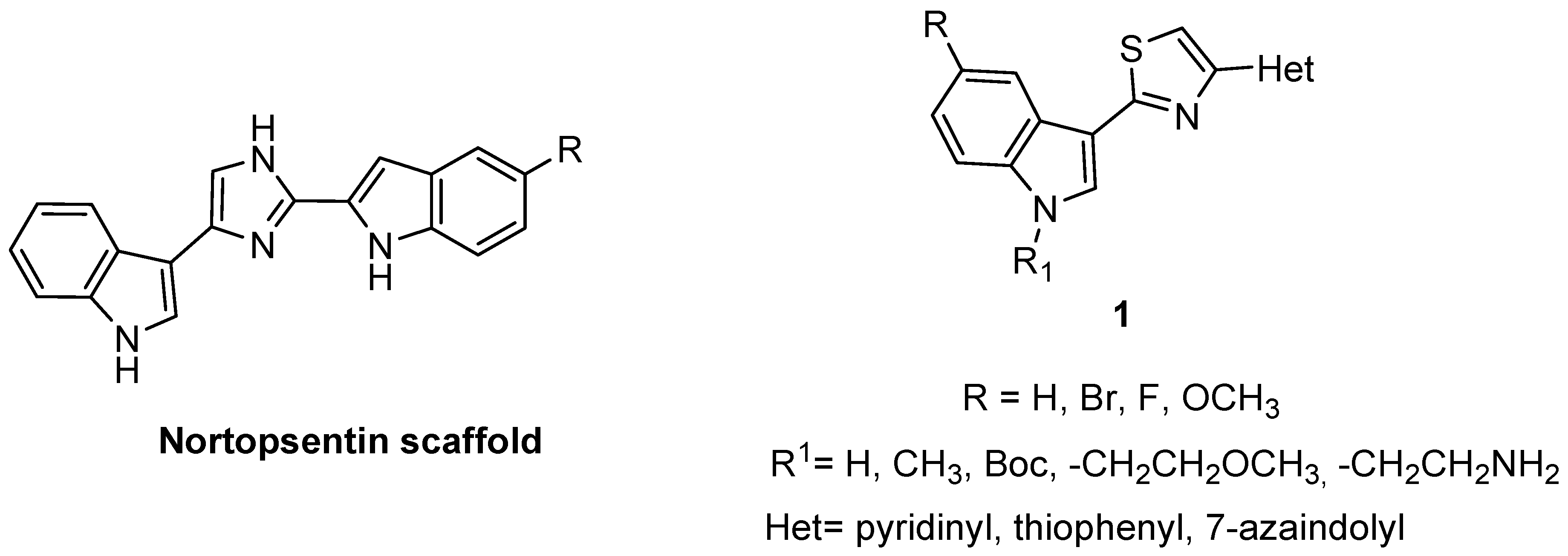
1. Introduction
2. Results and Discussion
2.1. Chemistry
2.2. Biology
2.2.1. Antibacterial Activity
2.2.2. Inhibition of Biofilm Formation
2.2.3. Dispersal Activity against Pre-Formed Biofilm
2.2.4. In Vivo Anti-Infective Evaluation of 8j
2.2.5. Toxicity Evaluation of 8j in In Vivo Model
3. Material and Methods
3.1. Chemistry
3.1.1. Synthesis of 1H-indole-3-carbonitriles (4b–e)
3.1.2. Synthesis of 1-methylindole-3-carbonitriles (5a–e)
3.1.3. Synthesis of 5-(1H-indol-3-yl)-1,3,4-thiadiazol-2-amines (6a–f)
3.1.4. Synthesis of Ethyl 3-oxo-3-(thiophen-3-yl)propanoate 7c
3.1.5. General Procedure for the Synthesis of -[1,3,4]thiadiazolo[3,2-a]pyrimidin-5-ones (8a–v)
- 2. -(1H-Indol-3-yl)-7-phenyl-5H-[1,3,4]thiadiazolo[3,2-a]pyrimidin-5-one (8a)
- 2. -(5-Methoxy-1H-indol-3-yl)-7-phenyl-5H-[1,3,4]thiadiazolo[3,2-a]pyrimidin-5-one (8b)
- 2. -(5-Bromo-1H-indol-3-yl)-7-phenyl-5H-[1,3,4]thiadiazolo[3,2-a]pyrimidin-5-one (8c)
- 2. -(5-Chloro-1H-indol-3-yl)-7-phenyl-5H-[1,3,4]thiadiazolo[3,2-a]pyrimidin-5-one (8d)
- 2. -(5-Fluoro-1H-indol-3-yl)-7-phenyl-5H-[1,3,4]thiadiazolo[3,2-a]pyrimidin-5-one (8e)
- 2. -(1-Methyl-1H-indol-3-yl)-7-phenyl-5H-[1,3,4]thiadiazolo[3,2-a]pyrimidin-5-one (8f)
- 2. -(5-Methoxy-1-methyl-1H-indol-3-yl)-7-phenyl-5H-[1,3,4]thiadiazolo[3,2-a]pyrimidin-5-one (8g)
- 2. -(5-Bromo-1-methyl-1H-indol-3-yl)-7-phenyl-[1,3,4]thiadiazolo[3,2-a]pyrimidin-5-one (8h)
- 2. -(5-Chloro-1-methyl-1H-indol-3-yl)-7-phenyl-5H-[1,3,4]thiadiazolo[3,2-a]pyrimidin-5-one (8i)
- 2. -(5-Fluoro-1-methyl-1H-indol-3-yl)-7-phenyl-5H-[1,3,4]thiadiazolo[3,2-a]pyrimidin-5-one (8j)
- 2. -(1H-Indol-3-yl)-7-methyl-5H-[1,3,4]thiadiazolo[3,2-a]pyrimidin-5-one (8k)
- 7. -Methyl-2-(1-methyl-1H-indol-3-yl)-5H-[1,3,4]thiadiazolo[3,2-a]pyrimidin-5-one (8l)
- 2. -(1H-Indol-3-yl)-7-thiophen-3-yl-[1,3,4]thiadiazolo[3,2-a]pyrimidin-5-one (8m)
- 2. -(5-Methoxy-1H-indol-3-yl)-7-thiophen-3-yl-[1,3,4]thiadiazolo[3,2-a]pyrimidin-5-one (8n)
- 2. -(5-Bromo-1H-indol-3-yl)-7-thiophen-3-yl-[1,3,4]thiadiazolo[3,2-a]pyrimidin-5-one (8o)
- 2. -(5-Chloro-1H-indol-3-yl)-7-thiophen-3-yl-[1,3,4]thiadiazolo[3,2-a]pyrimidin-5-one (8p)
- 2. -(5-Fluoro-1H-indol-3-yl)-7-thiophen-3-yl-[1,3,4]thiadiazolo[3,2-a]pyrimidin-5-one (8q)
- 2. -(1H-Indol-3-yl)-7-thiophen-3-yl-[1,3,4]thiadiazolo[3,2-a]pyrimidin-5-one (8r)
- 2. -(5-Methoxy-1-methyl-1H-indol-3-yl)-7-thiophen-3-yl-[1,3,4]thiadiazolo[3,2-a]pyrimidin-5-one (8s)
- 2. -(5-Bromo-1-methyl-1H-indol-3-yl)-7-thiophen-3-yl-[1,3,4]thiadiazolo[3,2-a]pyrimidin-5-one (8t)
- 2. -(5-Chloro-1-methyl-1H-indol-3-yl)-7-thiophen-3-yl-[1,3,4]thiadiazolo[3,2-a]pyrimidin-5-one (8u)
- 2. -(5-Fluoro-1-methyl-1H-indol-3-yl)-7-thiophen-3-yl-[1,3,4]thiadiazolo[3,2-a]pyrimidin-5-one (8v)
3.2. Biology
3.2.1. Determination of Minimum Inhibitory Concentrations (MICs)
3.2.2. Antibiofilm Activity
3.2.3. Effect of Compounds against Preformed Biofilm Biomasses
3.2.4. In Vivo Anti-Infective Activity and Toxicity Evaluation of Compound 8j
Insect Rearing and Preparation
In Vivo Bioassay
Insect Data Analysis
4. Conclusions
Author Contributions
Funding
Institutional Review Board Statement
Data Availability Statement
Conflicts of Interest
References
- Cascioferro, S.; Parrino, B.; Carbone, D.; Pecoraro, C.; Diana, P. Novel Strategies in the War against Antibiotic Resistance. Future Med. Chem. 2021, 13, 529–531. [Google Scholar] [CrossRef]
- Ventola, C.L. The Antibiotic Resistance Crisis. Pharm. Ther. 2015, 40, 277–283. [Google Scholar]
- Sulayyim, H.J.A.; Ismail, R.; Hamid, A.A.; Ghafar, N.A. Antibiotic Resistance during COVID-19: A Systematic Review. Int. J. Environ. Res. Public Health 2022, 19, 11931. [Google Scholar] [CrossRef]
- Romandini, A.; Pani, A.; Schenardi, P.A.; Pattarino, G.A.C.; De Giacomo, C.; Scaglione, F. Antibiotic Resistance in Pediatric Infections: Global Emerging Threats, Predicting the Near Future. Antibiotics 2021, 10, 393. [Google Scholar] [CrossRef]
- Jamal, M.; Ahmad, W.; Andleeb, S.; Jalil, F.; Imran, M.; Nawaz, M.A.; Hussain, T.; Ali, M.; Rafiq, M.; Kamil, M.A. Bacterial Biofilm and Associated Infections. J. Chin. Med. Assoc. 2018, 81, 7–11. [Google Scholar] [CrossRef]
- Pecoraro, C.; Carbone, D.; Deng, D.; Cascioferro, S.M.; Diana, P.; Giovannetti, E. Biofilm Formation as Valuable Target to Fight against Severe Chronic Infections. Curr. Med. Chem. 2022, 29, 4307–4310. [Google Scholar] [CrossRef]
- Singh, S.; Singh, S.K.; Chowdhury, I.; Singh, R. Understanding the Mechanism of Bacterial Biofilms Resistance to Antimicrobial Agents. Open Microbiol. J. 2017, 11, 53–62. [Google Scholar] [CrossRef]
- Wood, T.K.; Knabel, S.J.; Kwan, B.W. Bacterial Persister Cell Formation and Dormancy. Appl. Environ. Microbiol. 2013, 79, 7116–7121. [Google Scholar] [CrossRef]
- Sharma, S.; Mohler, J.; Mahajan, S.D.; Schwartz, S.A.; Bruggemann, L.; Aalinkeel, R. Microbial Biofilm: A Review on Formation, Infection, Antibiotic Resistance, Control Measures, and Innovative Treatment. Microorganisms 2023, 11, 1614. [Google Scholar] [CrossRef]
- Pecoraro, C.; Carbone, D.; Parrino, B.; Cascioferro, S.; Diana, P. Recent Developments in the Inhibition of Bacterial Adhesion as Promising Anti-Virulence Strategy. Int. J. Mol. Sci. 2023, 24, 4872. [Google Scholar] [CrossRef]
- Parrino, B.; Carbone, D.; Cascioferro, S.; Pecoraro, C.; Giovannetti, E.; Deng, D.; Di Sarno, V.; Musella, S.; Auriemma, G.; Cusimano, M.G.; et al. 1,2,4-Oxadiazole Topsentin Analogs as Staphylococcal Biofilm Inhibitors Targeting the Bacterial Transpeptidase Sortase A. Eur. J. Med. Chem. 2021, 209, 112892. [Google Scholar] [CrossRef]
- Hwang, H.-J.; Li, D.-D.; Lee, J.; Kang, M.K.; Moon, H.R.; Lee, J.-H. Compounds That Have an Anti-Biofilm Effect against Common Bacteria at Very Low Concentrations and Their Antibiotic Combination Effect. Antibiotics 2023, 12, 853. [Google Scholar] [CrossRef]
- Miller, T.; Waturangi, D.E. Yogiara Antibiofilm Properties of Bioactive Compounds from Actinomycetes against Foodborne and Fish Pathogens. Sci. Rep. 2022, 12, 18614. [Google Scholar] [CrossRef]
- Lahiri, D.; Nag, M.; Dey, A.; Sarkar, T.; Pati, S.; Nirmal, N.P.; Ray, R.R.; Upadhye, V.J.; Pandit, S.; Moovendhan, M.; et al. Marine Bioactive Compounds as Antibiofilm Agent: A Metabolomic Approach. Arch. Microbiol. 2023, 205, 54. [Google Scholar] [CrossRef]
- Carbone, A.; Cascioferro, S.; Parrino, B.; Carbone, D.; Pecoraro, C.; Schillaci, D.; Cusimano, M.G.; Cirrincione, G.; Diana, P. Thiazole Analogues of the Marine Alkaloid Nortopsentin as Inhibitors of Bacterial Biofilm Formation. Molecules 2021, 26, 81. [Google Scholar] [CrossRef]
- Natarajan, R.; Anthoni Samy, H.N.; Sivaperuman, A.; Subramani, A. Structure-Activity Relationships of Pyrimidine Derivatives and Their Biological Activity—A Review. Med. Chem. 2023, 19, 10–30. [Google Scholar] [CrossRef]
- El-Gendy, M.M.A.; Shaaban, M.; Shaaban, K.A.; El-Bondkly, A.M.; Laatsch, H. Essramycin: A First Triazolopyrimidine Antibiotic Isolated from Nature. J. Antibiot. 2008, 61, 149–157. [Google Scholar] [CrossRef]
- Azab, M.E.; Abdel-Wahab, S.S.; Mahmoud, N.F.; Elsayed, G.A. Novel Bridgehead Thiadiazolopyrimidine Derivatives with Antimicrobial and Antitumor Activities. J. Heterocycl. Chem. 2018, 55, 2349–2359. [Google Scholar] [CrossRef]
- Dehbanipour, R.; Ghalavand, Z. Anti-Virulence Therapeutic Strategies against Bacterial Infections: Recent Advances. Germs 2022, 12, 262–275. [Google Scholar] [CrossRef]
- Venditti, N.; Vergalito, F.; Magnifico, I.; Cutuli, M.A.; Pietrangelo, L.; Cozzolino, A.; Angiolillo, A.; Succi, M.; Petronio, G.P.; Di Marco, R. The Lepidoptera Galleria Mellonella “in Vivo” Model: A Preliminary Pilot Study on Oral Administration of Lactobacillus Plantarum (Now Lactiplantibacillus Plantarum). New Microbiol. 2021, 44, 42–50. [Google Scholar]
- Cutuli, M.A.; Petronio Petronio, G.; Vergalito, F.; Magnifico, I.; Pietrangelo, L.; Venditti, N.; Di Marco, R. Galleria Mellonella as a Consolidated in Vivo Model Hosts: New Developments in Antibacterial Strategies and Novel Drug Testing. Virulence 2019, 10, 527–541. [Google Scholar] [CrossRef]
- Ménard, G.; Rouillon, A.; Cattoir, V.; Donnio, P.-Y. Galleria Mellonella as a Suitable Model of Bacterial Infection: Past, Present and Future. Front. Cell Infect. Microbiol. 2021, 11, 782733. [Google Scholar] [CrossRef]
- Carbone, D.; Parrino, B.; Cascioferro, S.; Pecoraro, C.; Giovannetti, E.; Di Sarno, V.; Musella, S.; Auriemma, G.; Cirrincione, G.; Diana, P. 1,2,4-Oxadiazole Topsentin Analogs with Antiproliferative Activity against Pancreatic Cancer Cells, Targeting GSK3β Kinase. ChemMedChem 2021, 16, 537–554. [Google Scholar] [CrossRef]
- Cerca, N.; Martins, S.; Cerca, F.; Jefferson, K.K.; Pier, G.B.; Oliveira, R.; Azeredo, J. Comparative Assessment of Antibiotic Susceptibility of Coagulase-Negative Staphylococci in Biofilm versus Planktonic Culture as Assessed by Bacterial Enumeration or Rapid XTT Colorimetry. J. Antimicrob. Chemother. 2005, 56, 331–336. [Google Scholar] [CrossRef]
- da Silva, A.F.; da Rocha, C.Q.; da Silva, L.C.N.; Carvalho Júnior, A.R.; Mendes, I.N.F.V.; de Araruna, A.B.; Motta, E.P.; Silva, R.d.S.; Campos, C.D.L.; Farias, J.R.; et al. Antifungal and Antivirulence Activities of Hydroalcoholic Extract and Fractions of Platonia Insignis Leaves against Vaginal Isolates of Candida Species. Pathogens 2020, 9, 84. [Google Scholar] [CrossRef]
- Lade, H.; Chung, S.H.; Lee, Y.; Kumbhar, B.V.; Joo, H.-S.; Kim, Y.-G.; Yang, Y.-H.; Kim, J.-S. Thymol Reduces Agr-Mediated Virulence Factor Phenol-Soluble Modulin Production in Staphylococcus Aureus. Biomed. Res. Int. 2022, 2022, 8221622. [Google Scholar] [CrossRef]
- Zammuto, V.; Spanò, A.; Agostino, E.; Macrì, A.; De Pasquale, C.; Ferlazzo, G.; Rizzo, M.G.; Nicolò, M.S.; Guglielmino, S.; Gugliandolo, C. Anti-Bacterial Adhesion on Abiotic and Biotic Surfaces of the Exopolysaccharide from the Marine Bacillus Licheniformis B3-15. Mar. Drugs 2023, 21, 313. [Google Scholar] [CrossRef]
- Manandhar, S.; Singh, A.; Varma, A.; Pandey, S.; Shrivastava, N. Evaluation of Methods to Detect in Vitro Biofilm Formation by Staphylococcal Clinical Isolates. BMC Res. Notes 2018, 11, 714. [Google Scholar] [CrossRef]


| Compound | R | R1 | R2 |
|---|---|---|---|
| 8a | H | H | Ph |
| 8b | OCH3 | H | Ph |
| 8c | Br | H | Ph |
| 8d | Cl | H | Ph |
| 8e | F | H | Ph |
| 8f | H | CH3 | Ph |
| 8g | OCH3 | CH3 | Ph |
| 8h | Br | CH3 | Ph |
| 8i | Cl | CH3 | Ph |
| 8j | F | CH3 | Ph |
| 8k | H | H | CH3 |
| 8l | H | CH3 | CH3 |
| 8m | H | H | 3-thiophenyl |
| 8n | OCH3 | H | 3-thiophenyl |
| 8o | Br | H | 3-thiophenyl |
| 8p | Cl | H | 3-thiophenyl |
| 8q | F | H | 3-thiophenyl |
| 8r | H | CH3 | 3-thiophenyl |
| 8s | OCH3 | CH3 | 3-thiophenyl |
| 8t | Br | CH3 | 3-thiophenyl |
| 8u | Cl | CH3 | 3-thiophenyl |
| 8v | F | CH3 | 3-thiophenyl |
| Compound | MIC (µg/mL) | |||
|---|---|---|---|---|
| Pathogen | ||||
| S. aureus ATCC 25923 | P. aeruginosa ATCC 15442 | E. coli ATCC 25922 | E. faecalis ATCC 29212 | |
| 8a | >100 | >100 | >100 | >100 |
| 8b | >100 | >100 | >100 | >100 |
| 8c | >100 | >100 | >100 | >100 |
| 8d | >100 | >100 | >100 | >100 |
| 8e | 50 | >100 | >100 | >100 |
| 8f | 100 | >100 | >100 | >100 |
| 8g | >100 | >100 | >100 | >100 |
| 8h | >100 | >100 | >100 | >100 |
| 8i | >100 | >100 | >100 | >100 |
| 8j | >100 | >100 | >100 | 50 |
| 8k | 50 | >100 | >100 | 25 |
| 8l | 50 | >100 | >100 | >100 |
| 8m | >100 | >100 | >100 | >100 |
| 8n | >100 | >100 | >100 | >100 |
| 8o | >100 | >100 | >100 | >100 |
| 8p | >100 | >100 | >100 | >100 |
| 8q | >100 | >100 | >100 | >100 |
| 8r | >100 | >100 | >100 | >100 |
| 8s | >100 | >100 | >100 | >100 |
| 8t | >100 | >100 | >100 | >100 |
| 8u | >100 | >100 | >100 | >100 |
| 8v | >100 | >100 | >100 | >100 |
| Compounds | IBF (%, 100 µg/mL) | BIC50 µg/mL (µM) | Pathogen |
|---|---|---|---|
| 8c | 51 (±5.0) | 10.4 (24.5) | P. aeruginosa ATCC 15442 |
| 8e | 54 (±5.2) | 18.2 (50.2) | S. aureus ATCC 25923 |
| 8f | 75 (±7.3) | 7.6 (21.3) | S. aureus ATCC 25923 |
| 8j | 60 (±5.9) | 14.4 (38.4) | P. aeruginosa ATCC 15442 |
| 70 (±6.8) | 6.3 (16.7) | S. aureus ATCC 25923 | |
| 64 (±6.2) | 6.1 (16.2) | E. coli ATCC 25922 | |
| 8k | 64 (±6.1) | 21.1 (75.8) | S. aureus ATCC 25923 |
| 57 (±5.5) | 6.4 (22.6) | E. faecalis ATCC 29212 | |
| 8l | 53 (±5.1) | 14.5 (48.9) | P. aeruginosa ATCC 15442 |
| Compounds | S. aureus ATCC 25923 | P. aeruginosa ATCC 15442 | E. coli ATCC 25922 |
|---|---|---|---|
| 8a | 69 (±6.9) | 65 (±6.5) | 53 (±5.3) |
| 8b | 71 (±7.1) | 59 (±5.9) | 75 (±7.5) |
| 8c | 58 (±5.8) | 67 (±6.7) | 73 (±7.3) |
| 8d | 66 (±6.0) | 63 (±6.3) | 63 (±6.3) |
| 8e | 51 (±5.0) | 62 (±6.1) | 65 (±6.5) |
| 8f | 69 (±6.9) | 54 (±5.4) | 67 (±6.7) |
| 8g | 72 (±7.0) | 68 (±6.7) | 73 (±7.3) |
| 8h | 66 (±6.1) | 77 (±7.3) | 70 (±7.0) |
| 8i | 69 (±6.9) | 67 (±6.7) | 59 (±5.7) |
| 8j | 60 (±5.9) | 69 (±6.9) | 68 (±6.8) |
| 8k | 66 (±6.6) | 73 (±7.3) | 70 (±7.0) |
| 8l | 65 (±6.5) | 71 (±7.0) | 76 (±7.3) |
Disclaimer/Publisher’s Note: The statements, opinions and data contained in all publications are solely those of the individual author(s) and contributor(s) and not of MDPI and/or the editor(s). MDPI and/or the editor(s) disclaim responsibility for any injury to people or property resulting from any ideas, methods, instructions or products referred to in the content. |
© 2024 by the authors. Licensee MDPI, Basel, Switzerland. This article is an open access article distributed under the terms and conditions of the Creative Commons Attribution (CC BY) license (https://creativecommons.org/licenses/by/4.0/).
Share and Cite
Carbone, D.; Pecoraro, C.; Scianò, F.; Catania, V.; Schillaci, D.; Manachini, B.; Cascioferro, S.; Diana, P.; Parrino, B. Novel [1,3,4]Thiadiazole[3,2-a]pyrimidin-5-ones as Promising Biofilm Dispersal Agents against Relevant Gram-Positive and Gram-Negative Pathogens. Mar. Drugs 2024, 22, 133. https://0-doi-org.brum.beds.ac.uk/10.3390/md22030133
Carbone D, Pecoraro C, Scianò F, Catania V, Schillaci D, Manachini B, Cascioferro S, Diana P, Parrino B. Novel [1,3,4]Thiadiazole[3,2-a]pyrimidin-5-ones as Promising Biofilm Dispersal Agents against Relevant Gram-Positive and Gram-Negative Pathogens. Marine Drugs. 2024; 22(3):133. https://0-doi-org.brum.beds.ac.uk/10.3390/md22030133
Chicago/Turabian StyleCarbone, Daniela, Camilla Pecoraro, Fabio Scianò, Valentina Catania, Domenico Schillaci, Barbara Manachini, Stella Cascioferro, Patrizia Diana, and Barbara Parrino. 2024. "Novel [1,3,4]Thiadiazole[3,2-a]pyrimidin-5-ones as Promising Biofilm Dispersal Agents against Relevant Gram-Positive and Gram-Negative Pathogens" Marine Drugs 22, no. 3: 133. https://0-doi-org.brum.beds.ac.uk/10.3390/md22030133