Pregnancy with Heart Disease: Maternal Outcomes and Risk Factors for Fetal Growth Restriction


 , , and
Abstract
:1. Introduction
2. Methods
2.1. Study Design
2.2. Variables
2.3. Statistical Analysis
2.4. Ethical Approval
3. Results
4. Discussion
5. Conclusions
Author Contributions
Funding
Acknowledgments
Conflicts of Interest
References
- Koutrolou-Sotiropoulou, P.; Parikh, P.B.; Miller, C.; Lima, F.V.; Butler, J.; Stergiopoulos, K. Impact of heart disease on maternal and fetal outcomes in pregnant women. Am. J. Cardiol. 2015, 116, 474–480. [Google Scholar] [CrossRef] [PubMed]
- Mishra, R. Ian Donald’s Practical Obstetric Problems; BI Publications: New Delhi, India, 2010. [Google Scholar]
- Barbosa, P.J.B.; Lopes, A.A.; Feitosa, G.S.; Almeida, R.V.d.; Silva, R.M.d.; Brito, J.C.; Duarte, M.L.; Almeida, A.J.G. Prognostic factors of rheumatic mitral stenosis during pregnancy and puerperium. Arq. Bras. Cardiol. 2000, 75, 220–224. [Google Scholar] [CrossRef] [PubMed] [Green Version]
- Sawhney, H.; Aggarwal, N.; Suri, V.; Vasishta, K.; Sharma, Y.; Grover, A. Maternal and perinatal outcome in rheumatic heart disease. Int. J. Gynecol. Obstet. 2003, 80, 9–14. [Google Scholar] [CrossRef]
- Roos-Hesselink, J.W.; Ruys, T.P.; Stein, J.I.; Thilén, U.; Webb, G.D.; Niwa, K.; Kaemmerer, H.; Baumgartner, H.; Budts, W.; Maggioni, A.P. Outcome of pregnancy in patients with structural or ischaemic heart disease: Results of a registry of the European Society of Cardiology. Eur. Heart J. 2012, 34, 657–665. [Google Scholar] [CrossRef] [PubMed]
- Arafeh, J.M.; Baird, S.M. Cardiac disease in pregnancy. Crit. Care Nurs. Q. 2006, 29, 32–52. [Google Scholar] [CrossRef]
- Dobbenga-Rhodes, Y.A.; Prive, A.M. Assessment and evaluation of the woman with cardiac disease during pregnancy. J. Perinat. Neonatal Nurs. 2006, 20, 295–302. [Google Scholar] [CrossRef]
- Phuc, V.M.; Tin do, N.; Giang do, T.C. Challenges in the management of congenital heart disease in Vietnam: A single center experience. Ann. Pediatr. Cardiol. 2015, 8, 44–46. [Google Scholar]
- Davies, G.A.L.; Herbert, W.N.P. Assessment and management of cardiac disease in pregnancy. J. Obstet. Gynaecol. Can. JOGC 2007, 29, 331–336. [Google Scholar] [CrossRef]
- Pilliod, R.A.; Cheng, Y.W.; Snowden, J.M.; Doss, A.E.; Caughey, A.B. The risk of intrauterine fetal death in the small-for-gestational-age fetus. Am. J. Obstet. Gynecol. 2012, 207, e311–e316. [Google Scholar] [CrossRef]
- Pasupathy, D.; Wood, A.M.; Pell, J.P.; Fleming, M.; Smith, G.C. Rates of and factors associated with delivery-related perinatal death among term infants in Scotland. JAMA 2009, 302, 660–668. [Google Scholar] [CrossRef]
- Bukowski, R.; Burgett, A.D.; Gei, A.; Saade, G.R.; Hankins, G.D. Impairment of fetal growth potential and neonatal encephalopathy. Am. J. Obstet. Gynecol. 2003, 188, 1011–1015. [Google Scholar] [CrossRef] [PubMed]
- MacKay, D.F.; Smith, G.C.; Dobbie, R.; Pell, J.P. Gestational age at delivery and special educational need: Retrospective cohort study of 407,503 schoolchildren. PLoS Med. 2010, 7, e1000289. [Google Scholar] [CrossRef] [PubMed]
- Group, G.D. Guidelines: management of diabetes from preconception to the postnatal period: Summary of NICE guidance. BMJ Br. Med. J. 2008, 336, 714. [Google Scholar]
- Nguyễn, B.G. Nhận Xét Về Tình Hình Và Kết Quả Điều Trị Bệnh Tim Và Thai Nghén Tại Bệnh Viện Phụ Sản Trung Ương ( Từ Tháng 1/2000 Đến Tháng 9/2004); Trường Đại học Y Hà Nội: Hanoi, Vietnam, 2004. [Google Scholar]
- Gardosi, J. Customized fetal growth standards: Rationale and clinical application. Semin. Perinatol. 2004, 28, 33–40. [Google Scholar] [CrossRef] [PubMed]
- Quinn, J.-A.; Munoz, F.M.; Gonik, B.; Frau, L.; Cutland, C.; Mallett-Moore, T.; Kissou, A.; Wittke, F.; Das, M.; Nunes, T. Preterm birth: Case definition & guidelines for data collection, analysis, and presentation of immunisation safety data. Vaccine 2016, 34, 6047–6056. [Google Scholar] [PubMed] [Green Version]
- Pillutla, P.; Nguyen, T.; Markovic, D.; Canobbio, M.; Koos, B.J.; Aboulhosn, J.A. Cardiovascular and neonatal outcomes in pregnant women with high-risk congenital heart disease. Am. J. Cardiol. 2016, 117, 1672–1677. [Google Scholar] [CrossRef]
- Joshi, G.; Joshi, S.; Jha, S.; Singh, Y.; Joshi, A. Maternal heart disease and pregnancy outcome: Findings from a retrospective cohort in a tertiary care government hospital in Haldwani, Nainital. Niger. J. Cardiol. 2015, 12, 120–123. [Google Scholar] [CrossRef]
- Abbasi, S.; Siddiqua, S.F.; Rijvi, S.; Akhtar, S.; Haque, B.; Jesmin, S. Study of Maternal and Fetal Outcome in Pregnancy with Heart Disease. Anwer Khan Mod. Med Coll. J. 2017, 8, 112–116. [Google Scholar] [CrossRef] [Green Version]
- Salam, S.; Mushtaq, S.; Mohi-ud-Din, K.; Gul, I.; Ali, A. Maternal and fetal outcome in pregnancy with heart disease in tertiary care hospital in India. Int. J. Reprod. Contracept. Obstet. Gynecol. 2017, 6, 3947–3951. [Google Scholar] [CrossRef] [Green Version]
- Phạm, T.Q. Tình hình bệnh tim và thai nghén tại Viện bảo vệ bà mẹ và trẻ sơ sinh trong 5 năm 1995–1999. In Luận Văn Thạc sĩ y Học; Trường đại học Y Hà Nội: Hanoi, Vietnam, 2000. [Google Scholar]
- Savarese, G.; Lund, L.H. Global Public Health Burden of Heart Failure. Cardiac Fail. Rev. 2017, 3, 7–11. [Google Scholar] [CrossRef]
- Abdel-Hady, E.S.; El-Shamy, M.; El-Rifai, A.A.; Goda, H.; Abdel-Samad, A.; Moussa, S. Maternal and perinatal outcomes of pregnancies complicated by cardiac disease. Int. J. Gynaecol. Obstet. 2005, 90, 21–25. [Google Scholar] [CrossRef] [PubMed]
- Vasu, S.; Stergiopoulos, K. Valvular heart disease in pregnancy. Hellenic J. Cardiol. 2009, 50, 498–510. [Google Scholar] [PubMed]
- Blössner, M.; Villar, J. Levels and patterns of intrauterine growth retardation in developing countries. Eur. J. Clin. Nutr. 1998, 52, S5–S15. [Google Scholar]
- Swanson, A.M.; David, A.L. Animal models of fetal growth restriction: considerations for translational medicine. Placenta 2015, 36, 623–630. [Google Scholar] [CrossRef] [PubMed]
- Xiong, X.; Mayes, D.; Demianczuk, N.; Olson, D.M.; Davidge, S.T.; Newburn-Cook, C.; Saunders, L.D. Impact of pregnancy-induced hypertension on fetal growth. Am. J. Obstet. Gynecol. 1999, 180, 207–213. [Google Scholar] [CrossRef]
- Resnik, R. Intrauterine growth restriction. Obstet. Gynecol. 2002, 99, 490–496. [Google Scholar]
- Manandhar, T.P.B.; Nath, P.M. Risk Factors for Intrauterine Growth Restriction and Its Neonatal Outcome. Gynecol; 464, O. Risk Factors for Intrauterine Growth Restriction and Its Neonatal Outcome. Gynecol Obs. 2018, 8, 464. [Google Scholar]
- Muhammad, T.; Khattak, A.A.; Khan, M.A.; Khan, A.; Khan, M.A. Maternal factors associated with intrauterine growth restriction. J. Ayub Med Coll. Abbottabad 2010, 22, 64–69. [Google Scholar]
- Sharma, D.; Shastri, S.; Sharma, P. Intrauterine Growth Restriction: Antenatal and Postnatal Aspects. Clin. Med. Insights Pediatr. 2016, 10, 67–83. [Google Scholar] [CrossRef] [Green Version]


| Characteristics | n | % |
|---|---|---|
| Age group | ||
| <35 | 247 | 86.97 |
| ≥35 | 37 | 13.03 |
| Mean (SD *; Min–Max) | 28.18 (5.05; 18–44) | |
| Gravida | ||
| Primi | 135 | 47.54 |
| Gravida 2 | 121 | 42.61 |
| Gravida 3 or more | 28 | 9.86 |
| Diagnosis time | ||
| Before pregnancy | 229 | 80.63 |
| After Pregnancy | 55 | 19.37 |
| Occupation | ||
| Officer | 112 | 39.44 |
| Worker | 38 | 13.38 |
| Others | 134 | 47.18 |
| Area | ||
| Urban | 127 | 44.72 |
| Rural | 157 | 55.28 |
| The Types of Heart Disease | n | % |
|---|---|---|
| Rheumatic heart disease | ||
| Mitral valve stenosis | 65 | 22.89 |
| Mitral valve regurgitation | 114 | 40.14 |
| Mitral stenosis + Mitral valve regurgitation | 46 | 16.20 |
| Aortic valve regurgitation | 12 | 4.23 |
| Pulmonary stenosis | 17 | 5.99 |
| Congenital heart disease | ||
| ASD | 47 | 16.55 |
| VSD | 55 | 19.37 |
| Tetralogy of Fallot | 15 | 5.28 |
| Patent ductus arteriosus | 5 | 1.76 |
| Complications | n | % |
|---|---|---|
| Heart failure | 48 | 16.90 |
| Pulmonary edema | 4 | 1.41 |
| Heart arrhythmia | 55 | 19.37 |
| NYHA Classification | n | % |
|---|---|---|
| I Grade | 15 | 31.25 |
| II Grade | 24 | 50.00 |
| III Grade | 8 | 16.67 |
| IV Grade | 1 | 2.08 |
| Characteristics | FRG (n, %) | Without FRG (n, %) | p-Value | Fetal Growth Restriction (26, 9.15%) | |
|---|---|---|---|---|---|
| OR | 95% CI | ||||
| Age of mother | |||||
| <35 | 21 (8.50%) | 226 (91.50%) | 0.356 F | 1 | - |
| ≥35 | 5 (13.51%) | 32 (86.49%) | 1.08 | 0.24–4.84 | |
| Time diagnosed with heart disease | |||||
| Before pregnancy | 15 (6.55%) | 214 (93.45%) | 0.002 p | 1 | – |
| After pregnancy | 11 (20.00%) | 44 (80.00%) | 1.84 | 0.6–5.62 | |
| Gravida | |||||
| Primi | 13 (9.63%) | 122 (90.37%) | 0.164 F | 1 | - |
| Gravida 2 | 8 (6.61%) | 113 (93.39%) | 0.62 | 0.19–2.02 | |
| Gravida 3 or more | 5 (17.86%) | 23 (82.14%) | 0.91 | 0.2–4.24 | |
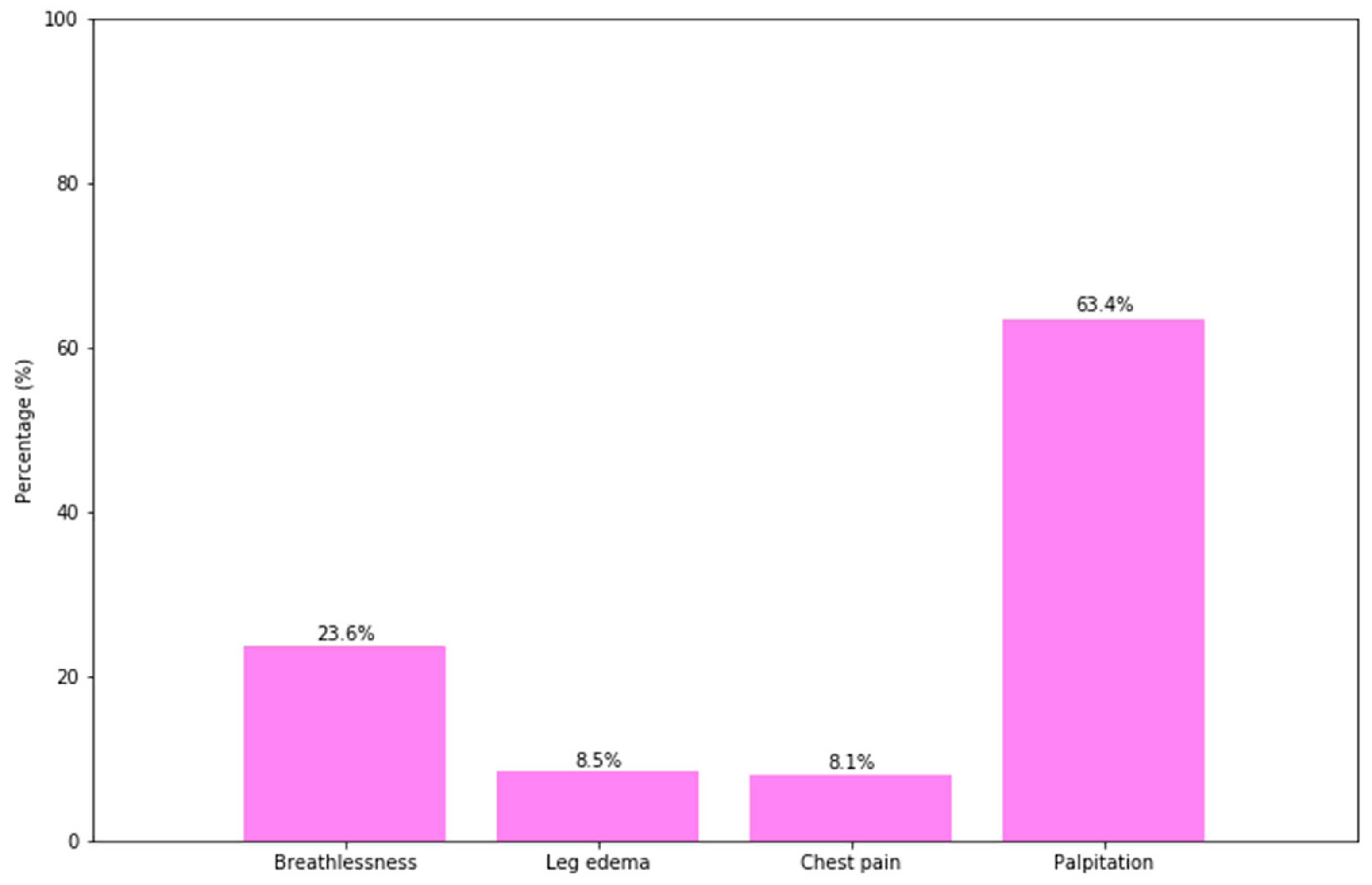
| Clinic presentation | |||||
| No | 8 (8.42%) | 87 (91.58%) | 0.831 p | 1 | - |
| Yes | 18 (9.52%) | 171 (90.48%) | 0.59 | 0.19–1.8 | |
| Hypertension | |||||
| No | 22 (7.97%) | 254 (92.03%) | 0.003 F | 1 | - |
| Yes | 4 (50.00%) | 4 (50.00%) | 59.75 *** | 9.1–392.17 | |
| Congenital heart diseases | |||||
| ASD | |||||
| No | 19 (8.02%) | 218 (91.98%) | 0.135 p | 1 | - |
| Yes | 7 (14.89%) | 40 (85.11%) | 4.27 * | 1.19–15.29 | |
| VSD | |||||
| No | 23 (10.04%) | 206 (89.96%) | 0.435 F | 1 | - |
| Yes | 3 (5.45%) | 52 (94.55%) | 0.28 | 0.05–1.7 | |
| Tetralogy of fallot | |||||
| No | 23 (8.55%) | 246 (91.45%) | 0.148 F | 1 | - |
| Yes | 3 (20.00%) | 12 (80.00%) | 6.82 * | 1.21–38.55 | |
| Complications | |||||
| Heart failure | |||||
| No | 15 (6.36%) | 221 (93.64%) | 0.000 p | 1 | - |
| Yes | 11 (22.92%) | 37 (77.08%) | 10.34 ** | 2.75–38.87 | |
| Pulmonary edema | |||||
| No | 23 (8.21%) | 257 (91.79%) | 0.003 F | 1 | - |
| Yes | 3 (75.00%) | 1 (25.00%) | 107.16 ** | 4.96–2313.93 | |
| Heart arrhythmia | |||||
| No | 21 (9.17%) | 208 (90.83%) | 1 F | 1 | - |
| Yes | 5 (9.09%) | 50 (90.91%) | 2.65 | 0.58–12.17 | |
© 2019 by the authors. Licensee MDPI, Basel, Switzerland. This article is an open access article distributed under the terms and conditions of the Creative Commons Attribution (CC BY) license (http://creativecommons.org/licenses/by/4.0/).
Share and Cite
Nguyen Manh, T.; Bui Van, N.; Le Thi, H.; Vo Hoang, L.; Nguyen Si Anh, H.; Trinh Thi Thu, H.; Nguyen Xuan, T.; Vu Thi, N.; Minh, L.B.; Chu, D.-T. Pregnancy with Heart Disease: Maternal Outcomes and Risk Factors for Fetal Growth Restriction. Int. J. Environ. Res. Public Health 2019, 16, 2075. https://0-doi-org.brum.beds.ac.uk/10.3390/ijerph16122075
Nguyen Manh T, Bui Van N, Le Thi H, Vo Hoang L, Nguyen Si Anh H, Trinh Thi Thu H, Nguyen Xuan T, Vu Thi N, Minh LB, Chu D-T. Pregnancy with Heart Disease: Maternal Outcomes and Risk Factors for Fetal Growth Restriction. International Journal of Environmental Research and Public Health. 2019; 16(12):2075. https://0-doi-org.brum.beds.ac.uk/10.3390/ijerph16122075
Chicago/Turabian StyleNguyen Manh, Thang, Nhon Bui Van, Huyen Le Thi, Long Vo Hoang, Hao Nguyen Si Anh, Huong Trinh Thi Thu, Thuc Nguyen Xuan, Nga Vu Thi, Le Bui Minh, and Dinh-Toi Chu. 2019. "Pregnancy with Heart Disease: Maternal Outcomes and Risk Factors for Fetal Growth Restriction" International Journal of Environmental Research and Public Health 16, no. 12: 2075. https://0-doi-org.brum.beds.ac.uk/10.3390/ijerph16122075