Article

Survival of Microorganisms on Nonwovens Used for the Construction of Filtering Facepiece Respirators

by
Katarzyna Majchrzycka
1,
Małgorzata Okrasa
1,*,
Justyna Szulc
2,
Anita Jachowicz
2 and
Beata Gutarowska
2
1
Department of Personal Protective Equipment, Central Institute for Labour Protection—National Research Institute, Wierzbowa 48, 90-133 Łódź, Poland
2
Institute of Fermentation Technology and Microbiology, Faculty Biotechnology and Food Science, Lodz University of Technology, Wólczańska 171/173, 90-924 Łódź, Poland
*
Author to whom correspondence should be addressed.
Int. J. Environ. Res. Public Health 2019, 16(7), 1154; https://0-doi-org.brum.beds.ac.uk/10.3390/ijerph16071154
Submission received: 15 February 2019 / Revised: 25 March 2019 / Accepted: 27 March 2019 / Published: 31 March 2019
(This article belongs to the Special Issue Environmental and Occupational Exposure to Microbial Contaminants)

Abstract

:
Filtering nonwovens that constitute the base material for filtering facepiece respirators (FFRs) used for the protection of the respiratory system against bioaerosols may, in favourable conditions, promote the development of harmful microorganisms. There are no studies looking at the impact that different types of filtering nonwovens have on microorganism survival, which is an important issue for FFR producers and users. Five commercial filtering nonwovens manufactured using diverse textile technologies (i.e., needle-punching, melt-blown, spun-bonding) with different structural parameters and raw material compositions were used within our research. The survival of microorganisms on filtering nonwovens was determined for E. coli, S. aureus, B. subtilis bacteria; C. albicans yeast and A. niger mould. Samples of nonwovens were collected immediately after inoculum application (at 0 h) and after 4, 8, 24, 48, 72, and 96 h of incubation. The tests were carried out in accordance with the AATCC 100-1998 method. Survival depended strongly on microorganism species. E. coli and S. aureus bacteria grew the most on all nonwovens tested. The structural parameters of the nonwovens tested (mass per unit area and thickness) and contact angle did not significantly affect microorganism survival.

1. Introduction

The ability to prevent the harmful impact of biological agents on humans depends largely on the efficacy and quality of the protection method or equipment used. Given that a significant fraction of pathogenic microorganisms is transmitted by dust or droplets, filtering facepiece respirators (FFRs), whose base material are filtering nonwovens, are an important method of protection. The use of respiratory protecting equipment of this type is common in many workplaces. The deployment of FFRs for work-unrelated applications is also becoming more common [1,2]. This is due to the rapidly spreading epidemics of influenza and other infectious diseases, as well as due to an increased public awareness concerning environmental threats.
Protection against biological agents is particularly important in health care, where the use of surgical respirators (SRs), which are manufactured from filtering nonwovens, is common. SRs have been used for over 100 years to protect patients from droplet-transmitted infections. Moreover, SRs are frequently perceived as personal protection equipment for medical personnel, although their protective efficacy is at a relatively low level compared to standard FFRs.
Filtering facepiece respirators have to comply with European standard requirements as a product ensuring human safety [3]. Unfortunately, the standards do not define requirements or research methods that would consider the specifics of biological factors. In particular, there are no standards for the assessment of microorganism survival rate on filtering material. Therefore, despite numerous research and development studies aimed at conferring biocidal properties to filtering nonwovens [4,5,6,7,8,9,10,11,12,13,14,15], there are no FFRs on the market that guarantee restricted growth of microorganisms accumulated on the filtering material during use. At the same time, many studies [16,17,18,19,20,21,22,23] confirmed that during prolonged FFR use against bioaerosols, bacterial numbers may increase, and biofilms may form on the filtering nonwoven, which may become a potential threat for the user. Likewise, it was found that during influenza A (H1N1) pandemics, viruses and microorganisms survived on the filtering material of SRs used by the personnel for several hours to several days [24,25,26]. Therefore, the knowledge of factors affecting dynamics of microorganism growth on filtering materials is particularly important to the FFR manufacturer and user.
Moisture and mineral dust from work environments may promote microorganism growth within filtering nonwovens [21,22,23]. Based on breathing simulation, Majchrzycka et al confirmed that moisture from exhaled air, which accumulate within FFRs creates favourable conditions for microorganism growth [21]. Such conditions persist over time when FFRs are in use, even if there are breaks. Environmental conditions have a significant impact on the survival of some microorganism species on nonwovens. There is a dearth of research concerning the influence of the type of filtering nonwovens on this phenomenon.
Filtering facepiece respirators are multilayer products. They consist of a thin layer of a protective nonwoven on both sides, i.e., both on the side exposed to bioaerosol influx from the working environment (external part of the equipment) and the one in contact with the user’s face (inside of the equipment). Its mass per unit area is usually between 20 and 50 g/m2. The next layer is a needled nonwoven responsible for pre-filtration. Its mass per unit area can be as high as 250 g/m2. It is often subjected to a high temperature calendering process that thickens the nonwoven structure and gives it rigidity. This helps form the appropriate facepiece shape and ensures that it does not change over time when the FFR is in use. The high efficiency melt-blown electret nonwoven (formed by blowing the melted polymer) constitutes the most important layer. It is responsible for the efficiency of filtration. Due to this function, it is usually an electret nonwoven (electrified by corona discharge). Nonwovens made of polypropylene (PP) are widely used in FFRs; albeit those made of polyacrylonitrile (PAN) or polyethylene (PE) also exist.
Mass per unit area, thickness, porosity, and diameter of elementary fibres are the basic parameters determining the properties of the nonwovens used for FFR construction. All of these can contribute to the survival of microorganisms on the filtering material. The hydrophobicity of the elementary fibre is another factor that may affect microorganism survival. Therefore, the aim of this study was to assess the survival of selected bacteria, yeasts, and moulds (E. coli, S. aureus, B. subtilis, C. albicans and A. niger) on five types of filtering nonwovens most often used for FFR construction over a duration corresponding to the time of use on consecutive working days. In addition, the article discusses a correlation between microorganism survival and the mass per unit area and thickness of the nonwoven and its contact angle.

2. Materials and Methods

2.1. Filtering Nonwovens

Five filtering nonwovens typically used for FFR construction described in Table 1 were used within the study.

2.2. Assessment of the Survival of Microorganisms on Nonwovens

The microorganisms stored in the Pure Culture Collection ŁOCK 105 were used to study survival on filtering nonwovens (Table 2). The selected microorganisms belonged to various taxonomic groups (bacteria, yeasts, moulds) and were characterized by diverse physiology of growth.
Inocula of bacteria and yeast were prepared by inoculating 20 ml of sterile tryptic soy broth (TSB, Merck, Kenilworth, NJ, USA) or malt extract broth (MEB, Merck) medium for bacteria or yeast, respectively. Inoculum suspensions were incubated at 30 ± 2 °C for 24 h. Mould inoculum was obtained by washing the spores of malt extract agar (MEA, Merck) slants containing 7-day A. niger cultures using MEB medium. In this way, microorganism suspensions of 3.79 × 10⁷–1.12 × 10⁹ CFU/mL density were obtained (Table 2). Then, 25 µL of the suspension inocula were applied homogenously onto the UV-disinfected nonwoven swatches of 4 cm2 (2 × 2 cm squares) surface area using pipette with sterile tips for the distribution of small water droplets over the whole samples. The swatches were then placed in sterile Petri dishes and incubated in Binder-720 climatic chamber at 28 ± 2 °C and relative humidity of 80%.
Quantitative static AATCC 100-1998 “Antimicrobial Finishes of Textile Materials” method was used to determine microorganism survival on the nonwovens [27]. Test samples were taken immediately after inoculum application (time 0h) and after 4, 8, 24, 48, 72 and 96 h of incubation. The test was performed according to methodology described in [21].
Nine results obtained in Grubbs test were selected for statistical analysis, after discarding uncertain results. Arithmetic mean and standard deviation of the number of microorganisms grown on the plates over consecutive hours of the experiment were calculated using Microsoft® Excel. In addition, statistical differences between microorganism number at t = 0 and after 4, 8, 24, 48, 72 and 96 h, and between the number of microorganisms over consecutive incubation hours were calculated (one-way analysis of variance (ANOVA), significance level α = 0.05). The normality was checked with Shapiro-Wilk test, which was followed by Levene’s test to assess the equality of variances. Statistically significant differences in microorganism number on different types of nonwovens incubated for the same length of time (one- and two-way ANOVA, significance level α = 0.05) were determined using the STATISTICA 13.1 software (StatSoft, Kraków, Poland). Ultimately, the results were compared using Tukey test (significance level α = 0.05). Microorganism survivability (N) was calculated after 4, 8, 24, 48, and 96 h of incubation as a ratio of the number of microorganisms after a given incubation time to the initial number of microorganisms.

2.3. Structural Parameters and Contact angle Determination

The mass per unit area of nonwovens was determined according to methodology described in EN 29073-1:1992 standard [28] using R160P laboratory balance (Sartorius GMBH, Goettingen, Germany). Their thickness was ascertained according to ISO 5084:1996 [29] using the J-40-T digital material thickness gauge (CheckLine, Bad Bentheim, Germany). The contact angle was determined using the Phoenic-Alpha contact angle apparatus (SEO, Suwon-si, Republic of Korea) equipped with an attachment for measuring contact angle at low and high temperatures. During the test, a drop of liquid was dispensed onto the sample surface using a syringe with a needle. The image of the drop was recorded using a high-resolution camera and then exported to ImageJ software for contact angle measurement.

3. Results and Discussion

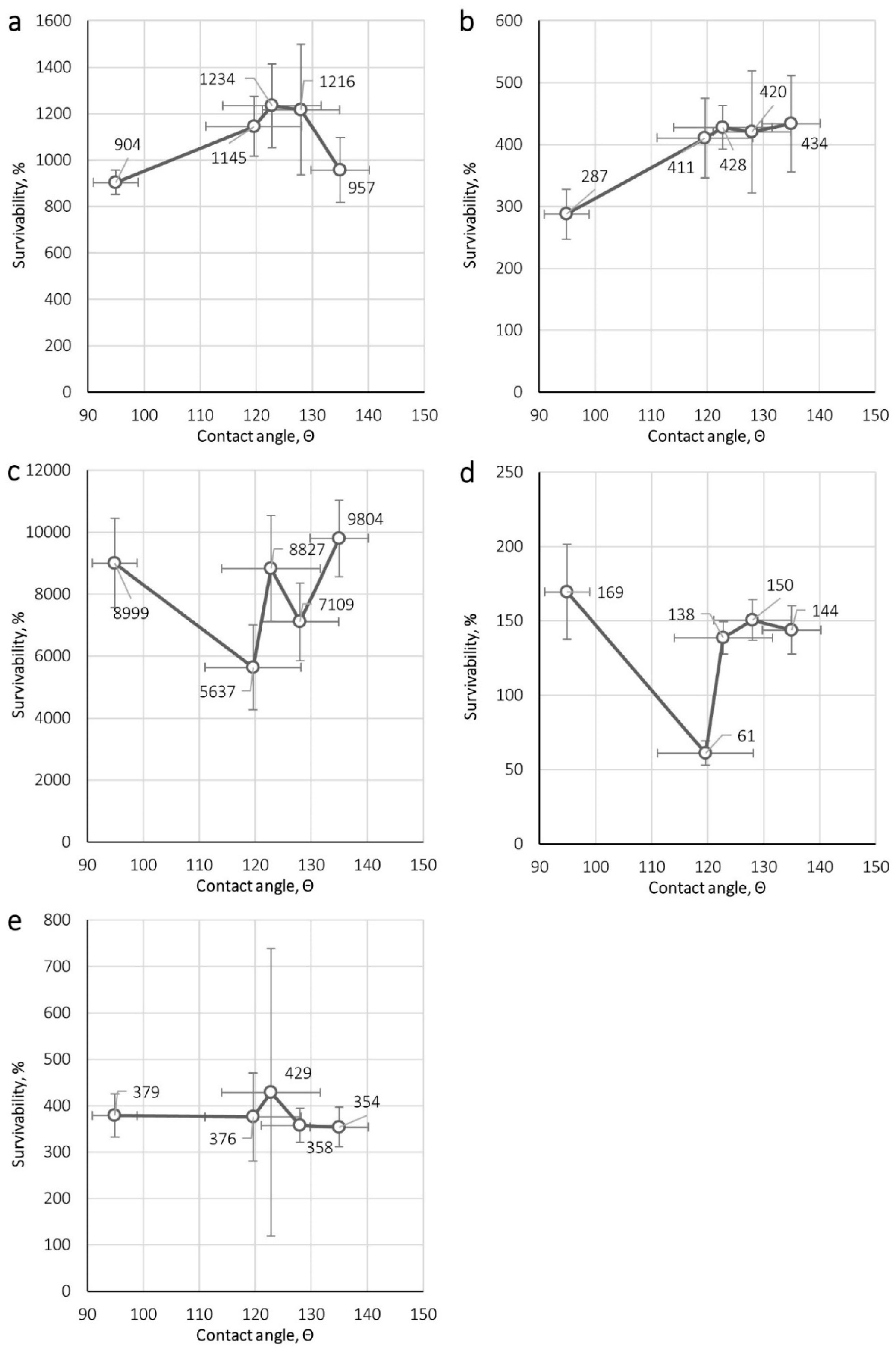
3.1. Assessment of the Survival of Microorganisms on Nonwovens

The number of microorganisms on the filtering nonwovens studied at 0, 4, 8, 24, 48 and 96 h of incubation is shown in Table S1, and microorganism survival at subsequent time points in Figure 1.
The number of E. coli bacteria on the nonwovens varied 5.07 × 10⁵–7.54 × 10⁶ CFU/sample depending on the nonwoven type and incubation time. Statistically significant differences were observed in the number of E. coli bacteria between the control sample (t = 0 h) and each subsequent incubation time for all nonwovens studied. Statistically significant differences were also seen in the number of E. coli bacteria on nonwovens A, B and E at incubation times of 8, 24, 72 and 96 h compared to the remaining samples.
The number of S. aureus bacteria on the nonwovens equalled 5.33 × 10⁵–2.45 × 10⁶ CFU/sample depending on the nonwoven type and incubation time. Statistically significant differences were observed in the number of S. aureus bacteria between the control sample (t = 0 h) and each subsequent time point for all nonwovens studied except for nonwovens A and D at 4 h of incubation. When comparing bacterial numbers for consecutive incubation times, statistically significant differences were observed for nonwovens A and E and nonwoven B following 8 h; nonwoven E and other nonwovens tested after 24 h; and for nonwovens E and A after 96 h incubation.
The number of B. subtilis bacteria on the nonwovens studied equalled 1.47 × 10⁴–1.43 × 10⁶ CFU/sample depending on the nonwoven type and incubation time, depending on the incubation time. Statistically significant differences were observed in bacterial numbers between the control sample (t = 0 h) and after 8, 24, 58, 72 and 96 h of incubation for each nonwoven studied. When comparing bacterial numbers for consecutive incubation times, statistically significant differences were observed for nonwoven B and other nonwovens following 72 h of incubation, and for nonwovens B and D following 96 h incubation.
The number of C. albicans yeast on the studied nonwovens was in the range 3.02 × 10⁴–5.49 × 10⁴ CFU/sample depending on the nonwoven type and the incubation time. In all nonwovens, the number of C. albicans increased with the length of incubation time. An exception was nonwoven B, where a gradual decrease was seen. Such changes were not always statistically significant. When comparing yeast numbers on nonwoven types for consecutive incubation times, statistically significant differences were observed between nonwovens A and B, and between nonwovens B and C following 24 h of incubation. In addition, statistically significant differences were also observed between nonwoven B and all the others following 48, 72, and 96 h of incubation.
The number of A. niger mould on the studied nonwovens equalled 5.00 × 10³–6.62 × 10⁴ CFU/sample depending on the nonwoven type and incubation time depending on the incubation time. In all nonwovens, the number of A. niger was the lowest after 24 h of incubation and subsequently increased, reaching a peak after 96 h of incubation. No statistically significant differences in mould numbers were detected for consecutive incubation times for the various types of nonwovens.
E. coli survival rate on the nonwovens ranged from 394 to 1234%. This index reached the highest values (904–1234%) after 24 h and the lowest (394–595%) after 4 and 96h of incubation. The survival rate of S. aureus bacteria was lower and between 119–434%. Depending on the type of nonwoven, the index reached the highest values (156–434%) following 8, 24, and 48 h of incubation. The lowest survival rate (119–139%) of S. aureus was recorded after 4h of incubation. The survival rate of B. subtilis was 105–9804%. The index reached the highest value (5637–9804%) after 72 h except for nonwoven B, for which the highest survival was documented after 48h of incubation (7539%). The lowest B. subtilis survival was recorded after 4 h of incubation for all nonwovens tested. For C. albicans yeast, the survival rate was in the range of 41–169%. This index reached the highest value (138–169%) after 72 h of incubation except for nonwoven B, for which the highest survival rate (109%) was found after 4 h. The lowest survival rate for C. albicans (101%–116%) was recorded after 4h incubation for nonwovens A, C, D and E and after 96h (41%) for nonwoven B. The survival rate of A. niger mould was 25–429%. The index reached the highest values (354–429%) after 96 h incubation, and the lowest (25–45%) after 24 h.
The survival of microorganisms on the nonwovens tested was the highest in the case of bacteria B. subtilis (105–9804%), E. coli (394–1234%) and S. aureus (119–439%). It was lower for of A. niger mould (25–429%) and lowest for C. albicans yeast (41–169%). The maximal survival rate was achieved the quickest (at 24h of incubation) by E. coli (904–1234%). S. aureus bacteria achieved maximal survival rate very quickly, as well (312–434% after 8, 24 and 48 h depending on the nonwoven). In contrast, the maximal survival rate for spore-forming B. subtilis bacteria (7109–9804%) and C. albicans yeast (138–1169%) was achieved relatively slowly, only after 72 h. Finally, A. niger moulds achieved maximal survival rate the slowest (354–429% after 72 and 96 h).
Statistical analysis of bacteria number for the same incubation times showed that nonwovens A (PP/PEL, needle-punched nonwoven), C (PPQ, corona charged melt-blown nonwoven) and D (PP, spun-bonded nonwoven) favoured growth the most. B. subtilis bacteria (after 72 h incubation) grew the least on nonwoven B (PET, needle-punched nonwoven). For S. aureus (after 8 h incubation) and E. coli (after 72 and 96 h) bacteria, reduced survival on nonwoven E (PP, calandered needle-punched nonwoven) was noted. In comparison to the other nonwovens tested, we found that nonwoven B was least favourable for C. albicans yeast growth. The decrease in survival rate from 108% (after 4 h of incubation) and to 41% (after 96 h) indicates a gradual dying of yeast on this nonwoven. The difference in the survival rate of A. niger was not statistically significant for all nonwovens tested.

3.2. Nonwovens Structural Parameters and Contact Angle

The results of mass per unit area, thickness and contact angle measurements for the nonwovens are presented in Table 3.
Figure 2 and Figure 3 show the correlation between microorganism survival on nonwovens and their mass per unit area, thickness and contact angle. The correlations were determined for the maximal survival achieved for each microorganism, i.e., 24 h for E. coli and S. aureus, 72 h for B. subtilis and yeast C. albicans, and 96 h for A. niger.
A particularly important conclusion from our research is the lack of a clear relationship between the degree of fibre wettability (evaluated by the contact angle) and their structural parameters, and the tendency for the microorganism to grow on their surface. In none of the cases, was there an unequivocal relationship between the structural parameters of a nonwoven and microorganism survival (Figure 2). It should be emphasized that from the point of view of filtration efficiency of biological particles, these parameters are of utmost importance. Based on the assumption that more effective filtering materials would accumulate more particles favouring microorganism growth, we used nonwovens endowed with significantly different mass per unit area and thickness, including a highly effective electret melt-blown nonwoven, for testing. However, contrary to expectations, even in the case of nonwovens made from the same raw material (nonwovens C, D and E made of PP), both downward (for S. aureus, B. subtilis bacteria and C. albicans yeast) and upward (for E. coli bacteria and A. niger mould) trends in microorganism survival were observed. This property is therefore not related to the structural parameters of nonwovens, and hence to their filtration efficiency.
The contact angle measurement results (Table 3) showed that it ranged from 94 to 135, indicating that the wettability was poor for all filtering nonwovens. The correlations depicted in Figure 3 show that there were no significant differences between microorganism survival and the determined contact angle. On the other hand, the conclusions of previously published studies [18,19,20,21], indicated that the moisture content in the filtering nonwovens (the total amount of water accumulated in the fibrous structure) has a dominant influence on microorganism survival. Taking the above into consideration, it can be stated that the characteristics linked to fibre wettability (such as chemical nature of the fibre surface and the fibre geometry) are less important in the context of microbial growth than the nonwoven’s ability to accumulate moisture under real-use conditions, which would depend not only on the wettability of the nonwoven but also on the characteristics of the fibrous structure and external conditions (e.g., relative humidity or the flow pattern and velocity of the passing air). Of course, to fully support this hypothesis experiments that more adequately mimic real-use conditions should be conducted, including the studies of the influence of such factors as ambient temperature and humidity conditions, content of inorganic or organic dust as well as the presence, acidic, or alkaline sweat on the microorganism’s survival on nonwoven materials used for construction of FFRs.
Commercial FFRs used at workplaces consist mostly of filtering nonwovens described within the study. In particular, this concerns high-efficiency PP melt-blown nonwovens, which are the basic structural material of FFRs. Translating the results of laboratory tests into the actual use, it should be stated that the more effective the FFRs are, the more microorganisms will accumulate inside them. Their growth can constitute a danger to the user [21,22,23]. Therefore, the use of standard FFRs throughout the working shift for protection against biological agents does not guarantee a sufficient protection of the user. A much safer solution is the application of nonwovens with biocidal properties to the construction of FFRs [4,5,6,7,12,13]. However, such FFRs are not widely available. Another option is the frequent replacement of standard FFRs or their disinfection.

4. Conclusions

Microorganism growth on all filtering nonwovens utilized for FFR construction was confirmed. Their survival on pure nonwovens strongly depended on the species. The highest survival rate over short incubation time was achieved by E. coli and S. aureus bacteria. Maximal survival was achieved at later time points by spore-forming B. subtilis bacteria and C. albicans yeast, while A. niger moulds took the longest, i.e., after 96 hours. These differences stemmed from the growth physiology of the microorganisms studied. Microorganism survival rate is negligibly impacted by the type of filtering nonwoven, as there was no obvious relationship between survival and nonwoven characteristics (mass per unit area, thickness and contact angle). Based on our research, we also established that the composition of nonwovens did not significantly affect microorganism survival.

Supplementary Materials

The following are available online at https://0-www-mdpi-com.brum.beds.ac.uk/1660-4601/16/7/1154/s1. Table S1: Number of microorganisms on the nonwovens depending on the incubation time.

Author Contributions

Conceptualization: K.M., M.O. and B.G.; Data curation: M.O. and A.J.; Investigation: M.O., J.S. and A.J.; Methodology: K.M., M.O. and B.G.; Supervision: K.M. and B.G.; Visualization: M.O.; Writing—original draft: K.M., M.O. and J.S.; Writing—review & editing: K.M., M.O., J.S., A.J. and B.G.

Funding

The publication is based on the results of a research task carried out within the scope of the fourth stage of the National Programme “Improvement of safety and working conditions” partly supported in 2017–2019—within the scope of research and development—by the Ministry of Science and Higher Education/National Centre for Research and Development. The Central Institute for Labour Protection—National Research Institute is the Programme’s main coordinator.

Acknowledgments

We would like to express our gratitude to Agnieszka Adamus-Włodarczyk, PhD (TUL, CIOP-PIB) for technical assistance during this research.

Conflicts of Interest

The authors declare no conflict of interest. The funders had no role in the design of the study; in the collection, analyses, or interpretation of data; in the writing of the manuscript, and in the decision to publish the results.

References

  1. Rubino, I.; Choi, H.-J. Respiratory Protection against pandemic and epidemic diseases. Trends Biotechnol. 2017, 35, 907–910. [Google Scholar] [CrossRef] [PubMed]
  2. Coulliette, A.D.; Perry, K.A.; Edwards, J.R.; Noble-Wang, J.A. Persistence of the 2009 pandemic influenza A (H1N1) virus on N95 respirators. Appl. Environ. Microbiol. 2013, 79, 2148–2155. [Google Scholar] [CrossRef] [PubMed]
  3. European Standard EN 149. Respiratory Protective Devices. Filtering Half Masks to Protect against Particles. Requirements, Testing, Marking; EN 149:2001+A1:2009; European Committee for Standardization: Bruxelles, Belgium, 2009. [Google Scholar]
  4. Majchrzycka, K.; Gutarowska, B.; Brochocka, A.; Brycki, B. New filtering antimicrobial nonwovens with various carriers for biocides as respiratory protective materials against bioaerosol. Int. J. Occup. Saf. Ergon. 2012, 3, 375–385. [Google Scholar] [CrossRef] [PubMed]
  5. Gutarowska, B.; Skóra, J.; Nowak, E.; Łysiak, I.; Wdówka, M. Antimicrobial activity and filtration effectiveness of nonwovens with Sanitized for respiratory protective equipment. Fibres Text. East. Eur. 2014, 22, 120–125. [Google Scholar]
  6. Gutarowska, B.; Michalski, A. Antimicrobial activity of filtrating meltblown nonwoven with the additions of silver ions. Fibres Text. East. Eur. 2009, 3, 23–28. [Google Scholar]
  7. Brochocka, A.; Majchrzycka, K. Technology for the production of bioactive melt-blown filtration materials applied to respiratory protective devices. Fibres Text. East. Eur. 2009, 5, 92–98. [Google Scholar]
  8. Rengasamy, S.; Fisher, E.; Shaffer, R.E. Evaluation of the survivability of MS2 viral aerosols deposited on filtering face piece respirator samples incorporating antimicrobial technologies. Am. J. Infect. Control 2010, 38, 9–17. [Google Scholar] [CrossRef]
  9. Kamiyama, Y.; Adachi, K.; Handharyani, E.; Soejoedono, R.D.; Kusano, T.; Inai, M.; Tsukamoto, M.; Kashiwagi, S.; Tsukamoto, Y. Protection from avian influenza H5N1 virus infection with antibody-impregnated filters. Virol. J. 2011, 8, 54. [Google Scholar] [CrossRef] [PubMed]
  10. Quan, F.S.; Rubio, I.; Lee, S.-H.; Koch, B.; Choi, H.-J. Universal and reusable virus deactivation system for respiratory protection. Sci. Rep. 2017, 7, 39956. [Google Scholar] [CrossRef] [Green Version]
  11. Nicas, M.; Best, D. A study quantifying the hand-to-face contact rate and its potential application to predicting respiratory tract infection. J. Occup. Environ. Hyg. 2008, 5, 347–352. [Google Scholar] [CrossRef]
  12. Majchrzycka, K.; Okrasa, M.; Szulc, J.; Brycki, B.; Gutarowska, B. Time-dependent antimicrobial activity of filtering nonwovens with gemini surfactant-based biocides. Molecules 2017, 22, 1620. [Google Scholar] [CrossRef] [PubMed]
  13. Majchrzycka, K.; Okrasa, M.; Brycki, B.; Skóra, J.; Gutarowska, B. Application of bioactive porous structures with time-dependent activity into high-efficiency filtering melt-blown nonwovens. Przem. Chem. 2017, 96, 534–538. [Google Scholar]
  14. Li, Y.; Leung, P.; Yao, L.; Song, Q.W.; Newton, E. Antimicrobial effect of surgical masks coated with nanoparticles. J. Hosp. Infect. 2006, 62, 58–63. [Google Scholar] [CrossRef] [PubMed]
  15. Tseng, C.C.; Pan, Z.-M.; Chang, C.-H. Application of a quaternary ammonium agent on surgical face masks before use for pre-decontamination of nosocomial infection-related bioaerosols. Aerosol Sci. Technol. 2016, 50, 199–210. [Google Scholar] [CrossRef] [Green Version]
  16. Szulc, J.; Otlewska, A.; Okrasa, M.; Majchrzycka, K.; Sulyok, M.; Gutarowska, B. Microbiological contamination at workplaces in a combined heat and power (CHP) station processing plant biomass. Int. J. Environ. Res. Public Health 2017, 14, 99. [Google Scholar] [CrossRef]
  17. Bonnevie Perrier, J.C.; Le Coq, L.; Andres, Y.; Le Cloire, P. SFGP 2007—Microbial growth onto filter media used in air treatment devices. Int. J. Chem. React. Eng. 2008, 6. [Google Scholar] [CrossRef]
  18. Brosseau, L.M.; McCullough, N.V.; Vesley, D. Bacterial survival on respirator filters and surgical masks. Appl. Biosaf. 1997, 2, 232–243. [Google Scholar] [CrossRef]
  19. Maus, R.; Goppelsroder, A.; Umhauer, H. Survival of bacterial and mold spores in air filter media. Atmos. Environ. 2001, 35, 105–113. [Google Scholar] [CrossRef]
  20. Pasanen, A.L.; Keinanen, J.; Kalliokoski, P.; Martikainen, P.I.; Ruuskanen, J. Microbial growth on respirator filters from improper storage. Scand. J. Work Environ. Health 1993, 19, 421–425. [Google Scholar] [CrossRef]
  21. Majchrzycka, K.; Okrasa, M.; Skóra, J.; Gutarowska, B. Evaluation of the survivability of microorganisms deposited on filtering respiratory protective devices under varying conditions of humidity. Int. J. Environ. Res. Public Health 2016, 13, 98. [Google Scholar] [CrossRef]
  22. Majchrzycka, K.; Okrasa, M.; Szulc, J.; Gutarowska, B. The impact of dust in filter materials of respiratory protective devices on the microorganisms viability. Int. J. Ind. Ergon. 2017, 58, 109–116. [Google Scholar] [CrossRef]
  23. Majchrzycka, K.; Okrasa, M.; Jachowicz, A.; Szulc, J.; Gutarowska, B. Microbial growth on dust-loaded filtering materials used for the protection of respiratory tract as a factor affecting filtration efficiency. Int. J. Environ. Res. Public Health 2018, 15, 1902. [Google Scholar] [CrossRef] [PubMed]
  24. Lai, A.C.K.; Poon, C.K.M.; Cheung, A.C.T. Effectiveness of facemasks to reduce exposure hazard for airborne infections among general populations. JRC Soc. Interface 2012, 9, 938–948. [Google Scholar] [CrossRef] [PubMed]
  25. Makison Booth, C.; Clayton, M.; Crook, B.; Gawn, J.M. Effectiveness of surgical masks against influenza bioaerosol. J. Hosp. Infect. 2013, 84, 22–26. [Google Scholar] [CrossRef] [PubMed]
  26. Sublett, J.L. Effectiveness of air filters and air cleaners in allergic respiratory diseases: A review of the recent literature. Curr. Allergy Asthma Rep. 2011, 11, 395–402. [Google Scholar] [CrossRef] [PubMed]
  27. AATCC Test Method 100-2004. Antibacterial Finishes on Textile Materials: Assessment of Antibacterial Finishes on Textile Materials. Technical Manual/2010. 2004. Available online: http://www. manufacturingsolutionscenter.org/aatcc-100-antibacterial-finishes-textile.html (accessed on 14 February 2019).
  28. European Standard EN 29073-1:1992. Methods of Test for Nonwovens. Methods of Test for Nonwovens. Determination of Mass per Unit Area; European Committee for Standardization: Bruxelles, Belgium, 1992. [Google Scholar]
  29. European Standard EN ISO 5084:1996. Textiles. Determination of Thickness of Textiles and Textile Products; European Committee for Standardization: Bruxelles, Belgium, 1996. [Google Scholar]
Figure 1. Survivability of microorganisms on nonwovens depending on incubation time; (a) E. coli, (b) S. aureus, (c) B. subtilis, (d) C. albicans, (e) A. niger.
Ijerph 16 01154 g001
Figure 2. Dependence of the survivability of microorganisms on nonwovens on the mass per unit area (left) and the thickness of nonwoven (right) for (a) E. coli, (b) S. aureus, (c) B. subtilis, (d) C. albicans, (e) A. niger.
Ijerph 16 01154 g002
Figure 3. Dependence of the survivability of microorganisms on nonwoven contact angle for (a) E. coli, (b) S. aureus, (c) B. subtilis, (d) C. albicans, (e) A. niger.
Ijerph 16 01154 g003
Table 1. Characteristics of filtering nonwovens.
NotationRaw Material CompositionType of NonwovenFunction in FFR
Apolypropylene/polyacrylonitrile (PP/PEL)needle-punched
nonwoven
stiffening of the FFR
structure, pre-filtration of coarse dust particles
Bpolyethylene (PET)
Cpolypropylene (PPQ)corona charged
melt-blown nonwoven
high-efficiency filtration of fine dust particles
Dpolypropylene (PP)spun-bonded
nonwoven
pre-filtration of coarse dust particles
Epolypropylene (PP)calandered needle-punched nonwovenstiffening of the FFR
structure,
FFR: filtering facepiece respirator.
Table 2. Characteristics of microorganisms.
MicroorganismsSpeciesCollection Reference NumberInoculum Density, CFU/mL
BacteriaEscherichia coliATCC 105361.12 × 109 ± 4.06 × 10⁸
Staphylococcus aureusATCC 65381.05 × 109 ± 3.60 × 10⁸
Bacillus subtilisNCAIM 016446.67 × 108 ± 1.06 × 10⁸
FungiYeastCandida albicansATCC 102311.32 × 108 ± 1.93 × 10⁷
MouldAspergillus nigerATCC 164043.70 × 107 ± 8.87 × 10⁶
ATCC: American Type Culture Collection; NACIM: National Collection of Agricultural and Industrial Microorganisms.
Table 3. Structural parameters and contact angle of nonwovens.
Type of NonwovenMass per unit Area, g/m2Thickness, mmContact Angle, Θ
A142.4 ± 8.82.72 ± 0.16128.0 ± 6.9
B174.9 ± 6.32.79 ± 0.14119.6 ± 8.6
C104.5 ± 7.81.59 ± 0.12135.0 ± 5.2
D15.1 ± 1.50.20 ± 0.01122.8 ± 8.8
E85.0 ± 9.60.69 ± 0.0294.9 ± 4.0

