Article

In Vitro Anti-Leptospiral Activity of Phyllanthus amarus Extracts and Their Combinations with Antibiotics

by
Che Ain Munirah Ismail
1,
Zakuan Zainy Deris
1,2,
Ruzilawati Abu Bakar
2,3 and
Nabilah Ismail
1,2,*
1
Department of Medical Microbiology & Parasitology, School of Medical Sciences, Health Campus, Universiti Sains Malaysia, Kubang Kerian, Kelantan 16150, Malaysia
2
Hospital Universiti Sains Malaysia, Universiti Sains Malaysia Kampus Kesihatan, Jalan Raja Perempuan Zainab 2, Kota Bharu, Kelantan 16150, Malaysia
3
Department of Pharmacology, School of Medical Sciences, Health Campus, Universiti Sains Malaysia, Kubang Kerian, Kelantan 16150, Malaysia
*
Author to whom correspondence should be addressed.
Int. J. Environ. Res. Public Health 2021, 18(6), 2834; https://0-doi-org.brum.beds.ac.uk/10.3390/ijerph18062834
Submission received: 15 February 2021 / Revised: 3 March 2021 / Accepted: 4 March 2021 / Published: 10 March 2021

Abstract

:
Despite modern medicine, there is an increasing trend for cases of the bacterial infection leptospirosis, and this has led to the exploration of alternative medicines from various sources including plants. The aim of this study was to investigate the in vitro anti-leptospiral activity of Phyllanthus amarus extracts alone and combined with penicillin G, ceftriaxone, and doxycycline. Antimicrobial susceptibility testing was performed using the microdilution broth technique upon methanol extract (ME), aqueous extract (AE), and antibiotics against the Leptospira interrogans serovars Australis, Bataviae, Canicola, and Javanica, to determine minimum inhibitory concentrations (MICs) and minimum bactericidal concentrations (MBCs). The results were analyzed using an ELISA microplate reader combined with microscopic analysis. Synergy testing using a checkerboard assay was performed to determine the fractional inhibitory concentration index values of extracts combined with antibiotics against leptospires. Scanning electron microscopy (SEM) was used to investigate morphological changes of leptospires caused by potential anti-leptospiral agents alone and combined with antibiotics. The MICs and MBCs for P. amarus extracts ranged from 100 to 400 µg/mL for AEs and from 400 to 800 µg/mL for MEs. Penicillin G was the most effective anti-leptospiral drug, with MICs and MBCs ranging from <0.01 to 0.78 and <0.01 to 3.13 µg/mL, respectively, followed by ceftriaxone, with both MICs and MBCs ranging from 0.05 to 0.78 µg/mL, and doxycycline, with MICs and MBCs ranging from 0.39 to 3.13 µg/mL and 12.5 to 25 µg/mL, respectively. Combinations of P. amarus extracts and antibiotics did not show synergistic effects on all tested Leptospira serovars, with some combinations demonstrating antagonistic effects. SEM analysis, however, showed distorted Leptospira surfaces. P. amarus AE performed better anti-leptospiral activity than P. amarus ME. The morphological effects of P. amarus extract alone and its combination with antibiotic on Leptospira cells revealed promising anti-leptospiral properties.

1. Introduction

Leptospirosis is a re-emerging bacterial infection causing serious concerns worldwide [1]. It occurs in urban environments of both industrialized and developing countries, as well as in rural areas [2]. Leptospirosis cases are endemic in tropical and subtropical regions, and become epidemic in monsoon seasons and flooding [3]. The incidence is significantly high in countries with warm climates compared to the temperate regions, causing the long survival rate of leptospires in the warm and humid conditions. The World Health Organization reported that leptospirosis cases commonly occur in the countries of South-East Asia, and their magnitude differs between the countries, depending on attitude and awareness of the public health care decision makers [4,5]. It is estimated that 1.03 million annual cases with 58,999 deaths occur worldwide due to leptospirosis [6].
Infection of leptospirosis ranges from mild febrile illness to severe fatal disease. The early phase of the infection (anicteric form) shows symptoms of fever, headache, chills, nausea, malaise, myalgia, vomiting, and conjunctival hyperemia. The severe form of leptospirosis (icteric form of leptospirosis known as Weil’s disease) [7] includes the symptoms of jaundice, myocarditis, meningitis, renal failure, uveitis, multi organ failure, and lethal pulmonary hemorrhage [8]. The increasing incidence of leptospirosis with high mortality rates underlines the need for research and updated information on its management and treatment [1]. As the available therapies for leptospirosis in modern medicine are limited, the search for alternative medicines to replace synthetic ones has attracted increasing interest in traditional medicines. Along with animals and microorganisms, plant-based natural products used in the medicinal field contain a wide range of bioactive components useful for treating various diseases [9]. However, research on natural products that can be used as anti-leptospiral agents is scarce and needs to be advanced urgently.
Phyllanthus amarus, commonly known as black catnip, carry me seed, or windbreaker, found in tropical and subtropical countries worldwide, has been used for treating problems of the stomach, spleen, kidney, liver, and genitourinary system [10]. In Ayurveda, P. amarus is used for treating dysentery, genital infections, gonorrhea, and urinary tract infections [11]. P. amarus has shown a wide range of antimicrobial activity. Methanol extracts (MEs) of P. amarus exert activity against Shigella spp., Vibrio cholera, Bacillus subtilis, Streptococcus pneumonia, S. mutans, S. salivarius, S. pyogenes, Penicillium spp., and Aspergillus niger [12,13,14,15,16]. Oluwafemi and Debiri found that the activity of MEs against the Salmonella serotype Typhi has comparable zone of inhibition to that of ciprofloxacin, a widely used antibiotic for typhoid fever [17]. P. amarus also has anti-leptospiral effects, damaging the DNA of the Leptospira serovars Icterohaemorrhagiae, Canicola, and Pomona [18]. However, research on the anti-leptospiral activity of P. amarus remains limited. Therefore, this study aims to investigate the in vitro anti-leptospiral activity of Phyllanthus amarus extracts alone and combined with penicillin G, ceftriaxone, and doxycycline.

2. Materials and Methods

2.1. Sampling and Plant Extraction

Mature whole P. amarus (Schumach. and Thonn.) plants were harvested from Peringat, Kota Bharu, Kelantan, Malaysia, and sent to the Forest Research Institute Malaysia for identification as PID 150616-14 (Supplementary Figure S1). Whole plants were then washed and dried in the shade, powdered, and stored in dry place at room temperature.
A ME was prepared by adding 30 g of plant powder to 250 mL of methanol (Merck, Darmstadt, Germany), with extraction at 56–60 °C for five hours using a Soxhlet apparatus (Electrothermal, Staffordshire, UK) to achieve optimal oil yield from the plant [19], followed by evaporation to dryness using a vacuum rotary evaporator (337 mbar) at 250 rpm and 40 °C (Buchi, Flawil, Switzerland), and then storage in an airtight container at room temperature. An AE was prepared by adding 60 g of plant powder to 200 mL of distilled water and soaking for three days with continuous stirring using a magnetic stirrer (500 rpm) at room temperature (Thermo Fisher Scientific, Shanghai, China) [20]. Concentrated filtered solutions were freeze-dried (Ilshin Lab. Co., Ltd., Gyeonggi-do, Korea) into a powder and stored. Extract stock solutions were prepared by dissolving 8 mg of plant powder in 1 mL of phosphate-buffered saline (PBS; Sigma-Aldrich, St. Louis, MO, USA). A filtered working solution (3200 µg/mL) was used immediately after preparation.

2.2. Ellinghausen McCullough Johnson Harris Broth Preparation

Ellinghausen McCullough Johnson Harris (EMJH) broth was prepared by dissolving DifcoTM Leptospira Medium Base EMJH (Becton, Dickinson and Company, Sparks, MD, USA) in distilled water (2.3 g/L), which was autoclaved (Tomy Kogyo Co., Ltd., Tokyo, Japan) and left to cool. DifcoTM Leptospira Enrichment EMJH (Becton, Dickinson, and Company, Sparks, MD, USA) (100 mL/L) and filtered (0.22-µm syringe filter; Bioflow, Kuala Lumpur, Malaysia), and 5-fluorouracil (5-FU; Intas Pharmaceuticals Ltd., Ahmedabad, India) (200 µg/mL) was added. The broth was transferred to 15-mL centrifuge tubes (Nest Biotech Co., Ltd., Wuxi, Jiangsu, China).
EMJH agar was prepared by dissolving DifcoTM Leptospira Medium Base EMJH (2.3 g/L) and BactoTM Agar (Becton, Dickinson and Company, Sparks, Maryland, USA) (9 g/mL) in distilled water, followed by autoclaving, leaving to cool, and then adding DifcoTM Leptospira Enrichment EMJH (100 mL/L) supplemented with filtered 5-FU (200 µg/mL). The solution was poured into plastic petri dishes. Both EMJH broth and agar were incubated at 30–35 °C for 24 h for sterility testing and stored in a cold room prior to use.

2.3. Antibiotic Solution Preparation

Penicillin G (Karnataka Antibiotics and Pharmaceuticals Limited, Karnataka, India), ceftriaxone (F. Hoffmann-La Roche Ltd., Basel, Switzerland), and doxycycline (Sigma-Aldrich, St. Louis, MO, USA) stock solutions were prepared by dissolving 1 g of powder to 1 mL of sterile distilled water. The working solutions (25 µg/mL) were used immediately after preparation.

2.4. Leptospira Inoculum Preparation

Reference strains of the Leptospira interrogans serovars Australis, Bataviae, and Canicola were obtained from Universiti Putra Malaysia, and L. interrogans serovar Javanica cultures were obtained from the Institute for Medical Research, Malaysia. The strains were subcultured and incubated with shaking (Jeio Tech, Daejeon, Korea) at 30 °C and 40 rpm for seven days. The resultant cultures and were diluted in EMJH broth to an optical density of 0.32 at 420 nm (approximately 1 × 10⁸ colony-forming unit per milliliter (CFU/mL)) by an indirect method using an ultraviolet-visible spectrophotometer (Shimadzu, Kyoto, Japan) and then further serial-diluted to 2 × 10⁶ CFU/mL in EMJH broth [21].

2.5. Antimicrobial Susceptibility Testing

The broth microdilution method was used to determine the susceptibility of Leptospira species to penicillin, ceftriaxone, and doxycycline. The results were interpreted based on minimum inhibitory concentrations (MICs) and minimum bactericidal concentrations (MBCs) [22]. One hundred microliters of EMJH medium broth was transferred into wells in 96-well plates (Nest Biotech Co., Ltd., Wuxi, Jiangsu, China), except for the first column. One hundred microliters of working extract or antibiotic solutions was added to the first column with a final volume of 200 µL and mixed well. Then, 100 µL from the first column was transferred to the second column with a final volume of 200 µL (two-fold dilution) and mixed well. The steps of two-fold serial dilution were repeated up to the tenth column with a final volume of 200 µL, producing concentration ranges of 0.01–25 µg/mL for the antibiotic solution and 1.56–3200 µg/mL for the extract solution. One hundred microliters from the tenth column was discarded with a final volume of 100 µL for each well. One hundred microliters of Leptospira culture was added into the first column until the tenth column with a final volume of 200 µL for each well. Positive (100 µL broth and 100 µL Leptospira) and negative control (200 µL broth) wells were included in the eleventh and twelfth columns, respectively. A growth control plate was prepared by replacing Leptospira cultures with broth. The plates were incubated at 30 °C and 40 rpm for seven days. The absorbance of the test plate was read by an automatic enzyme-linked immunosorbent assay (ELISA) tray reader (Thermo Labsystems, Helsinki, Finland) at 420 nm. To eliminate interferences, the control absorbance value was subtracted from the total absorbance values, and the pre-incubation absorbance values were subtracted from the post-incubation values. Turbidity and an obvious white dot at the bottom of wells showed Leptospira growth, and this was confirmed by examining a loopful of each well’s contents under a dark field microscope (Olympus, Tokyo, Japan) at 200× magnification, zooming in and out [23]. Wells of MICs and the next two dilutions of increased concentrations were proceeded to MBC determination by inoculating a loopful of each well onto the agar EMJH medium and incubating at 30 °C for two to three weeks. Each experiment was triplicated to ensure reliability.

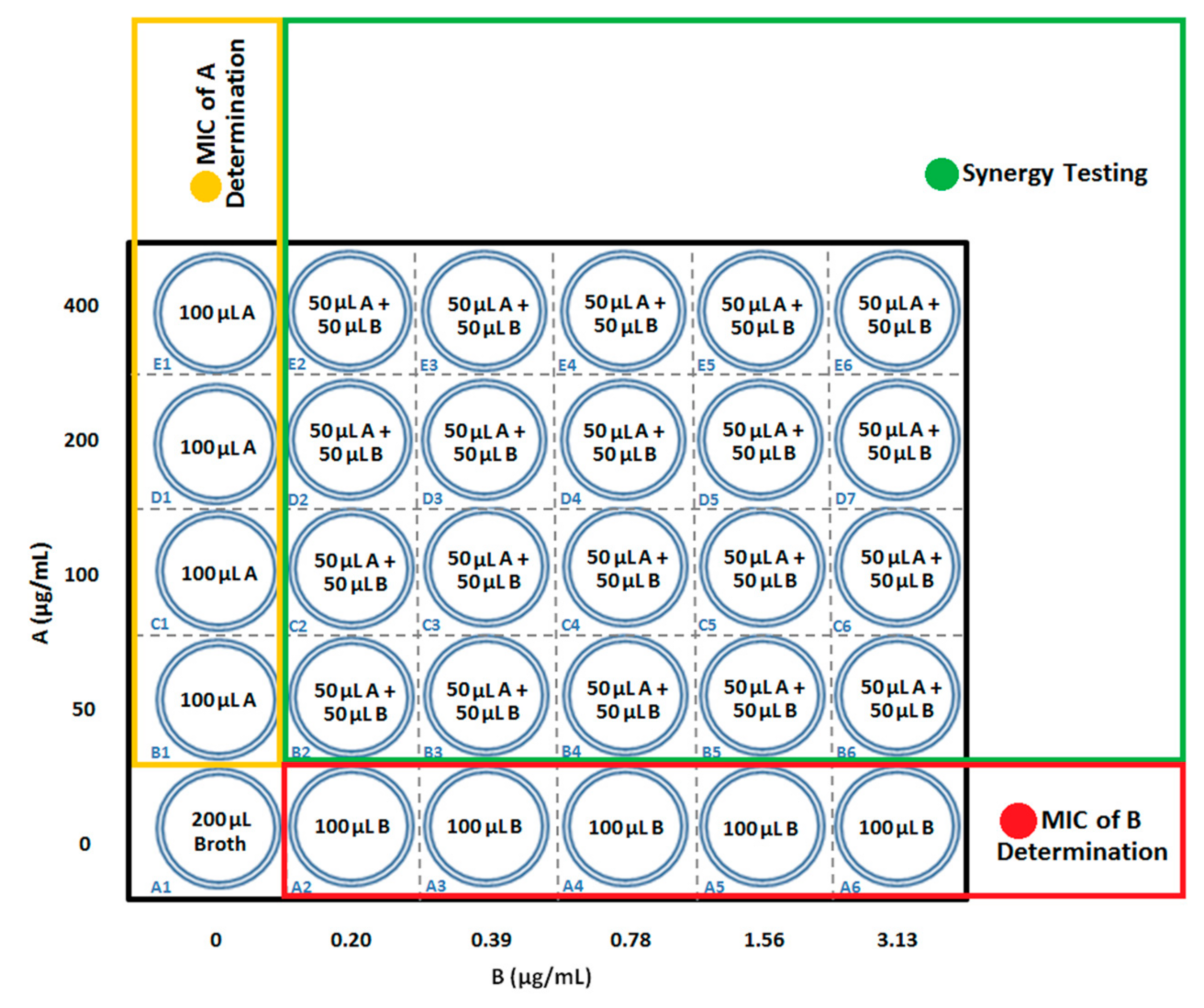
2.6. Synergy Testing by Checkerboard Assay

For MIC testing of the extract, 100 µL EMJH broth was added to the first column of a 96-well plate. One hundred microliters of extract was added to E1 well with a final volume of 200 µL. The extract was serially diluted by transferring 100 µL of the extract solution from E1 to the next well by two-fold. Then, 100 μL of Leptospira cultures was added to the first column of the 96-well plate (final volume of 200 μL per well). For MIC testing of the antibiotic, 100 µL EMJH broth was added to the first row of the 96-well plate. One hundred microliters of the antibiotic was added to A6 well with a final volume of 200 µL. The antibiotic was serially diluted by transferring 100 µL of the antibiotic solution from A6 to the next well by two-fold. Then, 100 μL Leptospira cultures were added to the first row of the 96-well plate (final volume of 200 μL per well). For synergy testing, the two-fold serial dilutions of 50 µL antibiotic and 50 µL extract were prepared and transferred into the second column until the twelfth column. Next, 100 µL of Leptospira culture was added into the respective columns (final volume of 200 µL per well) (Figure 1). Control plates were prepared with broth to replace Leptospira cultures. The plates were incubated at 30 °C and 40 rpm for seven days. The control absorbance value was subtracted from the total absorbance value determined by an ELISA reader, and the pre-incubation values were subtracted from the post-incubation values. Each of the synergy testing was duplicated. Turbidity and an obvious white dot at the bottom of wells showed Leptospira growth, and this was confirmed by examining a loopful of each well’s contents under a dark field microscope (Olympus, Tokyo, Japan) at 200× magnification, zooming in and out [23]. Fractional inhibitory concentration index (FICI) values were calculated for the wells with the lowest concentration showing no visible growth using formulas 1 and 2, and interpreted according to criteria of the American Society of Microbiology and the British Society of Antimicrobial Chemotherapy as follows: synergy (FICI ≤ 0.5), additive (FICI > 0.5–1.0), indifferent (FICI > 1.0–4.0), and antagonistic (FICI > 4.0) [24,25]:
FICI   of   well = FIC   of   Antibiotic + FIC   of   Extract = Concentration   of   Antibiotic MIC   of   Antibiotic + Concentration   of   Extract MIC   of   Extract ,
FICI   of   test = FICI   of   wells numbers   of   wells

2.7. Scanning Electron Microscope Analysis

The combinations with the lowest FICI values were examined using scanning electron microscopy (SEM). Leptospira cultures (2 × 10⁶ CFU/mL) were incubated at 30 °C and 40 rpm for seven days and treated with the MICs of the extracts and antibiotic solutions separately and combined for 18 h. A negative control (untreated Leptospira) sample was prepared. The samples were centrifuged (4560 rpm at 2000 g for 10 min) (Hettich, Westphalia Germany) and the pellets were fixed using McDowell-Trump’s fixative (Electron Microscopy Sciences, Hatfield, PA, USA) at 4 °C for two hours, centrifuged again, washed with 0.1 M PBS (Electron Microscopy Sciences, Hatfield, PA, USA), and fixed using 1% osmium tetroxide (Electron Microscopy Sciences, Hatfield, PA, USA) at 4 °C for one hour. The pellets were then washed with 0.1 M PBS and dehydrated using 50%, 75%, 95%, and 100% acetone (Electron Microscopy Sciences, Hatfield, PA, USA) for ten minutes each. Then, 100% hexamethyldisilazane (Electron Microscopy Sciences, Hatfield, PA, USA) was added to the pellets for 10 min and air-dried overnight. Specimens were then mounted on sample stubs and coated with gold (Electron Microscopy Sciences, Hatfield, PA, USA) for examination with a high-resolution versatile SEM instrument (FEI, Brno, Czech Republic).

2.8. Statistical Analysis

The data were analyzed using SPSS software version 26 (IBM, Armonk, NY, USA) and presented as mean ± SD of the triplicates for antimicrobial susceptibility testing and duplicates for synergy testing.

3. Results

3.1. Antimicrobial Susceptibility Testing

The MICs and MBCs of the extracts and antibiotics are shown in Table 1 and Table 2, respectively. Both P. amarus AEs and MEs exhibited anti-leptospiral effects. However, AEs were more effective, with both MICs and MBCs ranging from 100 to 400 µg/mL, whilst MEs ranged from 400 to 800 µg/mL. Penicillin G showed the strongest anti-leptospiral activity, with MICs and MBCs ranging from 0.01 to 0.78 µg/mL and 0.02 to 3.13 µg/mL, respectively, followed by ceftriaxone, with both MICs and MBCs ranging from 0.05 to 0.78 µg/mL, and doxycycline, with MICs and MBCs ranging from 0.39 to 3.13 µg/mL and 12.5 to 25 µg/mL.

3.2. Synergy Testing
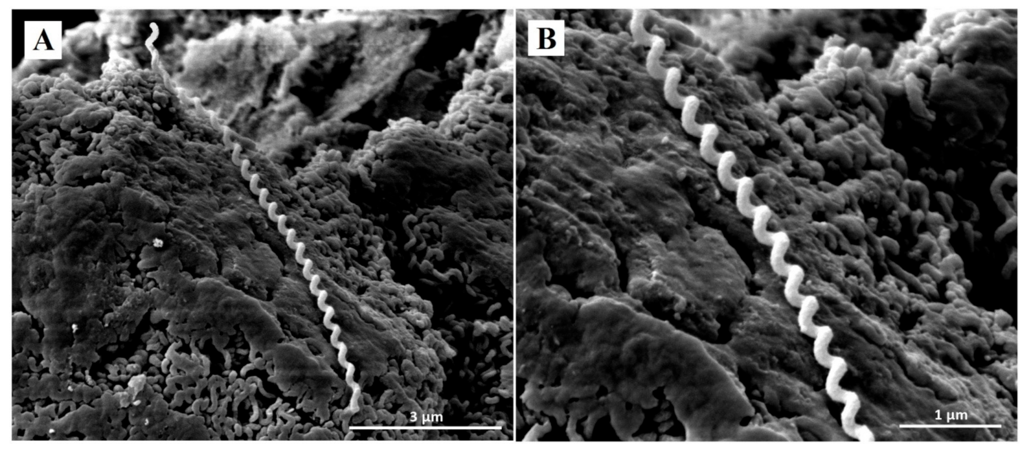
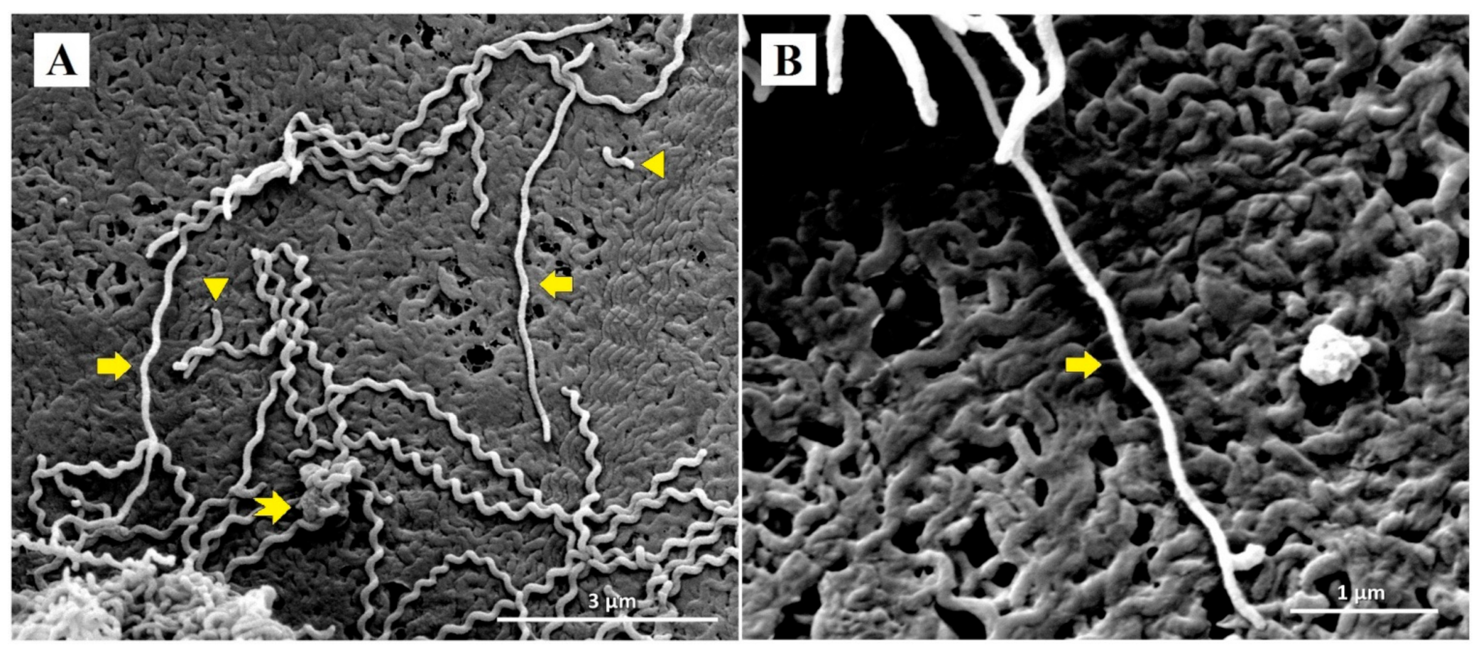
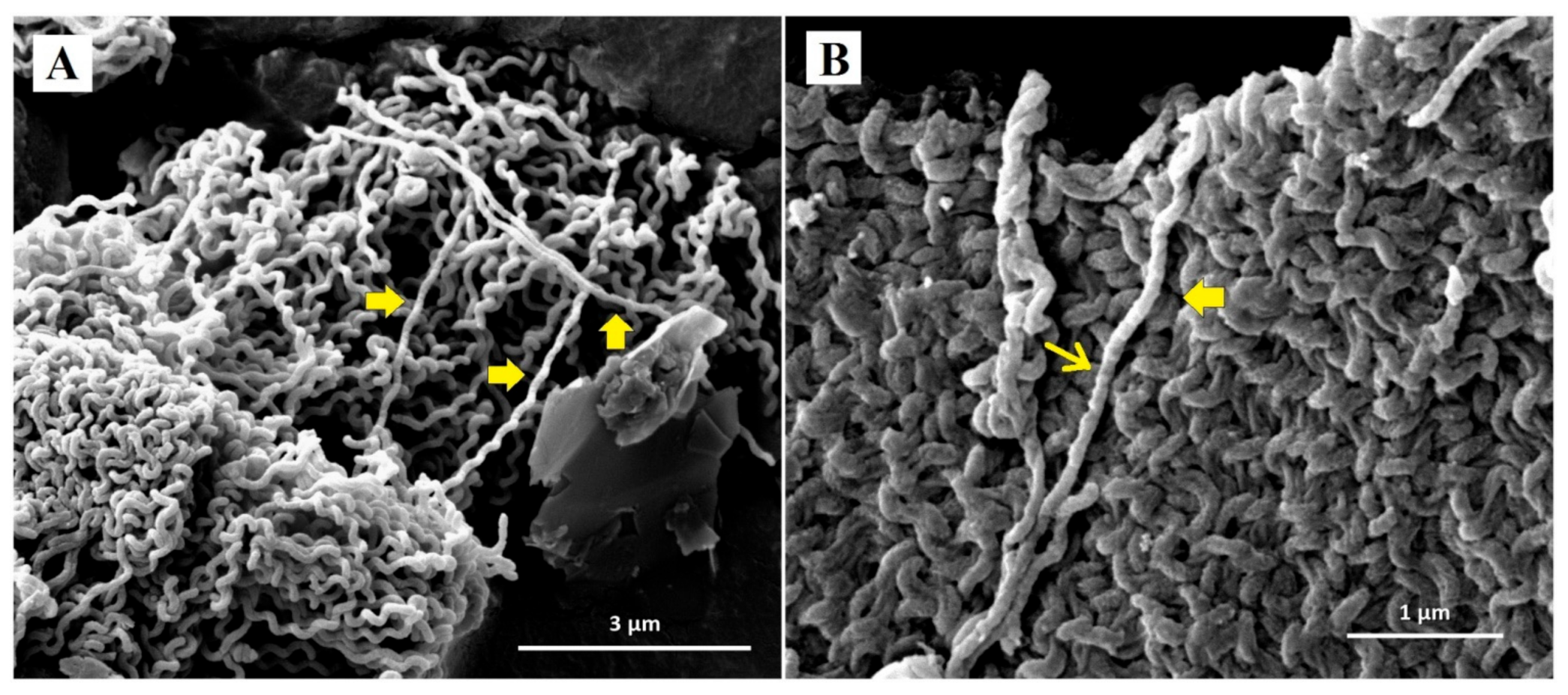
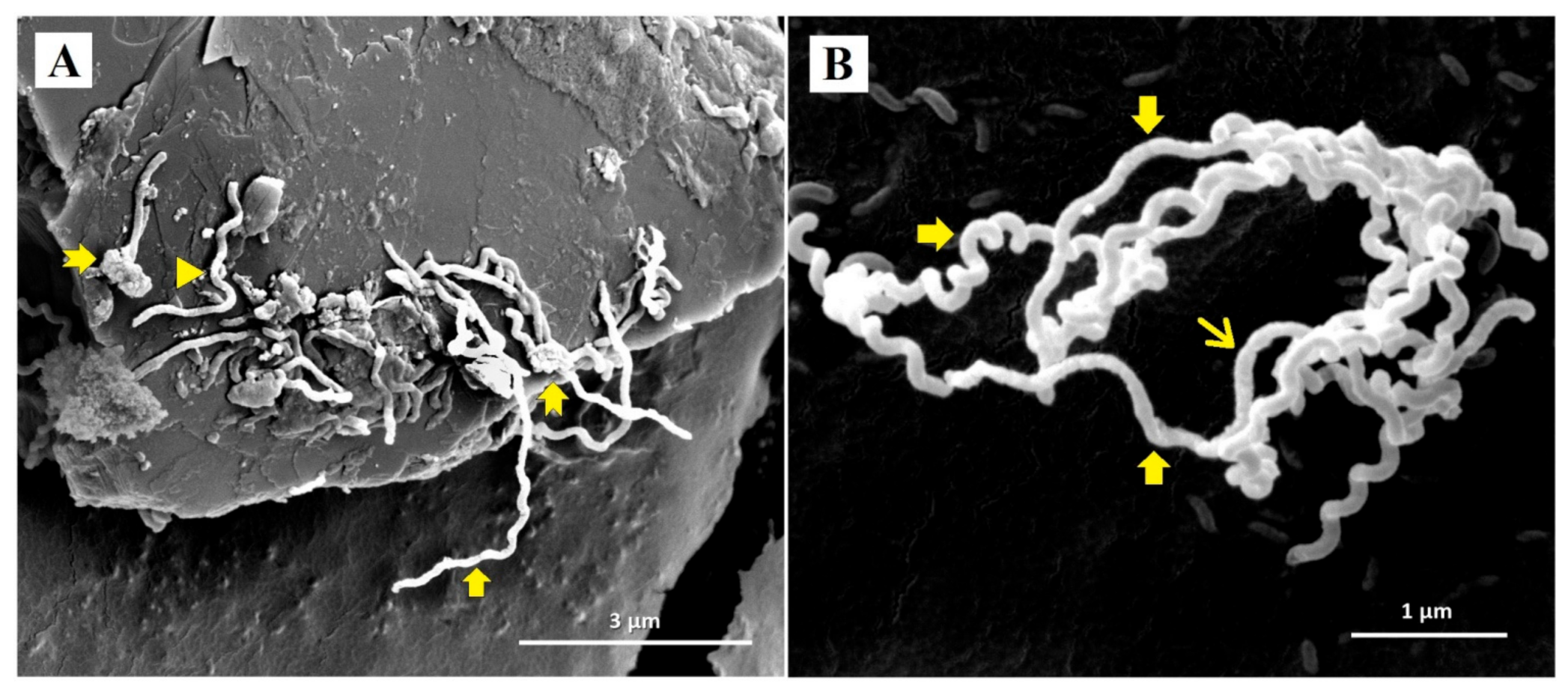
Table 3, Table 4, Table 5 show the FICI values and antibacterial effects of extracts combined with antibiotics. P. amarus AE combined with antibiotics exhibited indifferent effects against Leptospira serovars (FICI 1.59–3.41), while combinations of P. amarus ME with antibiotics exhibited indifferent or antagonistic activities (FICI 2.51–4.87). P. amarus AE combined with doxycycline against L. interrogans serovar Australis had the lowest FICI values (1.59), showing indifferent activity. P. amarus ME combined with doxycycline against L. interrogans serovar Bataviae and penicillin G against L. interrogans serovar Canicola had antagonistic effects (FICI 4.22 and 4.87, respectively).

3.3. SEM Analysis

Representative SEM micrographs revealed notable structural differences between untreated Leptospira and Leptospira treated with doxycycline and P. amarus AE. SEM micrographs of control Leptospira cells showed normal spirochaete morphology with helical-shaped and hooked-end structures (Figure 2). Leptospira treated with the MIC of doxycycline (0.78 µg/mL) exhibited thinning and less coiling (Figure 3), while Leptospira treated with the MIC of P. amarus AE (800 µg/mL) exhibited less coiling and irregular surfaces (Figure 4). Leptospira treated with P. amarus AE combined with doxycycline exhibited severe damage (Figure 5), with thinning, shortening, less and distorted coiling, and irregular surfaces with a blebby appearance.

4. Discussion

The results suggest that P. amarus AE exhibited stronger anti-leptospiral effects than ME. The MIC ranges of both AE and ME coincided with their MBC ranges, suggesting that the same concentration of P. amarus simultaneously inhibits and completely kills the Leptospira. Unlike our study, which found that P. amarus AE was more effective against L. interrogans than ME, other studies have reported that MEs are more effective than AEs against other bacteria, as well as fungi. MEs of aerial parts of the plant have shown more potent inhibitory effects against Bacillus subtilis, Escherichia coli, Pseudomonas aeruginosa, Staphylococcus aureus, Salmonella serotype Typhi, and Candida albicans than AEs [26]. Similarly, Sen and Batra reported that aqueous leaf extracts exhibited minimal activity against B. cereus, E. coli, S. aureus, P. aeruginosa, and Aspergillus niger, A. flavus, Fusarium oxysporum, and Rhizopus stolonifer [27]. Conversely, Oladosu et al. found no significant differences between the effects of AEs and MEs against P. aeruginosa, S. saprophyticus, S. aureus, Klebsiella pneumoniae, E. coli, Proteus mirabilis, and Enterococcus faecalis [12]. The differences between the antimicrobial effects observed in previous studies may be due to differences in plant extraction parameters, parts of the plant used, and geographical settings. Extracted phytochemicals even vary according to pre- and post-harvest stages [28].
Our preliminary research found differences in anti-leptospiral effects between different extracts. This suggests that the extraction process and choice of solvents play a vital role in extracting components that best inhibit the growth of Leptospira or kill it. Previous studies have found slight differences in phytochemical contents between AEs and MEs. Phytochemical analyses of AEs have detected the presence of primary (carbohydrates, proteins, and amino acids) and secondary metabolites (anthraquinone, steroids, including phytosterols and cardiac glycosides, alkaloids, terpenoids, including triterpenes and saponins, phenolic compounds, including flavonoids and lignins, and tannins, specifically phlobatannins) [12,16,17,26,29,30]. Analyses of MEs have revealed the presence of carbohydrates, alkaloids, saponins, terpenoids (triterpenes), steroids (cardiac glycosides), phenolic compounds (flavonoids), and tannins (phlobatannins) [12,16,30,31]. Shirahatti, Syed, and Elgorban investigated the anti-leptospiral effects of three novel biologically active compounds isolated from MEs. Of those, 4-(3-(3,4dimethoxybenzyl)-4-methoxy-2-(methoxymethyl)butyl)-3,6dimethoxybenzene-1,2-diol exhibited the most significant activity against L. interrogans serovars Icterohaemorrhagiae, Canicola, Pomona, Autumnalis, Javanica, Pyrogenes, Australis, and Hardjo, followed by 5-(3-(3,4-dimethoxybenzyl)-4-methoxy-2-(methoxymethyl)butyl)4,7-dimethoxybenzo[d][1,3]dioxole and 1-(3-(3,4-dimethoxybenzyl)-4methoxy-2-(methoxymethyl)butyl)-2,3,4,5tetramethoxybenzene [32].
Flavonoids, tannins, terpenoids, alkaloids, lignins, saponins, anthraquinones, and steroids exert antibacterial effects, mainly by disrupting the formation or structure of bacteria cells, causing them to increase their fluidity. These changes lead to an uncontrolled efflux of metabolites, ions, and membrane proteins, causing leakage of cell contents, resulting in cell damage, lysis, and eventually cell death. Flavonoids, saponins, tannins, and anthraquinones disrupt the microbial cell wall by increasing its permeability, leading to cytoplasm leakage. Terpenoids, alkaloids, tannins, and anthraquinones inhibit biofilm formation, thus reducing resistance to antibacterial agents and environmental conditions. Terpenoids, alkaloids, saponins, and tannins inhibit microbial growth by disrupting microbial cell physiology and metabolism, including the inhibition of protein, nucleic acid, and adenosine triphosphate synthesis [33,34,35,36,37,38,39,40]. In reference to mechanisms of action of phytochemicals found in both AEs and MEs against microorganisms, the actions might be applied to Leptospira as well. The effects observed in this study might be exerted not only by their major components separately, but also by their synergism.
Our study found that the tested antibiotics were more effective in inhibiting Leptospira growth than P. amarus extracts. This can be explained by the fact that they are in pure form, whereas crude plant extracts contain substances that may not be particularly effective in inhibiting Leptospira growth or killing the bacteria. Leptospira serovars were more susceptible to beta-lactam antibiotics, primarily penicillin G and to a lesser degree ceftriaxone, and less susceptible to doxycycline. Similar findings have been reported by previous studies [41,42,43]. In our study, doxycycline was most effective against L. interrogans serovar Bataviae, followed by serovars Australis, Canicola, and Javanica. A higher susceptibility of L. interrogans serovar Bataviae than serovar Canicola to doxycycline has also been observed in previous studies [43,44,45]. The high MBC ranges of doxycycline observed in this study suggest that higher concentration is required to kill the Leptospira completely.
In this study, ceftriaxone was most effective against L. interrogans serovar Bataviae, followed by serovars Australis, Canicola, and Javanica. Its MICs and MBCs against L. interrogans serovars Bataviae and Javanica coincided, suggesting that it inhibits and kills the Leptospira at the same concentrations. Penicillin G was found to be effective against all Leptospira spp. and most effective against L. interrogans serovar Australis, followed by serovars Bataviae, Canicola, and Javanica. In line with our findings, previous studies have reported that L. interrogans serovar Bataviae is more susceptible to penicillin G, as well as to doxycycline, than L. interrogans serovar Canicola [43,44,45].
Our study found indifferent inhibitory effects of combinations of antibiotics and extracts against Leptospira serovars, except for the antagonistic effect of P. amarus ME combined with doxycycline on L. interrogans serovar Bataviae and the antagonistic effect of P. amarus ME combined with penicillin G on L. interrogans serovar Canicola. The lowest FICI value was that of P. amarus AE combined with doxycycline against L. interrogans serovar Australis, which is close to additive effect values (>0.5–1.0), suggesting that P. amarus extracts could have stronger effects when in a purified form. There are limited data on the synergistic effects of plant extracts against Leptospira spp. and no data on P. amarus extracts. The only study on synergistic effects of plant extracts against Leptospira spp. found that purified xanthone γ-mangostin from Garcinia mangostana combined with penicillin G exhibited synergistic activity against L. interrogans serovars Autumnalis, Bataviae, and Javanica, no interaction activity against L. biflexa serovar Patoc, and antagonistic activity against L. interrogans serovar Saigon [46].
In this study, SEM revealed that doxycycline and AE alone, and their combination, caused damage to L. interrogans serovar Australis. Cell bulging or blebbing is a known effect of doxycycline. Kersten et al. and Meriläinen et al. reported similar morphological distortions of the spirochaete Borrelia burgdorferi when exposed to doxycycline [47,48]. Bacterial blebbing, which is one of the factors leading to cell lysis, is caused by physiological stress and turgor pressure, metabolite depletion, cell ageing, exposure to antibiotics, and pH changes that disturb the cell envelope [49,50]. In living cells, cell blebs transport microbial virulence factors and are involved in cell communication and division [49]. The mechanism of action of doxycycline against L. interrogans serovar Australis is associated with thinning, shortening, bleb formation, and reduced coiling. To our knowledge, this is the first study to investigate the effects of P. amarus extracts on bacterial morphology. The observed distorted Leptospira surface morphology could be due to phytochemical components of P. amarus extracts that mainly exert effects on the cellular membranes, which can be further analyzed in the future research. Considering the synergy testing and SEM results, P. amarus AE combined with doxycycline causes severe damage to Leptospira cells but with no enhancement on its inhibiting and killing abilities on Leptospira. Further evaluation of internal structural changes of leptospiral cells by transmission electron microscopy analyses is important to confirm anti-leptospiral activities of phytochemicals in P. amarus.

5. Conclusions

P. amarus showed promising anti-leptospiral properties, with AEs yielding better results than MEs. All tested Leptospira serovars were more susceptible to penicillin G than to the other tested antibiotics. Synergy testing revealed moderate effects, indicating a need for improvement in this area. However, P. amarus AE combined with doxycycline caused significant damage to Leptospira cell morphology, further suggesting the potential of P. amarus as an anti-leptospiral agent. Further evaluation of P. amarus in animal models study is needed, as they have potential anti-leptospiral property, as well as to investigate the whole effects and mechanisms on the subjects including its toxicity.

Supplementary Materials

The following are available online at https://0-www-mdpi-com.brum.beds.ac.uk/1660-4601/18/6/2834/s1, Figure S1: Plant identification.

Author Contributions

Conceptualization: N.I.; methodology: N.I., Z.Z.D., R.A.B., and C.A.M.I.; software: not applicable; validation: C.A.M.I., Z.Z.D., and N.I.; formal analysis: C.A.M.I.; investigation: C.A.M.I.; resources: N.I.; data curation: C.A.M.I.; writing—original draft preparation: C.A.M.I.; writing—review and editing: C.A.M.I., N.I., R.A.B., and Z.Z.D.; visualization: N.I.; supervision: N.I., R.A.B., and Z.Z.D.; project administration: N.I.; funding acquisition: N.I. All authors have read and agreed to the published version of the manuscript.

Funding

This study was funded by a Bridging Grant (304/PPSP/6316187).

Institutional Review Board Statement

Not applicable.

Informed Consent Statement

Not applicable.

Data Availability Statement

The data that support the findings of this study are available from the corresponding author, upon reasonable request.

Acknowledgments

The authors would like to thank the late Kamarudin Saleh from the Forest Research Institute Malaysia for help in identifying the plant species, staff at the Department of Medical Microbiology and Parasitology, Scanning Electron Microscope Laboratory, Pharmacology Laboratory, and Central Research Laboratory, Universiti Sains Malaysia for direct and indirect assistance throughout the research. The authors would also like to acknowledge Universiti Putra, Malaysia, and the Institute for Medical Research, Malaysia, for the Leptospira strains used in the study.

Conflicts of Interest

The authors declare no conflict of interest. The funders had no role in the design of the study; in the collection, analyses, or interpretation of data; in the writing of the manuscript; or in the decision to publish the results.

References

  1. Lim, V.K.E. Leptospirosis: A re-emerging infection. Malays. J. Pathol. 2011, 33, 1–5. [Google Scholar]
  2. Prabhakaran, S.G.; Shanmughapriya, S.; Dhanapaul, S.; James, A.; Natarajaseenivasan, K. Risk factors associated with rural and urban epidemics of leptospirosis in Tiruchirappalli district of Tamilnadu, India. J. Public Health 2014, 22, 323–333. [Google Scholar] [CrossRef]
  3. Haake, D.A.; Levett, P.N. Leptospirosis in humans. Curr. Top. Microbiol. Immunol. 2015, 387, 65–97. [Google Scholar] [CrossRef] [Green Version]
  4. World Health Organization. Leptospirosis: Fact Sheet; Regional Office for South-East Asia: New Delhi, India, 2009; pp. 1–12. [Google Scholar]
  5. Levett, P.N. Leptospirosis. Clin. Microbiol. Rev. 2001, 14, 296–326. [Google Scholar] [CrossRef] [PubMed] [Green Version]
  6. Costa, F.; Hagan, J.E.; Calcagno, J.; Kane, M.; Torgerson, P.; Martinez-Silveira, M.S.; Stein, C.; Abela-Ridder, B.; Ko, A.I. Global morbidity and mortality of leptospirosis: A Systematic Review. PLOS Negl. Trop. Dis. 2015, 9, e0003898. [Google Scholar] [CrossRef] [Green Version]
  7. Marchiori, E.; Lourenço, S.; Setúbal, S.; Zanetti, G.; Gasparetto, T.D.; Hochhegger, B. Clinical and imaging manifestations of hemorrhagic pulmonary leptospirosis: A state-of-the-art review. Lung 2011, 189, 1–9. [Google Scholar] [CrossRef]
  8. Fraga, T.R.; Carvalho, E.; Isaac, L.; Barbosa, A.S. Leptospira and leptospirosis. In Molecular Medical Microbiology; Yi-Wei, T., Liu, D., Schwartzman, J., Sussman, M., Poxton, I., Eds.; Academic Press: Amsterdam, The Netherlands, 2015; pp. 1973–1990. [Google Scholar]
  9. Gyawali, R.; Ibrahim, S.A. Natural products as antimicrobial agents. Food Control 2014, 46, 412–429. [Google Scholar] [CrossRef]
  10. Joseph, B.; Raj, S.J. Overview pharmacognostic properties of Phyllanthus Amarus Linn. Int. J. Pharmacol. 2011, 7, 40–45. [Google Scholar] [CrossRef]
  11. Sarin, B.; Verma, N.; Martín, J.P.; Mohanty, A. An overview of important ethnomedicinal herbs of Phyllanthus species: Present status and future prospects. Sci. World J. 2014, 2014, 839172. [Google Scholar] [CrossRef] [Green Version]
  12. Oladosu, S.A.; Coker, A.O.; Nwaokorie, F. Antibacterial effects of Phyllantus amarus on urinary tract pathogens. Int. Clin. Pathol. J. 2019, 7, 1–10. [Google Scholar] [CrossRef]
  13. Bharathi, T.; Kolanjinathan, K.; Saranraj, P. Antimicrobial activity of solvent extracts of Ocimum sanctum, Azadirachta indica and Phyllanthus amarus against clinical pathogens. Glob. J. Pharmacol. 2014, 8, 294–305. [Google Scholar] [CrossRef]
  14. Mazumder, A.; Mahato, A.; Mazumder, R. Antimicrobial potentiality of Phyllanthus amarus against drug resistant pathogens. Nat. Prod. Res. 2006, 20, 323–326. [Google Scholar] [CrossRef] [PubMed]
  15. Bhat, S.S.; Hegde, K.S.; Chandrashekhar, S.; Rao, S.N.; Manikkoth, S. Preclinical screening of Phyllanthus amarus ethanolic extract for its analgesic and antimicrobial activity. Pharmacogn. Res. 2015, 7, 378–384. [Google Scholar] [CrossRef] [Green Version]
  16. Sekar, D.; Kolanjinathan, K.; Saranraj, P.; Gajendiran, K. Screening of Phyllanthus amarus, Acalypha indica and Datura metel for its antimicrobial activity against selected pathogens. Int. J. Pharm. Biol. Arch. 2012, 3, 1231–1235. [Google Scholar]
  17. Oluwafemi, F.; Debiri, F. Antimicrobial effect of Phyllanthus amarus and Parquetina nigrescens on Salmonella typhi. Afr. J. Biomed. Res. 2008, 11, 215–219. [Google Scholar] [CrossRef]
  18. Chandan, S.; Umesha, S.; Balamurugan, V. Antileptospiral, antioxidant and dna damaging properties of Eclipta alba and Phyllanthus amarus. Open Access Sci. Rep. 2012, 1, 231. [Google Scholar] [CrossRef]
  19. Suwari, H.Z.K.; Buang, Y. Optimization of soxhlet extraction and physicochemical analysis of crop oil from seed kernel of Feun kase (Thevetia peruviana). AIP Conf. Proc. 2017, 1911, 1–7. [Google Scholar] [CrossRef]
  20. Azwanida, N.N. A review on the extraction methods use in medicinal plants, principle, strength and limitation. Med. Aromat. Plants 2015, 4, 196. [Google Scholar] [CrossRef]
  21. Murray, C.K.; Ellis, M.W.; Hospenthal, D.R. Susceptibility of Leptospira serovars to antimalarial agents. Am. J. Trop. Med. Hyg. 2004, 71, 685–686. [Google Scholar] [CrossRef]
  22. Drancourt, M.; Raoult, D. Methodology of antibiotics testing for intracellular pathogens. In Antimicrobial Agents and Intracellular Pathogens; Raoult, D., Ed.; CRC Press, Inc.: Boca Raton, FL, USA, 1993; pp. 73–85. [Google Scholar]
  23. European Committee for Antimicrobial Susceptibility Testing of the European Society of Clinical Microbiology Infectious Diseases. Determination of minimum inhibitory concentrations (mics) of antibacterial agents by broth dilution. Clin. Microbiol. Infect. 2003, 9, ix–xv. [Google Scholar] [CrossRef] [Green Version]
  24. Van Vuuren, S.; Viljoen, A. Plant-based antimicrobial studies–methods and approaches to study the interaction between natural products. Planta Med. 2011, 77, 1168–1182. [Google Scholar] [CrossRef] [PubMed] [Green Version]
  25. Den Hollander, J.G.; Mouton, J.W. The predictive value of laboratory tests for efficacy of antibiotic combination therapy. In Antimicrobial Pharmacodynamics in Theory and Clinical Practice; Nightingale, C.H., Ambrose, P.G., Drusano, G.L., Murakawa, T., Eds.; Informa Healthcare USA, Inc.: New York, NY, USA, 2007; pp. 103–127. [Google Scholar]
  26. Alli, A.I.; Ehinmidu, J.O.; Ibrahim, Y.K.E. Preliminary phytochemical screening and antimicrobial activities of some medicinal plants used in Ebiraland. Bayero J. Pure Appl. Sci. 2011, 4, 10–16. [Google Scholar] [CrossRef]
  27. Sen, A.; Batra, A. Determination of antimicrobial potentialities of different solvent extracts of the medicinal plant: Phyllanthus amarus Schum. and Thonn. Int. J. Gree Pharm. 2012, 6, 50–56. [Google Scholar] [CrossRef]
  28. Tiwari, U.; Cummins, E. Factors influencing levels of phytochemicals in selected fruit and vegetables during pre-and post-harvest food processing operations. Food Res. Int. 2013, 50, 497–506. [Google Scholar] [CrossRef]
  29. Ogunjobi, A.A.; Abiala, M.A. Antimicrobial activity of Senna alata and Phyllanthus amarus. Glob. J. Pharmacol. 2013, 7, 198–202. [Google Scholar] [CrossRef]
  30. Dhandapani, R.; Lakshmi, D.; Balakrishnan, V.; Jayakumar, S.; Kumar, A. Preliminary phytochemical investigation and antibacterial activity of Phyllanthus amarus Schum & Thorn. Anc. Sci. Life 2007, 27, 1–5. [Google Scholar]
  31. Zubair, M.; Atolani, O.; Ibrahim, S.; Adebisi, O.; Hamid, A.; Sowunmi, R. Chemical constituents and antimicrobial properties of Phyllanthus amarus (Schum & Thonn). Bayero J. Pure Appl. Sci. 2017, 10, 238–246. [Google Scholar] [CrossRef] [Green Version]
  32. Shirahatti, P.S.; Syed, A.; Elgorban, A.M. Potential antileptospiral constituents from Phyllanthus amarus. Pharmacogn. Mag. 2020, 16, S371–S378. [Google Scholar] [CrossRef]
  33. Górniak, I.; Bartoszewski, R.; Króliczewski, J. Comprehensive review of antimicrobial activities of plant flavonoids. Phytochem. Rev. 2019, 18, 241–272. [Google Scholar] [CrossRef] [Green Version]
  34. Lahiri, D.; Dash, S.; Dutta, R.; Nag, M. Elucidating the effect of anti-biofilm activity of bioactive compounds extracted from plants. J. Biosci. 2019, 44, 1–19. [Google Scholar] [CrossRef]
  35. Mahmood, Z.; Yameen, M.; Jahangeer, M.; Riaz, M.; Ghaffar, A.; Javid, I. Lignin as Natural Antioxidant Capacity. In Lignin—Trends and Application; Poletto, M., Ed.; IntechOpen: Rijeka, Croatia, 2018; pp. 181–205. [Google Scholar]
  36. Dong, S.; Yang, X.; Zhao, L.; Zhang, F.; Hou, Z.; Xue, P. Antibacterial activity and mechanism of action saponins from Chenopodium quinoa Willd. husks against foodborne pathogenic bacteria. Ind. Crops Prod. 2020, 149, 112350. [Google Scholar] [CrossRef]
  37. Yu, Z.-H.; Ding, X.-Z.; Xia, L.-Q.; Xiao, X.-Q.; Cao, Z.-P.; Xu, S.; Liu, S.; Liu, X.-M. Antimicrobial activity and mechanism of total saponins from Allium chinense. Food Sci. 2013, 34, 75–80. [Google Scholar] [CrossRef]
  38. Wei, Y.; Liu, Q.; Yu, J.; Feng, Q.; Zhao, L.; Song, H.; Wang, W. Antibacterial mode of action of 1, 8-dihydroxy-anthraquinone from Porphyra haitanensis against Staphylococcus aureus. Nat. Prod. Res. 2015, 29, 976–979. [Google Scholar] [CrossRef] [PubMed]
  39. Doğan, A.; Otlu, S.; Çelebi, Ö.; Aksu, P.; Sağlam, A.G.; Doğan, A.N.C.; Mutlu, N. An investigation of antibacterial effects of steroids. Turkish J. Vet. Anim. Sci. 2017, 41, 302–305. [Google Scholar] [CrossRef]
  40. Li, Y.; Jiang, J.-G. Health functions and structure–activity relationships of natural anthraquinones from plants. Food Funct. 2018, 9, 6063–6080. [Google Scholar] [CrossRef]
  41. Liegeon, G.; Delory, T.; Picardeau, M.A. Antibiotic susceptibilities of livestock isolates of Leptospira. Int. J. Antimicrob. Agents 2018, 51, 693–699. [Google Scholar] [CrossRef]
  42. Chitra, M.A. Susceptibility of Leptospira serotypes against various antimicrobials by broth microdilution method. Indian J. Anim. Sci. 2013, 83, 775–778. [Google Scholar]
  43. Ressner, R.A.; Griffith, M.E.; Beckius, M.L.; Pimentel, G.; Miller, R.S.; Mende, K.; Fraser, S.L.; Galloway, R.L.; Hospenthal, D.R.; Murray, C.K. Antimicrobial susceptibilities of geographically diverse clinical human isolates of Leptospira. Antimicrob. Agents Chemother. 2008, 52, 2750–2754. [Google Scholar] [CrossRef] [PubMed] [Green Version]
  44. Benacer, D.; Mohd Zain, S.N.; Ooi, P.T.; Thong, K.L. Antimicrobial susceptibility of Leptospira spp. isolated from environmental, human and animal sources in Malaysia. Indian J. Med. Microbiol. 2017, 35, 124–128. [Google Scholar] [CrossRef]
  45. Murray, C.K.; Hospenthal, D.R. Determination of susceptibilities of 26 Leptospira sp. serovars to 24 antimicrobial agents by a broth microdilution technique. Antimicrob. Agents Chemother. 2004, 48, 4002–4005. [Google Scholar] [CrossRef] [Green Version]
  46. Seesom, W.; Jaratrungtawee, A.; Suksamrarn, S.; Mekseepralard, C.; Ratananukul, P.; Sukhumsirichart, W. Antileptospiral activity of xanthones from Garcinia mangostana and synergy of gamma-mangostin with penicillin G. BMC Complement. Altern. Med. 2013, 13, 182. [Google Scholar] [CrossRef] [PubMed] [Green Version]
  47. Kersten, A.; Poitschek, C.; Rauch, S.; Aberer, E. Effects of Penicillin, Ceftriaxone, and Doxycycline on Morphology of Borrelia burgdorferi. Antimicrob. Agents Chemother. 1995, 39, 1127–1133. [Google Scholar] [CrossRef] [PubMed] [Green Version]
  48. Meriläinen, L.; Herranen, A.; Schwarzbach, A.; Gilbert, L. Morphological and biochemical features of Borrelia burgdorferi pleomorphic forms. Microbiology 2015, 161, 516–527. [Google Scholar] [CrossRef] [PubMed]
  49. Toyofuku, M.; Nomura, N.; Eberl, L. Types and origins of bacterial membrane vesicles. Nat. Rev. Microbiol. 2019, 17, 13–24. [Google Scholar] [CrossRef] [PubMed]
  50. Singh, S.K.; Girschick, H.J. Lyme borreliosis: From infection to autoimmunity. Clin. Microbiol. Infect. 2004, 10, 598–614. [Google Scholar] [CrossRef] [Green Version]
Figure 1. Arrangement of synergy testing in a 96-well plate. A: Extract; B: Antibiotic.
Ijerph 18 02834 g001
Figure 2. SEM micrographs of normal L. interrogans serovar Australis at 30,000× (A) and 60,000× (B) magnifications with perfect helical-shaped and hooked-end structures.
Ijerph 18 02834 g002
Figure 3. SEM micrographs of L. interrogans serovar Australis treated with MIC doxycycline at 30,000× (A) and 60,000× (B) magnifications. Block arrows: less coiling and thinning in Leptospira diameters; notched arrow: blebbing; arrowhead: shortened.
Ijerph 18 02834 g003
Figure 4. SEM micrographs of L. interrogans serovar Australis treated with MIC AE at 30,000× (A) and 60,000× (B) magnifications. Block arrows: less coiling; arrow: irregular surfaces of Leptospira cells.
Ijerph 18 02834 g004
Figure 5. SEM micrographs of L. interrogans serovar Australis treated with the MIC of doxycycline combined with the MIC of AE at 30,000× (A) and 60,000× (B) magnifications. Block arrows: distorted and less coiling; arrow: thinning; arrowhead: shortened; notched arrows: irregular and blebbing appearances.
Ijerph 18 02834 g005
Table 1. MIC and MBC values (mean ± SD) of P. amarus extracts on Leptospira serovars in triplicate.
Leptospira SerovarsAE (µg/mL)ME (µg/mL)
MICMBCMICMBC
L. interrogans serovar Australis400 ± 0.0400 ± 0.0800 ± 0.0800 ± 0.0
L. interrogans serovar Bataviae100 ± 0.0100 ± 0.0400 ± 0.0400 ± 0.0
L. interrogans serovar Caniola100 ± 0.0100 ± 0.0400 ± 0.0400 ± 0.0
L. interrogans serovar Javanica200 ± 0.0200 ± 0.0800 ± 0.0800 ± 0.0
AE: Aqueous Extract; ME: Methanol Extract; MIC: Minimum Inhibitory Concentration; MBC: Minimum Bactericidal Concentration.
Table 2. MIC and MBC values (mean ± SD) of antibiotics on Leptospira serovars in triplicate.
Leptospira SerovarsDoxycycline
(µg/mL)
Penicillin G
(µg/mL)
Ceftriaxone
(µg/mL)
MICMBCMICMBCMICMBC
L. interrogans serovar Australis0.78 ± 0.0012.50 ± 0.00<0.01 ± 0.00<0.01 ± 0.000.20 ± 0.000.39 ± 0.00
L. interrogans serovar Bataviae0.39 ± 0.0012.50 ± 0.00<0.01 ± 0.000.05 ± 0.000.05 ± 0.000.05 ± 0.00
L. interrogans serovar Canicola1.56 ± 0.0012.50 ± 0.000.10 ± 0.000.20 ± 0.000.20 ± 0.000.39 ± 0.00
L. interrogans serovar Javanica3.13 ± 0.0025.00 ± 0.000.78 ± 0.003.13 ± 0.000.78 ± 0.000.78 ± 0.00
MIC: Minimum Inhibitory Concentration; MBC: Minimum Bactericidal Concentration.
Table 3. FICI values of AE and ME in combination with doxycycline against Leptospira serovars by checkerboard assays and the activities. The test was duplicated and reported in mean ± SD.
Leptospira SerovarsCombination of AE and DoxycyclineCombination of ME and Doxycycline
MIC AE (µg/mL)MIC DOX (µg/mL)FICIActivityMIC ME (µg/mL)MIC DOX (µg/mL)FICIActivity
L. interrogans serovar Australis800 ± 0.000.78 ± 0.001.59 ± 0.00Indifferent800 ± 0.000.39 ± 0.002.82 ± 0.00Indifferent
L. interrogans serovar Bataviae50 ± 0.000.78 ± 0.003.09 ± 0.00Indifferent200 ± 0.000.39 ± 0.004.22 ± 0.00Antagonistic
L. interrogans serovar Canicola100 ± 0.001.56 ± 0.002.09 ± 0.00Indifferent400 ± 0.001.56 ± 0.002.65 ± 0.00Indifferent
L. interrogans serovar Javanica100 ± 0.001.56 ± 0.002.46 ± 0.00Indifferent400 ± 0.000.78 ± 0.003.58 ± 0.00Indifferent
AE: Aqueous Extract; ME: Methanol Extract; DOX: Doxycycline; MIC: Minimum Inhibitory Concentration.
Table 4. FICI values of AE and ME in combination with penicillin G against Leptospira serovars by checkerboard assays and their activities. The test was duplicated and reported in mean ± SD.
Leptospira SerovarsCombination of AE and Penicillin GCombination of ME and Penicillin G
MIC AE (µg/mL)MIC PEN (µg/mL)FICIActivityMIC ME (µg/mL)MIC PEN (µg/mL)FICIActivity
L. interrogans serovar Australis200 ± 0.000.02 ± 0.002.92 ± 0.00Indifferent800 ± 0.000.01 ± 0.003.32 ± 0.00Indifferent
L. interrogans serovar Bataviae50 ± 0.000.02 ± 0.003.41 ± 0.00Indifferent400 ± 0.000.01 ± 0.002.95 ± 0.00Indifferent
L. interrogans serovar Canicola100 ± 0.000.05 ± 0.002.42 ± 0.00Indifferent400 ± 0.000.05 ± 0.004.87 ± 0.00Antagonistic
L. interrogans serovar Javanica100 ± 0.000.78 ± 0.002.61 ± 0.00Indifferent400 ± 0.000.78 ± 0.003.38 ± 0.00Indifferent
AE: Aqueous Extract; ME: Methanol Extract; PEN: Penicillin G; MIC: Minimum Inhibitory Concentration.
Table 5. FICI values of AE and ME in combination with ceftriaxone against Leptospira serovars by checkerboard assays and their activities. The test was duplicated and reported in mean ± SD.
Leptospira SerovarsCombination of AE and CeftriaxoneCombination of ME and Ceftriaxone
MIC AE (µg/mL)MIC CEF (µg/mL)FICIActivityMIC ME (µg/mL)MIC CEF (µg/mL)FICIActivity
L. interrogans serovar Australis400 ± 0.000.10 ± 0.002.39 ± 0.00Indifferent400 ± 0.000.20 ± 0.003.19 ± 0.00Indifferent
L. interrogans serovar Bataviae100 ± 0.000.05 ± 0.002.24 ± 0.00Indifferent400 ± 0.000.02 ± 0.002.51 ± 0.00Indifferent
L. interrogans serovar Canicola100 ± 0.000.20 ± 0.002.36 ± 0.00Indifferent100 ± 0.000.10 ± 0.003.22 ± 0.00Indifferent
L. interrogans serovar Javanica200 ± 0.001.56 ± 0.002.40 ± 0.00Indifferent800 ± 0.001.56 ± 0.002.75 ± 0.00Indifferent
AE: Aqueous Extract; ME: Methanol Extract; CEF: Ceftriaxone; MIC: Minimum Inhibitory Concentration.
Publisher’s Note: MDPI stays neutral with regard to jurisdictional claims in published maps and institutional affiliations.

