
Medial Sigmoid Depression of the Mandibular Ramus as a Lesion-Mimicking Anatomical Variation: A Systematic Review


Abstract
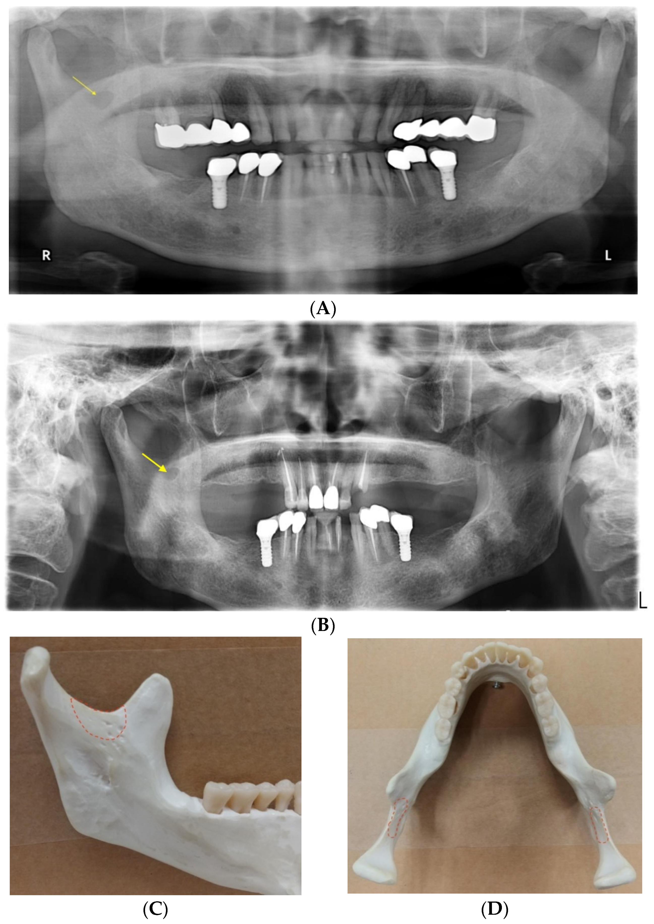
:1. Introduction
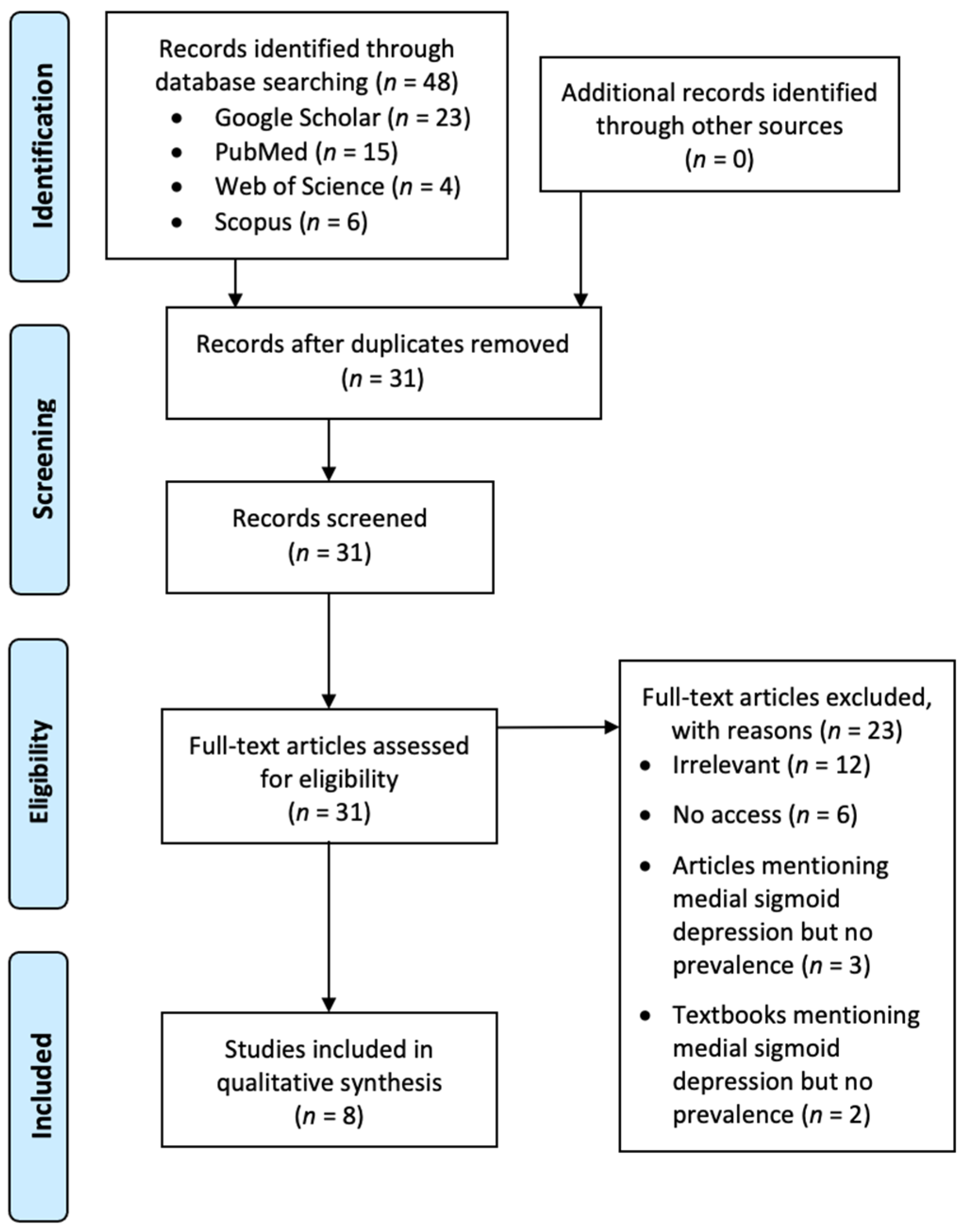
2. Materials and Methods
3. Results
4. Discussion
5. Conclusions
Author Contributions
Funding
Institutional Review Board Statement
Informed Consent Statement
Data Availability Statement
Acknowledgments
Conflicts of Interest
References
- Langlais, R.P.; Glass, B.J.; Bricker, S.L.; Miles, D.A. Medial sigmoid depression: A panoramic pseudoforamen in the upper ramus. Oral Surg. Oral Med. Oral Pathol. 1983, 55, 635–638. [Google Scholar] [CrossRef]
- Whaites, E.; Drage, N. Essentials of Dental Radiography and Radiology; Elsevier Health Sciences: Amsterdam, The Netherlands, 2013. [Google Scholar]
- White, S.; Pharaoh, M. Oral Radiology Principles and Interpretation, 5th ed.; Mosby: Maryland Heights, MO, USA, 2004. [Google Scholar]
- Koenig, L.J.; Tamimi, D.; Petrikowski, C.G.; Perschbacher, S.E. Diagnostic Imaging: Oral and Maxillofacial; Elsevier Health Sciences: Amsterdam, The Netherlands, 2017. [Google Scholar]
- Carvalho, I.; Damante, J.; Tallents, R.; Ribeiro-Rotta, R. An anatomical and radiographic study of medial depression of the human mandibular ramus. Dentomaxillofacial Radiol. 2001, 30, 209–213. [Google Scholar] [CrossRef]
- Philipsen, H.; Takata, T.; Reichart, P.; Sato, S.; Suei, Y. Lingual and buccal mandibular bone depressions: A review based on 583 cases from a world-wide literature survey, including 69 new cases from Japan. Dentomaxillofacial Radiol. 2002, 31, 281–290. [Google Scholar] [CrossRef] [PubMed]
- Adisen, M.Z.; Okkesim, A.; Misirlioglu, M. A possible association between medial depression of mandibular ramus and maximum bite force. Folia Morphol. 2018, 77, 711–716. [Google Scholar] [CrossRef] [PubMed] [Green Version]
- Asdullah, M.; Aggarwal, A.; Khawja, K.J.; Khan, M.H.; Gupta, J.; Ratnakar, K. An anatomic and radiographic study of medial sigmoid depression in human mandible. J. Indian Acad. Oral Med. Radiol. 2019, 31, 123–127. [Google Scholar] [CrossRef]
- Dalili, Z.; Mohtavipour, S. Frequency of medial sigmoid depression in panoramic view of orthodontic patients based on facial skeletal classification. J. Guilan Univ. Med Sci. 2003, 12, 16–23. [Google Scholar]
- Kang, B.-C. The Medial Sigmoid Depression: Its Anatomic and Radiographic Considerations. J. Korean Acad. Maxillofac. Radiol. 1991, 21, 7–13. [Google Scholar]
- Özkan, G.; Sessiz AK, R. Evaluation of the frequency of medial sigmoid depression using panoramic radiographs: A retrospective study. J. Dent. Fac. Atatürk Univ. 2020, 20, 552–556. [Google Scholar]
- Sudhakar, S.; Kumar, N.; Prabhat, M.; Nalini, J. Characteristics of medial depression of the mandibular ramus in patients with orthodontic treatment needs: A panoramic radiography study. J. Clin. Diagn. Res. 2014, 8, ZC100–ZC104. [Google Scholar]
- Yeung, A.W.K. The “As Low As Reasonably Achievable” (ALARA) principle: A brief historical overview and a bibliometric analysis of the most cited publications. Radioprotection 2019, 54, 103–109. [Google Scholar] [CrossRef]
- Yeung, A.W.K.; Jacobs, R.; Bornstein, M.M. Novel low-dose protocols using cone beam computed tomography in dental medicine: A review focusing on indications, limitations, and future possibilities. Clin. Oral Investig. 2019, 23, 2573–2581. [Google Scholar] [CrossRef] [PubMed]
- Dave, M.; Horner, K. Challenges in X-ray diagnosis: A review of referrals for specialist opinion. Br. Dent. J. 2017, 222, 431–437. [Google Scholar] [CrossRef] [PubMed]
- Harvey, S.; Ball, F.; Brown, J.; Thomas, B. ‘Non-standard’ panoramic programmes and the unusual artefacts they produce. Br. Dent. J. 2017, 223, 248–252. [Google Scholar] [CrossRef]
- Wood, N.K.; Goaz, P.W. Differential Diagnosis of Oral and Maxillofacial Lesions; Mosby: Maryland Heights, MO, USA, 1997. [Google Scholar]
- William Jr, G.; Merritt, C.R. Reeder and Felson’s Gamuts in Radiology: Comprehensive Lists of Roentgen Differential Diagnosis; Springer Science & Business Media: Amsterdam, The Netherlands, 2006. [Google Scholar]
- Abramovitch, K.; Langlais, R.P.; Dolwick, M.F. Panoramic radiography for temporomandibular joint arthrography: A description of arthropanoramograms. Oral Surg. Oral Med. Oral Pathol. 1989, 67, 775–780. [Google Scholar] [CrossRef]
- Chudhry, A.; Sankireddy, S. Variants of major salivary gland related bone defects—a case series. Int. J. Maxillofac. Imaging 2016, 2, 69–72. [Google Scholar]
- Hisatomi, M.; Munhoz, L.; Asaumi, J.; Arita, E.S. Parotid mandibular bone defect: A case report emphasizing imaging features in plain radiographs and magnetic resonance imaging. Imaging Sci. Dent. 2017, 47, 269–273. [Google Scholar] [CrossRef] [PubMed] [Green Version]
- Miles, D.A. Clinical Experience with Cone-beam Volumetric Imaging—Report of Findings in 381 Cases. Comput. Tomogr. 2005, 20, 416–424. [Google Scholar]
- Miles, D.A. Interpreting the cone beam data volume for occult pathology. Semin. Orthod. 2009, 15, 70–76. [Google Scholar] [CrossRef]
- Clark, M.J.; McAnear, J.T. Pseudocyst in the coronoid process of the mandible. Oral Surg. Oral Med. Oral Pathol. 1984, 57, 231. [Google Scholar] [CrossRef]
- Subhan, N.F.C. Bilateral ‘coronoid foramina’with accessory foramina on the ‘lateral aspect of ramus’ of mandible: An unseen variance discovery in humans. Surg. Radiol. Anat. 2018, 40, 641–646. [Google Scholar] [CrossRef]
- Firdoose, N. Concurrent ‘coronoid foramen’with trifid mandibular canal in a live human: CBCT exploration of a unilateral variant. Eur. J. Anat. 2020, 24, 229–234. [Google Scholar]


| Prevalence | |||||||||||||||||
|---|---|---|---|---|---|---|---|---|---|---|---|---|---|---|---|---|---|
| Reference | Year | No. of Dry mandibles | No. of Patient Panoramic Radiographs | No. of MSD Reported | MSD | Male | Female | Unilateral MSD 1 | Bilateral MSD 1 | Angle Class I | Angle Class II | Angle Class III | Tear-drop 2 | Semilunar 2 | Circular 2 | Triangular 2 | No. of Citations |
| [11] | 2020 | / | 1000 | 298 | 0.234 | 0.220 | 0.242 | 0.170 | 0.064 | 0.289 | 0.326 | 0.077 | 0.309 | 0 | |||
| [8] | 2019 | 50 | 76 | 0.820 | 0.120 | 0.700 | 0.158 | 0.342 | 0.053 | 0.447 | 1 | ||||||
| 50 | 64 3 | 0.700 | 0.760 | 0.640 | 0.160 | 0.540 | 0.094 | 0.641 | 0.078 | 0.188 | |||||||
| [7] | 2018 | / | 110 | 83 | 0.500 | 0.525 | 0.484 | 0.487 | 0.120 | 0.361 | 0.289 | 0.229 | 2 | ||||
| [12] | 2014 | / | 300 | 106 3 | 0.233 | 0.117 | 0.117 | 0.190 | 0.280 | 0.230 | 0.210 | 0.343 | 0.171 | 0.286 | 3 | ||
| [9] | 2003 | / | 465 | 0.242 4 | 0.389 4 | 0.313 4 | 2 | ||||||||||
| [5] | 2001 | 251 | 118 | 0.339 | 0.131 | 0.208 | 8 | ||||||||||
| 2067 | 586 | 0.202 | 0.183 | 0.220 | 0.120 | 0.082 | 0.124 | 0.329 | 0.321 | 0.200 | 0.314 | 0.089 | 0.397 | ||||
| [10] | 1991 | 78 | / | 0.282 | 0.333 | 2 | |||||||||||
| [1] | 1983 | 88 | 76 | 0.557 | 0.250 | 0.307 | 12 | ||||||||||
| 1986 | 226 | 0.082 | 0.050 | 0.032 | |||||||||||||
Publisher’s Note: MDPI stays neutral with regard to jurisdictional claims in published maps and institutional affiliations. |
© 2021 by the authors. Licensee MDPI, Basel, Switzerland. This article is an open access article distributed under the terms and conditions of the Creative Commons Attribution (CC BY) license (https://creativecommons.org/licenses/by/4.0/).
Share and Cite
Yeung, A.W.K.; Wong, N.S.M. Medial Sigmoid Depression of the Mandibular Ramus as a Lesion-Mimicking Anatomical Variation: A Systematic Review. Int. J. Environ. Res. Public Health 2021, 18, 4271. https://0-doi-org.brum.beds.ac.uk/10.3390/ijerph18084271
Yeung AWK, Wong NSM. Medial Sigmoid Depression of the Mandibular Ramus as a Lesion-Mimicking Anatomical Variation: A Systematic Review. International Journal of Environmental Research and Public Health. 2021; 18(8):4271. https://0-doi-org.brum.beds.ac.uk/10.3390/ijerph18084271
Chicago/Turabian StyleYeung, Andy Wai Kan, and Natalie Sui Miu Wong. 2021. "Medial Sigmoid Depression of the Mandibular Ramus as a Lesion-Mimicking Anatomical Variation: A Systematic Review" International Journal of Environmental Research and Public Health 18, no. 8: 4271. https://0-doi-org.brum.beds.ac.uk/10.3390/ijerph18084271