
Sudden Cardiac Arrest in an Adult with Anomalous Origin of the Left Coronary Artery from the Pulmonary Artery (ALCAPA): Case Report

 , ,
, , 
Abstract
:1. Introduction
2. Case Report
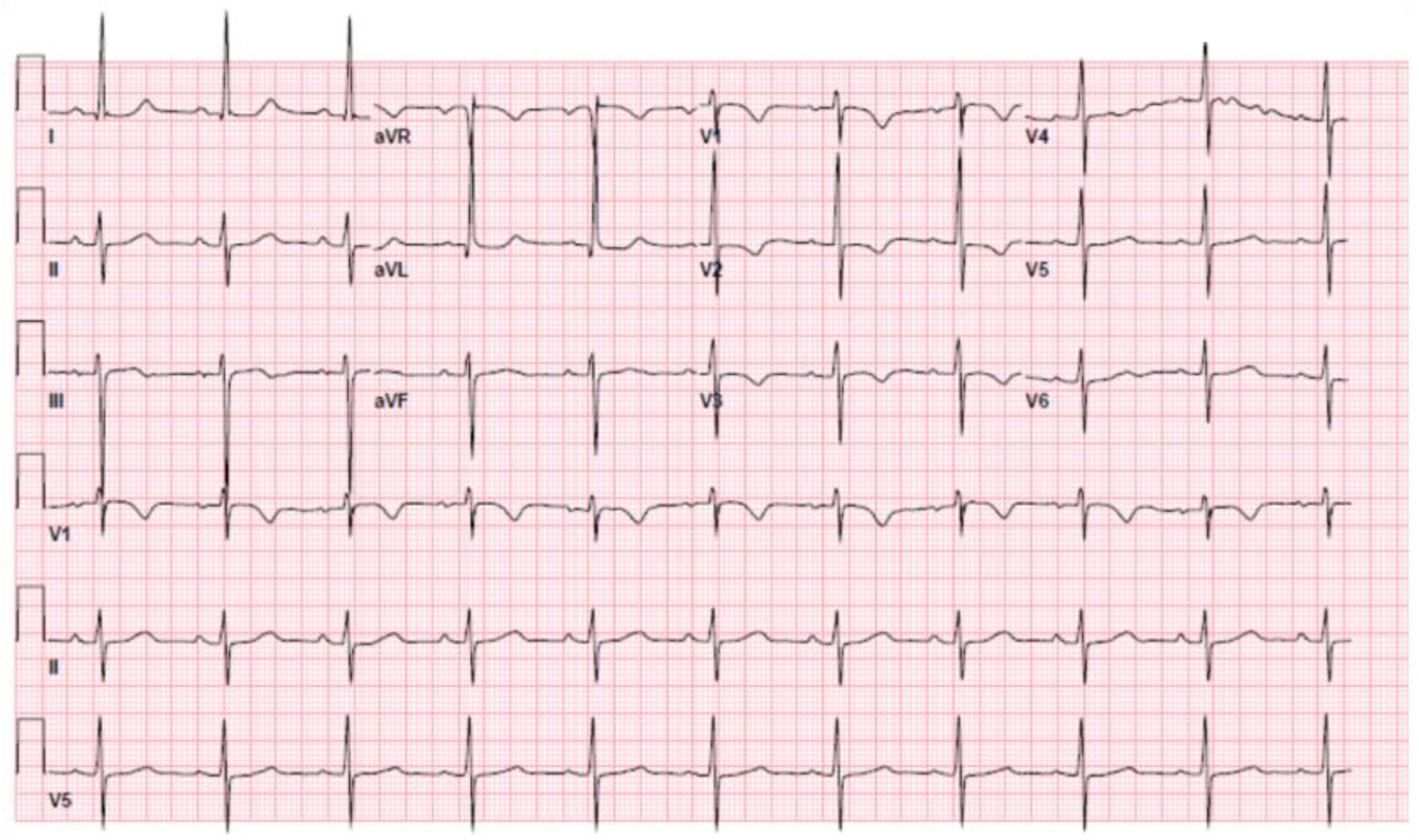
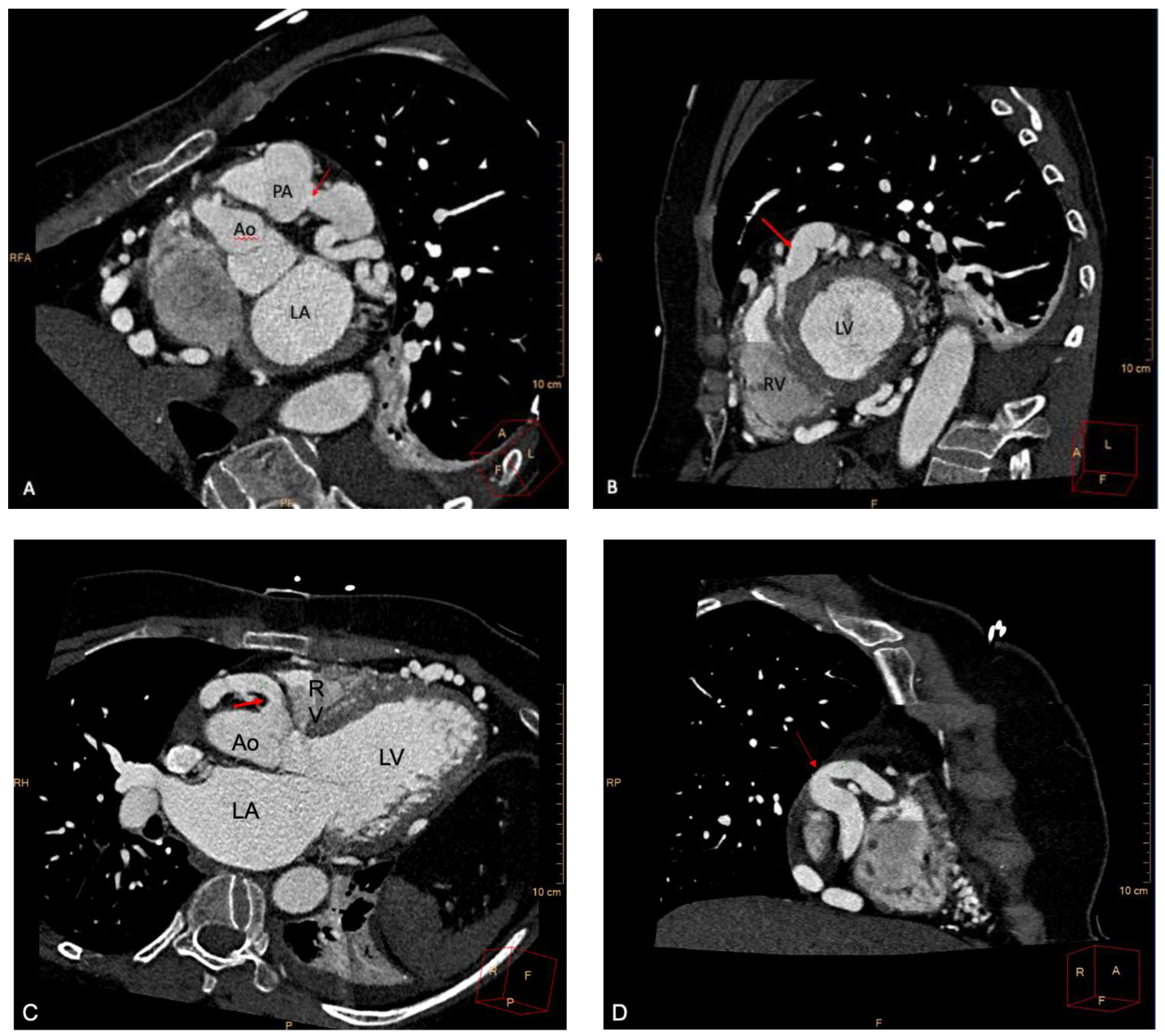
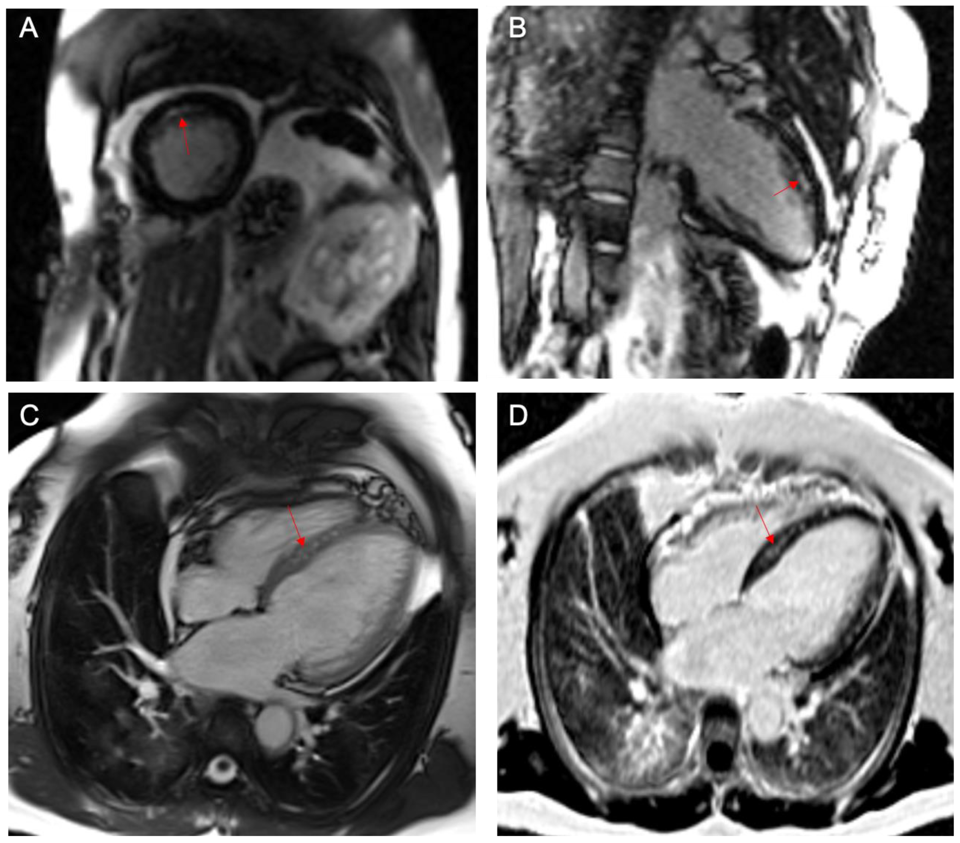
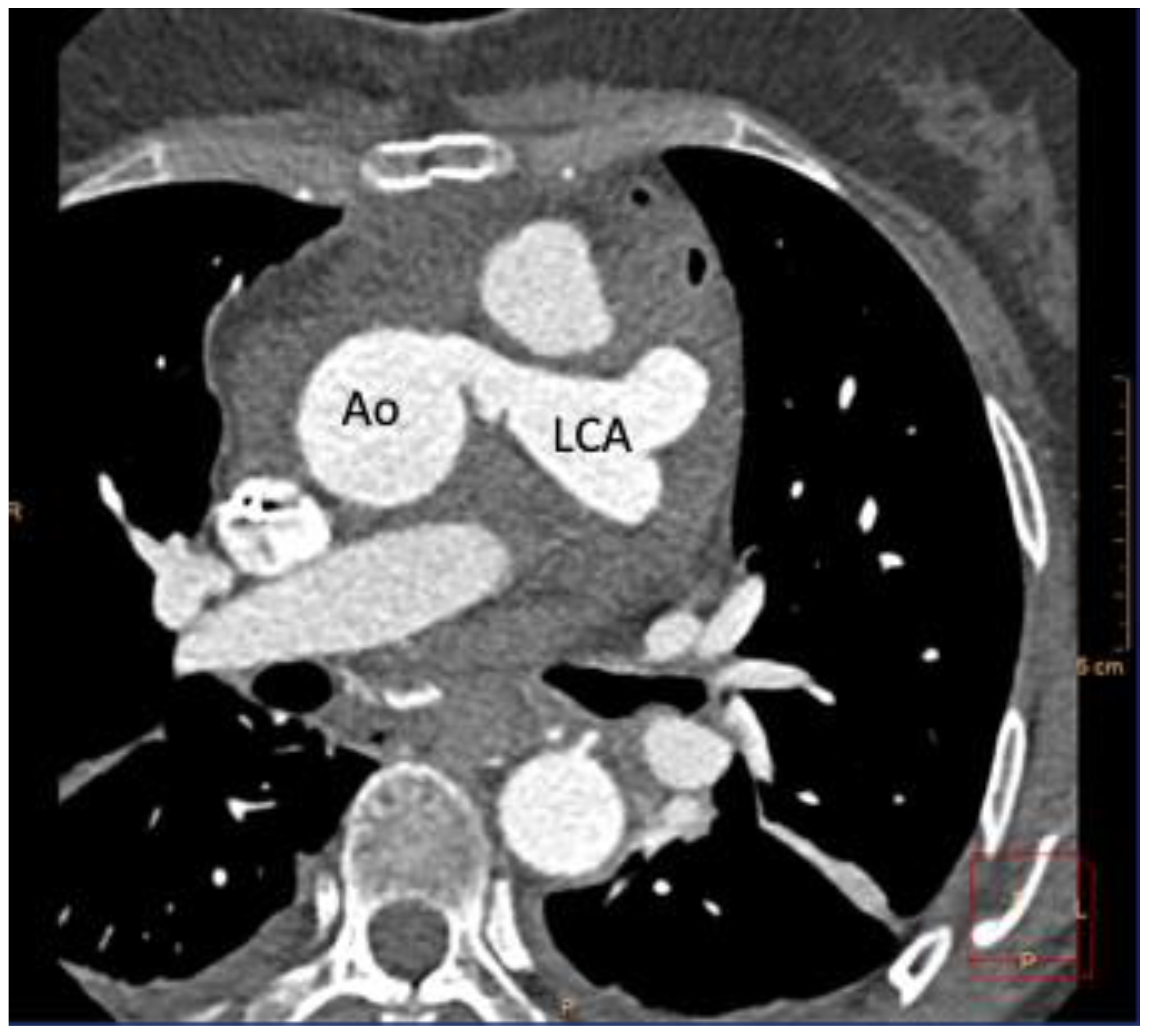
Case Presentation
3. Discussion
4. Conclusions
Supplementary Materials
Author Contributions
Funding
Institutional Review Board Statement
Informed Consent Statement
Data Availability Statement
Conflicts of Interest
References
- Angelini, P.; Velasco, J.A.; Flamm, S. Coronary anomalies: Incidence, pathophysiology, and clinical relevance. Circulation 2002, 105, 2449–2454. [Google Scholar] [CrossRef] [PubMed]
- Villa, A.D.M.; Sammut, E.; Nair, A.; Rajani, R.; Bonamini, R.; Chiribiri, A. Coronary artery anomalies overview: The normal and the abnormal. World J. Radiol. 2016, 8, 537–555. [Google Scholar] [CrossRef] [PubMed] [Green Version]
- Al-Dairy, A.; Rezaei, Y.; Pouraliakbar, H.; Mahdavi, M.; Bayati, P.; Dehaki, M.G. Surgical Repair for Anomalous Origin of the Right Coronary Artery from the Pulmonary Artery. Korean Circ. J. 2017, 47, 144–147. [Google Scholar] [CrossRef] [PubMed] [Green Version]
- Boutsikou, M.; Shore, D.; Li, W.; Rubens, M.; Pijuan, A.; Gatzoulis, M.A.; Babu-Narayan, S.V. Anomalous left coronary artery from the pulmonary artery (ALCAPA) diagnosed in adulthood: Varied clinical presentation, therapeutic approach and outcome. Int. J. Cardiol. 2018, 261, 49–53. [Google Scholar] [CrossRef]
- Yamanaka, O.; Hobbs, R.E. Coronary artery anomalies in 126595 patients undergoing coronary arteriography. Catheter. Cardiovasc. Diagn. 1990, 21, 28–40. [Google Scholar] [CrossRef]
- Regeer, M.V.; Bondarenko, O.; Zeppenfeld, K.; Egorova, A.D. Anomalous left coronary artery from the pulmonary artery: A rare cause of an out-of-hospital cardiac arrest in an adult—A case report. Eur. Heart J.-Case Rep. 2020, 4, 1–5. [Google Scholar] [CrossRef]
- Williams, I.A.; Gersony, W.M.; Hellenbrand, W.E. Anomalous right coronary artery arising from the pulmonary artery: A report of 7 cases and a review of the literature. Am. Heart J. 2006, 152, 9–17. [Google Scholar] [CrossRef]
- Mazurak, M.; Kusa, J. The radiologist’s tragedy, or Bland-White-Garland syndrome (BWGS). On the 80(th) anniversary of the first clinical description of ALCAPA (anomalous left coronary artery from the pulmonary artery). Kardiochir Torakochirurgia Pol. 2014, 11, 225–229. [Google Scholar]
- Stout, K.K.; Daniels, C.J.; Aboulhosn, J.A.; Bozkurt, B.; Broberg, C.S.; Colman, J.M. 2018 AHA/ACC guideline for the management of adults with congenital heart disease: A report of the American College of Cardiology/American Heart Association Task Force on Clinical Practice Guidelines. Circulation 2018, 139, e637–e697. [Google Scholar]
- Virmani, R.; Burke, A.P.; Farb, A. The pathology of sudden cardiac death in athletes. In The Athlete and Heart Disease; Williams, R.A., Ed.; Lippincott Williams & Wilkins: Philadelphia, PA, USA, 2000; pp. 249–272. [Google Scholar]
- Maron, B.J.; Thompson, P.D.; Puffer, J.C.; McGrew, C.A.; Strong, W.B.; Douglas, P.S.; Epstein, A.E. Cardiovascular preparticipation screening of competitive athletes: A statement for health professionals from the Sudden Death Committee (Clinical Cardiology) and Congenital Cardiac Defects Committee (Cardiovascular Disease in the Young), American Heart Association. Circulation 1996, 94, 850–856. [Google Scholar]
- Vergara-Uzcategui, C.E.; Neves, B.D.; Salinas, P.; Fernández-Ortiz, A.; Núñez-Gil, I.J. Anomalous origin of coronary arteries from pulmonary artery in adults: A case series. Eur. Heart J.-Case Rep. 2020, 4, 1–5. [Google Scholar] [CrossRef] [PubMed] [Green Version]
- Cannata’, A.; Pareek, N.; Cannata, S.; Emmanuel, Y.; Piper, S.; Dworakowski, R.; Austin, C.; MacCarthy, P. Out-of-Hospital cardiac arrest caused by ALCAPA syndrome in adulthood. Eur. Heart J. 2021, 42, 1118. [Google Scholar] [CrossRef] [PubMed]
- Parizek, P.; Haman, L.; Harrer, J.; Tauchman, M.; Rozsival, V.; Varvarovsky, I.; Pleskot, M.; Mestan, M.; Stasek, J. Bland-White-Garland syndrome in adults: Sudden cardiac death as a first symptom and long-term follow-up after successful resuscitation and surgery. Europace 2010, 12, 1338–1340. [Google Scholar] [CrossRef] [PubMed]
- Kristensen, T.; Kofoed, K.F.; Helqvist, S.; Helvind, M.; Søndergaard, L. Anomalous origin of the left coronary artery from the pulmonary artery (ALCAPA) presenting with ventricular fibrillation in an adult: A case report. J. Cardiothorac. Surg. 2008, 3, 33. [Google Scholar] [CrossRef] [Green Version]
- Dilaveris, P.; Koutagiar, I.; Alexopoulos, N.; Tsiachris, D.; Gatzoulis, K. Secondary prevention of sudden cardiac death in a 65 year untreated ALCAPA patient. Int. J. Cardiol. 2014, 176, e73–e74. [Google Scholar] [CrossRef]
- Patrianakos, A.P.; Hatzidakis, A.; Marketou, M. Parthenakis Adult-type ALCAPA syndrome: A rare coronary artery anomaly. Echocardiography 2018, 35, 1056–1059. [Google Scholar] [CrossRef] [PubMed]
- Baumgartner, H.; De Backer, J.; Babu-Narayan, S.V.; Budts, W.; Chessa, M.; Diller, G.P.; Lung, B.; Kluin, J.; Lang, I.M.; Meijboom, F.; et al. 2020 ESC Guidelines for the management of adult congenital heart disease: The Task Force for the management of adult congenital heart disease of the European Society of Cardiology (ESC). Endorsed by: Association for European Paediatric and Congenital Cardiology (AEPC), International Society for Adult Congenital Heart Disease (ISACHD). Eur. Heart J. 2021, 42, 563–645. [Google Scholar]
- Rajbanshi, B.G.; Burkhart, H.M.; Schaff, H.; Daly, R.C.; Phillips, S.D.; Dearani, J.A. Surgical strategies for anomalous origin of coronary artery from pulmonary artery in adults. J. Thorac. Cardiovasc. Surg. 2014, 148, 220–224. [Google Scholar] [CrossRef] [Green Version]
- Lotman, E.M.; Karu, K.; Mikkel, M.; Elmet, M. Late adult presentation of ALCAPA syndrome: Need for a new clinical classification? A case report and literature overview. Eur. Heart J. Case Rep. 2020, 4, 1–5. [Google Scholar] [CrossRef]
- Priori, S.G.; Blomstrom-Lundqvis, C.; Mazzanti, A.; Blom, N.; Borggrefe, M.; Camm, J.; Elliott, P.M.; Fitzsimons, D.; Robert, H.; ESC Scientific Document Group; et al. 2015 ESC Guidelines for the management of patients with ventricular arrhythmias and the prevention of sudden cardiac death: The Task Force for the Management of Patients with Ventricular Arrhythmias and the Prevention of Sudden Cardiac Death of the European Society of Cardiology (ESC). Eur. Heart J. 2015, 36, 2793–2867. [Google Scholar]
- American College of Cardiology/American Heart Association Task Force on Clinical Data Standards (ACC/AHA/HRS Writing Committee to Develop Data Standards on Electrophysiology); Buxton, A.E.; Calkins, H.; Callans, D.J.; DiMarco, J.P.; Fisher, J.D.; Greene, H.L.; Haines, D.E.; Hayes, D.L.; Heidenreich, P.A.; et al. ACC/AHA/HRS 2006 key data elements and definitions for electrophysiological studies and procedures: A report of the American College of Cardiology/American Heart Association Task Force on Clinical Data Standards (ACC/AHA/HRS Writing Committee to Develop Data Standards on Electrophysiology). Circulation 2006, 114, 2534–2570. [Google Scholar] [CrossRef] [PubMed] [Green Version]
- Fratz, S.; Hager, A.; Schreiber, C.; Schwaiger, M.; Hess, J.; Stern, H.C. Long-Term myocardial scarring after operation for anomalous left coronary artery from the pulmonary artery. Ann. Thorac. Surg. 2011, 92, 1761–1765. [Google Scholar] [CrossRef] [PubMed]
- van der Lingen, A.C.J.; Becker, M.A.J.; Kemme, M.J.B.; Rijnierse, M.T.; Spoormans, E.M.; Timmer, S.A.J.; van Rossum, A.C.; van Halm, V.P.; Germans, T.; Allaart, C.P. Reversible Cause of Cardiac Arrest and Secondary Prevention Implantable Cardioverter Defibrillators in Patients With Coronary Artery Disease: Value of Complete Revascularization and LGE-CMR. J. Am. Heart Assoc. 2021, 10, e019101. [Google Scholar] [CrossRef]
- Yau, J.M.; Singh, R.; Halpern, E.J.; Fischman, D. Anomalous origin of the left coronary artery from the pulmonary artery in adults: A comprehensive review of 151 adult cases and a new diagnosis in a 53-year-old woman. Clin. Cardiol. 2011, 34, 204210. [Google Scholar] [CrossRef]


Publisher’s Note: MDPI stays neutral with regard to jurisdictional claims in published maps and institutional affiliations. |
© 2022 by the authors. Licensee MDPI, Basel, Switzerland. This article is an open access article distributed under the terms and conditions of the Creative Commons Attribution (CC BY) license (https://creativecommons.org/licenses/by/4.0/).
Share and Cite
Prandi, F.R.; Zaidi, A.N.; LaRocca, G.; Hadley, M.; Riasat, M.; Anastasius, M.O.; Moreno, P.R.; Sharma, S.; Kini, A.; Murthy, R.; et al. Sudden Cardiac Arrest in an Adult with Anomalous Origin of the Left Coronary Artery from the Pulmonary Artery (ALCAPA): Case Report. Int. J. Environ. Res. Public Health 2022, 19, 1554. https://0-doi-org.brum.beds.ac.uk/10.3390/ijerph19031554
Prandi FR, Zaidi AN, LaRocca G, Hadley M, Riasat M, Anastasius MO, Moreno PR, Sharma S, Kini A, Murthy R, et al. Sudden Cardiac Arrest in an Adult with Anomalous Origin of the Left Coronary Artery from the Pulmonary Artery (ALCAPA): Case Report. International Journal of Environmental Research and Public Health. 2022; 19(3):1554. https://0-doi-org.brum.beds.ac.uk/10.3390/ijerph19031554
Chicago/Turabian StylePrandi, Francesca Romana, Ali N. Zaidi, Gina LaRocca, Michael Hadley, Maria Riasat, Malcolm O. Anastasius, Pedro R. Moreno, Samin Sharma, Annapoorna Kini, Raghav Murthy, and et al. 2022. "Sudden Cardiac Arrest in an Adult with Anomalous Origin of the Left Coronary Artery from the Pulmonary Artery (ALCAPA): Case Report" International Journal of Environmental Research and Public Health 19, no. 3: 1554. https://0-doi-org.brum.beds.ac.uk/10.3390/ijerph19031554