Article

The Role of a Navigational Radiofrequency Ablation Device and Concurrent Vertebral Augmentation for Treatment of Difficult-to-Reach Spinal Metastases

1
Regional Referral Center for Oncologic Disease, Department of Oncological and Interventional Radiology, Businco Hospital, A.O. Brotzu, 09100 Cagliari, Italy
2
Institute for Global Health, UCL, Royal Free Campus Rowland Hill Street, London NW3 2PF, UK
3
Division of Nuclear Medicine, Businco Hospital, Regional Referral Center for Oncologic Disease, A.O. Brotzu, 09100 Cagliari, Italy
4
Unit of General Surgery, Department of Clinical and Experimental Medicine, University of Sassari, 07100 Sassari, Italy
*
Author to whom correspondence should be addressed.
Submission received: 17 September 2021 / Revised: 3 October 2021 / Accepted: 5 October 2021 / Published: 8 October 2021
(This article belongs to the Special Issue Treatment of Bone Metastasis)

Abstract

:
Aims: The purpose of this study was to assess the effectiveness of a navigational radiofrequency ablation device with concurrent vertebral augmentation in the treatment of posterior vertebral body metastatic lesions, which are technically difficult to access. Primary outcomes of the study were evaluation of pain palliation and radiologic assessment of local tumor control. Materials and Methods: Thirty-five patients with 41 vertebral spinal metastases involving the posterior vertebral body underwent computed tomography-guided percutaneous targeted radiofrequency ablation, with a navigational radiofrequency ablation device, associated with vertebral augmentation. Twenty-one patients (60%) had 1 or 2 metastatic lesions (Group A) and fourteen (40%) patients had multiple (>2) vertebral lesions (Group B). Changes in pain severity were evaluated by visual analog scale (VAS). Metastatic lesions were evaluated in terms of radiological local control. Results: The procedure was technically successful in all the treated vertebrae. Among the symptomatic patients, the mean VAS score dropped from 5.7 (95% CI 4.9–6.5) before tRFA and to 0.9 (95% CI 0.4–1.3) after tRFA (p < 0.001). The mean decrease in VAS score between baseline and one week follow up was 4.8 (95% CI 4.2–5.4). VAS decrease over time between one week and one year following radiofrequency ablation was similar, suggesting that pain relief was immediate and durable. Neither patients with 1–2 vertebral metastases, nor those with multiple lesions, showed radiological signs of local progression or recurrence of the tumor in the index vertebrae during a median follow up of 19 months (4–46 months) and 10 months (4–37 months), respectively. Conclusion: Treatment of spinal metastases with a navigational radiofrequency ablation device and vertebral augmentation can be used to obtain local tumor control with immediate and durable pain relief, providing effective treatment in the multimodality management of difficult-to-reach spinal metastases.

1. Introduction

The incidence of bone metastases in patients with cancer is extremely high: 84% for prostate, 72% for breast, 60% for thyroid, 37% for kidney, 33% for pancreas, and 31% for lung [1,2]. In particular, vertebral metastases occur in approximately 60–70% of patients with a primary tumor [3]. This high frequency of spine involvement can be due to the high hematopoietic activity and vascularization of the vertebrae [4]. Metastatic spinal lesions can involve one or two vertebrae or, in some cases, can affect multiple vertebrae. Conventional treatment regimens such as radiotherapy, chemotherapy, and analgesic drugs may have shortcomings in the treatment of these lesions [4,5,6]. Surgical decompression and stabilization represent an option for patients with spine instability and neurologic symptoms, especially in those with a longer life expectancy [4,7]. Several tumor ablation techniques, such as cryoablation, radiofrequency, microwave ablation, and laser interstitial thermal therapy, have been developed for percutaneous treatment of vertebral metastases and osseus metastatic lesions in general, and each of these methods has specific indications and advantages [4,8,9,10,11]. In particular, radiofrequency ablation (RFA) associated to vertebral augmentation (VA) has been gaining popularity in the multidisciplinary treatment of spinal metastases without spinal cord compression [5,12,13,14,15]. In addition, the combined treatment with RFA and VA of spinal metastases improves oncological outcomes in comparison with vertebroplasty or radiofrequency ablation alone [2,4,5,11]. However, the standard RFA involves the use of straight needles through a transpeduncolar approach, which can be inadequate for obtaining ablation of tumor tissue located in difficult-to-reach metastases, such as those located in the posterior part of the vertebral body (Figure 1). Recently, this important disadvantage of the use of straight needles has been overcome by targeted radiofrequency ablation (tRFA). In tRFA, a navigational radiofrequency ablation device is used, which contains an articular extensible electrode that allows treatment of posterior spinal metastases with a curved needle (Figure 2) [4,5,13,14,15]. To date, few authors have reported on the use of a navigational radiofrequency ablation device for the treatment of difficult-to-reach spinal metastases, and many aspects need to be clarified [4,5,13,14,15] (Table 1). The purpose of this study was to assess the effectiveness of tRFA and VA using a navigational radiofrequency ablation device in the treatment of posterior vertebral body metastatic lesions that are technically difficult to access via a transpedicular approach. Primary outcomes of the study were evaluation of pain palliation, and radiologic assessment of local tumor control.

2. Material and Methods

2.1. Study Population and Inclusion Criteria

Patients who underwent minimally invasive CT fluoroscopy-guided percutaneous tRFA and VA between November 2013 and March 2017 were identified. Patients were included in the study if they had vertebral lesions from T1 to L5, with evidence of osteolytic or mixed metastatic lesions in the posterior vertebral body (i.e., in the posterior half of the vertebra), irrespective of whether pathologic fractures occurred. Patients with disruption of the posterior cortical wall were also included. Patients with metastases located in the spinal cord and/or nerve root compression were excluded.

2.2. Group Definition and Preoperative Assessment

Patients were classified into two groups according to the number of metastases located in the spine:
(1)
Group A: patients with 1 or 2 vertebral lesions.
(2)
Group B: patients having more than 2 metastatic spine lesions.
All patients were assessed prior to surgery with a CT scan or MRI and, in some cases, PET scan in order to study the metastatic vertebral lesion and to plan the percutaneous intervention. CT scan, MRI, or PET scan were performed at 1, 6, and 12 months post tRFA treatment to assess the local activity of the vertebral metastases and/or residual viable tumor, and/or recurrence/growth of the spinal metastases (Figure 2). Pain severity was documented using a visual analog scale (VAS—scaled from a minimum of 0 to a maximum of 10) [16] the day before the treatment, and then one week, six months, and twelve months after tRFA and VA treatment.

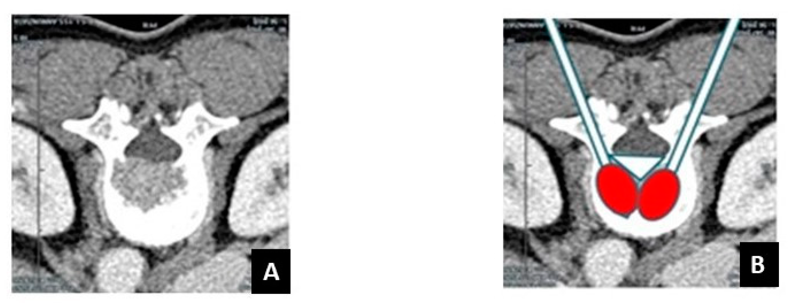
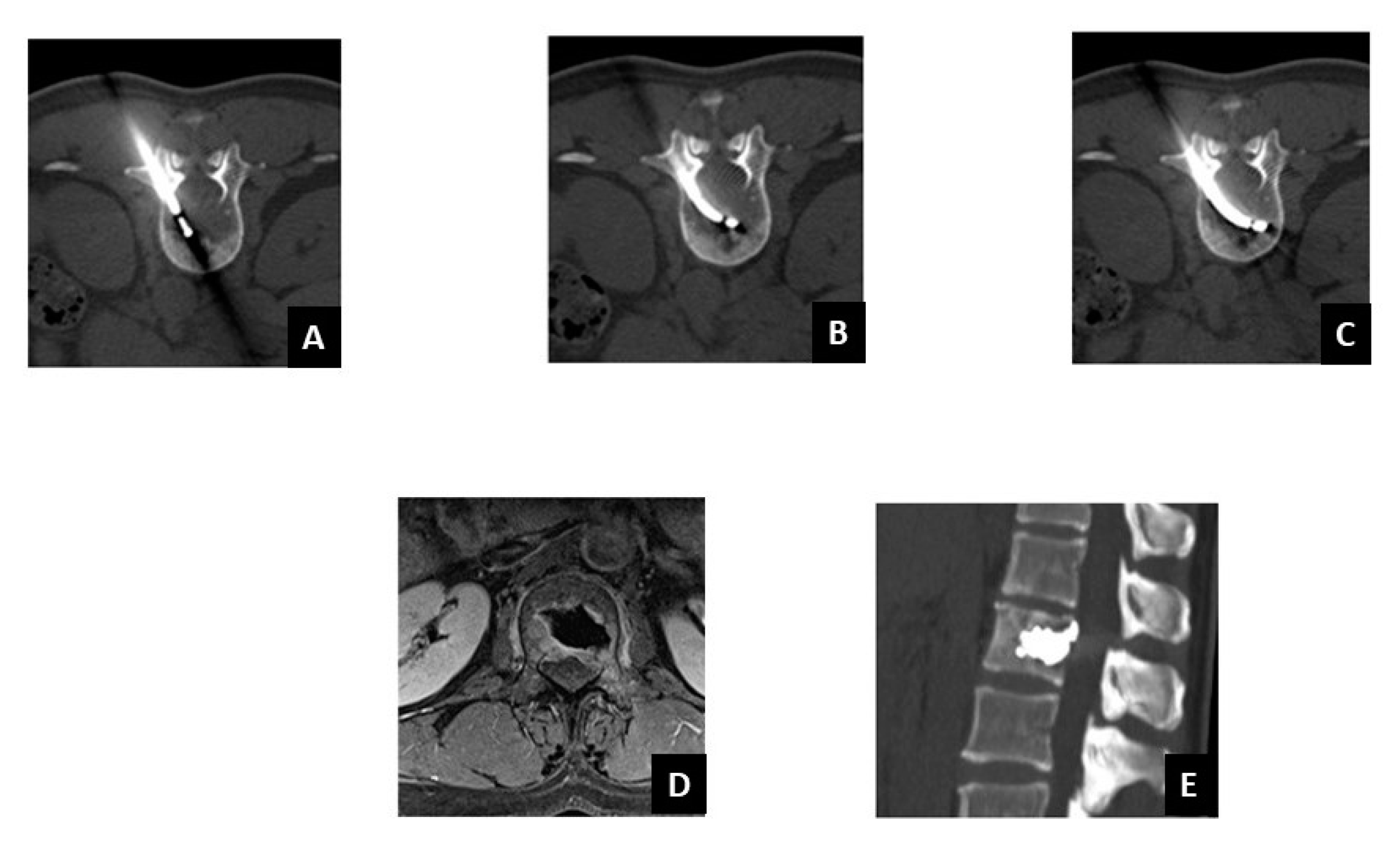
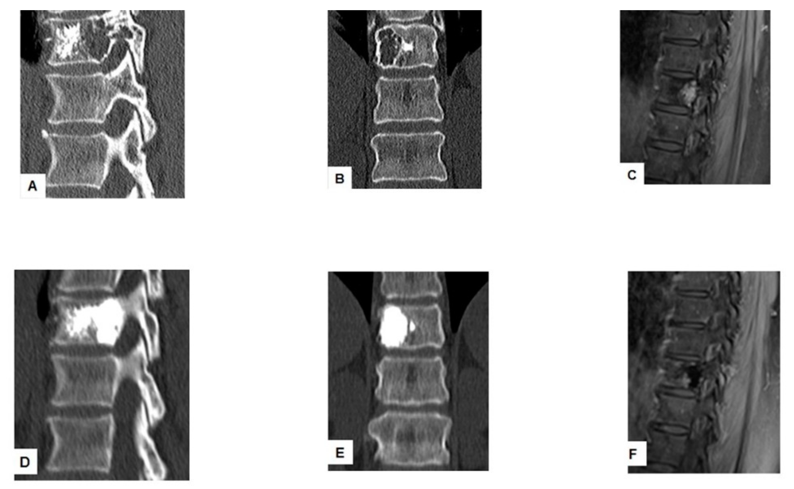
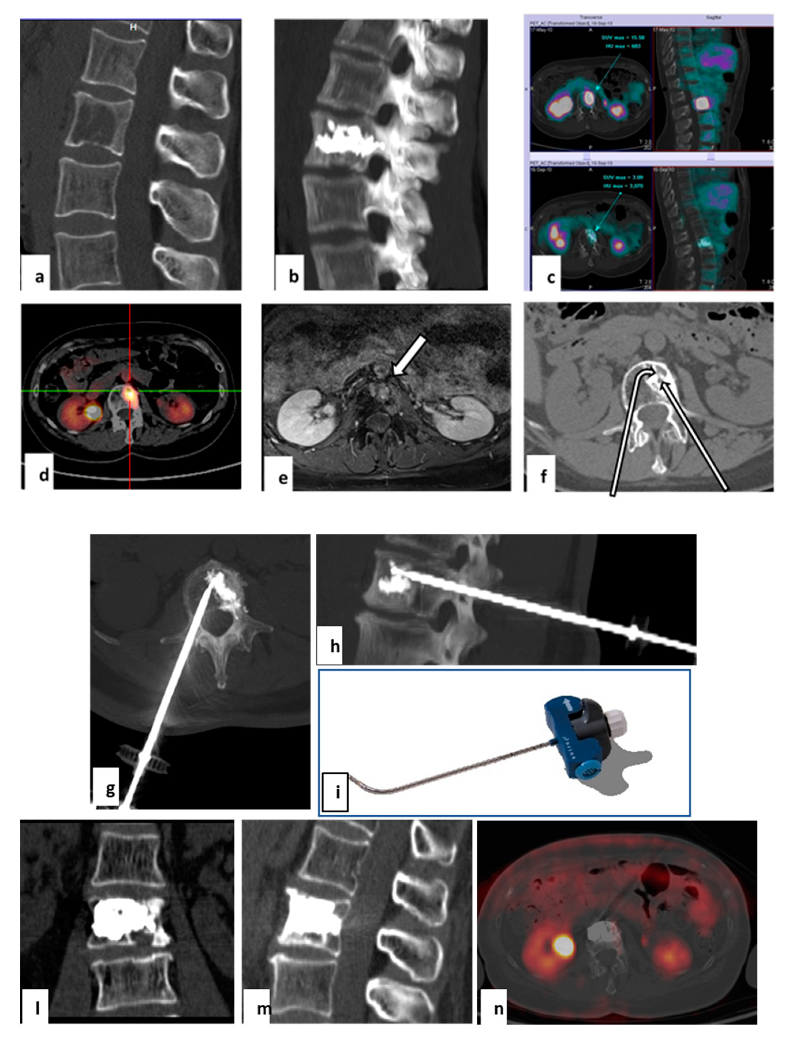
2.3. Target Radiofrequency Ablation (tRFA) and Vertebral Augmentation (VA) Procedure

Using CT fluoroscopy guidance (SOMATOM Sensation, Siemens, AG, Forchheim, Germany), the spine lesion was identified, measured, and marked to establish and validate the desired needle pathway. The procedures were performed according to the following steps: (i) anesthesia: conscious sedation with intravenous infusion of fentanyl citrate (0.1 mg/2 mL diluted with saline solution) followed by subcutaneous injection of lidocaine hydrochloride (2%) performed until the pedicle periosteal and the bone cortex. (ii) tRFA: under CT scan, a 10-G vertebroplasty needle was introduced and, coaxially to it, the bipolar tRFA electrode (STAR, Merit Medical Systems, Inc., South Jordan, UT, USA) was placed into the vertebral lesion to deliver the RF energy and ablate the metastases properly. The instrument has two thermocouples (the distal at 10 mm and the proximal at 15 mm from the ablation core) which actively monitor the temperature of the tumor avoiding undesired burns around the lesion. (iii) Vertebral augmentation: using the same vertebroplasty needle, in order to minimize cement leakage risk, ultra-high viscosity bone cement was inserted through a controller under low-pressure injection (STABILIT, Merit Medical Systems, Inc.) for optimal filling of the vertebral lesion (Figure 3).

2.4. Postoperative Follow Up

CT scan, MRI, or PET (in selected cases) were performed at 1, 6, and 12 months after tRFA-VA treatment to assess local activity of the vertebral metastases and/or residual viable tumor, as well as recurrence/growth of the spinal metastases. Tumor progression was defined as an increase of the size of metastasis on CT or MRI. Stable disease was considered as a stable metastasis diameter along with no contrast enhancement. Recurrence at follow-up imaging was defined as contrast enhancement or enlargement at the site of successfully treated lesion, and/or the occurrence of osteolytic lesions near the treated areas.

2.5. Statistical Analysis
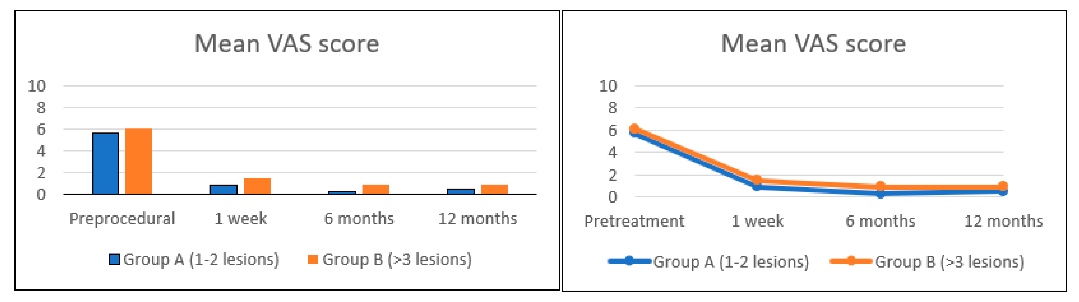
Changes over time in the VAS score were evaluated using a linear mixed-effect model to account for the correlation between VAS scores within the same patient. Changes in the VAS score before and one week after tRFA were modeled with a dummy variable, while scores from one week after tRFA onwards were modeled as a linear function of time. All statistical analyses were performed using R Statistical Software (version 3.3.3) and p < 0.05 was considered significant.

3. Results

Forty-one vertebral metastases were treated in thirty-five patients (22 females and 13 males, median (IQR) age 59 (56–70) years). Breast cancer was the most common type of primary tumor (n = 16, 46%), followed by lung (n = 5, 14%), colon (n = 4, 11%), prostate (n = 3, 9%), and kidney (n = 2, 6%). None of the patients had signs of neurological deficits, nor required an open surgical stabilization. Twenty-one (60%) patients belonged to Group A (1 or 2 vertebral metastatic lesions) (Table 2), the remaining fourteen (40%) to Group B (multiple metastases in the spine) (Table 3). In one case, tRFA and VA were successfully used after a tumor recurrence after a vertebral microwave ablation (Figure 4). Two patients belonging to Group A were asymptomatic. All the procedures were uneventful and well tolerated, without complications related to tRFA and concurrent VA. Asymptomatic cement leakage occurred in 3 of 41 treated vertebrae (7.3%). Ten patients (29%) received radiotherapy before tRFA, five (14%) after tRFA, and twenty (57%) patients did not receive radiotherapy at all. No local relapse nor tumor progression was observed in both Group A during a median follow up of 12 months (4–46 months), and Group B during a median follow up of 10.5 months (4–37 months). Eight patients in Group B and two patients in Group A died as a result of disease progression. In the 33 patients with painful metastases, the mean value of VAS score before the procedure, after 1 week, 6 months, and 12 months was 5.7 ± 1.6, 0.9 ± 0.7, 0.3 ± 0.4, 0.5 ± 0.6, and 6.1 ± 1.8, 1.5 ± 1.0, 0.9 ± 0.7, 0.9 ± 0.7 in Group A and Group B, respectively (Figure 5). Considering the patients as a whole, the mean VAS score dropped from 5.7 (95% CI 4.9–6.5) before tRFA to 0.9 (95% CI 0.4–1.3) after tRFA (Figure 5). The mean decrease in the VAS score between baseline and follow up was 4.8 (4.2–5.4, p < 0.0001). With the numbers available, we found no difference in the VAS score over time from one week up to one year after tRFA and VA, suggesting that pain relief was immediate and durable. There was no difference in the VAS scores before and after surgery between the two groups of the study. Use of steroids and non-steroidal anti-inflammatory drugs taken by patients in both groups were reduced or completely suspended after the procedure. Patient characteristics and results are summarized in Table 2 and Table 3.

4. Discussion

The use of tRFA associated to vertebroplasy with VA has been gaining importance in the management of spine metastases [5,13,14,17]. However, classical RFA with straight needles usually permits the treatment of lesions located in the anterior part of the vertebral body. In this study, we have evaluated the usefulness of tRFA with a navigation radiofrequency ablation device, which overcomes some limitations of conventional RFA [5,13,14,15]. Our results demonstrated that tRFA with a dedicated navigation radiofrequency device, with concurrent VA, can obtain satisfactory results in terms of pain reduction and local tumor control.
This study has some limitations, the main being its retrospective nature and the small sample size. Moreover, the overlapping with radiation therapy in some patients and the use of drugs for pain relief, may have influenced the VAS score. However, this study has some points of strengths, in that it supports the use of a navigation radiofrequency ablation device for the treatment of metastases located in the posterior part of the vertebrae. As we reported in Table 1, only few studies have addressed the use of that approach, and some authors limited their experience to patients with vertebral osteoid osteoma [15]. To the best of our knowledge, this is the first single-institution report from a European institution on the use of a navigational system for the tRFA of vertebral metastases.
Spinal metastases represent a complex clinical scenario in patients affected from cancer, especially in those with decreased life expectancy [5,14,18]. Those lesions can result in severe complications such as pain, vertebral fractures, hypercalcemia, spinal instability, and neurological symptoms due to spinal cord and/or nerve root compression [13,14,15,19]. tRFA with concurrent vertebroplasty has shown effectiveness for vertebral lesions without spinal cord compression. Normally, RFA treatment in the spine is used for lesions located in the anterior vertebral body and far from neurovascular structures, where a conventional RF ablation straight device can easily reach the metastases [13,20]. The navigational radiofrequency ablation device differs in that it has the ability to properly curve the needle, which allows navigation of the instrument; this permits the obtaining of necrosis of tumor cells even in difficult locations of the vertebral body. Furthermore, the steerable needle is able to approach vertebral lesions that are technically challenging to access with straight needles, such as metastases close to the posterior vertebral wall and spinal cord. To note, all patients in the present study had one or more metastatic lesions located in the posterior vertebral body, without signs of spinal cord compression. The latter commonly represents a contraindication for percutaneous ablation as upfront treatment, being corticosteroids, surgery, radiotherapy, and systemic therapy the preferred options [12,21,22].
One of the purposes of our work was to evaluate the role of tRFA with concurrent VA in the control of pain, using a dedicated navigational device. Cancer-induced bone pain recognizes multiple mechanisms, including disruption of bone homeostasis, release of neurochemicals that modulate pain, and microglia activation. Furthermore, instability of a vertebral body usually causes pain, which is poorly localized, and sometimes becomes intractable [23]. It has shown that VA is an effective option for pain control and stabilization of pathologic fractures of the spine [5,14,24,25,26]. Progressive pain is a frequent symptom with important repercussions in the quality of life. tRFA in combination with VA is the preferred approach in certain situations, such as single vertebral metastases, spinal active residual tumor tissue, and occurrence of vertebral compression fractures with cortical interruptions after radiotherapy [24,25,26]. However, our results confirm that pain control is achieved by using tRFA-VA also in patients with multiple localizations. In fact, the VAS scores indicated that pain symptoms were durably relieved in both the study groups. To note, the mean decrease in the VAS score between baseline and follow up was 4.8 (p < 0.0001), and no difference was observed in the VAS score over time from one week up to one year after tRFA and VA, suggesting that pain relief was immediate and durable. These findings were consistent to previous studies. In the multicentric study from Anchala and colleagues, significant (p < 0.01) decrease in the VAS scores at one week, one month, and six months was observed, although only 62% of the spinal lesions in the largest institution participating in the study was located in the posterior vertebral body [14]. Reyes et al. reported that mean VAS scores decreased from 7.9 pre-procedure to 3.5 post-procedure (p < 0.0001) in their multicentric study involving almost 50 patients [4].
The second purpose of the present study was to evaluate the metastatic lesions in terms of radiological local control after tRFA and VA treatment. Our results reinforce the concept that treating patients with a navigational radiofrequency device with the goal to achieve tumor control is feasible [5]. Twenty-one patients with 1 or 2 metastatic lesions showed no local relapse or tumor progression during a median follow up of 19 months. Similarly, none of the 14 patients with multiple lesions developed local relapse or tumor progression after a follow up of 10 months. These findings may be partially explained by the small sample size as well as the short follow-up duration. However, data regarding follow-up imaging and local relapse after the use of a navigational radiofrequency device for spinal metastases are very scarce. In the study from Anchala et al., only 13 out of 92 patients received follow-up postoperative imaging, of whom 3 developed tumor progression at the treated site [14]. Hillen et al. reported on the use of a navigational radiofrequency ablation device on 26 patients with metastatic posterior vertebral body. Of them, 13 patients underwent follow-up imaging, of whom 3 developed tumor progression at the treated site [13]. In the multicentric study from Reyes et al., 49 patients received navigational radiofrequency ablation and VA for vertebral metastases. Among those who had postoperative follow-up imaging, MRI showed tumor extension into epidural space and neural foramen in one case, and a new epidural extension from a different primary tumor in another case [4].
As described elsewhere, the goal of performing tRFA before vertebroplasty is to destroy tumor tissue, to reduce the intrametastatic pressure, and to thrombose intravertebral venous plexus [27]. The purpose of vertebroplasty with VA is to stabilize the vertebra by stabilization of vertebral microfractures with cement [28]. Literature reports described that polymethylmethacrylate alone and the exothermic reaction generated during its polymerization are not able to completely destroy tumor cells [8,29]. Some authors even reported that the injection of the cement in a metastatic vertebral body increases the intrametastatic pressure with a risk of dissemination of tumor cells [30]. Four patients out of thirty-five enrolled in this study were treated with tRFA after they underwent previous vertebroplasty alone, and they developed new signs or reappearance of the metastatic vertebral lesion associated with pain (Figure 3). Although this phenomenon involves only a few patients, it underlines that vertebroplasty alone is not efficient in local tumor control. Two (2) asymptomatic patients were treated with tRFA in combination with vertebroplasty, which is a further example of the effectiveness of the ablation treatment. In fact, a goal of tRFA-VA is to obtain local control of the bone disease by destroying the spinal metastases, avoiding extension into the neural structures, and limiting vertebral fracture risk associated with radiotherapy, as reported in the literature [24,25]. Furthermore, this approach may avoid skeletal-related events associated with standard treatments (chemotherapy, radiotherapy, bisphosphonates, and surgery), which include pain, risk of fracture, and nerve or spinal cord compression [27]. In addition, it has been reported that tRFA-VA may improve anti-tumor local response in association with radiotherapy [31].
External-beam radiation therapy is another option to treat pain and achieve local control and represents the standard treatment for pain relief and local control of spinal metastases [7,12,13]. However, radiotherapy has certain limitations as only a few tumor types are considered to be highly radiosensitive. Furthermore, pain relief obtained with radiotherapy may be transient and delayed [13,27,32,33]. In our study, 10 patients received low-dose palliative radiotherapy before tRFA and vertebroplasty treatment. Unfortunately, our data did not provide insight about the best timing of tRFA treatment in the oncologic patient care pathway. Optimal radiotherapy timing in combination of tRFA and vertebroplasty remains unclear and needs to be assessed in future studies. Interestingly, the effectiveness of radiation therapy after surgical fixation of impending or actual pathologic fractures in the long bones has been questioned. A metanalysis involving two studies has shown conflicting results, and the authors concluded that data were insufficient to conclude whether postoperative RT after surgical stabilization should be standard care for long bone metastases [34]. Nonetheless, in current literature there is evidence that both radiotherapy and percutaneous termoablation modalities such as tRFA have been shown to be safe and effective in the treatment of painful vertebral metastases, and thus a synergic effect can be hypothesized in a multidisciplinary approach to vertebral metastases [5,35]. Our data corroborate that the sequence tRFA-VA and radiotherapy can be helpful in cases where pain still persists after percutaneous treatment; in fact, in five cases of the present series where cancer-related pain was consistent, radiotherapy was used to optimize pain control after tRFA-VA.
It is our thought that the goal of the tRFA in treating patients with metastatic spinal tumors should be not only the pain relief, but also the achievement of an oncological outcome in terms of stopping lesions from spreading into the rest of the vertebral body to avoid neurological damage. Our study indicates that percutaneous treatment of spinal metastases with tRFA and vertebroplasty can be used in a multimodality therapeutic approach. In addition, it may bridge the gaps reported for conventional treatment methods in vertebral metastases management. Radiation therapy, chemotherapy, “open” spinal surgeries, vertebroplasty, and “conservative” treatment are treatments that must be tailored for each patient, considering the nature of the tumor, life expectancy, pain, metastases at high risk of fracture, and compression of neurovascular structures [36].
In conclusion, tRFA using a dedicated radiofrequency ablation device, with concurrent VA, can achieve satisfactory results in terms of pain palliation and local tumor control in patients with posterior vertebral body metastases. That approach may widen the field of application of termoablation in the management of spinal metastases. Further studies with larger cohorts are necessary to establish the optimal role of tRFA and vertebroplasty in association with radiotherapy in the multidisciplinary treatment of vertebral metastases.

Author Contributions

Conceptualization, C.P. and A.F.; methodology, D.D.F.; formal analysis, D.D.F.; investigation, N.B. and L.M.; writing—original draft preparation, C.P. and D.D.F.; writing—review and editing, A.F.; visualization, N.B. and L.M. All authors have read and agreed to the published version of the manuscript.

Funding

This research received no external funding.

Institutional Review Board Statement

The study was conducted according to the guidelines of the Declaration of Helsinki and approved by the Institutional Review Board of Businco Hospital, Regional Referral Center for Oncologic Disease, A.O. Brotzu. Cagliari, Zip code 09100, Italy (protocol number 108/21).

Informed Consent Statement

The indications, risks, and benefits of the procedure were discussed with all the patients, and informed consent to perform this treatment was obtained in all cases.

Data Availability Statement

Data will be available under reasonable request.

Conflicts of Interest

The authors declare no conflict of interest.

References

  1. Coleman, R. Metastatic bone disease: Clinical features, pathophysiology and treatment strategies. Cancer Treat. Rev. 2001, 27, 165–176. [Google Scholar] [CrossRef]
  2. Yang, P.-L.; He, X.-J.; Li, H.-P.; Zang, Q.-J.; Wang, G.-Y. Image-guided minimally invasive percutaneous treatment of spinal metastasis. Exp. Ther. Med. 2017, 13, 705–709. [Google Scholar] [CrossRef] [Green Version]
  3. Shah, L.M.; Salzman, K.L. Imaging of Spinal Metastatic Disease. Int. J. Surg. Oncol. 2011, 2011, 769753. [Google Scholar] [CrossRef] [PubMed] [Green Version]
  4. Reyes, M.; Georgy, M.; Brook, L.; Ortiz, O.; Brook, A.; Agarwal, V.; Muto, M.; Manfre, L.; Marcia, S.; Georgy, B.A. Multicenter clinical and imaging evaluation of targeted radiofrequency ablation (t-RFA) and cement augmentation of neoplastic vertebral lesions. J. Neurointerv. Surg. 2018, 10, 176–182. [Google Scholar] [CrossRef] [PubMed]
  5. Ma, Y.; Wallace, A.N.; Madaelil, T.P.; Jennings, J.W. Treatment of osseous metastases using the Spinal Tumor Ablation with Radiofrequency (STAR) system. Expert Rev. Med. Devices 2016, 13, 1137–1145. [Google Scholar] [CrossRef] [PubMed]
  6. Dosani, M.; Lucas, S.; Wong, J.; Weir, L.; Lomas, S.; Cumayas, C.; Fisher, C.; Tyldesley, S. Impact of the Spinal Instability Neoplastic Score on Surgical Referral Patterns and Outcomes. Curr. Oncol. 2018, 25, 53–58. [Google Scholar] [CrossRef] [PubMed] [Green Version]
  7. Bollen, L.; Van Der Linden, Y.M.; Pondaag, W.; Fiocco, M.; Pattynama, B.P.M.; Marijnen, C.; Nelissen, R.; Peul, W.C.; Dijkstra, P.S. Prognostic factors associated with survival in patients with symptomatic spinal bone metastases: A retrospective cohort study of 1 043 patients. Neuro-Oncology 2014, 16, 991–998. [Google Scholar] [CrossRef] [Green Version]
  8. Pusceddu, C.; Sotgia, B.; Fele, R.M.; Ballicu, N.; Melis, L. Combined Microwave Ablation and Cementoplasty in Patients with Painful Bone Metastases at High Risk of Fracture. Cardiovasc. Interv. Radiol. 2016, 39, 74–80. [Google Scholar] [CrossRef]
  9. Tomasian, A.; Jennings, J.W. Percutaneous minimally invasive thermal ablation for management of osseous metastases: Recent advances. Int. J. Hyperth. 2019, 36, 3–12. [Google Scholar] [CrossRef]
  10. Pusceddu, C.; Dessì, G.; Melis, L.; Fancellu, A.; Ruggiu, G.; Sailis, P.; Congia, S.; Derudas, D.; Cau, R.; Senis, I.; et al. Combined Microwave Ablation and Osteosynthesis for Long Bone Metastases. Medicina 2021, 57, 825. [Google Scholar] [CrossRef]
  11. Delank, K.S.; Wendtner, C.; Eich, H.T.; Eysel, P. The treatment of spinal metastases. Dtsch. Arztebl. Int. 2011, 108, 71–79; quiz 80. [Google Scholar] [CrossRef]
  12. NCCN Guidelines Version 2.2021. Metastatic Spine Tumors. Available online: https://www.nccn.org/professionals/physician_gls/pdf/cns.pdf (accessed on 15 June 2021).
  13. Hillen, T.J.; Anchala, P.; Friedman, M.V.; Jennings, J.W. Treatment of metastatic posterior vertebral body osseous tumors by using a targeted bipolar radiofrequency ablation device: Technical note. Radiology 2014, 273, 261–267. [Google Scholar] [CrossRef] [PubMed] [Green Version]
  14. Anchala, P.R.; Irving, W.D.; Hillen, T.J.; Friedman, M.V.; Georgy, B.A.; Coldwell, D.M.; Tran, N.D.; Vrionis, F.D.; Brook, A.; Jennings, J.W. Treatment of metastatic spinal lesions with a navigational bipolar radiofrequency ablation device: A multicenter retrospective study. Pain Physician 2014, 17, 317–327. [Google Scholar] [PubMed]
  15. Tomasian, A.; Jennings, J.W. Spinal Osteoid Osteoma: Percutaneous Radiofrequency Ablation Using a Navigational Bipolar Electrode System. AJR Am. J. Roentgenol. 2018, 211, 856–860. [Google Scholar] [CrossRef] [PubMed]
  16. Delgado, D.A.; Lambert, B.S.; Boutris, N.; Mcculloch, P.; Robbins, A.B.; Moreno, M.; Harris, J.D. Validation of Digital Visual Analog Scale Pain Scoring with a Traditional Paper-based Visual Analog Scale in Adults. J. Am. Acad. Orthop. Surg. Glob. Res. Rev. 2018, 2, e088. [Google Scholar] [CrossRef]
  17. Deschamps, F.; Farouil, G.; Ternes, N.; Gaudin, A.; Hakime, A.; Tselikas, L.; Teriitehau, C.; Baudin, E.; Auperin, A.; De Baere, T. Thermal ablation techniques: A curative treatment of bone metastases in selected patients? Eur. Radiol. 2014, 24, 1971–1980. [Google Scholar] [CrossRef]
  18. Khan, L.; Mitera, G.; Probyn, L.; Ford, M.; Christakis, M.; Finkelstein, J.; Donovan, A.; Zhang, L.; Zeng, L.; Rubenstein, J.; et al. Inter-Rater Reliability between Musculoskeletal Radiologists and Orthopedic Surgeons on Computed Tomography Imaging Features of Spinal Metastases. Curr. Oncol. 2011, 18, e282–e287. [Google Scholar] [CrossRef] [PubMed] [Green Version]
  19. Clemons, M.; Gelmon, K.A.; Pritchard, K.I.; Paterson, A.H. Bone-targeted agents and skeletal-related events in breast cancer patients with bone metastases: The state of the art. Curr. Oncol. 2012, 19, 259–268. [Google Scholar] [CrossRef] [Green Version]
  20. Muto, M.; Guarnieri, G.; Giurazza, F.; Manfrè, L. What’s new in vertebral cementoplasty? Br. J. Radiol. 2016, 89, 20150337. [Google Scholar] [CrossRef] [Green Version]
  21. Robson, P. Metastatic spinal cord compression: A rare but important complication of cancer. Clin. Med. 2014, 14, 542–554. [Google Scholar] [CrossRef]
  22. Boussios, S.; Cooke, D.; Hayward, C.; Kanellos, F.S.; Tsiouris, A.K.; Chatziantoniou, A.A.; Zakynthinakis-Kyriakou, N.; Karathanasi, A. Metastatic Spinal Cord Compression: Unraveling the Diagnostic and Therapeutic Challenges. Anticancer. Res. 2018, 38, 4987–4997. [Google Scholar] [CrossRef] [Green Version]
  23. Coleman, R.E.; Croucher, P.I.; Padhani, A.R. Bone metastases. Nat. Rev. Dis. Primers 2020, 15, 83. [Google Scholar] [CrossRef] [PubMed]
  24. Sahgal, A.; Whyne, C.M.; Ma, L.; Larson, D.A.; Fehlings, M.G. Vertebral compression fracture after stereotactic body radiotherapy for spinal metastases. Lancet Oncol. 2013, 14, e310–e320. [Google Scholar] [CrossRef]
  25. Sahgal, A.; Atenafu, E.G.; Chao, S.; Al-Omair, A.; Boehling, N.; Balagamwala, E.H.; Cunha, M.; Thibault, I.; Angelov, L.; Brown, P.; et al. Vertebral compression fracture after spine stereotactic body radiotherapy: A multi-institutional analysis with a focus on radiation dose and the spinal instability neoplastic score. J. Clin. Oncol. 2013, 31, 3426–3431. [Google Scholar] [CrossRef] [PubMed]
  26. Tomasian, A.; Gangi, A.; Wallace, A.N.; Jennings, J.W. Percutaneous Thermal Ablation of Spinal Metastases: Recent Advances and Review. AJR Am. J. Roentgenol. 2018, 210, 142–152. [Google Scholar] [CrossRef] [PubMed]
  27. Georgy, B.A. Metastatic Spinal Lesions: State-of-theArt, treatment options and future trends. AJNR Am. J. Neuroradiol. 2008, 29, 1605–1611. [Google Scholar] [CrossRef] [Green Version]
  28. Deschamps, F.; de Baere, T. Cementoplasty of bone metastases. Diagn. Interv. Imaging 2012, 93, 685–689. [Google Scholar] [CrossRef] [Green Version]
  29. Anselmetti, G.C.; Manca, A.; Kanika, K.; Murphy, K.; Eminefendic, H.; Masala, S.A.; Regge, D. Temperature Measurement During Polymerization of Bone Cement in Percutaneous Vertebroplasty: An In Vivo Study in Humans. Cardiovasc. Interv. Radiol. 2009, 32, 491–498. [Google Scholar] [CrossRef]
  30. Mohme, M.; Riethdorf, S.; Dreimann, M.; Werner, S.; Maire, C.L.; Joosse, S.; Bludau, F.; Mueller, V.; Neves, R.; Stoecklein, N.; et al. Circulating Tumour Cell Release after Cement Augmentation of Vertebral Metastases. Sci. Rep. 2017, 7, 7196. [Google Scholar] [CrossRef] [Green Version]
  31. Greenwood, T.J.; Wallace, A.; Friedman, M.V.; Hillen, T.J.; Robinson, C.G.; Jennings, J.W. Combined Ablation and Radiation Therapy of Spinal Metastases: A Novel Multimodality Treatment Approach. Pain Physician 2015, 18, 573–581. [Google Scholar] [CrossRef]
  32. Wallace, A.N.; Tomasian, A.; Vaswani, D.; Vyhmeister, R.; Chang, R.O.; Jennings, J.W. Radiographic Local Control of Spinal Metastases with Percutaneous Radiofrequency Ablation and Vertebral Augmentation. AJNR Am. J. Neuroradiol. 2016, 37, 759–765. [Google Scholar] [CrossRef] [PubMed] [Green Version]
  33. Patel, B.; DeGroot, H., III. Evaluation of the risk of pathologic fractures secondary to metastatic bone disease. Orthopedics 2001, 24, 612–617. [Google Scholar] [CrossRef] [PubMed]
  34. Willeumier, J.J.; van der Linden, Y.M.; Dijkstra, P.D. Lack of clinical evidence for postoperative radiotherapy after surgical fixation of impending or actual pathologic fractures in the long bones in patients with cancer; a systematic review. Radiother. Oncol. 2016, 121, 138–142. [Google Scholar] [CrossRef] [PubMed]
  35. Di Staso, M.; Zugaro, L.; Gravina, G.L.; Bonfili, P.; Marampon, F.; di Nicola, L.; Conchiglia, A.; Ventura, L.; Franzese, P.; Gallucci, M.; et al. A feasibility study of percutaneous Radiofrequency Ablation followed by Radiotherapy in the management of painful osteolytic bone metastases. Eur. Radiol. 2011, 21, 2004–2010. [Google Scholar] [CrossRef]
  36. Cappuccio, M.; Bandiera, S.; Babbi, L. Management of bone metastases. Eur. Rev. Med. Pharmacol. Sci. 2010, 14, 407–414. [Google Scholar] [PubMed]
Figure 1. Limits of the use of conventional radiofrequency straight needles for the treatment of difficult-to-reach vertebral metastases. (A) Breast cancer metastasis in a 61-year-old patient. Axial CT scan shows a lytic lesion of in the posterior wall of the vertebral body L2 with cortical disruption. (B) Conventional RFA with straight needles is not able to ablate the entire lesion (white triangle).
Curroncol 28 00340 g001
Figure 2. Advantages of the use of the navigational radiofrequency ablation device using steerable needles instead of conventional straight ones. L2 breast cancer metastasis (same patient from Figure 1). (AC) Placement of targeted radiofrequency ablation device to obtain complete necrosis of the tumor lesion located in a difficult-to-access location. (D,E) Follow-up imaging after one month after the tRFA-VA treatment. (D) MRI showed a layer of granulation tissue around the ablated zone. This is the typical inflammatory reaction after tRFA ablation. (E) CT scan showed the right position of the polymethylmethacrylate used for VA. After tRFA, vertebroplasty was performed to strengthen the bone in order to avoid pathological fracture.
Curroncol 28 00340 g002
Figure 3. Successful treatment with tRFA and vertebroplasty with VA of a painful vertebral metastasis in a 74-year-old man with metastatic colon cancer. The lesion located in the posterior vertebral wall of T10 was initially treated with vertebroplasty (A,B). MRI scan T1 fat suppression shows progression of the lesion after vertebroplasty (C). tRFA was performed in the posterior vertebral body: axial and coronal CT scan reconstruction after tRFA followed by vertebroplasty (D,E). Postprocedural MRI scan T1 fat suppression after tRFA and vertebroplasty showed lack of contrast enhancement of the lesion (F).
Curroncol 28 00340 g003
Figure 4. Treatment with tRFA and VA of a recurrent metastasis in a 57-year-old woman with a single L2 lytic metastasis from breast cancer. The metastasis had been treated previously with microwave ablation. Sagittal CT reconstruction showed a large lytic metastasis of L2 treated with microwave ablation (a). Sagittal CT obtained with MIP technique demonstrated the results of vertebral augmentation (b). PET-FDG performed before and 4 months after treatment demonstrates complete absence of metastasis uptake (c). Vertebral tumor recurrence 12 months after the microwave ablation treatment. FDG-PET demonstrated intense uptake of the vertebral body, anterior to the cemented area (d). T1 MRI with fat suppression acquired after gadolinium infusion revealed an area of contrast enhancement related to recurrence (e). Cement interposition hindered access to metastasis by left transpeduncular route with straight needle (straight arrow). The lesion can be reached by contralateral transpeduncular approach with a steerable needle (tSTAR) (f). Axial and coronal CT scans reconstructed with MIP technique (g,h) after tRFA navigation system placement (i). Coronal and sagittal CT scans after treatment of the second vertebroplasty (l,m). PET-FDG performed after 6 months demonstrates absence of metastasis uptake (n).
Curroncol 28 00340 g004
Figure 5. Evaluation of VAS score in symptomatic patients across Group A and Group B, before tRFA, 1 week, 6 months, and 12 months after tRFA.
Curroncol 28 00340 g005
Table 1. Summary of studies reporting on the use of a navigational radiofrequency ablation device for the treatment of spinal lesions.
AuthorCountryYearNo of Patients/No of Treated LesionsInclusion CriteriaTreatment of Spinal LesionsMain Conclusion(s)
Anchala [14] *US201492/128Spine metastatic lesionstRFA ± VAThe STAR System is an RFA device that was safely and effectively used in the treatment of spine metastatic osseous lesions.
Hillen [13]US201426/47Posterior vertebral body tumorstRFA + VATargeted RFA with a newly developed articulating device is both feasible and safe for the treatment of painful posterior vertebral body metastatic tumors.
Reyes [4] *US/Italy201749/72Vertebral metastasestRFA + VAtRFA followed by vertebral augmentation in malignant vertebral lesions resulted in significant pain reduction and functional status improvement.
Tomasian [15]US20187/7Spinal osteoid osteomastRFASafe and effective percutaneous CT-guided radiofrequency ablation of spinal osteoid osteomas can be performed using a targeted navigational bipolar electrode system.
* Multicentric study.
Table 2. Group A (patients with one or two vertebral metastatic lesions).
3Primary TumorBaseline ImagingSite of Treated VertebraeBaseline LXD (cm × cm)RT before/after tRFAComplicationVAS before/after tRFAVAS 6 MonthsVAS 12 MonthsImaging Post tRFAStatus-Follow Up (Months)
49-FMelanomaCT-PET T10 1.5 × 1.2Nono3/000CT-Scintigraphyalive—46
51-FBreastCT- PET L1 3.2 × 2Nono3/000MRIalive—45
39-FBreastScintigraphy-MRIT8 1.8 × 1.6Afterno4/000MRIalive—40
58-FBreastPET-CT-MRI L2, L42.1 × 1.8–2 × 1.4Afterno4/000MRIalive—40
47-FBreastMRI-CT-PECTT9 2.6 × 2 Beforeno5/000CT-Scintigraphyalive—34
57-FBreastCT-MRI-PETL22 × 1 Beforeno2/000MRI-PETalive—28
78-MProstateCTT3 1.8 × 1.5Nono5/000CT-MRIdeath—7
66-FBreastCT-MRI-PETL31.2 × 1.3Nono5/000MRI-PETalive—22
72-FColonCT-PETL32.4 × 2 Beforeno6/000CTdeath—10
65-FBreastCT-PETL32.3 × 2 Beforeno7/200MRIalive—21
70-MProstate CT-PETT102 × 2.2Nono6/000CT-MRIalive—19
70-MColonCTT122.9 × 2.4 Beforeno4/000MRIalive—19
56-FBreastCTL33 × 2.3Nono7/200CT-MRIalive—19
62-FLungCT-MRIL12.4 × 1.6Nono7/000CTalive—15
76-FBreastCTT7, T102.1 × 1.8–2 × 1Nono8/200CTalive—13
56-FBreastCT-MRIT91.3 × 1.1Nono4/000CT-PETalive—12
57-FBreast CT-MRI-PETL21 × 1Nono4/000MRI-PETalive—8
71-FMultiple myelomaCT-MRIS1 5 × 4Nono7/20N/ACT-MRIalive—10
54-MLungCTT83.2 × 1.2Nono8/0N/AN/ACTalive—4
57-FBreastCTT6, T82 × 2–2 × 2.05Nono8/0N/AN/ACTalive—4
55-MProstate CTT113 × 3 Nono6/2N/AN/ACTalive—4
Table 3. Group B (patients with more than two vertebral metastatic lesions).
Age-GenderPrimary TumorBaseline ImagingSite of Treated VertebraeBaseline LXD (cm × cm)RT before/after tRFAComplicationVAS before/after tRFAVAS 6 MonthsVAS 12 MonthsImaging Post tRFAStatus-Follow Up (Months)
67-FBreastCT-PET-MRIL21.9 × 2 beforeNo4/000PET alive—33
57-FBreastCT-MRI T81.06 × 1NoNo5/000MRI-CTalive—37
59-MLungCTL13 × 3.5beforeNo6/000CT-Scintigraphy death—4
57-MColonCTL54.5 × 3.8afterNo8/33N/AMRIdeath—12
70-FThyroidCT-ScintigraphyT102.3 × 0.7NoNo3/000MRI-Scintigraphyalive—24
68-MKidneyCTT11, L22.4 × 2.2–2.4 × 1.5beforeposterior leakage8/235MRIdeath—19
41 -FSarcomaCTL3, L42.2 × 1.7–2 × 1.4NoNo8/320CTdeath—12
79-MLungCTT43.1 × 2.5NoNo8/5N/AN/ACTdeath—4
40-MAdrenal glandCTL12.9 × 3.1NoNo7/100CTdeath—4
71-MLungCT-PETL44.6 × 3.7Afterposterior leakage8.5/3N/AN/ACTdeath—5
66-MKidneyCTT125 × 3Afterlateral leakage9/300CTalive—11
65-MColonCT-MRIL11.8 × 1.3 beforeNo6/000CTdeath—6
71-FBreastCT-MRIL2, L33 × 2.7–2.9 × 2.8 beforeNo6/033CTalive—10
58-FBreastCTT121.7 × 1.5NoNo6/0N/AN/ACTalive—5
Publisher’s Note: MDPI stays neutral with regard to jurisdictional claims in published maps and institutional affiliations.

