Article

Filtration Materials Modified with 2D Nanocomposites—A New Perspective for Point-of-Use Water Treatment

by
Michał Jakubczak
1,*,
Ewa Karwowska
2,*,
Anita Rozmysłowska-Wojciechowska
1,
Mateusz Petrus
1,
Jarosław Woźniak
1,
Joanna Mitrzak
1 and
Agnieszka M. Jastrzębska
1
1
Faculty of Materials Science and Engineering, Warsaw University of Technology, Wołoska 141, 02-507 Warsaw, Poland
2
Faculty of Building Services, Hydro and Environmental Engineering, Warsaw University of Technology, Nowowiejska 20, 00-653 Warsaw, Poland
*
Authors to whom correspondence should be addressed.
Submission received: 11 November 2020 / Revised: 18 December 2020 / Accepted: 28 December 2020 / Published: 2 January 2021

Abstract

:
Point-of-use (POU) water treatment systems and devices play an essential role in limited access to sanitary safe water resources. The filtering materials applied in POU systems must effectively eliminate contaminants, be readily produced and stable, and avoid secondary contamination of the treated water. We report an innovative, 2D Ti3C2/Al2O3/Ag/Cu nanocomposite-modified filtration material with the application potential for POU water treatment. The material is characterized by improved filtration velocity relative to an unmodified reference material, effective elimination of microorganisms, and self-disinfecting potential, which afforded the collection of 99.6% of bacteria in the filter. The effect was obtained with nanocomposite levels as low as 1%. Surface oxidation of the modified material increased its antimicrobial efficiency. No secondary release of the nanocomposites into the filtrate was observed and confirmed the stability of the material and its suitability for practical application in water treatment.

Graphical Abstract

1. Introduction

Low microbiological water quality remains a real problem, especially in regions with limited access to safe drinking water sources, some developing and low-income countries, and areas affected by natural disasters or terrorist attacks [1,2,3,4]. Bacteriological water contamination poses a serious threat of waterborne diseases such as diarrhea, cholera, or dysentery [5]. On-site, low cost, and easy maintenance point-of-use (POU) water treatment systems can help to deal with the problem to some extent. For effective application, POU devices must provide drinking water of sufficient microbiological quality, rapidly and independently from available water sources. These systems must be reliable, simple, and reusable [1,2,6].
Recently, many approaches have targeted the use of nanomaterials for water treatment. They cover modifications of existing water treatment methods including filtration or membrane processes [7,8,9,10]. Some promising attempts to apply the nanocomposite-based materials suitable for the POU water treatment exist. Jain and Pradeep [11] investigated polyurethane foam with nano-silver additives. Zhang and Oyanedel-Craver [10] described ceramic filters modified with nano-silver that effectively removed of E. coli from water. However, the need for readily available and effective materials for POU water treatment devices remains.
Two-dimensional (2D) nanocomposite structures have attracted research efforts due to their numerous potential applications. Their structures, called MXenes, come from a parental MAX phase—a hexagonal, ternary compound that forms laminar structures, built of carbides and/or nitrides, and defined by the formula Mn+1AXn, where ‘M’ is a transition metal, ‘A’ refers to elements from groups 13 and 14 on the periodic table, ‘X’ is carbon and/or nitrogen, and n = 1, 2 or 3 [12,13,14,15,16]. By selective etching, using hydrofluoric acid, for instance, the ‘A’ element is removed from the MAX phase and individual sheets with additional ligands are stacked together in book-like structures. An additional treatment such as sonication affords single sheets on a nanoscale thickness [12,15,17]. Due to their combination of metal and ceramic properties, MXenes were used as heavy metal adsorbents, dyes, phosphates, and radionuclides in water treatment technologies as the membrane filtration base and electrodes for electrochemical separation and deionization processes [18,19,20,21].
Some studies have reported antibacterial activity for MXenes as well as some information concerning the ability of MXenes to decrease biofilm formation. Membranes activated with MXenes helped eliminate bacterial cells from filtered water [16,22]. Surface modification with other nanoparticles such as nano-zinc, nano-titanium, nano-manganese, or nano-niobium improved the antimicrobial properties of MXenes [16,22,23,24,25,26,27]. This research project developed an innovative 2D nanocomposite-modified material with potential application in the POU water treatment. Filtration materials based on polypropylene fabrics modified with Ti3C2 MXene, aluminum oxide, nano-Ag, and nano-Cu were tested for their antimicrobial properties, “self-disinfection” abilities, filtration efficiencies, and material stability (lack of nanocomponent release into the filtrate). The influence of surface oxidation of MXene Ti3C2/Al2O3/Ag/Cu-modified polypropylene fabric on its properties was also studied.

2. Materials and Methods

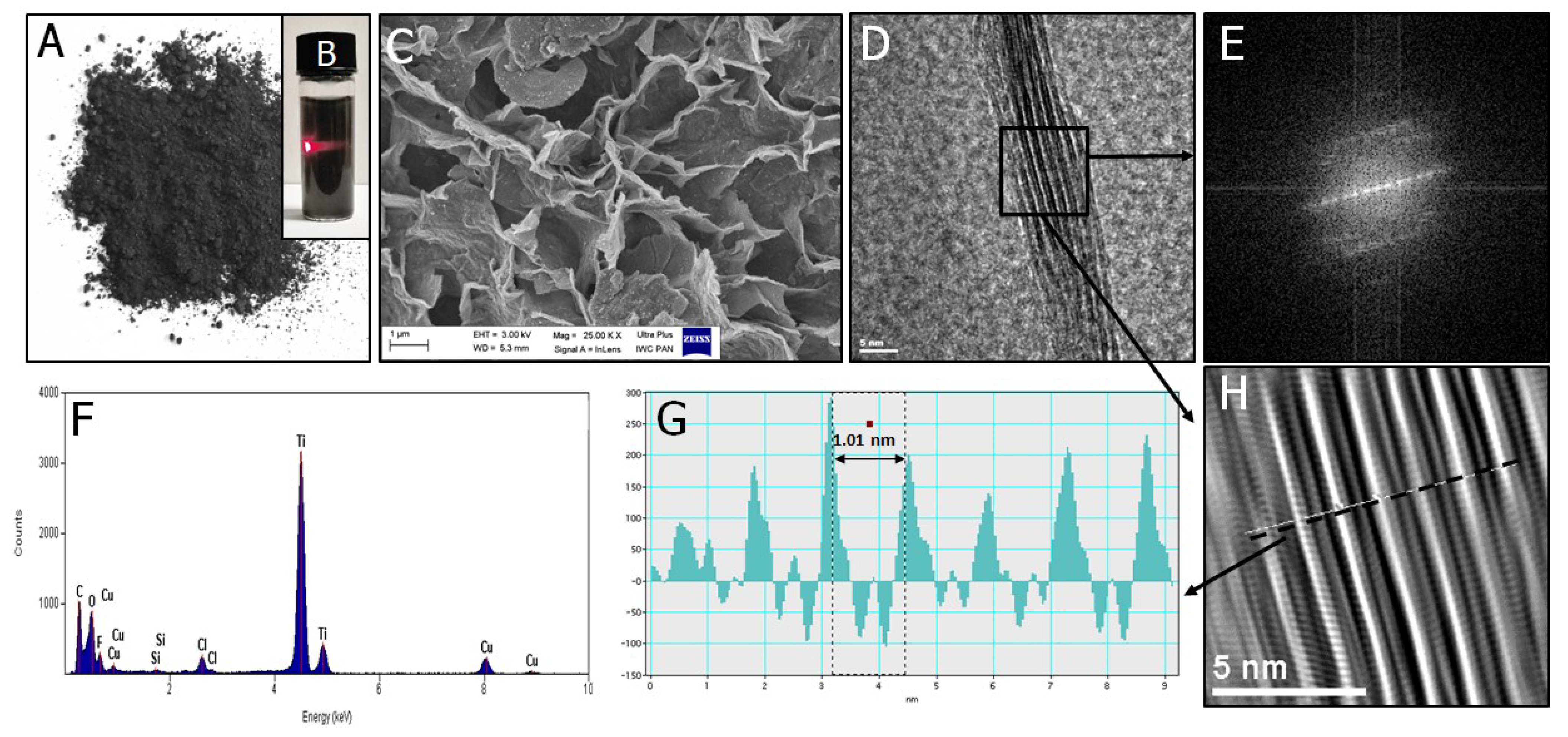
2.1. Synthesis and Characterization of Nanocomposites

The Ti3AlC2 MAX phase for MXenes was produced by powder metallurgy and Spark Plasma Sintering (SPS) and described in our previous work [28]. Titanium powder (Goodfellow, Great Britain), aluminum powder (Benda-Lutz Skawina, Skawina, Poland), and synthetic graphite powder (Sigma Aldrich, St. Louis, MO, USA) were ball milled, dried, and sieved (# = 300 µm). The powders were placed in a specially designed graphite die for pressureless synthesis (molar ratio Ti:Al:C = 3:1:1.9). The SPS synthesis parameters were as follows: temperature 1300 °C; heating rate, 250 °C min−1; vacuum, 5 × 10⁻² mbar. The MAX phase was ground using an automatic mortar grinder (Retsch KM100, Retsch GmbH, Haan, Germany), sieved (# = 300 µm), and etched using a 48% (v/v) concentrated hydrofluoric acid solution (24 h at room temperature, with constant stirring, 250 rpm). During this process, Al layers were removed from the MAX structure to obtain the Ti3C2 MXene. After washing with deionized water and drying at room temperature, MXenes were delaminated. The MXenes phase was magnetically stirred with a water solution of tetramethylammonium hydroxide (TMAOH) (1 mg: 1 mL: 10 mg ratio) for 24 h at room temperature and then subjected to periodical tip sonication for 6 h (1 s working/1 sec resting) with Ar bubbling (VCX 750 ultrasonic processor; Sonics, Leicestershire, UK) and subsequent washing using serial centrifugation water changing. The final 2D Ti3C2 powder was obtained after 24 h of freeze-drying (1 mbar pressure) from the suspension.
The delaminated 2D Ti3C2 MXene was further surface-modified with Al2O3, Ag, and Cu nanoparticles using chemical precursor reagents. The Ti3C2 portion was resuspended in 5 cm3 of isopropanol and treated with a mixture of other reagents (see Table 1) that included aluminum isopropoxide (C9H21O3Al), silver acetate (C2H3AgO2), and copper (II) acetate (C4H6O4Cu) in 15 cm3 of isopropanol (all reagents from Sigma-Aldrich, Poznan, Poland) and stirred for 2 days (250 rpm) in closed vessels using a magnetic mixer. The samples were opened for solvent evaporation. Powder materials such as Ti3C2/Al2O3/Ag/Cu (2 wt %), Ti3C2/Al2O3/Ag/Cu (4 wt %), and Ti3C2/Al2O3/Ag/Cu (8 wt %) were subjected to further examination. It is noted that each wt % value corresponds directly to nano-metals content that were present in a 1:1 proportion.

2.2. Development of the Modified Polypropylene Filter Fabric

The nanocomposite filtration materials were elaborated based on the pristine polypropylene fabric (fiber diameter = 1 µm) made by melt blowing. The nanocomposite with the highest antibacterial activity (Ti3C2/Al2O3/Ag/Cu with 8 wt % nano-metal content) was selected for material modification. Polypropylene fabric sheets were cut into 90 mm × 150 mm fragments and placed into 50 mL of an isopropanol suspension containing the nanocomponent in a flat reaction vessel. The samples were placed in a fume hood for 2 days so the volatiles would evaporate. The nanocomposite level in the filtered material was determined by weight, and the samples were stored at 4 °C.
A part of the MXene Ti3C2/Al2O3/Ag/Cu-modified polypropylene fabric was subjected to oxidation to obtain the nanocomposite enriched with TiO2 (labeled as o-Ti3C2/Al2O3/Ag/Cu). It was accomplished via material incubation for 7 days at 37 °C.
The wt % of the nanocomposite content in the modified polypropylene fabric was based on the weight difference of the fabric before and after synthesis (see Equation (1)).
added   amount   ( wt . % ) = w f w i w i × 100
where wi is the weight of propylene material before synthesis, and wf is the weight of material after synthesis.

2.3. Characterization of the Morphology and Chemical Composition of MXene-Based Composites and Surface-Modified Polypropylene Materials

The morphology of the modified polypropylene material surface was analyzed by scanning electron microscopy (SEM). The samples were coated with a layer of carbon powder using a BAL-TEC SCD 005 sputter coater and analyzed with a Zeiss Ultra Plus (Zeiss, San Diego, CA, USA) microscope at an accelerating voltage of 2.0 kV and varying magnifications.
Fourier transform infrared spectroscopy (FTIR; Nicolet iS5 FTIR Spectrometer; Thermo Scientific, Waltham, MA, USA) identified the organic, polymeric and inorganic components of the nanocomposite-modified polypropylene fabrics. The elemental compositions of all nanomaterials and composites were investigated using X-ray fluorescence (XRF, PI 100; Polon-Izot; Warsaw, Poland). The Supplementary Materials contains those results.
The presence of nanocomposite in polypropylene materials was determined with a PI 100 benchtop X-ray fluorescence spectrometer (XRF, Polon-Izot, Warsaw, Poland), equipped with a silicon drift detector (SSD) of 125–140 eV resolution, test tube with rhodium (Rh) anode, and a multilayer monochromator of 50 keV. The measurements were performed in an air atmosphere using a measurement time of 300 s.

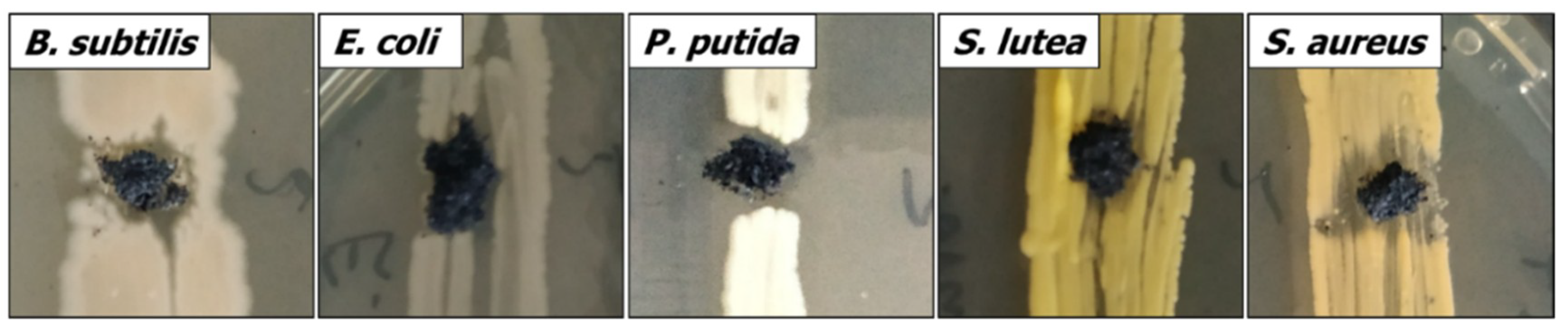
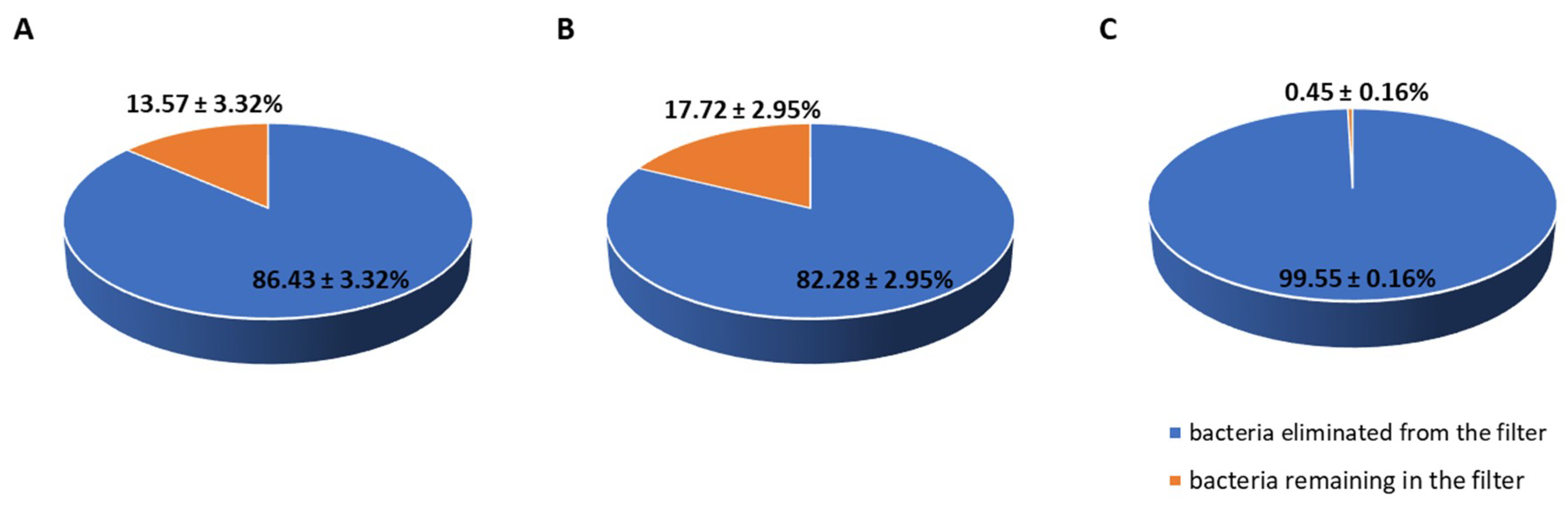
2.4. The Evaluation of Antimicrobial Properties of Nanocomposites

Pre-selection of the nanocomposites for their antibacterial properties was accomplished by diffusion. The bacterial strains used in the test (Gram-positive: Bacillus subtilis, Sarcina lutea, Staphylococcus aureus and Gram-negative: Pseudomonas putida, Escherichia coli) were obtained from the private collection of the Biology Department, Faculty of Building Services, Hydro, and Environmental Engineering, Warsaw University of Technology.
Microorganisms were inoculated in a form of a line on the surface of a solid nutritive culture medium (Nutrient LAB-AGAR™, Biocorp, Warsaw, Poland). The nanopowder samples were placed on the bacterial growth surface, and their cultures were incubated for 48 h at 26 °C (Bacillus subtilis, Sarcina lutea, and Pseudomonas putida) or 37 °C (Escherichia coli and Staphylococcus aureus). After incubation, the cultures were photographed, and the growth inhibition zones around the samples were measured. Ten measurements were taken for each nanocomponent, and the average with standard deviation was reported.
The dilution test was accomplished using two bacterial strains: Escherichia coli and Staphylococcus aureus. The experiment was carried out in a nutrient broth medium (Biocorp), diluted 1:1 with the nanocomposite suspension in a dilution ratio of q = 2, and nanocomposite concentrations from 0–500 mg L−1. After incubation (37 °C, 48 h), the optical densities of the cultures were measured in a MARCEL spectrophotometer at a wavelength of 610 nm, and the bacterial growth inhibition percentages were evaluated.

2.5. Filtration Tests

Filtration tests were carried out by applying three variants of the filtration materials:
  • Pristine (unmodified) polypropylene fabrics,
  • Polypropylene fabrics modified with Ti3C2/Al2O3/Ag/Cu (8 wt %),
  • Polypropylene filter subjected to surface oxidation after nanocomposite enrichment with TiO2 crystals (labeled as o-Ti3C2/Al2O3/Ag/Cu).
The 30 mm × 90 mm filter material sheets were rolled and slid into filter columns (200 mm × 10 mm) with a uniform bed compression in all variants. The filtration beds (height, 50 mm; diameter, 10 mm), were rinsed with ≈50 cm3 of sterile tap water, and the dense bacterial suspension (1.3 × 10⁷ CFU ml−1) in dechlorinated tap water was gradually dosed with an automatic pipette. Escherichia coli and Staphylococcus aureus strains were used to approximate microbiological water contamination. The process was conducted for 150 min. Filtrate samples were collected every 30 min, and the filtration speed was periodically measured.
The zeta electrokinetic potential (ζ) of the filtrates was analyzed with a Zetasizer Nano ZS (Malvern Instruments, Malvern, UK), equipped with an MPT-2 automatic titrator and a titration media degasser, using standard operating procedures. Simultaneously, the hydrodynamic size of the nanoparticles and the intensity of agglomerate formation in the filtrate samples were determined using dynamic light scattering (DLS).
Pearson’s linear correlation coefficient was applied to analyze the relationship between the number of bacterial cells and the zeta potential values in the filtrate. A non-parametric coefficient significance test assessed the significance of the correlation between the data.
UV-Vis absorption of the filtrates was studied using an Evolution 210 UV-Vis spectrometer (Thermo Scientific, Waltham, MA, USA). The number of bacteria in the filtrates was determined with a pour plate technique, on Nutrient LAB-AGAR™ (Biocorp), after 48 h incubation at 37 °C, and presented as the number of colony-forming units in 1 mL of the filtrate (CFU/mL).
The “self-disinfection” properties of the filtering materials were analyzed based on microbial ability to survive in the fabric after filtration. The bacteria concentration in the fabrics was determined immediately upon filtering and after 24 h of bacterial contact with the modified material. The 3 cm2 fabric samples were cut using sterile scissors and washed thoroughly by shaking in a nutrient broth medium (Biocorp). The number of bacteria was determined by the pour plate method after incubation for 48 h at 37 °C. The results were given as the number of colony-forming units per cm2 of fabric. The efficiency of the elimination of bacteria was calculated and compared to the pristine polypropylene material.

3. Results

The present work examined the antibacterial activity of nanocomposite structures upon the addition of Ti3C2 MXene. We initially performed the analysis using the starting MXene nanoflakes, as its morphology, shape, and size influence the nanocomposite bioactivity. Characterization of unmodified, pristine Ti3C2 MXene is shown in Figure 1. Their structure and morphology were characterized using SEM, and the flakes had irregular shapes and sharp edges after freeze-drying. When redispersed in isopropyl alcohol, they formed a stable nanocolloidal solution that showed a Tyndall effect. Its presence indicated the formation of a homogeneous nanoflake nanodispersion. The high-resolution transmission electron microscopy (HRTEM) taken for a randomly chosen sheet of Ti3C2 MXene revealed its characteristic multilayered 2D structure and agreed with works reported by Naguib et al. [13], Jastrzębska et al. [25], and Rozmysłowska-Wojciechowska et al. [27]. The layered pattern was confirmed by fast Fourier Transform (FFT) and inverse fast Fourier Transform (IFFT) imaging, in which alternating layers of light and dark bands correspond to the stacking of several single Ti3C2 monolayers together with increased spacing between them. Energy dispersive spectroscopy (EDS) analysis additionally confirmed the presence of Ti and C predominantly in investigated material. The distance between maximal intensities for two adjacent Ti3C2 monolayers was 1.01 nm and agreed with our previous results [25]. SEM analyses of the 2D materials confirmed the nanometric sizes of the nanocomposites, preservation of the MXene single sheet structures, as well as the presence of nano-metallic components (see Figure S1). The nanocomponent content in the propylene material was 1.15 ± 0.2 wt %.
Preliminary experiments based on diffusion tests (see Figure 2 and Table 2, for details, check Tables S1 and S4–S8) revealed that all nanocomposites inhibited bacterial growth to some extent. The antibacterial activity increased with the increasing content of nanometals in the composite and depended on the type of bacteria used in the test.
Dilution tests with Escherichia coli and Staphylococcus aureus confirmed the relationship between the nano-metal content and the intensity of microbial growth inhibition. The antibacterial activity was higher toward Gram-positive bacteria (Staphylococcus aureus), while results obtained for Gram-negative Escherichia coli were not as obvious and likely related to the protecting effect of the outer membrane of Gram-negative bacteria. For both test bacteria, inhibiting effects of the nanocomposite with 8 wt % noble metals started from 3.91 mg L−1, while for 4 wt %, it was 15.63 mg L−1. In the case of a 2 wt % sample, the inhibiting effect started from 250 mg L−1. The maximum inhibition values were 27.84% for E. coli and 34.65% for S. aureus. Increasing the nanocomposite concentration led to nanoparticle agglomeration and limited the antibacterial effect (the highest inhibition values were observed for the nanocomposite concentrations 31.25–62.5 mg L−1).

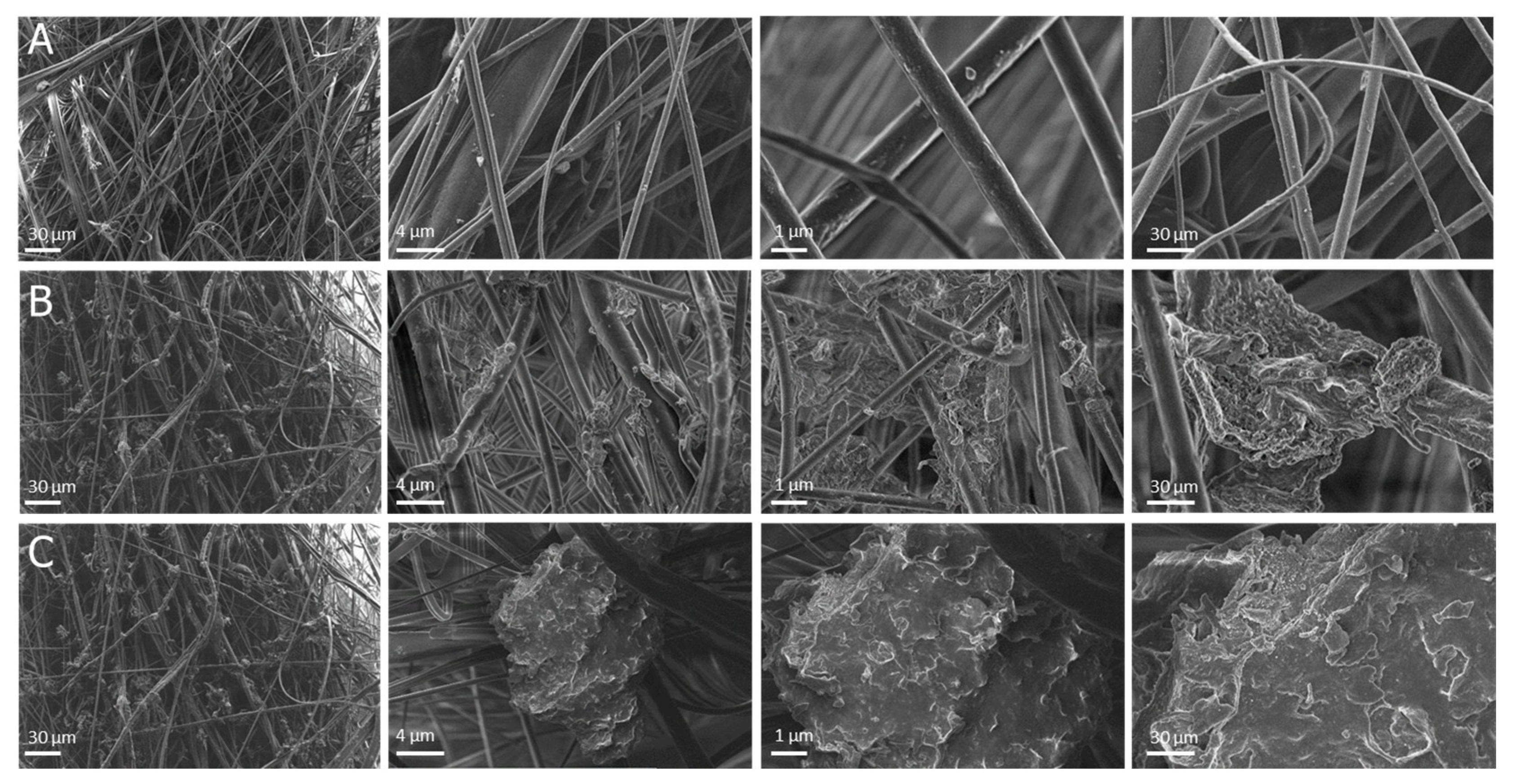
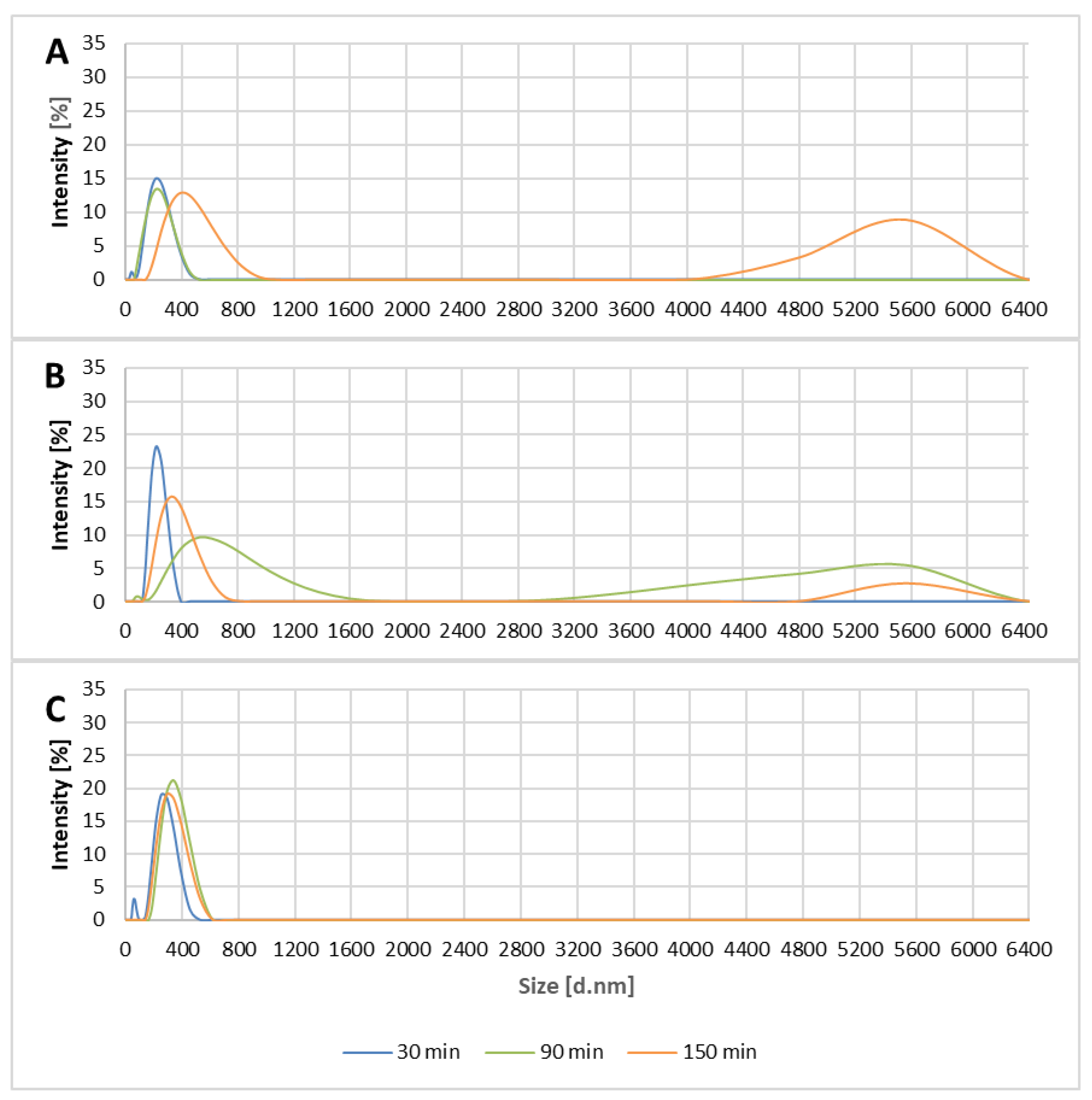
3.1. Structure and Properties of Modified Polypropylene Materials

Based on the above results, the Ti3C2/Al2O3/Ag/Cu containing 8 wt % Ag and Cu nanoparticles (see Figure 1C) was selected for polypropylene modification. XRF results revealed that Ti3C2/Al2O3/Ag/Cu nanostructures successfully covered the polypropylene fibers (SI; see Figures S2–S4). High-magnification SEM images showed that the polypropylene fibers were joined by the agglomerates formed by the introduced nanocomposites. The individual sheets of Ti3C2 MXene, covered with the other applied nanocomponents, were visible in the material modified with Ti3C2/Al2O3/Ag/Cu nanocomposite (see Figure 3).
The oxidation process changed the morphology and structure of polypropylene fabrics modified with the Ti3C2/Al2O3/Ag/Cu nanocomposite. In the non-oxidized material, the surface of the nanocomposite agglomerates was rather smooth (Figure 3B), while in the oxidized material, it appeared more waved and sharp (see Figure 3C). High-magnification SEM images showed that the oxidation process also resulted in diminishing the size of the nanocomposite agglomerates in the material.
FTIR analysis of the pristine polypropylene fabric used as a base for nanocomposite modifications showed peaks corresponding to C-C, C-H, CH2, and CH3 bonds. Stretching vibrations at 808, 972, and 997 cm−1 may be due to C-C stretches, while rocking and wagging ones occur at 841 and 1166 cm−1. Asymmetrical stretching vibrations at 2916 cm−1 correspond to CH2 as well as bands indicating the presence of CH3 groups (rocking vibrations at 841, 997, and 1166 cm−1, symmetrical bending at 1375 and 1453 cm−1, stretching at 2865 cm−1, and asymmetrical stretching at 2948 cm−1) were also detected.
FTIR peaks of fabrics modified with Ti3C2/Al3O3/Ag/Cu nanocomposite changed slightly with additional peaks at 1573 cm−1 and more intense ones at 1375–3342 cm−1. The symmetrical band detected at 1573 cm−1 may reflect the formation of C-O and C=O bonds, while additional bands between 500 and 1000 cm−1 that correspond to the Ti-O bond were not present.
Analysis of the o-Ti3C2/Al2O3/Ag/Cu-modified fabric FTIR spectra (after oxidation) revealed some changes. The bands at 809, 840, 943, 978, and 997 cm−1 were slightly higher, while the ones at 1376, 1453, 2837, 2866, 2916, and 2949 cm−1 were significantly lower, which reflects a decrease in the number of C-C and C-H functional groups, to which forming TiO2 bonds, a process that consumes oxygen atoms, contributed.

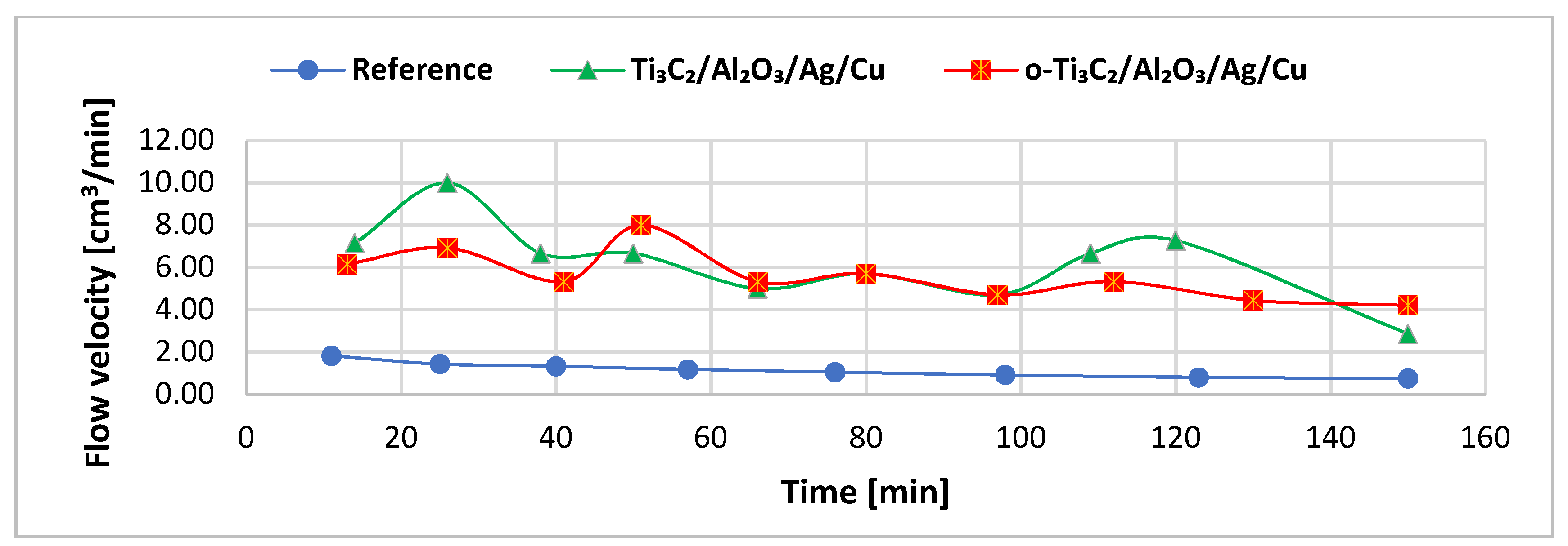
3.2. Filtration Process

The filtration process was carried out in three variants: unmodified polypropylene and filters modified with Ti3C2/Al2O3/Ag/Cu (8 wt %) and o-Ti3C2/Al2O3/Ag/Cu (8 wt %). Nanocomposite filter materials were characterized by a significantly higher flow velocity compared to the unmodified polypropylene fabric (see Figure 4). The average flow velocities were material modified with Ti3C2/Al2O3/Ag/Cu—6.27 ± 3.57 cm3 min−1, the oxidized material modified with the nanocomposite—5.62 ± 1.91 cm3 min−1, and reference polypropylene filter—1.16 ± 0.54 cm3 min−1. Initially, the flow velocity of the modified filters appeared less stable compared to the pristine polypropylene, but after 60 min, it stabilized in the o-Ti3C2/Al2O3/Ag/Cu-modified filter.
Modification of the filter fabrics by nanocomposite incorporation did not significantly influence the bacteria removal efficiency from the filtered suspension—details are presented in Supplementary Materials (Tables S2 and S3)—exceeding 90% (for the initial concentration of bacteria in filtered water over 107 CFU mL−1). It should be stressed that for the tested nanocomposite-based materials, this efficiency was achieved with filtration velocities three to four times higher compared to the reference material. For the oxidized Ti3C2/Al2O3/Ag/Cu-modified material, the curve describing the changes in the process efficiency was very similar to the reference filter after 1 h, while at the beginning of the process, it was approximately 30% higher.
The potential “self-disinfecting” properties were evaluated based on the bacteria content in filters immediately after filtration and after 24 h of storage at room temperature (22–24 °C). Samples were stored wet, in the filtration system, and exposed to light; therefore, the gradual evaporation of water that occurred must be considered. The results revealed the large antimicrobial potential of the oxidized material modified with Ti3C2/Al2O3/Ag/Cu, which eliminated 99.6% of all bacteria present in the fabric (see Figure 5; for details, check Table S9). This was not observed for the unoxidized nanocomposite material.

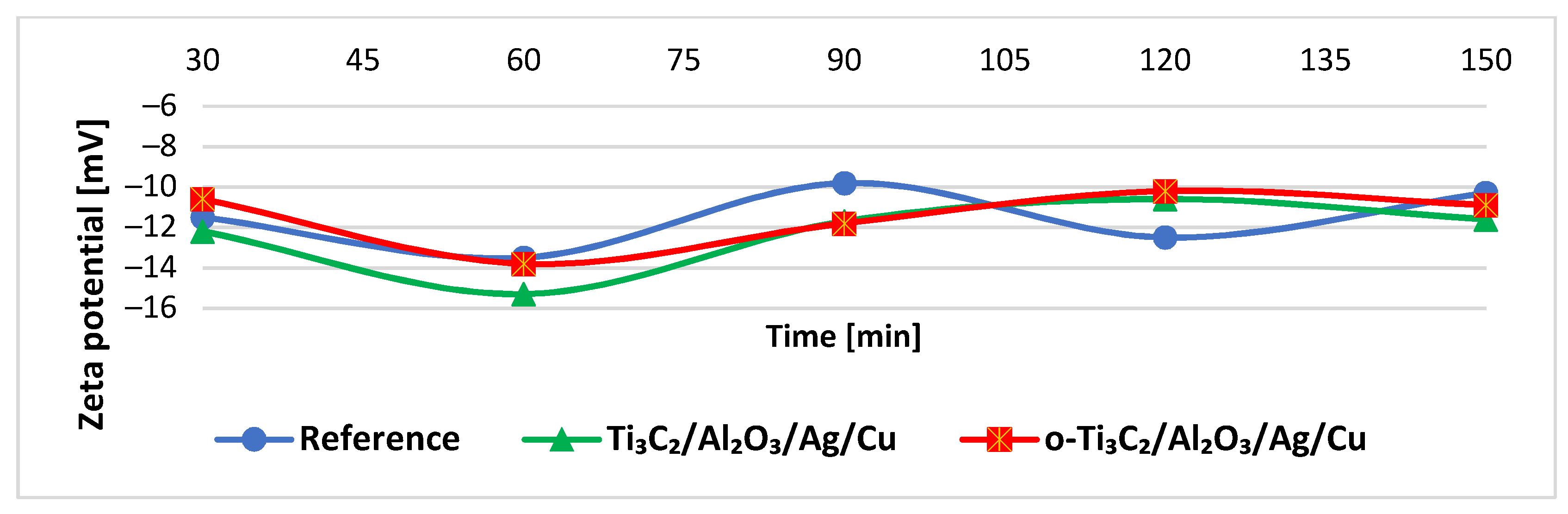
3.3. Filtrate Parameters

For all filtrates, the zeta potential was negative and varied with time, from ≈−16 to −10 mV, most intensely for the filtrate from the unmodified material (see Figure 6). It is worth noting the zeta potential of the filtrate samples collected from the nanocomposite-modified filters approximated those obtained from the reference material; this indicated that the nanocomponents were not flushed out intensively from the material during filtration.
The particle size distribution was similar in filtrates for both reference and modified filters. We assumed that the particles present in the filtrates were related to the pristine polypropylene matrix or the bacterial cell fragments passing through the filter. The signals detected might be also due to the presence of the nano-sized particles in tap water. Furthermore, smaller agglomerates (below 1000 nm) that appeared in all filtrates and were observed both in the tested and the reference material may be due to the presence of nanoparticles in the used test suspension. The second group of agglomerates (over 2800 nm) can be due to secondary contamination of the filtrate with the material collected on the filters. However, it should be stressed that the group of the larger clusters was not detected in filtrates collected from the nanocomposite filtration material modified with oxidized Ti3C2 MXene; this suggested a limited influence of the material on biological material leakage from the filter (see Figure 7). This effect was not observed in the unoxidized material.

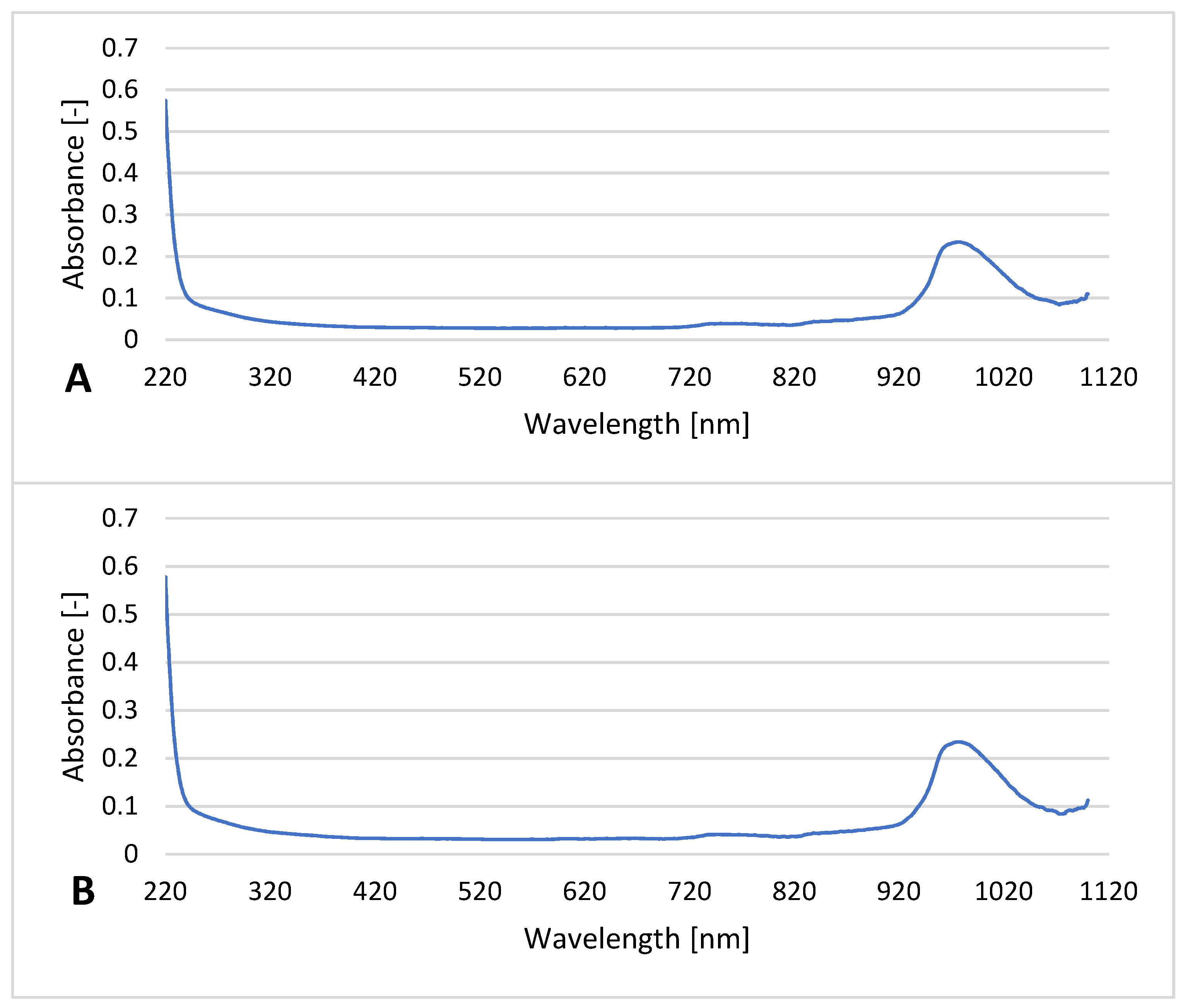
3.4. UV-Vis Study of The Filtrate

UV-Vis analysis of the filtrates revealed similar spectra for all filtrates with one significant peak, with a λmax (~0.24) at ≈970 nm and some slight fluctuations in the region between 720–820 nm (absorbances < 0.05; see Figure 8). The extremely consistent spectra confirmed that the nanocomponents introduced remained in the filters. The peak observed in all filtrates (including the reference) between 940 and 1040 nm may be related to the presence of bacterial cell fragments or bacterial suspension compounds.

4. Discussion

The nanomaterials characterized by a specific surface offers the potential for their functionalization with a variety of particles and structures, which confers the base material with new and desirable properties. We focused on the application of 2D structure-based composites to create a novel filtering material for “point-of-use” water treatment systems.
The first milestone was the synthesis of the series of innovative nanoproducts composed of elements with potential antimicrobial activity. Their synthesis should be relatively straightforward based on commercially available reagents, and their combination should reveal their antimicrobial impacts.
For nanocomposite construction, MXenes were selected due to the possibility of precise pore size control and hydrophilic properties that allow MXenes to contribute to liquid transport efficiency and ion release, fouling reduction, and biofilm formation. All attributes result in longer-lived filtration units [20,24,26,29]. The applied noble metal nano-metals had been used previously as antibacterial agents in different applications [30]. In addition, the antimicrobial properties of Al2O3 had been observed in several studies [31,32,33].
Diffusion and dilution tests confirmed that the novel nanocomponents limited bacterial growth. The nanocomposites with 8 wt % of metallic nanoparticles were the most effective toward bacteria, especially Gram-positive strains. An increase in the antibacterial effect related to the nanometal amount suggested that the diffusion of Ag and Cu ions from the nanocomposite was the main mechanism of the antimicrobial action of the powdery nanocomponents, while Ti3C2 MXene and Al2O3 nanoparticles played minor roles. The action of nano-Ag and nano-Cu is related to the adhesion of nanoparticles to the cell membrane and following interactions with its components resulting in changes of the cell permeability and affecting cellular respiration, enzymatic, gene expression, and metabolic processes [34,35,36,37]. The induction of oxidative stress associated with the generation of reactive oxygen species (ROS) and silver ion release should also be considered [38,39,40,41]. On the other hand, the tendency of Ti3C2 MXenes to agglomerate [42] due to strong van der Waals interactions helps keep the metal nanoparticles inside the agglomerates, which decreases their diffusion potential and impact on bacterial cells.
The production of the POU filtration material needs a simple, available base suitable for effective modifications. The synthetic fibers serve both as a base material and an effective nanocomponent carrier. The impregnation of polyester fibers with copper ions (3–10 wt %) produced the active antibacterial, anti-fungal, and anti-virus material [43]. The antibacterial effect was observed for nano-Ag-modified nylon and polyester [44,45], polypropylene [46], and polyamide 6™ [47].
In this research, the polypropylene base was used for nanocomposite modifications. The synthetic procedure was not complicated and very effective at producing the desired material. The modification did not change the chemical structure of the carrier fiber, which remained typical for the pristine material [48]. FTIR spectra showed that peak assignments typical for the pure polypropylene also occurred in the modified material; however, additional C-O and C=O bonds confirmed the appearance of Ti3C2 MXene. Successful modification of the polypropylene fabrics was also confirmed by SEM analysis and revealed the nanostructure presence. The visible changes that resulted from the oxidation of Ti3C2/Al2O3/Ag/Cu nanocomposite toward Ti3C2/TiO2/Al2O3/Ag/Cu on the surface of the polypropylene material resulted in the formation of anatase crystals (TiO2).
Our studies showed that the modified polypropylene fabrics can create a non-compressible, solid bed. Moreover, the modifications changed the structure of the material and caused a noticeable increase in the flow velocity. This astonishing effect brings an essential advantage for the material, solving one of the problems for point-of-use devices [49].
The effective “transfer” of antibacterial activity from the powder nanocomposite to the modified polypropylene material was another accomplishment of this research. The nanocomposite with the highest levels of metallic nanoparticles (8 wt %) was applied in the filtration experiments. In contrast to tests with powdery nanocomposites, the presence of Ti3C2/Al2O3/Ag/Cu did not improve the filtration efficiency of the bacterial suspension (artificially “contaminated water”). For filter fabrics modified with oxidized Ti3C2/Al2O3/Ag/Cu, a slight improvement in bacteria removal was observed compared to the pristine polypropylene fabric, especially at the beginning of the process. However, the material with the oxidized Ti3C2 MXene nanocomposite worked effectively over the entire experiment with superior antimicrobial activity that diminished bacterial numbers by two orders of magnitude.
Recently, the self-cleaning properties of filtration membranes and filters have been investigated. A majority of these works focused on “in situ” decomposition of dyes and chemicals. However, the aspect of killing residual surface bacteria was marginalized. The self-cleaning activity of BC-SiO2-TiO2 (where BC stands for bacterial cellulose) photocatalytic membranes enhanced by Ag doping was studied by Rahman et al. [50]. They reported a 41% efficiency increase of crystal violet dye decomposition relative to the reference sample. Furthermore, the samples showed some antibacterial activity toward Bacillus subtilis, Klebsiella pneumoniae, salmonella arizonae, E. coli, and Kluyvera spp. strains caused by the presence of nanosilver [50]. Self-cleaning MIL-125(Ti)/PVDF (polyvinylidene fluoride) hybrid membranes were obtained by Zhou et al. [51], who also tested their activity in contact with dyes and bacteria cells. Selected samples showed extraordinary efficiency (up to 100%, depending on the Ti content) during three filtration cycles and antibacterial properties against E. coli bacteria [51]. Ma et al. [52] investigated the efficiency of oil/water separation with nanofibrous membranes, which was characterized by remarkable recyclability, antibacterial properties, as well as environmentally friendly and low-cost features [52]. Li et al. [53] observed the ability of graphitic carbon nitride (g-C3N4) nanosheets to form self-cleaning membranes with antibacterial activity (≈100%). However, after several cycles, defects appeared that resulted in higher membrane permeability [53]. The self-disinfection properties were also tested by Lei et al. [54]. Filters were designed based on aminopyridine conjugated microporous polymer nanotubes to capture the PM 2.5 dust fraction. The antibacterial activity against E. coli enabled the rapid neutralization of tested bacteria and killed residual bacteria. This prevented secondary contamination of the filtrate. On the other hand, frequently used filtration systems differ significantly from the one presented here, which can be applied at places that lack sanitation [54]. Stan et al. [55] removed residual bacteria of the cotton fabrics by coating them with graphene oxide/TiO2. After 2 h contact time, the growth of S. aureus was inhibited slightly, although E. faecalis was not affected. Fortunately, after 24 h, both bacterial strains were reduced significantly. It is worth noting that these properties were achieved under static conditions, and their performance in dynamic filtration systems has not been studied [55].
There is evidence that the antimicrobial activity of nanoparticle-modified membranes makes them less sensitive to biological fouling [56]. This research obtained a material with “self-disinfecting” properties, which should help with repeated use of the filter-equipped devices. After 24 h, the number of bacteria dropped by 99.6 wt % in a novel filter modified with the TiO2 enriched nanocomposite. The formation of titanium dioxide (anatase crystals) on the surface of Ti3C2, acting as a robust antibacterial agent, was reported by Rasool et al. [16] and suspected to be the predominant factor of the antibacterial properties of Ti3C2 MXene. Antimicrobial properties of titanium dioxide have been reported in several works [29,57], but its activity was strongly related to UV irradiation [58]. In this research, antibacterial effects were readily visible despite the absence of UV stimulation.
The most essential quality for the practical usage of the filtration material is to procure a safe, high-quality filtrate. The effective elimination of microbiological contaminants must be accompanied by material stability, which is critical especially for nanocomponent-modified filters, because it avoids secondary contamination of the filtrate with nanoparticles.
The measurement of the electrochemical (zeta) potential of the filtrates helped evaluate the electrostatic interactions between the solution and the particles (e.g., bacteria cells), including the effects of the bacterial adsorption on the surface charge of bacterial cells [59]. The bacteria applied in the filtration experiment—E. coli and S. aureus, possess a negative zeta potential in drinking water (~−40 and −17 mV, respectively) [60]. The zeta potentials measured correspond to the bacterial cells that remained in the filtrate samples. According to literature data, Ti3C2 MXene, Al2O3, Ag, and Cu nanoparticles all contribute to negative zeta potentials in water, especially at alkaline pH [27,61,62,63]. Therefore, the release of nanocomponents was readily observed and yielded information regarding the instability of the material. In our research, this was not observed; the zeta values were similar for both unmodified polypropylene and nanocomposite-modified materials. The effect was also confirmed by UV-Vis spectral analyses of the filtrates that revealed no significant differences between filtrates collected after modified filters and the unmodified reference material. A peak detected in each sample was probably related to the presence of the biological fragments.
The DLS analysis revealed that certain levels of small particles and agglomerates were present in all filtrates. These likely included cells and their organelles [64], residues of the microbiological cultivation media, and the particles present in tap water used for the bacterial suspension preparation. However, they were also present in reference samples, so they were not related to the nanocomponents flushed from the filtration bed.

5. Conclusions

The filtration materials applied in point-of-use water treatment systems must meet certain basic requirements: synthetic and manufacturing ease, chemical and physical stability, and effectiveness in the elimination of microorganisms. The material presented in this work yielded additional advantages: improved filtration velocity with an undisturbed or improved elimination efficiency of microorganisms as compared to pristine polypropylene. The elaborated filtration material modified with Ti3C2/Al2O3/Ag/Cu was obtained using commercial reagents and a readily available and cheap polypropylene base. The 1 wt % content of the nanocomposite did not increase the fabric cost significantly. The filter collected almost ≈105 bacterial cells per 1 cm2 of material, which makes it suitable for the effective removal of typical microbiological water contaminants. The best effect was achieved by oxidation of the modified material, which increased its antimicrobial impact. The self-disinfecting properties eliminated ≈99.6 wt % of bacteria collected on the filter for both Escherichia coli (typical for fecal contamination) and potentially pathogenic Staphylococcus aureus. The material is environmentally safe due to its stability and lack of nanocomposite material release. This work clearly showed that 2D MXene nanocomposites are a promising new perspective for point-of-use water treatment.

Supplementary Materials

The following are available online at https://0-www-mdpi-com.brum.beds.ac.uk/1996-1944/14/1/182/s1; Table S1. The growth inhibition zones (mm) in the diffusion test of the Ti3C2 MXene and Al2O3 nanoparticles; Table S2. The number of colony-forming units (CFU) found in filtrate after certain time; Table S3. Percentage efficiency of filtration process; Table S4. Growth inhibition zones (mm) in the diffusion test of nanopowders with different levels of metallic nanoparticles, and with calculated statistics for Bacillus subtilis; Table S5. Growth inhibition zones (mm) in the diffusion test of nanopowders with different levels of metallic nanoparticles, and with calculated statistics for Escherichia coli; Table S6. Growth inhibition zones (mm) in the diffusion test of nanopowders with different levels of metallic nanoparticles, and with calculated statistics for Pseudomonas putida; Table S7. Growth inhibition zones (mm) in the diffusion test of nanopowders with different levels of metallic nanoparticles, and with calculated statistics for Sarcina lutea; Table S8. Growth inhibition zones (mm) in the diffusion test of nanopowders with different levels of metallic nanoparticles, and with calculated statistics for Staphylococcus aureus; Table S9. Results for “self-disinfection” properties investigation, as well as its statistical analysis; Figure S1. SEM images of the nanocomposite powders: Ti3C2/Al2O3/Ag/Cu (2 wt %) (A), Ti3C2/Al2O3/Ag/Cu (4 wt %) (B), Ti3C2/Al2O3/Ag/Cu (8 wt %) (C). The insets correspond to BSE imaging of metal particles; Figure S2. XRF spectra of reference polypropylene material; Figure S3. XRF spectra of Ti3C2/Al2O3/Ag/Cu-modified material; Figure S4. XRF spectra of o-Ti3C2/Al2O3/Ag/Cu-modified material.

Author Contributions

M.J. prepared and characterized studied samples, performed, analyzed and commented bactericidal analyses and filtration test, collected and analyzed the obtained results, designed and prepared figures, and also prepared and corrected the original manuscript; E.K. designed the concept and content of experiment, analyzed the obtained results, analyzed and commented on bactericidal analyses designed figures, coordinated and supervised the preparation of the manuscript as well as coordinated the whole research, A.R.-W. characterized studied samples, M.P. and J.W. synthesized the starting MAX phase, J.M. characterized studied samples, A.M.J. acquired founds, carried out MXene delamination process, coordinated and supervised the preparation of the manuscript, corrected the original manuscript and participated in discussion on obtained results. All authors have read and agreed to the published version of the manuscript.

Funding

The study was accomplished thanks to the funds allotted by the National Science Centre, within the framework of the research project ‘SONATA BIS 7’ (UMO-2017/26/E/ST8/01073) and ‘OPUS-18’ (UMO-2019/35/B/ST5/02538). The additional financial support from the Warsaw University of Technology, Faculty of Materials Science and Engineering as well as Faculty of Building Services, Hydro and Environmental Engineering is also acknowledged.

Data Availability Statement

The data presented in this study are partially available in supplementary material.

Conflicts of Interest

The authors declare no conflict of interest.

References

  1. Pooi, C.K.; Ng, H.Y. Review of low-cost point-of-use water treatment systems for developing communities. npj Clean Water 2018, 1. [Google Scholar] [CrossRef]
  2. Bitton, G. Microbiology of Drinking Water: Production and Distribution; Wiley-Blackwell: Hoboken, NJ, USA, 2014. [Google Scholar]
  3. Gleick, P.H. Water and terrorism. Water Policy 2006, 8, 481–503. [Google Scholar] [CrossRef] [Green Version]
  4. Li, Q.; Mahendra, S.; Lyon, D.Y.; Brunet, L.; Liga, M.V.; Li, D.; Alvarez, P.J.J. Antimicrobial nanomaterials for water disinfection and microbial control: Potential applications and implications. Water Res. 2008, 42, 4591–4602. [Google Scholar] [CrossRef] [PubMed]
  5. World Health Organization Regional Office for Africa. Available online: https://www.afro.who.int/health-topics/water (accessed on 9 November 2020).
  6. Sobsey, M.D.; Stauber, C.E.; Casanova, L.M.; Brown, J.M.; Elliott, M.A. Response to comment on “Point of use household drinking water filtration: A practical, effective solution for providing sustained access to safe drinking water in the developing world”. Environ. Sci. Technol. 2009, 43, 970–971. [Google Scholar] [CrossRef]
  7. Heidarpour, F.; Wan Ab Karim Ghani, W.; Ahmadun, F.-R.; Sobri, S.; Zargar, M.; Mozafari, M. Nano silver-coated polypropylene water filter: II. evaluation of antimicrobial efficiency. Dig. J. Nanomater. Biostructures 2010, 5, 797–804. [Google Scholar]
  8. Heidarpour, F.; Wan Ab Karim Ghani, W.A.; Fakhru’L-Razi, A.; Sobri, S.; Heydarpour, V.; Zargar, M.; Mozafari, M.R. Complete removal of pathogenic bacteria from drinking water using nano silver-coated cylindrical polypropylene filters. Clean Technol. Environ. Policy 2011, 13, 499–507. [Google Scholar] [CrossRef]
  9. Srinivasan, N.R.; Shankar, P.A.; Bandyopadhyaya, R. Plasma treated activated carbon impregnated with silver nanoparticles for improved antibacterial effect in water disinfection. Carbon N. Y. 2013, 57, 1–10. [Google Scholar] [CrossRef]
  10. Zhang, H.; Oyanedel-Craver, V. Comparison of the bacterial removal performance of silver nanoparticles and a polymer based quaternary amine functiaonalized silsesquioxane coated point-of-use ceramic water filters. J. Hazard. Mater. 2013, 260, 272–277. [Google Scholar] [CrossRef]
  11. Jain, P.; Pradeep, T. Potential of silver nanoparticle-coated polyurethane foam as an antibacterial water filter. Biotechnol. Bioeng. 2005, 90, 59–63. [Google Scholar] [CrossRef] [Green Version]
  12. Ortiz Balbuena, J.; Tutor De Ureta, P.; Rivera Ruiz, E.; Mellor Pita, S. Enfermedad de Vogt-Koyanagi-Harada. Med. Clin. (Barc) 2016, 146, 93–94. [Google Scholar] [CrossRef]
  13. Naguib, M.; Kurtoglu, M.; Presser, V.; Lu, J.; Niu, J.; Heon, M.; Hultman, L.; Gogotsi, Y.; Barsoum, M.W. Two-dimensional nanocrystals produced by exfoliation of Ti 3AlC 2. Adv. Mater. 2011, 23, 4248–4253. [Google Scholar] [CrossRef] [PubMed] [Green Version]
  14. Naguib, M.; Presser, V.; Lane, N.; Tallman, D.; Gogotsi, Y.; Lu, J.; Hultman, L.; Barsoum, M.W. Synthesis of a new nanocrystalline titanium aluminum fluoride phase by reaction of Ti 2AlC with hydrofluoric acid. RSC Adv. 2011, 1, 1493–1499. [Google Scholar] [CrossRef]
  15. Naguib, M.; Presser, V.; Tallman, D.; Lu, J.; Hultman, L.; Gogotsi, Y.; Barsoum, M.W. On the topotactic transformation of Ti2AlC into a Ti-C-O-F cubic phase by heating in molten lithium fluoride in air. J. Am. Ceram. Soc. 2011, 94, 4556–4561. [Google Scholar] [CrossRef] [Green Version]
  16. Rasool, K.; Mahmoud, K.A.; Johnson, D.J.; Helal, M.; Berdiyorov, G.R.; Gogotsi, Y. Efficient Antibacterial Membrane based on Two-Dimensional Ti3C2Tx (MXene) Nanosheets. Sci. Rep. 2017, 7, 1–11. [Google Scholar] [CrossRef] [PubMed]
  17. Sokol, M.; Natu, V.; Kota, S.; Barsoum, M.W. On the Chemical Diversity of the MAX Phases. Trends Chem. 2019, 1, 210–223. [Google Scholar] [CrossRef]
  18. Zhang, H.; Yang, G.; Zuo, X.; Tang, H.; Yang, Q.; Li, G. Computational studies on the structural, electronic and optical properties of graphene-like MXenes (M2CT2, M = Ti, Zr, Hf; T = O, F, OH) and their potential applications as visible-light driven photocatalysts. J. Mater. Chem. A 2016, 4, 12913–12920. [Google Scholar] [CrossRef]
  19. Zhang, Q.; Teng, J.; Zou, G.; Peng, Q.; Du, Q.; Jiao, T.; Xiang, J. Efficient phosphate sequestration for water purification by unique sandwich-like MXene/magnetic iron oxide nanocomposites. Nanoscale 2016, 8, 7085–7093. [Google Scholar] [CrossRef]
  20. Rasool, K.; Pandey, R.P.; Rasheed, P.A.; Buczek, S.; Gogotsi, Y.; Mahmoud, K.A. Water treatment and environmental remediation applications of two-dimensional metal carbides (MXenes). Mater. Today 2019, 30, 80–102. [Google Scholar] [CrossRef]
  21. Shahzad, A.; Rasool, K.; Miran, W.; Nawaz, M.; Jang, J.; Mahmoud, K.A.; Lee, D.S. Two-Dimensional Ti3C2Tx MXene Nanosheets for Efficient Copper Removal from Water. ACS Sustain. Chem. Eng. 2017, 5, 11481–11488. [Google Scholar] [CrossRef]
  22. Li, J.; Li, X.; Van der Bruggen, B. An MXene-based membrane for molecular separation. Environ. Sci. Nano 2020, 7, 1289–1304. [Google Scholar] [CrossRef]
  23. Rasool, K.; Helal, M.; Ali, A.; Ren, C.E.; Gogotsi, Y.; Mahmoud, K.A. Antibacterial Activity of Ti3C2Tx MXene. ACS Nano 2016, 10, 3674–3684. [Google Scholar] [CrossRef] [PubMed] [Green Version]
  24. Jiang, X.; Kuklin, A.V.; Baev, A.; Ge, Y.; Ågren, H.; Zhang, H.; Prasad, P.N. Two-dimensional MXenes: From morphological to optical, electric, and magnetic properties and applications. Phys. Rep. 2020, 848, 1–58. [Google Scholar] [CrossRef]
  25. Jastrzębska, A.M.; Karwowska, E.; Wojciechowski, T.; Ziemkowska, W.; Rozmysłowska, A.; Chlubny, L.; Olszyna, A. The Atomic Structure of Ti 2 C and Ti 3 C 2 MXenes is Responsible for Their Antibacterial Activity Toward E. coli Bacteria. J. Mater. Eng. Perform. 2019, 28, 1272–1277. [Google Scholar] [CrossRef]
  26. Arabi Shamsabadi, A.; Sharifian, M.; Anasori, B.; Soroush, M. Antimicrobial Mode-of-Action of Colloidal Ti3C2Tx MXene Nanosheets. ACS Sustain. Chem. Eng. 2018, 6, 16586–16596. [Google Scholar] [CrossRef]
  27. Rozmysłowska-Wojciechowska, A.; Karwowska, E.; Poźniak, S.; Wojciechowski, T.; Chlubny, L.; Olszyna, A.; Ziemkowska, W.; Jastrzębska, A.M. Influence of modification of Ti 3 C 2 MXene with ceramic oxide and noble metal nanoparticles on its antimicrobial properties and ecotoxicity towards selected algae and higher plants. RSC Adv. 2019, 9, 4092–4105. [Google Scholar] [CrossRef] [Green Version]
  28. Rozmysłowska-Wojciechowska, A.; Mitrzak, J.; Szuplewska, A.; Chudy, M.; Woźniak, J.; Petrus, M.; Wojciechowski, T.; Vasilchenko, A.S.; Jastrzębska, A.M. Engineering of 2D Ti3C2 MXene Surface Charge and its Influence on Biological Properties. Materials 2020, 13, 2347. [Google Scholar] [CrossRef]
  29. Wang, L.; Hu, C.; Shao, L. The-antimicrobial-activity-of-nanoparticles--present-situati. Int. J. Nanomedicine 2017, 12, 1227–1249. [Google Scholar] [CrossRef] [Green Version]
  30. Karwowska, E. Antibacterial potential of nanocomposite-based materials—A short review. Nanotechnol. Rev. 2017, 6, 243–254. [Google Scholar] [CrossRef]
  31. Mukherjee, A.; Mohammed Sadiq, I.; Prathna, T.C.; Chandrasekaran, N. Antimicrobial activity of aluminium oxide nanoparticles for potential clinical applications. Sci. against Microb. Pathog. Commun. Curr. Res. Technol. Adv. 2011, 245–251. [Google Scholar]
  32. Ansari, M.A.; Khan, H.M.; Khan, A.A.; Cameotra, S.S.; Saquib, Q.; Musarrat, J. Interaction of Al2O3 nanoparticles with Escherichia coli and their cell envelope biomolecules. J. Appl. Microbiol. 2014, 116, 772–783. [Google Scholar] [CrossRef]
  33. Ansari, M.A.; Khan, H.M.; Khan, A.A.; Pal, R.; Cameotra, S.S. Antibacterial potential of Al2O3 nanoparticles against multidrug resistance strains of Staphylococcus aureus isolated from skin exudates. J. Nanoparticle Res. 2013, 15. [Google Scholar] [CrossRef]
  34. Nakkala, J.R.; Mata, R.; Sadras, S.R. Green synthesized nano silver: Synthesis, physicochemical profiling, antibacterial, anticancer activities and biological in vivo toxicity. J. Colloid Interface Sci. 2017, 499, 33–45. [Google Scholar] [CrossRef] [PubMed]
  35. Salomoni, R.; Léo, P.; Montemor, A.F.; Rinaldi, B.G.; Rodrigues, M.F.A. Antibacterial effect of silver nanoparticles in Pseudomonas aeruginosa. Nanotechnol. Sci. Appl. 2017, 10, 115–121. [Google Scholar] [CrossRef] [PubMed] [Green Version]
  36. Harikumar, P.S. Antibacterial Activity of Copper Nanoparticles and Copper Nanocomposites against Escherichia Coli Bacteria. Int. J. Sci. 2016, 2, 83–90. [Google Scholar] [CrossRef] [Green Version]
  37. Zia, R.; Riaz, M.; Farooq, N.; Qamar, A.; Anjum, S. Antibacterial activity of Ag and Cu nanoparticles synthesized by chemical reduction method: A comparative analysis. Mater. Res. Express 2018, 5. [Google Scholar] [CrossRef]
  38. Ruddaraju, L.K.; Pallela, P.N.V.K.; Pammi, S.V.N.; Padavala, V.S.; Kolapalli, V.R.M. Synergetic antibacterial and anticarcinogenic effects of Annona squamosa leaf extract mediated silver nano particles. Mater. Sci. Semicond. Process. 2019, 100, 301–309. [Google Scholar] [CrossRef]
  39. Anuj, S.A.; Gajera, H.P.; Hirpara, D.G.; Golakiya, B.A. Bactericidal assessment of nano-silver on emerging and re-emerging human pathogens. J. Trace Elem. Med. Biol. 2019, 51, 219–225. [Google Scholar] [CrossRef]
  40. Kruk, T.; Szczepanowicz, K.; Stefańska, J.; Socha, R.P.; Warszyński, P. Synthesis and antimicrobial activity of monodisperse copper nanoparticles. Colloids Surf. B Biointerfaces 2015, 128, 17–22. [Google Scholar] [CrossRef]
  41. Ulloa-Ogaz, A.L.; Piñón-Castillo, H.A.; Muñoz-Castellanos, L.N.; Athie-García, M.S.; Ballinas-Casarrubias, M.D.L.; Murillo-Ramirez, J.G.; Flores-Ongay, L.Á.; Duran, R.; Orrantia-Borunda, E. Oxidative damage to Pseudomonas aeruginosa ATCC 27833 and Staphylococcus aureus ATCC 24213 induced by CuO-NPs. Environ. Sci. Pollut. Res. 2017, 24, 22048–22060. [Google Scholar] [CrossRef]
  42. Tontini, G.; Greaves, M.; Ghosh, S.; Bayram, V.; Barg, S. MXene-based 3D porous macrostructures for electrochemical energy storage. J. Phys. Mater. 2020, 3, 022001. [Google Scholar] [CrossRef]
  43. Gabbay, J.; Borkow, G.; Mishal, J.; Magen, E.; Zatcoff, R.; Shemer-Avni, Y. Copper oxide impregnated textiles with potent biocidal activities. J. Ind. Text. 2006, 35, 323–335. [Google Scholar] [CrossRef]
  44. Perelshtein, I.; Applerot, G.; Perkas, N.; Guibert, G.; Mikhailov, S.; Gedanken, A. Sonochemical coating of silver nanoparticles on textile fabrics (nylon, polyester and cotton) and their antibacterial activity. Nanotechnology 2008, 19. [Google Scholar] [CrossRef] [PubMed]
  45. Yang, Z.; Peng, H.; Wang, W.; Liu, T. Crystallization behavior of poly(ε-caprolactone)/layered double hydroxide nanocomposites. J. Appl. Polym. Sci. 2010, 116, 2658–2667. [Google Scholar] [CrossRef]
  46. Yeo, S.Y.; Lee, H.J.; Jeong, S.H. Preparation of nanocomposite fibers for permanent antibacterial effect. J. Mater. Sci. 2003, 38, 2143–2147. [Google Scholar] [CrossRef]
  47. Damm, C.; Münstedt, H.; Rösch, A. Long-term antimicrobial polyamide 6/silver-nanocomposites. J. Mater. Sci. 2007, 42, 6067–6073. [Google Scholar] [CrossRef]
  48. Fang, J.; Zhang, L.; Sutton, D.; Wang, X.; Lin, T. Needleless melt-electrospinning of polypropylene nanofibres. J. Nanomater. 2012, 2012. [Google Scholar] [CrossRef]
  49. Al-Yaseri, I.; Morgan, S.; Retzlaff, W. Assessment of Current Models Ability to Describe Chlorine Decay and Appraisal of Water Spectroscopic Data as Model Inputs. J. Environ. Eng. 2013, 139, 1152–1161. [Google Scholar] [CrossRef]
  50. Rahman, K.U.; Ferreira-Neto, E.P.; Rahman, G.U.; Parveen, R.; Monteiro, A.S.; Rahman, G.; Van Le, Q.; Domeneguetti, R.R.; Ribeiro, S.J.L.; Ullah, S. Flexible bacterial cellulose-based BC-SiO2-TiO2-Ag membranes with self-cleaning, photocatalytic, antibacterial and UV-shielding properties as a potential multifunctional material for combating infections and environmental applications. J. Environ. Chem. Eng. 2020, 104708. [Google Scholar] [CrossRef]
  51. Zhou, S.; Gao, J.; Zhu, J.; Peng, D.; Zhang, Y.; Zhang, Y. Self-cleaning, antibacterial mixed matrix membranes enabled by photocatalyst Ti-MOFs for efficient dye removal. J. Memb. Sci. 2020, 610, 118219. [Google Scholar] [CrossRef]
  52. Ma, W.; Ding, Y.; Zhang, M.; Gao, S.; Li, Y.; Huang, C.; Fu, G. Nature-inspired chemistry toward hierarchical superhydrophobic, antibacterial and biocompatible nanofibrous membranes for effective UV-shielding, self-cleaning and oil-water separation. J. Hazard. Mater. 2020, 384, 121476. [Google Scholar] [CrossRef]
  53. Li, R.; Ren, Y.; Zhao, P.; Wang, J.; Liu, J.; Zhang, Y. Graphitic carbon nitride (g-C3N4) nanosheets functionalized composite membrane with self-cleaning and antibacterial performance. J. Hazard. Mater. 2019, 365, 606–614. [Google Scholar] [CrossRef] [PubMed]
  54. Lei, Y.; Tian, Z.; Sun, H.; Zhu, Z.; Liang, W.; Li, A. Self-cleaning and flexible filters based on aminopyridine conjugated microporous polymers nanotubes for bacteria sterilization and efficient PM2.5 capture. Sci. Total Environ. 2020, 142594. [Google Scholar] [CrossRef]
  55. Stan, M.S.; Nica, I.C.; Popa, M.; Chifiriuc, M.C.; Iordache, O.; Dumitrescu, I.; Diamandescu, L.; Dinischiotu, A. Reduced graphene oxide/TiO2 nanocomposites coating of cotton fabrics with antibacterial and self-cleaning properties. J. Ind. Text. 2019, 49, 277–293. [Google Scholar] [CrossRef]
  56. Choi, H.; Antoniou, M.G.; de la Cruz, A.A.; Stathatos, E.; Dionysiou, D.D. Photocatalytic TiO2 films and membranes for the development of efficient wastewater treatment and reuse systems. Desalination 2007, 202, 199–206. [Google Scholar] [CrossRef]
  57. Joost, U.; Juganson, K.; Visnapuu, M.; Mortimer, M.; Kahru, A.; Nõmmiste, E.; Joost, U.; Kisand, V.; Ivask, A. Photocatalytic antibacterial activity of nano-TiO2 (anatase)-based thin films: Effects on Escherichia coli cells and fatty acids. J. Photochem. Photobiol. B Biol. 2015, 142, 178–185. [Google Scholar] [CrossRef] [PubMed]
  58. Hajipour, M.J.; Fromm, K.M.; Akbar Ashkarran, A.; Jimenez de Aberasturi, D.; de Larramendi, I.R.; Rojo, T.; Serpooshan, V.; Parak, W.J.; Mahmoudi, M. Antibacterial properties of nanoparticles. Trends Biotechnol. 2012, 30, 499–511. [Google Scholar] [CrossRef] [PubMed] [Green Version]
  59. Jastrzębska, A.; Karwowska, E.; Olszyna, A. Influence of the Staphylococcus aureus Bacteria cells on the Zeta Potential of Graphene Oxide Modified with Alumina Nanoparticles in Electrolyte and Drinking Water Environment. In Proceedings of the 2nd International Congress on Energy Efficiency and Energy Related Materials (ENEFM2014), Oludeniz, Fethiye/Mugla, Turkey, 16–19 October 2014; pp. 245–250. [Google Scholar] [CrossRef]
  60. Jastrzebska, A.; Karwowska, E.; Basiak, D.; Zawada, A.; Ziemkowska, W.; Wojciechowski, T.; Jakubowska, D.; Olszyna, A. Biological activity and bio-sorption properties of the Ti2C studied by means of zeta potential and SEM. Int. J. Electrochem. Sci. 2017, 12, 2159–2172. [Google Scholar] [CrossRef] [Green Version]
  61. Sankar, R.; Rahman, P.K.S.M.; Varunkumar, K.; Anusha, C.; Kalaiarasi, A.; Shivashangari, K.S.; Ravikumar, V. Facile synthesis of Curcuma longa tuber powder engineered metal nanoparticles for bioimaging applications. J. Mol. Struct. 2017, 1129, 8–16. [Google Scholar] [CrossRef] [Green Version]
  62. Ali, N.; Teixeira, J.A.; Addali, A. A Review on Nanofluids: Fabrication, Stability, and Thermophysical Properties. J. Nanomater. 2018, 2018. [Google Scholar] [CrossRef]
  63. Salih, H.H.M.; El Badawy, A.M.; Tolaymat, T.M.; Patterson, C.L. Removal of Stabilized Silver Nanoparticles from Surface Water by Conventional Treatment Processes. Adv. Nanoparticles 2019, 08, 21–35. [Google Scholar] [CrossRef] [Green Version]
  64. Kiefer, J.; Ebel, N.; Schlücker, E.; Leipertz, A. Characterization of Escherichia coli suspensions using UV/Vis/NIR absorption spectroscopy. Anal. Methods 2010, 2, 123–128. [Google Scholar] [CrossRef]
Figure 1. Characterization of the starting 2D Ti3C2 MXene flakes comprising (A) photograph of the 2D material after freeze-drying; (B) the observed Tyndall effect after redispersion in isopropyl alcohol; (C) SEM image; (D) high-resolution transmission electron microscopy (HRTEM) image of the edge-viewed 2D flake together with corresponding; (E) fast Fourier transform (FFT) image; (F) results of the energy dispersive spectroscopy (EDS) analysis; (G) intensity pattern; and (H) inverse fast Fourier Transfor (IFFT) image.
Materials 14 00182 g001
Figure 2. An example of the antibacterial effect of Ti3C2/Al2O3/Ag/Cu (8 wt %) in a diffusion test.
Materials 14 00182 g002
Figure 3. SEM images of the polypropylene filtration materials: nonmodified reference sample (A), and material obtained by the surface modification with Ti3C2/Al2O3/Ag/Cu (B), Ti3C2/Al2O3/Ag/Cu after oxidation to Ti3C2/TiO2/Al2O3/Ag/Cu (o-Ti3C2/Al2O3/Ag/Cu) (C).
Materials 14 00182 g003
Figure 4. Flow velocity of the nanocomposite filtration materials tested.
Materials 14 00182 g004
Figure 5. The survival of bacteria accumulated in the filter after the 24 h storage at room temperature: nonmodified polypropylene (A), with Ti3C2/Al2O3/Ag/Cu (B), with Ti3C2/Al2O3/Ag/Cu after oxidation to Ti3C2/TiO2/Al2O3/Ag/Cu (o-Ti3C2/Al2O3/Ag/Cu) (C) (one trial, four samples per trial).
Materials 14 00182 g005
Figure 6. Zeta potential of the filtrates collected from the tested filtration materials.
Materials 14 00182 g006
Figure 7. The agglomerate appearance intensity in the collected filtrates: reference polypropylene material (A), Ti3C2/Al2O3/Ag/Cu-modified (B), and o-Ti3C2/Al2O3/Ag/Cu-modified (C).
Materials 14 00182 g007
Figure 8. UV-Vis spectra of the filtrates from unmodified polypropylene (A) and material modified with o-Ti3C2/Al2O3/Ag/Cu (B).
Materials 14 00182 g008
Table 1. Reagent amounts used for nanocomponent syntheses with different levels of metallic nanoparticles.
Ti3C2/Al2O3/Ag/CuAl2O3/Ag/Cu
Content of Metallic Nanoparticles2 wt %4 wt %8 wt %2 wt %4 wt %8 wt %
C9H21O3Al [mg]202020378356312
C2H3AgO2 [mg]102040102040
C4H6O4Cu [mg]122448122448
Ti3C2 [mg]378356312
Table 2. Growth inhibition zones (mm) in the diffusion test of nanopowders with different levels of metallic nanoparticles.
BacteriaThe Growth Inhibition Zones (mm) for Different Noble Metal Contents in Ti3C2/Al2O3/Ag/Cu Nanocomposite
2 wt %4 wt %8 wt %
Escherichia coli0.70 ± 0.101.31 ± 0.182.40 ± 0.17
Pseudomonas putida1.70 ± 0.101.93 ± 0.164.47 ± 0.25
Sarcina lutea1.50 ± 0.140.29 ± 0.080.60 ± 0.06
Staphylococcus aureus2.02 ± 0.081.08 ± 0.102.42 ± 0.09
Bacillus subtilis0.69 ± 0.090.27 ± 0.032.43 ± 0.23
Publisher’s Note: MDPI stays neutral with regard to jurisdictional claims in published maps and institutional affiliations.

