
An In Vitro Stereomicroscopic Evaluation of Bioactivity between Neo MTA Plus, Pro Root MTA, BIODENTINE & Glass Ionomer Cement Using Dye Penetration Method

 ,
, 
 ,
, 
Abstract
:1. Introduction
2. Materials and Methods
2.1. Sampling Method
2.2. Evaluation
2.3. Statistical Analysis
3. Results
4. Discussion
5. Limitations
6. Conclusions
Author Contributions
Funding
Institutional Review Board Statement
Informed Consent Statement
Data Availability Statement
Conflicts of Interest
References
- Chong, B.S.; Ford, T.P.; Watson, T.F.; Wilson, R.F. Sealing ability of potential retrograde root filling materials. Dent. Traumatol. 1995, 11, 264–269. [Google Scholar] [CrossRef] [PubMed]
- Kokate, S.R.; Pawar, A.M. An in vitro comparative stereomicroscopic evaluation of marginal seal between MTA, glass inomer cement and BIODENTINE as root end filling materials using 1% methylene blue as tracer. Endodontology 2012, 24, 36–42. [Google Scholar]
- Chong, B.S.; Ford, T.P.; Watson, T.F. The adaptation and sealing ability of light-cured glass ionomer retrograde root fillings. Int. Endod. J. 1991, 24, 223–232. [Google Scholar] [CrossRef] [PubMed]
- Sousa, C.J.; Loyola, A.M.; Versiani, M.A.; Biffi, J.C.; Oliveira, R.P.; Pascon, E.A. A comparative histological evaluation of the biocompatibility of materials used in apical surgery. Int. Endod. J. 2004, 37, 738–748. [Google Scholar] [CrossRef] [PubMed]
- Ingle, J.I.; Bakland, L.K. Endodontics, 5th ed.; BC Decker: Hamilton, ON, Canada, 2002. [Google Scholar]
- Dorn, S.O.; Gartner, A.H. Retrograde filling materials: A retrospective success-failure study of amalgam, EBA, and IRM. J. Endod. 1990, 16, 391–393. [Google Scholar] [CrossRef]
- Shokouhinejad, N.; Nekoofar, M.H.; Razmi, H.; Sajadi, S.; Davies, T.E.; Saghiri, M.A.; Gorjestani, H.; Dummer, P.M. Bioactivity of EndoSequence root repair material and bioaggregate. Int. Endod. J. 2012, 45, 1127–1134. [Google Scholar] [CrossRef]
- Hsu, Y.H.; Turner, I.G.; Miles, A.W. A commentary on “Evaluation of the in vitro bioactivity of bioceramics”. Bone Tissue Regen. Insights 2010, 3, 1. [Google Scholar]
- Johnson, B.R. Considerations in the selection of a root-end filling material. Oral Surg. Oral Med. Oral Pathol. Oral Radiol. Endodontol. 1999, 87, 398–404. [Google Scholar] [CrossRef]
- Kokub, T. Bioactive glass ceramics properties and applications. Biomaterials 1990, 12, 155–163. [Google Scholar] [CrossRef]
- Subramaniam, P.; Pandey, A. Assessment of microleakage of a composite resin restoration in primary teeth following class III cavity preparation using Er,Cr:YSGG laser: An invitro study. J. Lasers. Med. Sci. 2016, 7, 172–176. [Google Scholar] [CrossRef] [Green Version]
- Barthel, C.R.; Moshonov, J.; Shuping, G.; Orstavik, D. Bacterial leakage versus dye leakage in obturated root canals. Int. Endod. J. 1999, 32, 370–375. [Google Scholar] [CrossRef]
- Bhavsar, B. A Comparative stereomicroscopic evaluation of bioactivity of different biomimetic root end filling materials—An in vitro study. Int. J. Med Sci. Diagn. Res. 2020, 30, 4. [Google Scholar]
- Dammaschke, T.; Gerth, H.U.; Zuchner, H.; Schafer, E. Chemical and physical surface and bulk material characterization of white pro root MTA and two portland cements. Dent. Mater. 2005, 21, 731–738. [Google Scholar] [CrossRef] [PubMed]
- Li, Q.; Coleman, N.J. The hydration chemistry of pro root, M.T.A. Dent. Mater. J. 2015, 34, 458–465. [Google Scholar] [CrossRef] [PubMed] [Green Version]
- Chen, I.; Karabucak, B.; Wang, C.; Wang, H.G.; Koyama, E.; Kohli, M.R.; Nah, H.D.; Kim, S. Healing after root-end microsurgery by using mineral trioxide aggregate and a new calcium silicate–based bioceramic material as root-end filling materials in dogs. J. Endod. 2015, 41, 389–399. [Google Scholar] [CrossRef] [Green Version]
- Kubo, C.H.; Gomes, A.P.; Mancini, M.N. Invitroevaluation of apical sealing in root apex treated with demineralization agents and retrofilled with mineral trioxide aggregate through marginal dye leakage. Braz. Dent. J. 2005, 16, 187–191. [Google Scholar] [CrossRef] [Green Version]
- Sarkar, N.K.; Caicedo, R.; Ritwik, P.; Moiseyeya, R.; Kawashima, I. Physicochemical basis of the biologic properties of mineral trioxide aggregate. J. Endod. 2005, 31, 97–100. [Google Scholar] [CrossRef]
- Yatsushiro, J.D.; Baumgartner, J.C.; Tinkle, J.S. Longitudinal study of the microleakage of two root-end filling materials using a fluid conductive system. J. Endod. 1998, 24, 716–719. [Google Scholar] [CrossRef]
- Podríguez-Lozano, F.J.; López-García, S.; Garcia-Bernal, D.; Sanz, J.L.; Lozano, A.; Pecci-Lloret, M.P.; Melo, M.; López-Ginés, C.; Forner, L. Cytocompatibility and bioactive properties of the new dual-curing resin-modified calcium silicate-based material for vital pulp therapy. Clin. Oral Investig. 2021. [Google Scholar] [CrossRef]
- Kunert, M.; Lukomska-Szymanska, M. Bio-Inductive Materials in Direct and Indirect Pulp Capping-A Review Article. Materials 2020, 13, 1204. [Google Scholar] [CrossRef] [PubMed] [Green Version]
- Tomás-Catalá, C.J.; Collado-González, M.; García-Bernal, D.; Oñate-Sánchez, R.E.; Forner, L.; Llena, C.; Lozano, A.; Moraleda, J.M.; Rodríguez-Lozano, F.J. Biocompatibility of New Pulp-capping Materials NeoMTA Plus, MTA Repair HP, and BIODENTINE on Human Dental Pulp Stem Cells. J. Endod. 2018, 44, 126–132. [Google Scholar] [CrossRef] [PubMed] [Green Version]


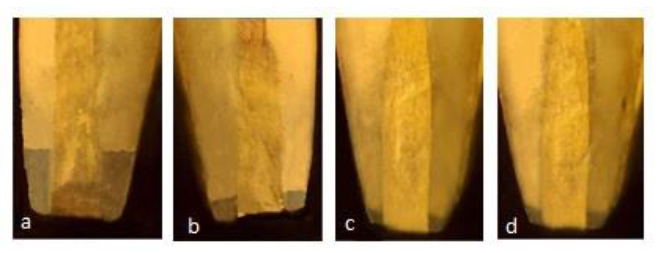
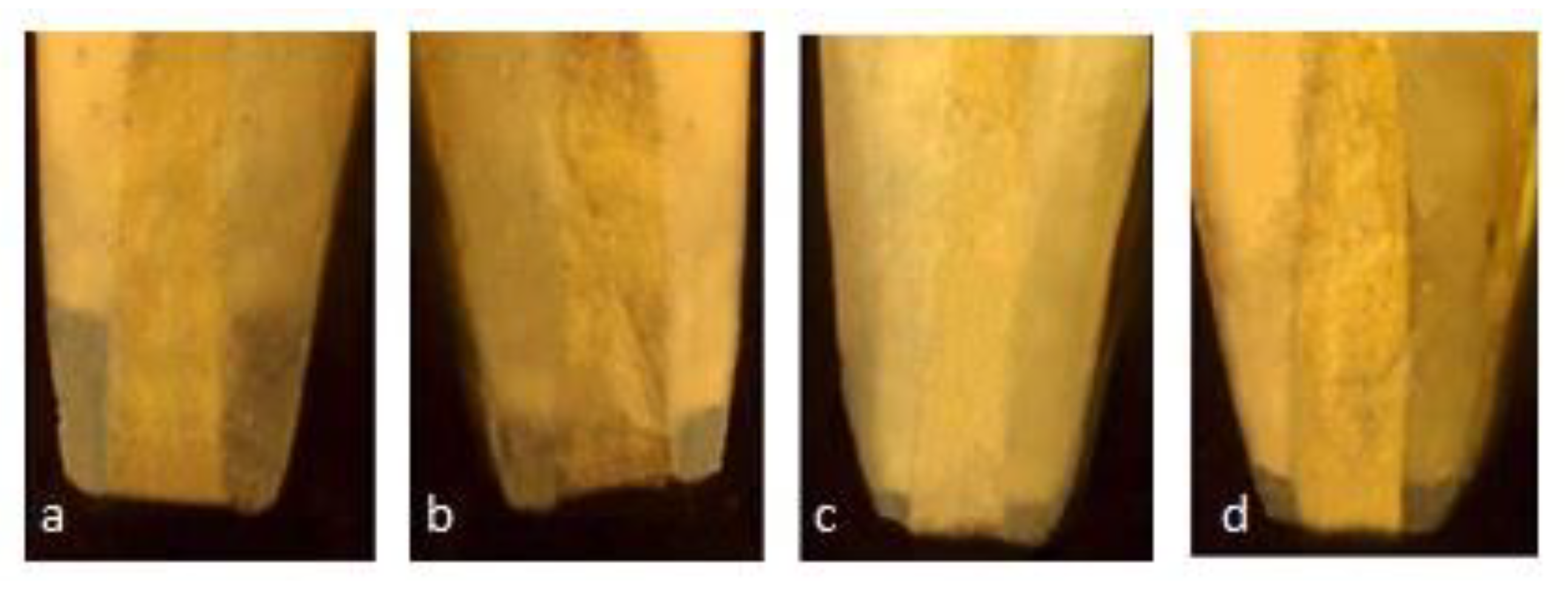
| Groups (n-20) | After 15 Days (n-10) | After 30 Days (n-10) |
|---|---|---|
| GIC | 1a | 1b |
| Pro Root MTA | 2a | 2b |
| Neo MTA plus | 3a | 3b |
| Biodentin | 4a | 4b |
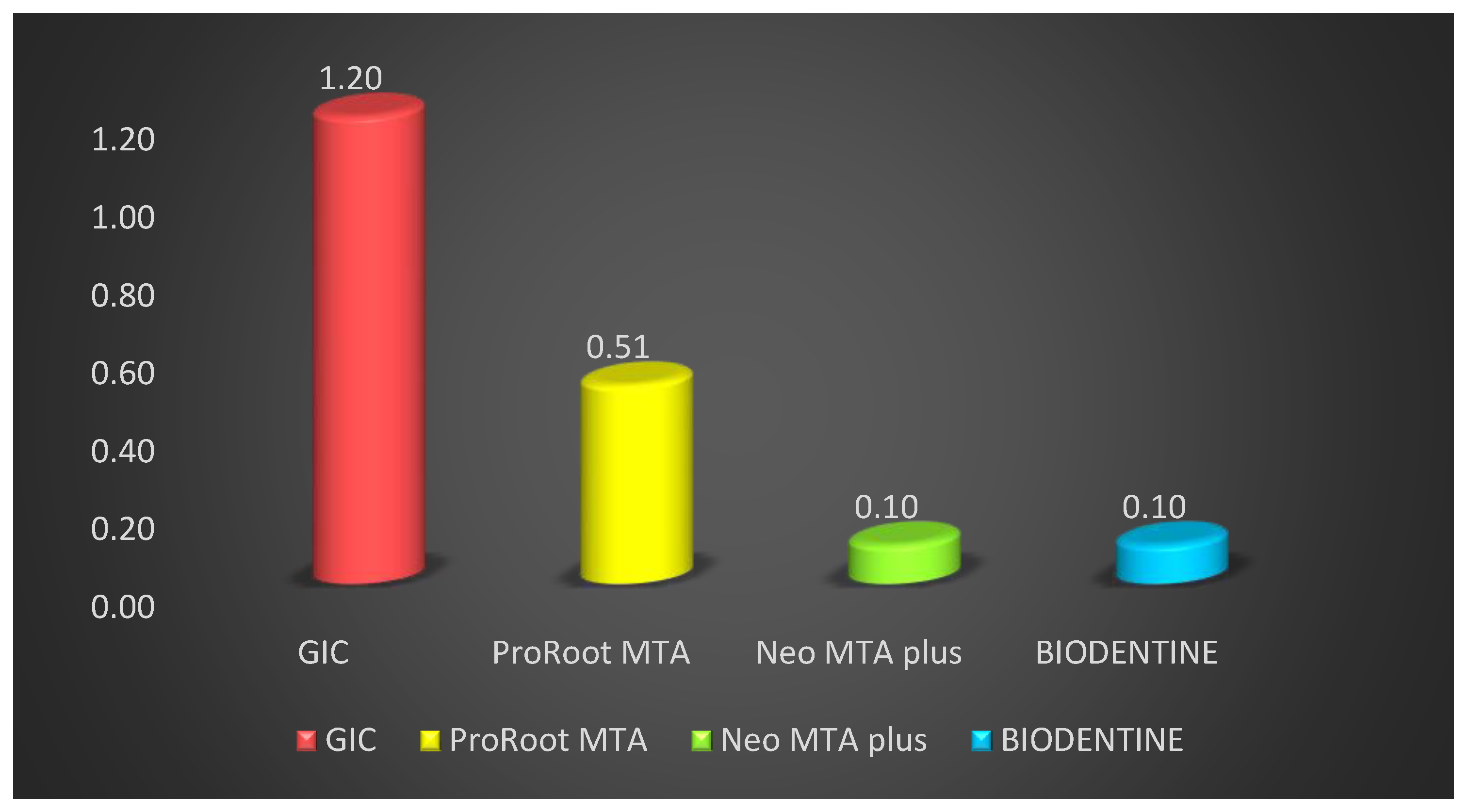
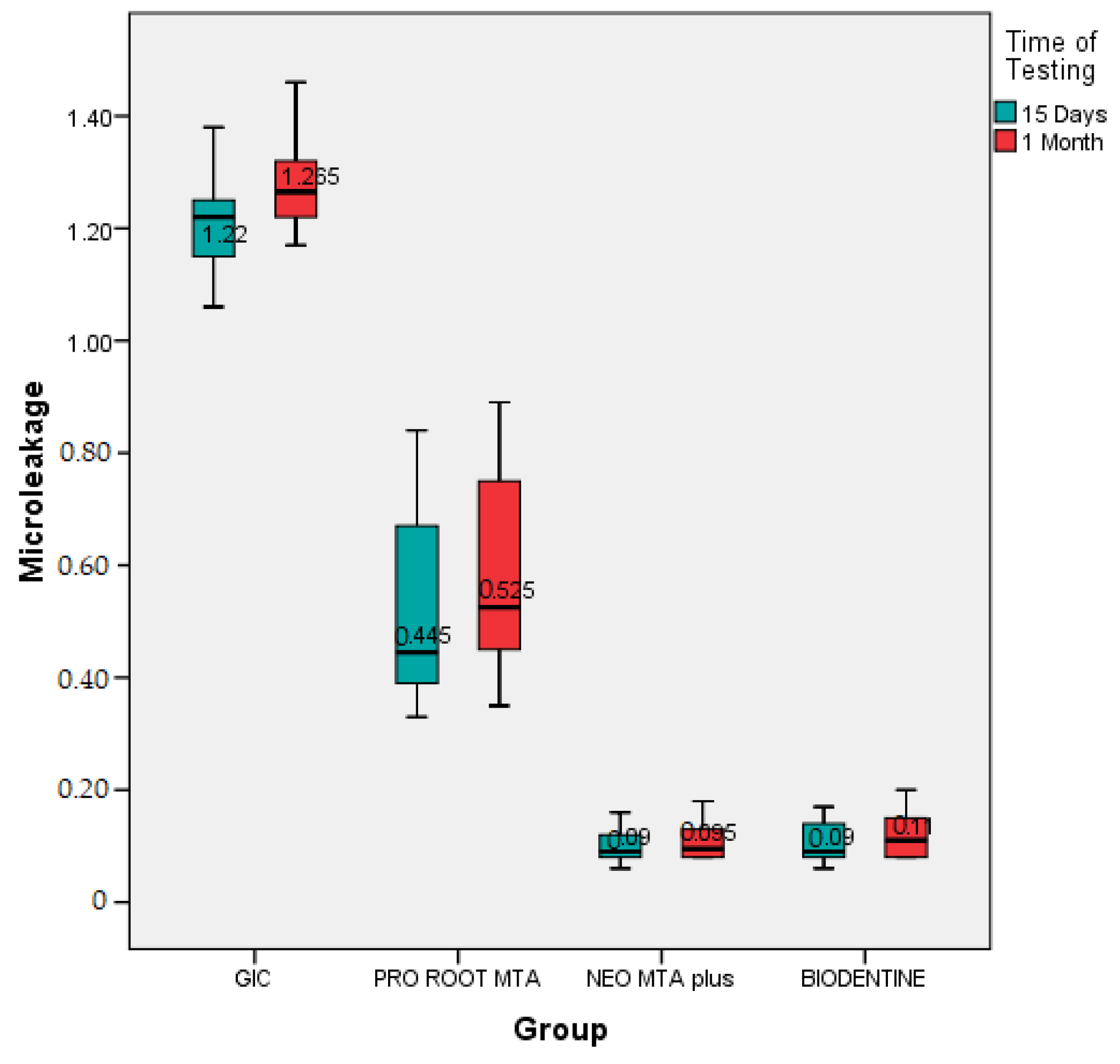
| Groups | n | Mean | Std. Deviation | F Value | p Value | Post Hoc Tukey Test |
|---|---|---|---|---|---|---|
| GIC | 10 | 1.20 | 0.10 | 248.03 | 0.01 (HS) | GIC > Pro Root MTA > BIODENTINE ≈ Neo MTA plus |
| ProRoot MTA | 10 | 0.51 | 0.16 | |||
| Neo MTA plus | 10 | 0.10 | 0.03 | |||
| BIODENTINE | 10 | 0.10 | 0.04 | |||
| Total | 40 | 0.42 | 0.36 |
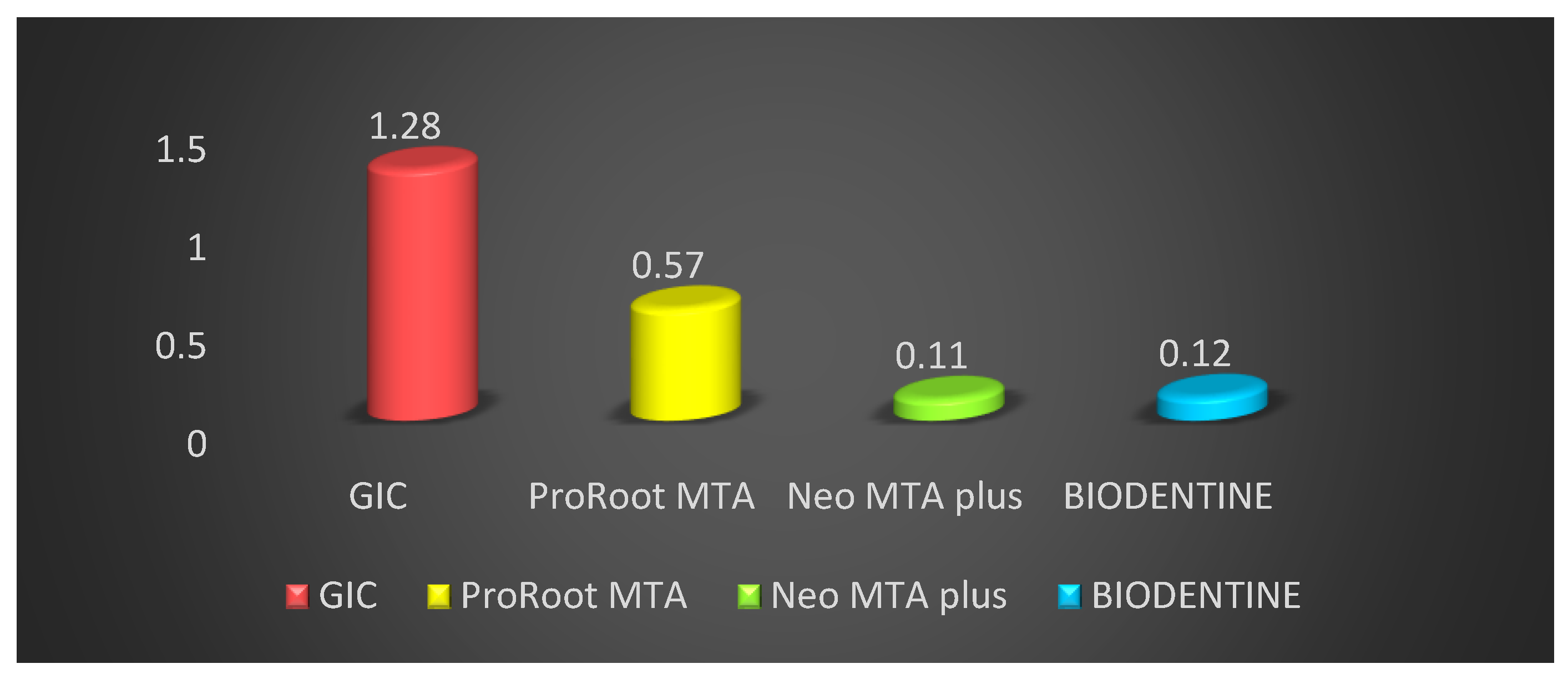
| Groups | n | Mean | Std. Deviation | F Value | p Value | LSD Post Hoc Test |
|---|---|---|---|---|---|---|
| GIC | 10 | 1.28 | 0.08 | - | - | GIC > Pro Root MTA > BIODENTINE ≈ Neo MTA plus |
| ProRoot Mta | 10 | 0.57 | 0.18 | - | - | |
| NEO MTA plus | 10 | 0.11 | 0.03 | - | - | |
| BIODENTINE | 10 | 0.12 | 0.04 | 279.30 | 0.01 |
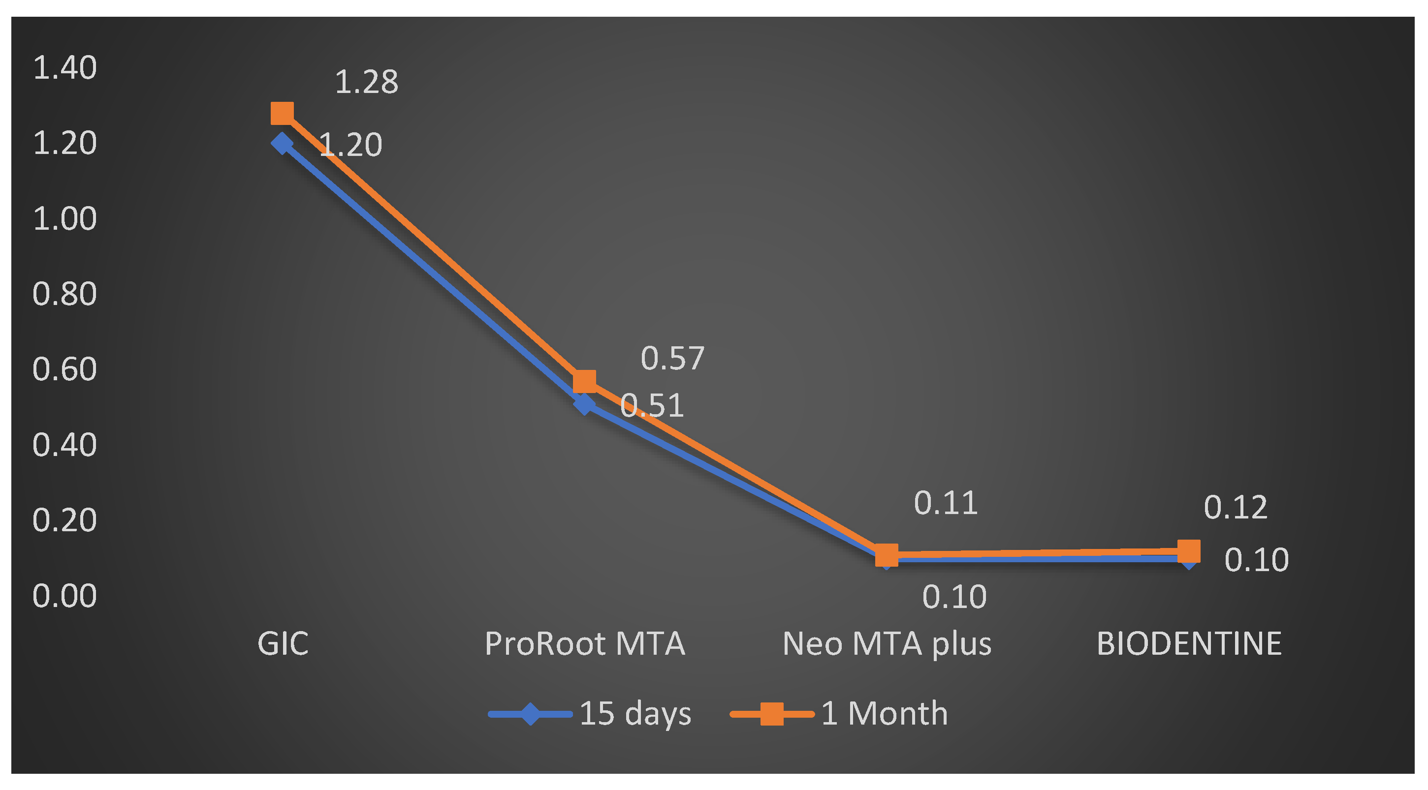
| Groups | Time Period | Mean | Std. Deviation | t Value | Sig. |
|---|---|---|---|---|---|
| GIC | 15 Days | 1.20 | 0.13 | 1.90 | 0.07 |
| 1 Month | 1.28 | 0.09 | |||
| PRO ROOT MTA | 15 Days | 0.51 | 0.14 | 0.87 | 0.67 |
| 1 Month | 0.57 | 0.18 | |||
| NEO MTA plus | 15 Days | 0.10 | 0.03 | 0.06 | 0.91 |
| 1 Month | 0.11 | 0.02 | |||
| BIODENTINE | 15 Days | 0.10 | 0.03 | 0.10 | 0.89 |
| 1 Month | 0.12 | 0.02 |
Publisher’s Note: MDPI stays neutral with regard to jurisdictional claims in published maps and institutional affiliations. |
© 2021 by the authors. Licensee MDPI, Basel, Switzerland. This article is an open access article distributed under the terms and conditions of the Creative Commons Attribution (CC BY) license (https://creativecommons.org/licenses/by/4.0/).
Share and Cite
Karobari, M.I.; Basheer, S.N.; Sayed, F.R.; Shaikh, S.; Agwan, M.A.S.; Marya, A.; Messina, P.; Scardina, G.A. An In Vitro Stereomicroscopic Evaluation of Bioactivity between Neo MTA Plus, Pro Root MTA, BIODENTINE & Glass Ionomer Cement Using Dye Penetration Method. Materials 2021, 14, 3159. https://0-doi-org.brum.beds.ac.uk/10.3390/ma14123159
Karobari MI, Basheer SN, Sayed FR, Shaikh S, Agwan MAS, Marya A, Messina P, Scardina GA. An In Vitro Stereomicroscopic Evaluation of Bioactivity between Neo MTA Plus, Pro Root MTA, BIODENTINE & Glass Ionomer Cement Using Dye Penetration Method. Materials. 2021; 14(12):3159. https://0-doi-org.brum.beds.ac.uk/10.3390/ma14123159
Chicago/Turabian StyleKarobari, Mohmed Isaqali, Syed Nahid Basheer, Fazlur Rahman Sayed, Sufiyan Shaikh, Muhammad Atif Saleem Agwan, Anand Marya, Pietro Messina, and Giuseppe Alessandro Scardina. 2021. "An In Vitro Stereomicroscopic Evaluation of Bioactivity between Neo MTA Plus, Pro Root MTA, BIODENTINE & Glass Ionomer Cement Using Dye Penetration Method" Materials 14, no. 12: 3159. https://0-doi-org.brum.beds.ac.uk/10.3390/ma14123159