
Debunking the Concept of Dentinal Tubule Penetration of Endodontic Sealers: Sealer Staining with Rhodamine B Fluorescent Dye Is an Inadequate Method

 , and
, and 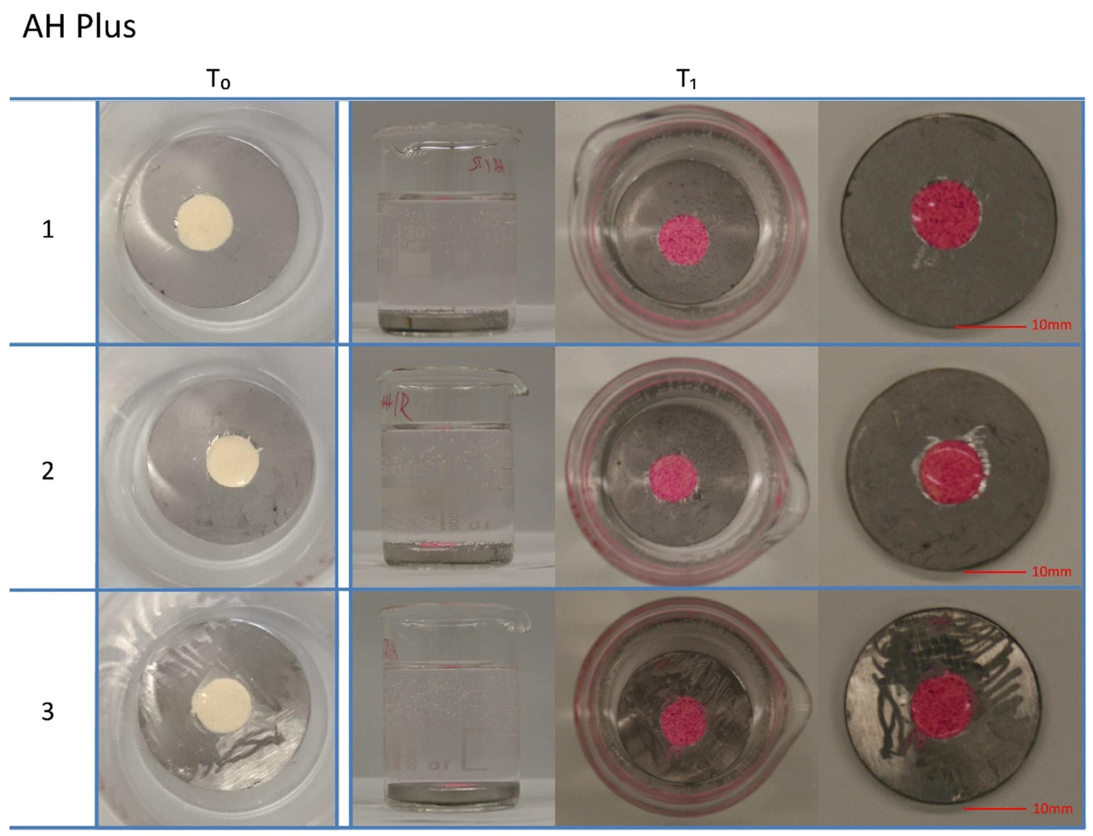
Abstract
:1. Introduction
2. Material & Methods
2.1. Experimental Setup 1
2.2. Experimental Setup 2
2.3. Experimental Setup 3
3. Results
3.1. Experimental Setup 1
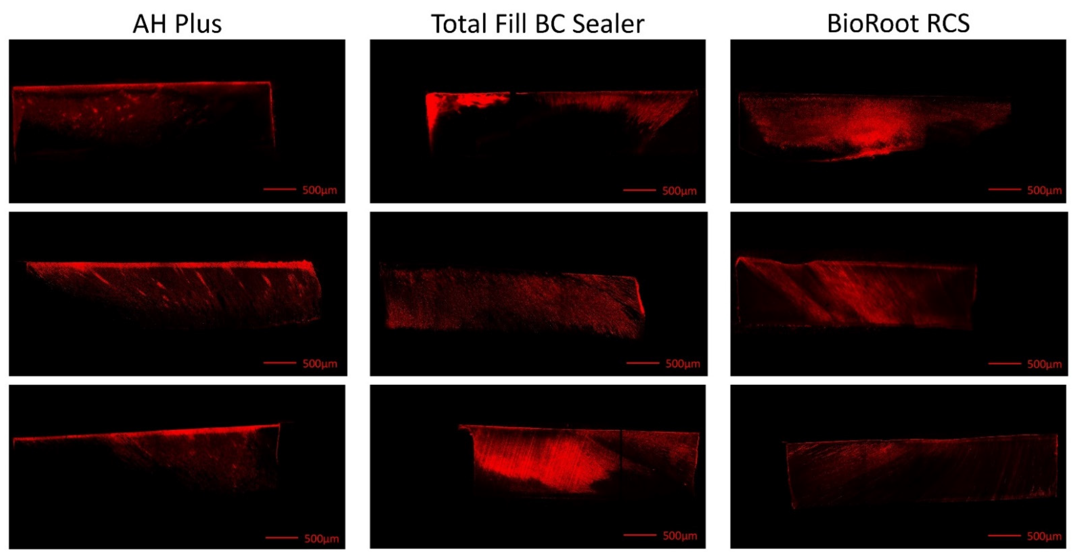
3.2. Experimental Setup 2
3.3. Experimental Setup 3
4. Discussion
5. Conclusions
Author Contributions
Funding
Institutional Review Board Statement
Informed Consent Statement
Data Availability Statement
Conflicts of Interest
References
- Orstavik, D. Materials used for root canal obturation: Technical, biological and clinical testing. Endod. Top. 2005, 12, 25–38. [Google Scholar] [CrossRef]
- Trope, M.; Bunes, A.; Debelian, G. Root filling materials and techniques: Bioceramics a new hope? Endod. Top. 2015, 32, 86–96. [Google Scholar] [CrossRef]
- Reynolds, J.Z.; Augsburger, R.A.; Svoboda, K.K.H.; Jalali, P. Comparing dentinal tubule penetration of conventional and “HiFlow” bioceramic sealers with resin-based sealer: An in vitro study. Aust. Endod. J. 2020, 46, 387–393. [Google Scholar] [CrossRef]
- Uzunoglu-Özyürek, E.; Erdoğan, Ö.; Aktemur, T.S. Effect of Calcium Hydroxide Dressing on the Dentinal Tubule Penetration of 2 Different Root Canal Sealers: A Confocal Laser Scanning Microscopic Study. J. Endod. 2018, 44, 1018–1023. [Google Scholar] [CrossRef]
- De-Deus, G.; Brandão, M.C.; Souza, E.M.; Reis, C.; Reis, K.; Machado, R.; Neelakantan, P. Epoxy resin-based root canal sealer penetration into dentin tubules does not improve root filling dislodgement resistance. Eur. Endod. J. 2017, 2, 7. [Google Scholar] [CrossRef] [Green Version]
- El Hachem, R.; Khalil, I.; Le Brun, G.; Pellen, F.; Le Jeune, B.; Daou, M.; El Osta, N.; Naaman, A.; Abboud, M. Dentinal tubule penetration of AH Plus, BC Sealer and a novel tricalcium silicate sealer: A confocal laser scanning microscopy study. Clin. Oral Investig. 2019, 23, 1871–1876. [Google Scholar] [CrossRef] [PubMed]
- Ricucci, D.; Siqueira, J.F. Fate of the Tissue in Lateral Canals and Apical Ramifications in Response to Pathologic Conditions and Treatment Procedures. J. Endod. 2010, 36, 1–15. [Google Scholar] [CrossRef]
- Weine, F.S. The enigma of the lateral canal. Dent. Clin. N. Am. 1984, 28, 833–852. [Google Scholar]
- van Meerbeek, B.; Vargas, M.; Inoue, S.; Yoshida, Y.; Perdigão, J.; Lambrechts, P.; Vanherle, G. Microscopy investigations. Techniques, results, limitations. Am. J. Dent. 2000, 13, 3D–18D. [Google Scholar]
- Jeong, J.W.; DeGraft-Johnson, A.; Dorn, S.O.; di Fiore, P.M. Dentinal Tubule Penetration of a Calcium Silicate–based Root Canal Sealer with Different Obturation Methods. J. Endod. 2017, 43, 633–637. [Google Scholar] [CrossRef]
- Coronas, V.S.; Villa, N.; Nascimento, A.L.D.; Duarte, P.H.M.; da Rosa, R.A.; Só, M.V.R. Dentinal tubule penetration of a calcium silicate-based root canal sealer using a specific calcium fluorophore. Braz. Dent. J. 2020, 31, 109–115. [Google Scholar] [CrossRef] [PubMed]
- Furtado, T.C.; de Bem, I.A.; Machado, L.S.; Pereira, J.R.; Marcus Vinícius Reis, S.; da Rosa, R.A. Intratubular penetration of endodontic sealers depends on the fluorophore used for CLSM assessment. Microsc. Res. Tech. 2021, 84, 305–312. [Google Scholar] [CrossRef] [PubMed]
- Donnermeyer, D.; Bürklein, S.; Dammaschke, T.; Schäfer, E. Endodontic sealers based on calcium silicates: A systematic review. Odontology 2019, 107, 421–436. [Google Scholar] [CrossRef] [PubMed]
- Hergt, A.; Wiegand, A.; Hülsman, M.; Rödig, T. AH Plus root canal sealer—An updated literature review. ENDO (Lond. Engl.) 2015, 9, 245–265. [Google Scholar]
- Siboni, F.; Taddei, P.; Zamparini, F.; Prati, C.; Gandolfi, M.G. Properties of bioroot RCS, a tricalcium silicate endodontic sealer modified with povidone and polycarboxylate. Int. Endod. J. 2017, 50, e120–e136. [Google Scholar] [CrossRef] [PubMed] [Green Version]
- Weis, M.V.; Parashos, P.; Messer, H.H. Effect of obturation technique on sealer cement thickness and dentinal tubule penetration. Int. Endod. J. 2004, 37, 653–663. [Google Scholar] [CrossRef] [PubMed] [Green Version]
- de Deus, G.A.; Gurgel-Filho, E.D.; Maniglia-Ferreira, O.; Coulinho-Filho, T. The influence of filling technique on depth of tubule penetration by root canal sealer: A study using light microscopy and digital image processing. Aust. Endod. J. 2004, 30, 23–28. [Google Scholar] [CrossRef]
- Patel, D.V.; Sherriff, M.; Ford, T.R.P.; Watson, T.F.; Mannocci, F. The penetration of RealSeal primer and Tubliseal into root canal dentinal tubules: A confocal microscopic study. Int. Endod. J. 2007, 40, 67–71. [Google Scholar] [CrossRef]
- del Monaco, R.J.; de Oliveira, M.T.; de Lima, A.F.; Navarro, R.S.; Zanetti, R.V.; da Silva, D.d.F.T.; Horliana, A.C.R.T. Influence of Nd:YAG laser on the penetration of a bioceramic root canal sealer into dentinal tubules: A confocal analysis. PLoS ONE 2018, 13, e0202295. [Google Scholar] [CrossRef] [PubMed] [Green Version]
- Aydın, Z.U.; Özyürek, T.; Keskin, B.; Baran, T. Effect of chitosan nanoparticle, QMix, and EDTA on TotalFill BC sealers’ dentinal tubule penetration: A confocal laser scanning microscopy study. Odontology 2019, 107, 64–71. [Google Scholar] [CrossRef]
- Gharib, S.R.; Tordik, P.A.; Imamura, G.M.; Baginski, T.A.; Goodell, G.G. A confocal laser scanning microscope investigation of the epiphany obturation system. J. Endod. 2007, 33, 957–961. [Google Scholar] [CrossRef] [PubMed]
- Pioch, T.; Stotz, S.; Buff, E.; Duschner, H.; Staehle, H.J. Influence of different etching times on hybrid layer formation and tensile bond strength. Am. J. Dent. 1998, 11, 202–206. [Google Scholar] [PubMed]
- Pioch, T. Novel feasibilities for visualizing the contact zone between dentin and resin by application of Leica CLSM. Leica Sci Tech. Inf. 1996, 11, 80–83. [Google Scholar]
- Jung, M.; Lommel, D.; Klimek, J. The imaging of root canal obturation using micro-CT. Int. Endod. J. 2005, 38, 617–626. [Google Scholar] [CrossRef]
- Linde, A.; Goldberg, M. Dentinogenesis. Crit. Rev. Oral Biol. Med. 1994, 4, 679–728. [Google Scholar] [CrossRef] [PubMed]


Publisher’s Note: MDPI stays neutral with regard to jurisdictional claims in published maps and institutional affiliations. |
© 2021 by the authors. Licensee MDPI, Basel, Switzerland. This article is an open access article distributed under the terms and conditions of the Creative Commons Attribution (CC BY) license (https://creativecommons.org/licenses/by/4.0/).
Share and Cite
Donnermeyer, D.; Schmidt, S.; Rohrbach, A.; Berlandi, J.; Bürklein, S.; Schäfer, E. Debunking the Concept of Dentinal Tubule Penetration of Endodontic Sealers: Sealer Staining with Rhodamine B Fluorescent Dye Is an Inadequate Method. Materials 2021, 14, 3211. https://0-doi-org.brum.beds.ac.uk/10.3390/ma14123211
Donnermeyer D, Schmidt S, Rohrbach A, Berlandi J, Bürklein S, Schäfer E. Debunking the Concept of Dentinal Tubule Penetration of Endodontic Sealers: Sealer Staining with Rhodamine B Fluorescent Dye Is an Inadequate Method. Materials. 2021; 14(12):3211. https://0-doi-org.brum.beds.ac.uk/10.3390/ma14123211
Chicago/Turabian StyleDonnermeyer, David, Sina Schmidt, Arno Rohrbach, Johannes Berlandi, Sebastian Bürklein, and Edgar Schäfer. 2021. "Debunking the Concept of Dentinal Tubule Penetration of Endodontic Sealers: Sealer Staining with Rhodamine B Fluorescent Dye Is an Inadequate Method" Materials 14, no. 12: 3211. https://0-doi-org.brum.beds.ac.uk/10.3390/ma14123211