
Structure, Luminescence, and Magnetic Properties of Crystalline Manganese Tungstate Doped with Rare Earth Ion


Division of Materials Science and Engineering, Silla University, Busan 46958, Korea
*
Author to whom correspondence should be addressed.
Materials 2021, 14(13), 3717; https://0-doi-org.brum.beds.ac.uk/10.3390/ma14133717
Submission received: 3 June 2021
/
Revised: 30 June 2021
/
Accepted: 30 June 2021
/
Published: 2 July 2021
(This article belongs to the Special Issue Synthesis, Characterization, and Degradation of Advanced Optical and Photo-Active Materials)
Abstract
:The precursor prepared by co-precipitation method was sintered at various temperatures to synthesize crystalline manganese tungstate (MnWO4). Sintered MnWO4 showed the best crystallinity at a sintering temperature of 800 °C. Rare earth ion (Dysprosium; Dy3+) was added when preparing the precursor to enhance the magnetic and luminescent properties of crystalline MnWO4 based on these sintering temperature conditions. As the amount of rare earth ions was changed, the magnetic and luminescent characteristics were enhanced; however, after 0.1 mol.%, the luminescent characteristics decreased due to the concentration quenching phenomenon. In addition, a composite was prepared by mixing MnWO4 powder, with enhanced magnetism and luminescence properties due to the addition of dysprosium, with epoxy. To one of the two prepared composites a magnetic field was applied to induce alignment of the MnWO4 particles. Aligned particles showed stronger luminescence than the composite sample prepared with unsorted particles. As a result of this, it was suggested that it can be used as phosphor and a photosensitizer by utilizing the magnetic and luminescent properties of the synthesized MnWO4 powder with the addition of rare earth ions.
1. Introduction
Recently, metal tungstate (MXO4, M = Ba, Ca, Mn, Sr, X = Mo, W) has attracted a lot of attention because of its applicability as a multiferroic, light-emitting material, light-emitting diode, and laser [1,2]. Among tungstate materials, the tetragonal scheelite-like structure is a comporting phosphor host material and photocatalytic material because the group shows a good absorption rate in the ultraviolet (UV) and blue ranges. This produces a specific emission band through energy transfer from the group to the RE ion [3,4,5]. In particular, manganese tungstate (MnWO4) crystal is a suitable parent material for doping rare earth and metal ions because of its excellent thermal stability and high energy transfer efficiency from tungsten ions to activator ions; also, rare earth and metal ions are generated by energy transfer between 4f-4f shells [6]. This material has the advantage of generating a high emission intensity with a narrow band gap and a variety of emission wavelengths [7]. The kind and positional symmetry of the activator ions doped in the thermally and chemically stable parent grid are important factors in implementing various types of electrochemical, laser, multiferroic, and display devices [8,9]. The diversity of the MnWO4 depends on the type and concentration of activator ions, the sintering temperature, the size of the crystal grain, and the synthesis conditions. Martinez et al. proposed that MWO4 (M = Ni, Co, Mn, Cu) can be synthesized using the dissolution-precipitation method and applied to the photocatalytic evaluation field through structural and UV absorbance characteristics analysis [10]. Li et al. synthesized MnWO4 nanoparticles using co-precipitation and suggested that electrochemical capacitive performance could be investigated by galvanostatic charge/discharge (GV), cycle electrochemical impedance spectroscopy (EIS), and cyclic voltammetry (CV) [11]. It has been reported that by changing the amount of and type of rare earth ions added by changing the structure and luminescence properties of BaWO4, it is possible to synthesize a phosphor with good crystallinity and capable of implementing various colors using the co-precipitation method [10]. In addition, the BaWO4:Ln3+ (Ln = Eu, Tb, and Dy) powders synthesized via a solid-state reaction, which showed green, yellow, and red emissions [12]. As in the previous literature, various synthesis methods and application cases of tungstate materials have been reported. In this study, the MnWO4 precursor was prepared using the co-precipitation method and the optimum synthesis temperature was investigated by varying the sintering temperature. In addition, it was suggested that magnetic and luminescent properties can be realized through the addition of rare earth ions, and that MnWO4 can be applied as a fluorescent and photosensitizing material.
2. Materials and Methods
2.1. Synthesis of MnWO4 and Rare Earth Doped with MnWO4
Materials: manganese (Ⅱ) nitrate (Mn(NO3)2·xH2O), sodium tungstate (Na2WO4), dysprosium (Ⅲ) nitrate (Dy(NO3)3·xH2O, Dy3+), and Allied epoxy set were used in this study.
First, two beakers ‘A’ and ‘B’ were prepared. In beaker ‘A’, 1 mmol of Mn(NO3)2·xH2O was added to 50 mL of distilled water (D.I water) and stirred until completely dissolved. In beaker ‘B’, 50 mL of distilled water was added with the same moles of Na2WO4 and stirred until dissolved. When reagents in both beakers had dissolved, beaker ‘B’ solution was slowly poured into stirring beaker ‘A’ and stirred for about 30 min. After that, powder was obtained using a centrifuge, and the precursor was prepared by rinsing with D.I water twice to remove the remaining sodium. The prepared precursors were dried in an 80 °C oven for about 18 h. The dried precursors were heat-treated at various sintering temperatures (80, 400, 600, 800, 900, and 1000 °C) and then structural characteristics were investigated. In addition, to synthesize MnWO4 having magnetic and luminescent properties in the same manner, MnWO4:Dy3+ powder was synthesized by varying the amount of Dy(NO3)3·xH2O added to beaker ‘A’ when preparing the precursor, as shown in Figure 1.
2.2. Chraraterization
The structural properties of the MnWO4 and MnWO4:Dy3+ powders were obtained by X-ray diffraction analysis (XRD; Rigaku Ultima IV, Tokyo, Japan). Raman spectra were obtained using a Raman spectrometer (LabRam-HR 800, Horiba Jobin-Yvon, France), equipped with a 514 nm laser as excitation source. The magnetic properties of the samples were measured using a vibrating sample magnetometer with MnWO4 and MnWO4:Dy3+ samples after magnetization with 6 T pulsing magnetic field. The chemical composition and oxidation state of the synthesized phosphors were investigated by X-ray photoelectron spectroscopy (XPS, ESCALAB 250XI, Waltham, MA, USA). The peak position of the insulating samples was calibrated using a C1 of 285 eV. The surface morphology and microstructure were observed by field emission scanning electron microscopy (FE-SEM, SU-8220, Hitachi, Tokyo, Japan) and transmission electron microscopy (TEM, JEM 2100F, JEOL, Japan). Photoluminescence spectra were obtained through a photomultiplier tube operating at 250 V, a fluorescence spectrophotometer (Scinco, FS-2, Seoul, Korea), and an optical microscope (OM, BX53M, OLYMPUS, Shinjuku, Japan).
2.3. Fabrication of MnWO4:Dy3+ Epoxy Composite
A composite was prepared by mixing with epoxy to find changes in the luminescence properties of MnWO4 particles aligned by magnetic field influence. An epoxy resin and a hardener were prepared at a weight ratio of 10:1, and 3 wt.% of MnWO4:Dy3+ powder was added and stirred for 1 h. The mixture was poured into a mold and air bubbles were removed in a vacuum desiccator for 1 h. After that, one specimen was hardened as is; the other specimen was hardened by generating a magnetic field by installing magnets on both sides of the mold to align the MnWO4:Dy3+ particles.
3. Results and Discussion
3.1. Crystallinity of MnWO4 According to Various Sintering Temperatures
The precursor prepared by co-precipitation method was heat-treated at various temperatures to determine the crystallinity and structure of the synthesized MnWO4, followed by XRD analysis, with results as shown in Figure 2a. A clear XRD pattern could not be confirmed at relatively low heat treatment temperature, but it was found that MnWO4 could be synthesized even at low temperature. At the heat treatment temperature of 600 °C, crystalline MnWO4 was confirmed, as in the results of the International Center for Diffraction Data (ICDD 01-080-0133, monoclinic, P2/c) reference. In particular, main peaks of the (111), (011), (002), and (130) phases were identified [13]. In addition, as the sintering temperature increased, the full width at half maximum (FWHM) of the main peaks decreased and showed a tendency to decrease significantly at 800 °C (Figure 2b). As a result of this, it is thought that the increase of the sintering temperature increases the crystallinity of MnWO4 [14]. However, there was no significant change after the sintering temperature of 800 °C and, for energy saving, the optimum sintering temperature was determined to be 800 °C.
3.2. Crystallinity, and Magnetic and Chemical State of MnWO4 Doped with Dy3+ Ions
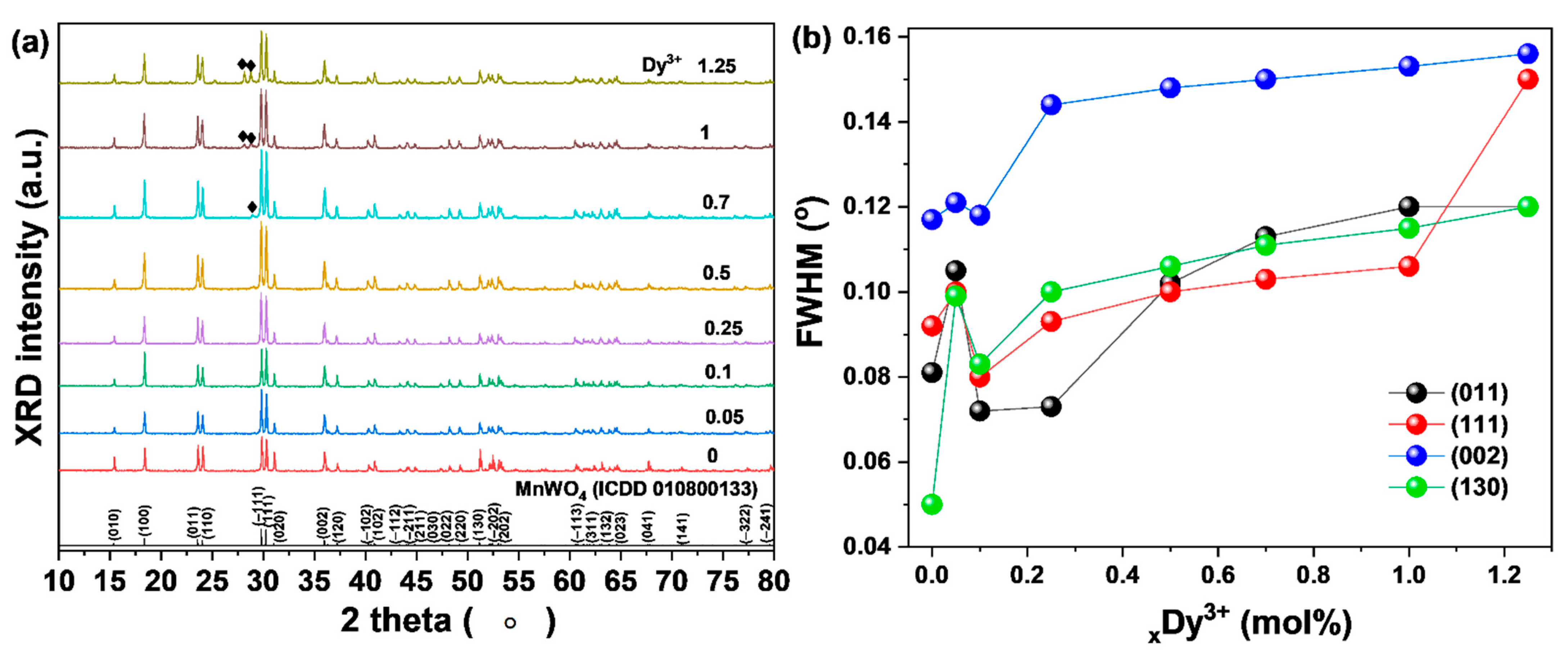
To observe changes in the characteristics of MnWO4 with the addition of rare earth materials, precursors were prepared by changing the amounts of dysprosium ions (Dy3+; 0.05, 0.1, 0.25, 0.5, 0.7, 1, 1.25 mol.%) added in the process of preparing the precursor by the same experimental method. The prepared precursor was sintered at 800 °C. When the added amount of Dy3+ ions was small (0.1 mol.%), the main peaks of the (−111), (011), (002), and (130) phases of the FWHM tended to increase. This is thought to be a phenomenon caused by the addition to the lattice of rare earth ions with relatively large ionic radii. In addition, when the amount of added rare earth material was 0.25 mol.%, the FWHM decreased (Figure 3b), which is thought to have the effect of enhancing the crystallinity [15]. However, as the amount of rare earth ions increased, the FWHM and secondary phase were found. The secondary phase (Figure 3a, black diamond symbol) was identified as a dysprosium oxide phase. When the added amount of rare earth ions was 0.25 mol.% or more, the crystallinity of MnWO4 decreased and a secondary phase formed; the critical doping concentration was considered to be 0.25 mol.%.
Raman analysis was performed to double check the XRD data obtained for Dy3+ ion-doped crystalline MnWO4. The vibration bands of MnWO4 shown in Figure 4a show six Raman active modes at 200, 320, 391, 539, 691, and 878 cm−1, This may be due to the ν(Ag), r(Bg), δ(Ag), symmetric Ag, (W2O4)n chain, νas(Bg) of the Mn cation, and the symmetric Ag oscillations of the two terminal WO groups, respectively. There is crystalline MnWO4, which correlates accurately with the literature [16]. The very strong band appearing at 878 cm-1 corresponds to the strong symmetrical stretching of the WO2 group in MnWO4. The bands at 769 and 691 cm−1 indicate the existence of weak asymmetric and symmetric tensile vibration modes of W–O–W bonds [17]. The peak at 539 cm−1 is attributed to the tensile vibration of Mn–O [18]. The band at 391 cm−1 indicates that there is a symmetric stretch of W–O–W [19]. The band at 320 cm−1 checked the moderate shear of WO2 and W–O–W [20]. The weak vibrating band appears at 251 cm−1, indicating the bending mode of [WO6]6 and the twisting vibrating mode of the WO2 group [21]. The two vibrating bands at 158 and 121 cm−1 are translational modes of tungsten [22]. In Raman analysis, no significant change was observed between the MnWO4:Dy3+ and MnWO4 samples. To identify changes in the magnetic properties according to the amount of Dy3+ added to the synthesized crystalline MnWO4, vibrating sample magnetometer (VSM) analysis was performed (Figure 4b).
In addition, it was shown that the magnetic properties increased as the amount of added Dy3+ ions increased. Due to its 4f orbital, the Dy3+ ion is a prototype of a highly correlated electronic system. It is partially occupied by the f shell, and the 4f orbital is split into seven non-degenerate orbitals. According to Hund’s law, the magnetic moments are caused by the 4f and 5d orbitals, which are consistent with the magnetic moments of the corresponding Dy3+ ions [23].
To determine MnWO4:Dy3+ samples of binding energy, oxidation, and chemical state, XPS analysis was performed on MnWO4:Dy3+ (Figure 5). Results show Mn 2p; two peaks can be observed corresponding to Mn 2p3/2 and Mn 2p1/2 at 641 eV and 653 eV, respectively. This indicates that the Mn present in the sample is in +2 oxidation state [24]. The XPS spectrum of W 4f is shown in Figure 5b. It has two peaks, corresponding to W 4f7/2 and W 4f5/2 at 34.88 and 36.98 eV, respectively. The W 4f7/2 and W 4f5/2 doublets’ spin-orbit is at 2.1 eV, and the oxidation state of W can be specified as +6 [25]. The O 1s peak shows a main component with a central energy of 530 eV and a lower binding energy, which monitored to the formation of O2 oxide-coupled manganese and tungsten elements (Mn–O–W), as shown in Figure 5c. In Figure 5e, the RE3+ 3d spectrum can be observed for the MnWO4 sample doped with RE3 +. The Dy3+ 3d spectrum is visible at 1317 eV and 1335 eV; these can be assigned to the RE3+ 3d5/2 and 3d3/2 states, respectively, based on the Dy–O bond [26].
3.3. Luminescence and Morphology Properties of MnWO4 Doped with Dy3+ Ions
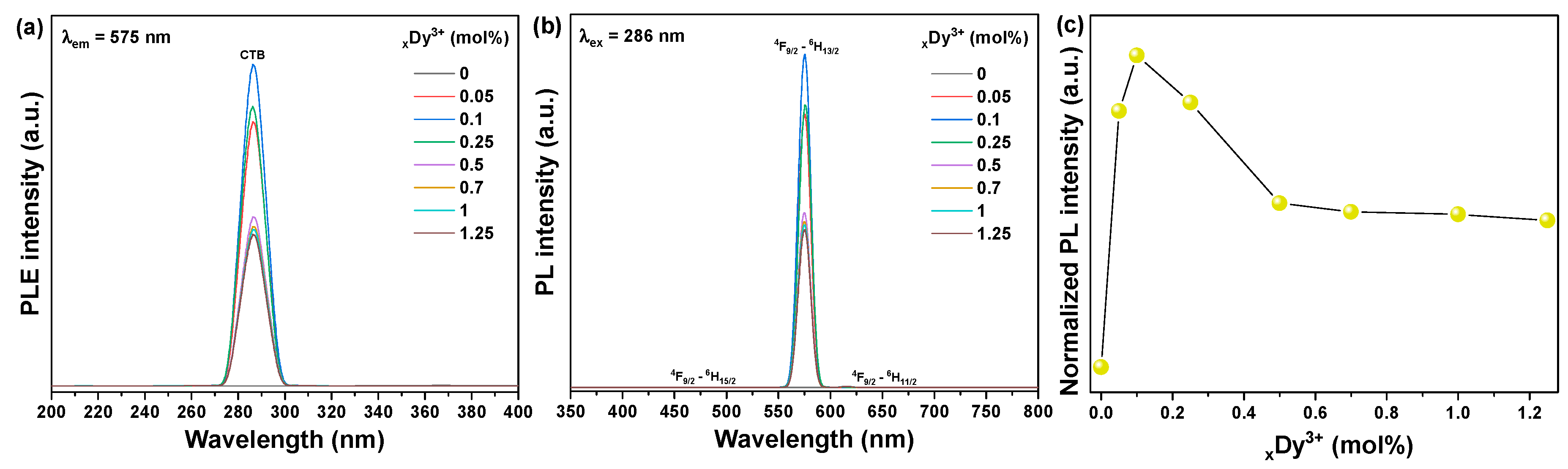
The photoluminescence excitation (PLE) and photoluminescence (PL) spectra of MnWO4: Dy3+ powder synthesized with activator Dy3+ ions of different doping concentrations were obtained. The excitation spectrum monitored at 575 nm consists of peaks in the range of 200 to 400 nm. The PLE spectrum almost covers the ultraviolet region. The excitation band is wide in the range of 270–300 nm to 286 nm, corresponding to the Dy3+-O2− charge transfer band (CTB) in the matrix crystal [27,28]. When the concentration of Dy3+ increased from 0.05 to 0.25 mol.%, the intensity of all excitation bands increased rapidly, reaching a maximum at 0.1 mol.%, and then decreased significantly within the Dy3+ concentration range of 0.5 to 1.25 mol.%, as shown in Figure 6a. For MnWO4: Dy3+ powder, the emission spectrum under 286 nm excitation shows two main emission bands at 480 and 575 nm, corresponding to the 4F9/2 → 6H15/2 magnetic dipole (MD) transition and the transition of the electric dipole (ED) of 4F9/2 → 6H13/2, respectively. [29] The emission intensity at 575 nm (ED) is very obvious. This result shows that when Dy3+ concentration is 0.1 mol.%, the position of the Dy3+ ion in the MnWO4 host lattice shifts from a non-antisymmetric position to an antisymmetric position. Among all the emission transitions of Dy3+, the strongest yellow emission originated from the 4F9/2 → 6H13/2 ED transition. As the concentration of Dy3+ ions increased from 0.5 to 1.25 mol.%, the intensity of the main 4F9/2 → 6H13/2 transition rapidly decreased due to the concentration quenching effect (Figure 6c), mainly due to non-radiant energy transfer between Dy3+ activator ions. The critical distance Rc between the Dy3+ activator ions can be calculated using the following equation presented by Blasse [30],
where V is the volume of the unit cell, Xc is the critical concentration (Dy3+ ions), and Z is the number of host cations in the unit cell. For the MnWO4 host, Å3, and . Therefore, Rc was estimated to be about 11.65 Å. It is well known that there are three types of interactions in which electric multipolar interaction is involved in the energy transfer: dipole–dipole, dipole–quadrupole, and quadrupole–quadrupole interactions.
To observe the shape and morphology of the synthesized MnWO4:Dy3+ particles, FE-SEM and TEM analyses were performed and results are shown in Figure 7. MnWO4:Dy3+ was generally clustered in the shape of a long, round column in the vertical direction; the size was about 4 µm in length and about 2 µm in width. In high-resolution analysis using TEM, the interplanar distance of the (−111) phase was observed to be about 0.213 nm. This was similar to the value calculated from the XRD data (d(−111) spacing; 0.299 nm). In addition, Mn, W, O, and Dy were detected in EDX component analysis; components of synthesized MnWO4:Dy3+ were confirmed.
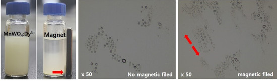
3.4. MnWO4:Dy3+ Particle Aligned in Epoxy Composite by Magnetic Field
As can be seen in Figure 8a, the synthesized MnWO4:Dy3+ powder was dispersed in ethanol and then a magnet was installed. It was confirmed that the powders moved in the direction of the magnet and can be used as a paramagnetic material. In addition, movements of particles in the mixture made with epoxy polymer were observed through an optical microscope according to the presence or absence of a magnetic field (Figure 8b). MnWO4:Dy3+ particles were confirmed to align in the direction of the magnetic field. Using these characteristics, we made an epoxy composite and investigated changes in luminescence characteristics according to alignment of particles according to presence or absence of magnetic field (Figure 8c). The composite in which the particles were aligned by exposure to the magnetic field showed a strong luminescence intensity of about 10% (Figure 8d). This is thought to be a phenomenon that occurs because the energy transfer of the aligned particles works slightly more efficiently than in particles in which light energy introduced from the outside is agglomerated. It seems that, utilizing these characteristics, this material can be applied to fields such as display and medical engineering.
4. Conclusions
We propose that MnWO4 powder synthesis is possible in an easy way. After preparing the MnWO4 precursor using the co-precipitation method, the crystallinity change of MnWO4 according to various synthesis temperatures was observed. The best crystallinity was exhibited when the heat treatment temperature was 800 °C. At this time, dysprosium, a rare earth ion, was added to enhance light emission and magnetic properties to synthesize MnWO4:Dy3+. According to the amount of Dy3+ ions added, the magnetic properties were enhanced and the luminescence properties were enhanced. When producing an epoxy composite using these properties, it was found that the luminescent properties were enhanced by about 10% as the particles were aligned in the magnetic field direction. It was suggested that the synthesized MnWO4:Dy3+ powder can be used as a paramagnetic material and can be applied to display and medical industries.
Author Contributions
Conceptualization, J.-Y.J.; D.-H.H.; methodology, J.-Y.J.;C.-S.S.; formal analysis, J.-Y.J., and D.-H.H.; investigation, J.-Y.J., and S.-S.Y.; writing—original draft preparation, J.-Y.J.; writing—review and editing, J.-Y.J., S.-S.Y., D.-H.H., and C.-S.S.; visualization, J.-Y.J.; supervision, J.-Y.J.;D.-H.H.; project administration, J.-Y.J., C.-S.S. All authors have read and agreed to the published version of the manuscript.
Funding
This research was supported by Basic Science Research Program through the National Research Foundation of Korea (NRF) funded by the Ministry of Education (grant number) (NRF-2019R1A6A3A01095400) and (NRF-2020R1F1A1072616) and by the National Research Foundation of Korea (NRF) grant funded by the Korea government (MSIT) (No. NRF-2018R1A5A1025594).
Institutional Review Board Statement
Not applicable.
Informed Consent Statement
Not applicable.
Data Availability Statement
The data presented in this study are available in the database of the authors at the Faculty of Materials Science and Engineering.
Conflicts of Interest
The authors declare no conflict of interest.
References
- Downing, E.; Hesselink, L.; Ralston, J.; Macfarlane, R. A Three-Color, Solid-State, Three-Dimensional Display. Science 1996, 273, 1185–1189. [Google Scholar] [CrossRef]
- Cooper, T.; De Leeuw, N. A combined ab initio and atomistic simulation study of the surface and interfacial structures and energies of hydrated scheelite: Introducing a CaWO4 potential model. Surf. Sci. 2003, 531, 159–176. [Google Scholar] [CrossRef]
- Nivetha, P.; Kavitha, B.; Kalanithi, M. Investigation of photocatalytic and antimicrobial activities of BaWO4–MoS2 nanoflowers. J. Sci. Adv. Mater. Devices 2021, 6, 65–74. [Google Scholar] [CrossRef]
- Kuzmin, A.; Purans, J. Local atomic and electronic structure of tungsten ions in AWO4 crystals of scheelite and wolframite types. Radiat. Meas. 2001, 33, 583–586. [Google Scholar] [CrossRef]
- Koepke’, C.; Wojtowicz, A.J.; Lempicki, A. Luminescence Excited-state absorption in excimer-pumped CaWO4 crystals. J. Lumin. 1993, 54, 345–355. [Google Scholar] [CrossRef]
- Grobelna, B.; Lipowska, B.; Kłonkowski, A.M. Energy transfer in calcium tungstate doped with Eu(III) or Tb(III) ions incorporated into silica xerogel. J. Alloys Compd. 2006, 419, 191–196. [Google Scholar] [CrossRef]
- Naik, S.; Salker, A. Solid state studies on cobalt and copper tungstates nano materials. Solid State Sci. 2010, 12, 2065–2072. [Google Scholar] [CrossRef]
- Rajagopal, S.; Nataraj, D.; Mangalaraj, D.; Djaoued, Y.; Robichaud, J.; Khyzhun, O.Y. Controlled Growth of WO3 Nanostructures with Three Different Morphologies and Their Structural, Optical, and Photodecomposition Studies. Nanoscale Res. Lett. 2009, 4, 1335–1342. [Google Scholar] [CrossRef] [Green Version]
- López, X.A.; Fuentes, A.F.; Zaragoza, M.M.; Guillén, J.A.D.; Gutiérrez, J.S.; Ortiz, A.L.; Collins-Martínez, V. Synthesis, characterization and photocatalytic evaluation of MWO4 (M = Ni, Co, Cu and Mn) tungstates. Int. J. Hydrogen Energy 2016, 41, 23312–23317. [Google Scholar] [CrossRef]
- Li, F.; Xu, X.; Huo, J.; Wang, W. A simple synthesis of MnWO4 nanoparticles as a novel energy storage material. Mater. Chem. Phys. 2015, 167, 22–27. [Google Scholar] [CrossRef]
- Jung, J.-Y.; Kim, J.; Shim, Y.-S.; Hwang, D.; Son, C. Structure and Photoluminescence Properties of Rare-Earth (Dy3+, Tb3+, Sm3+)-Doped BaWO4 Phosphors Synthesized via Co-Precipitation for Anti-Counterfeiting. Materials 2020, 13, 4165. [Google Scholar] [CrossRef] [PubMed]
- Sun, X.; Sun, X.; Li, X.; He, J.; Wang, B. Synthesis and Luminescence of BaWO4:Ln3+ (Ln = Eu, Tb, and Dy) Powders. J. Electron. Mater. 2014, 43, 3534–3538. [Google Scholar] [CrossRef]
- Chen, S.; Chen, X.; Xue, Z.; Zhou, J.; Li, J.; Hong, J.; You, X. Morphology control of MnWO4 nanocrystals by a solvothermal route. J. Mat. Chem. 2003, 13, 1132–1135. [Google Scholar] [CrossRef]
- Shen, Y.-J.; Zhang, Y.; Gao, F.; Yang, G.-S.; Lai, X.-P. Influence of Temperature on the Microstructure Deterioration of Sandstone. Energies 2018, 11, 1753. [Google Scholar] [CrossRef] [Green Version]
- Akhtar, M.N.; Hussain, T.; Khan, M.A.; Ahmad, M. Structural, magnetic, dielectric and high frequency response of synthesized rare earth doped bismuth nano garnets (BIG). Results Phys. 2018, 10, 784–793. [Google Scholar] [CrossRef]
- Tanaka, T.; Hirai, H.; Matsuoka, T.; Ohishi, Y.; Yagi, T.; Ohtake, M.; Yamamoto, Y.; Nakano, S.; Irifune, T. Phase changes of filled ice Ih methane hydrate under low temperature and high pressure. J. Chem. Phys. 2013, 139, 104701. [Google Scholar] [CrossRef]
- Dai, R.; Ding, X.; Wang, Z.; Zhang, Z. Pressure and temperature dependence of Raman scattering of MnWO4. Chem. Phys. Lett. 2013, 586, 76–80. [Google Scholar] [CrossRef]
- Iliev, M.; Gospodinov, M.M.; Litvinchuk, A.P. Raman spectroscopy of MnWO4. Phys. Rev. B 2009, 80, 212302. [Google Scholar] [CrossRef]
- Maczka, M.; Ptak, M.; Da Silva, K.P.; Freire, P.D.T.C.; Hanuza, J. High-pressure Raman scattering and an anharmonicity study of multiferroic wolframite-type Mn0.97Fe0.03WO4. J. Physics Condens. Matter 2012, 24, 345403. [Google Scholar] [CrossRef] [PubMed]
- Ruiz-Fuertes, J.; Friedrich, A.; Gomis, O.; Errandonea, D.; Morgenroth, W.; Sans, J.A.; Santamaria-Perez, D. High-pressure structural phase transition inMnWO4. Phys. Rev. B 2015, 91, 104109. [Google Scholar] [CrossRef] [Green Version]
- Macavei, J.; Schulz, H. The crystal structure of wolframite type tungstates at high pressure. Z. Krist. Cryst. Mater. 1993, 207, 193–208. [Google Scholar] [CrossRef]
- Iliev, M.N.; Guo, H.; Gupta, A. Raman spectroscopy evidence of strong spin-phonon coupling in epitaxial thin films of the double perovskite La2NiMnO6. Appl. Phys. Lett. 2007, 90, 151914. [Google Scholar] [CrossRef]
- Chen, X.-Y.; Long, M.-Q.; Wang, Y.-P. Paramagnetic phases of two-dimensional magnetic materials. Phys. Rev. B 2020, 102, 214417. [Google Scholar] [CrossRef]
- Jiang, F.; Pang, Z.; Yuan, H.; Wei, Z.; Xie, W.; Wu, Z.; Han, S. Room temperature ferromagnetic properties of dysprosium-doped tris(8-hydroxyquinoline) aluminum: Experimental and theoretical investigation. RSC Adv. 2016, 6, 43780–43785. [Google Scholar] [CrossRef]
- Muthamizh, S.; Suresh, R.; Giribabu, K.; Manigandan, R.; Kumar, S.P.; Munusamy, S.; Narayanan, V. MnWO4 nanocapsules: Synthesis, characterization and its electrochemical sensing property. J. Alloys Compd. 2015, 619, 601–609. [Google Scholar] [CrossRef]
- Dong, F.; Sattayasamitsathit, S.; Zhang, Y.X.; Zhou, Y. Materials Chemistry for Sustainability and Energy. J. Chem. 2014, 2014, 1–3. [Google Scholar] [CrossRef]
- Gokhale, S.; Ahmed, N.; Mahamuni, S.; Rao, V.; Nigavekar, A.; Kulkarni, S. XPS and XRD investigations of Dy/Si interface. Surf. Sci. 1989, 210, 85–98. [Google Scholar] [CrossRef]
- Rydberg, S.; Engholm, M.; Rydberg, S.; Engholm, M. Charge transfer processes and ultraviolet induced absorption in Yb:YAG single crystal laser materials Charge transfer processes and ultraviolet induced absorption in Yb:YAG single crystal laser ma-terials. J. Appl. Phys. 2013, 11, 223510. [Google Scholar] [CrossRef] [Green Version]
- Sharma, S.; Brahme, N.; Bisen, D.P.; Dewangan, P. Cool white light emission from Dy3+ activated alkaline alumino silicate phosphors. Opt. Express 2018, 26, 29495–29508. [Google Scholar] [CrossRef]
- Sun, J.; Zhang, X.; Xia, Z.; Du, H. Synthesis and luminescence properties of novel LiSrPO4:Dy3+ phosphor. Mater. Res. Bull. 2011, 46, 2179–2182. [Google Scholar] [CrossRef]
Figure 1.
Schematic of MnWO4 powder synthesis experimental procedure.

Figure 2.
(a) XRD patterns of MnWO4 and (b) FWHM of main peaks according to various sintering temperatures.
Figure 2.
(a) XRD patterns of MnWO4 and (b) FWHM of main peaks according to various sintering temperatures.

Figure 3.
(a) XRD patterns of MnWO4:Dy3+ and (b) change of FWHM.

Figure 4.
(a) Raman shift of MnWO4 and MnWO4:Dy3+, (b) magnetization properties of MnWO4:Dy3+ performed at room temperature, and (c) scale of M(10 kOe) vs. Dy3+ ions content.
Figure 4.
(a) Raman shift of MnWO4 and MnWO4:Dy3+, (b) magnetization properties of MnWO4:Dy3+ performed at room temperature, and (c) scale of M(10 kOe) vs. Dy3+ ions content.

Figure 5.
XPS data of MnWO4:Dy3+: (a) survey, (b) W 4f, (c) O 1s, (d) Mn 2p, and (e) Dy 3d.

Figure 6.
Luminescence properties of MnWO4:Dy3+: (a) PLE spectra under 575 nm, (b) PL spectra under 286 nm, and (c) integrated PL intensity.
Figure 6.
Luminescence properties of MnWO4:Dy3+: (a) PLE spectra under 575 nm, (b) PL spectra under 286 nm, and (c) integrated PL intensity.

Figure 7.
(a) SEM image of MnWO4:Dy3+, (b) TEM image of MnWO4:Dy3+, (c) high-resolution image, and (d) EDX composition analysis result.
Figure 7.
(a) SEM image of MnWO4:Dy3+, (b) TEM image of MnWO4:Dy3+, (c) high-resolution image, and (d) EDX composition analysis result.

Figure 8.
(a) Photograph of MnWO4:Dy3+ powder moved to magnet in ethanol, (b) OM images of MnWO4:Dy3+ particle behavior with magnetic field, (c) PL spectra, and (d) integrated PL intensity with magnetic field.
Figure 8.
(a) Photograph of MnWO4:Dy3+ powder moved to magnet in ethanol, (b) OM images of MnWO4:Dy3+ particle behavior with magnetic field, (c) PL spectra, and (d) integrated PL intensity with magnetic field.

Publisher’s Note: MDPI stays neutral with regard to jurisdictional claims in published maps and institutional affiliations. |
© 2021 by the authors. Licensee MDPI, Basel, Switzerland. This article is an open access article distributed under the terms and conditions of the Creative Commons Attribution (CC BY) license (https://creativecommons.org/licenses/by/4.0/).
Share and Cite
MDPI and ACS Style
Jung, J.-Y.; Yi, S.-S.; Hwang, D.-H.; Son, C.-S. Structure, Luminescence, and Magnetic Properties of Crystalline Manganese Tungstate Doped with Rare Earth Ion. Materials 2021, 14, 3717. https://0-doi-org.brum.beds.ac.uk/10.3390/ma14133717
AMA Style
Jung J-Y, Yi S-S, Hwang D-H, Son C-S. Structure, Luminescence, and Magnetic Properties of Crystalline Manganese Tungstate Doped with Rare Earth Ion. Materials. 2021; 14(13):3717. https://0-doi-org.brum.beds.ac.uk/10.3390/ma14133717
Chicago/Turabian StyleJung, Jae-Young, Soung-Soo Yi, Dong-Hyun Hwang, and Chang-Sik Son. 2021. "Structure, Luminescence, and Magnetic Properties of Crystalline Manganese Tungstate Doped with Rare Earth Ion" Materials 14, no. 13: 3717. https://0-doi-org.brum.beds.ac.uk/10.3390/ma14133717
Note that from the first issue of 2016, this journal uses article numbers instead of page numbers. See further details here.