
Development of Multifunctional Materials Based on Poly(ether ether ketone) with Improved Biological Performances for Dental Applications

 , ,
, ,
Abstract
:1. Introduction

| Research | Main Findings | Ref. |
|---|---|---|
| Toxicity of zinc oxide nanoparticles towards bacterial systems | 3.4 mM of ZnO (~13 nm), 100% inhibition of both bacteria Escherichia coli (E. coli) and Staphylococcus aureus (S. aureus) was reported. | [17] |
| Zinc-modified titanium surface for dental applications | ZnO nanoparticles have been shown to exert an osteoconductive and osteoinductive effect on mesenchymal cells, where the presence of zinc ions significantly increases the expression of genes related to osteoblasts. | [22] |
| Modified Poly (lactic acid) (PLA) and poly(3-hydroxybutirate) (PHB) with ZnO nanoparticles | High degree of distribution with an acceptable dispersion of the ZnO nanoparticles (~12 nm) in both polymeric matrices, without significant losses in mechanical properties, exhibited excellent antibacterial performance in E. coli and S. aureus strains for PHB matrix. | [18,19] |
| Influence of ZnO nanoparticles on the performance of poly(caprolactone) (PCL) based fibers for wound healing applications | No PCL/ZnO material (ZnO nanoparticle size around 60 nm) presented cytotoxicity against adult goat fibroblasts and the ability of the composite to heal wounds on the skin of animals without presenting tissue inflammation was shown for a nanoparticle content equal to or less than 4 wt. %. | [23] |
| Antibacterial properties of PEEK-ZnO matrices | Antimicrobial studies of PEEK-ZnO nanocomposites (ZnO nanoparticle size around 60 nm) revealed that the antibacterial activity of PEEK-ZnO nanocomposites against E. coli and S. aureus was enhanced with ZnO content, and the best antibacterial property was obtained at 7.5 wt. % silane-modified nanoparticles. | [24,25] |
| Dual Ag/ZnO-Sulfonated PEEK (SPEEK) with superior antibacterial capability and biocompatibility. | The antibacterial test showed that Ag-SPEEK and Ag/ZnO-SPEEK effectively inhibited E. coli and S. aureus reproduction. Additionally, the Ag/ZnO-SPEEK systems promoted an excellent osteoblast cell response evaluated through different tests such as: cell viability, proliferation, spreading, osteo-differentiation, and maturation. | [26] |
2. Materials and Methods
2.1. ZnO Nanoparticles Synthesis
2.2. PEEK-ZnO Matrices Preparation
2.3. PEEK-ZnO Matrices Characterization
2.3.1. Crystalline Structure and Thermal Properties
2.3.2. Sample Preparation before Surface Analysis and Biological Assay
2.3.3. Dynamic Water Contact Angle (WCA)
2.3.4. Surface Morphology by Atomic Force Microscopy (AFM)
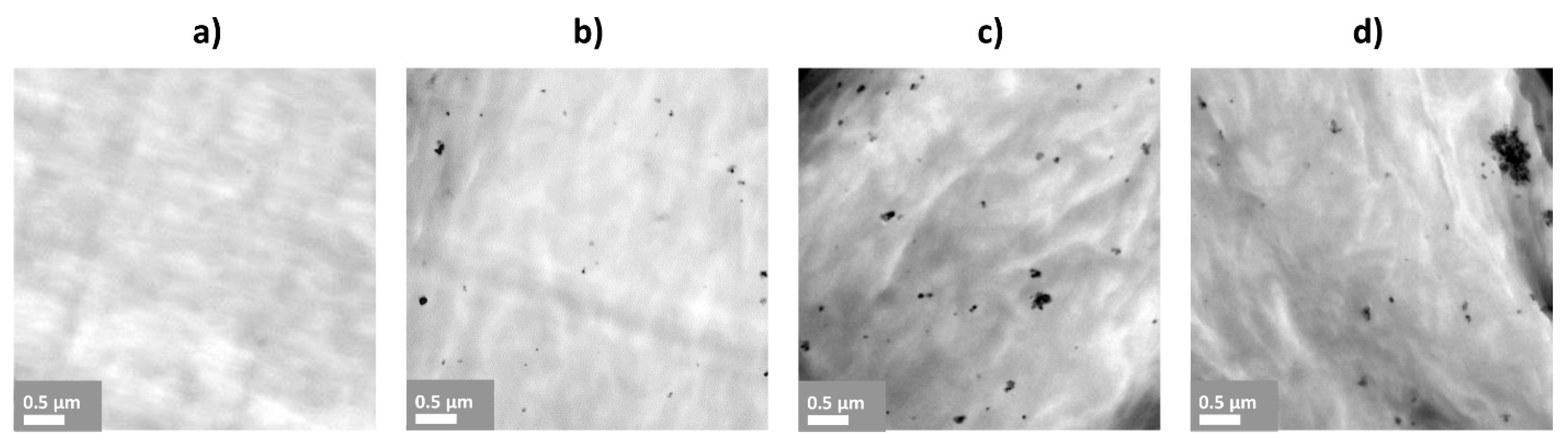
2.3.5. Scanning Electron Microscopy (SEM) and Transmission Electron Microscopy (TEM)
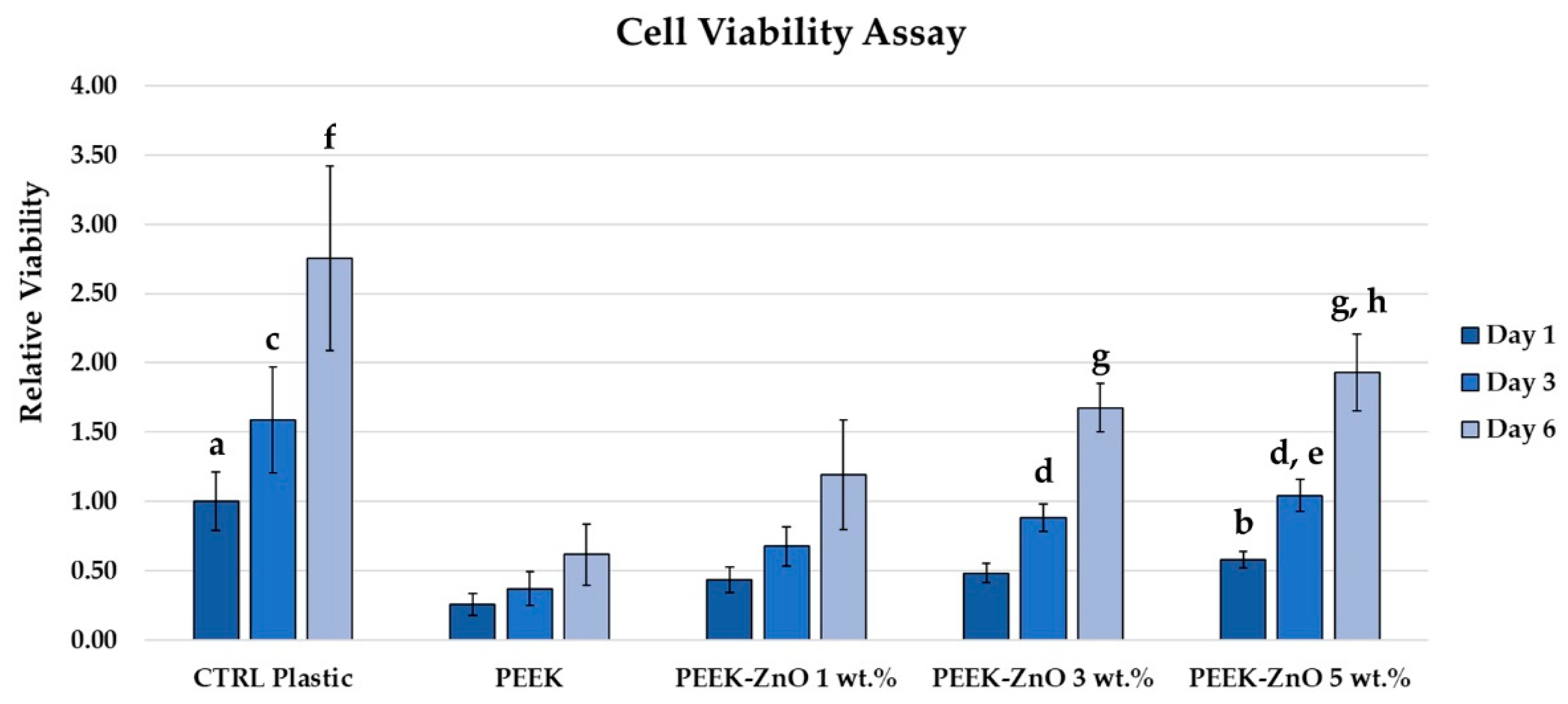
2.3.6. Cell Viability
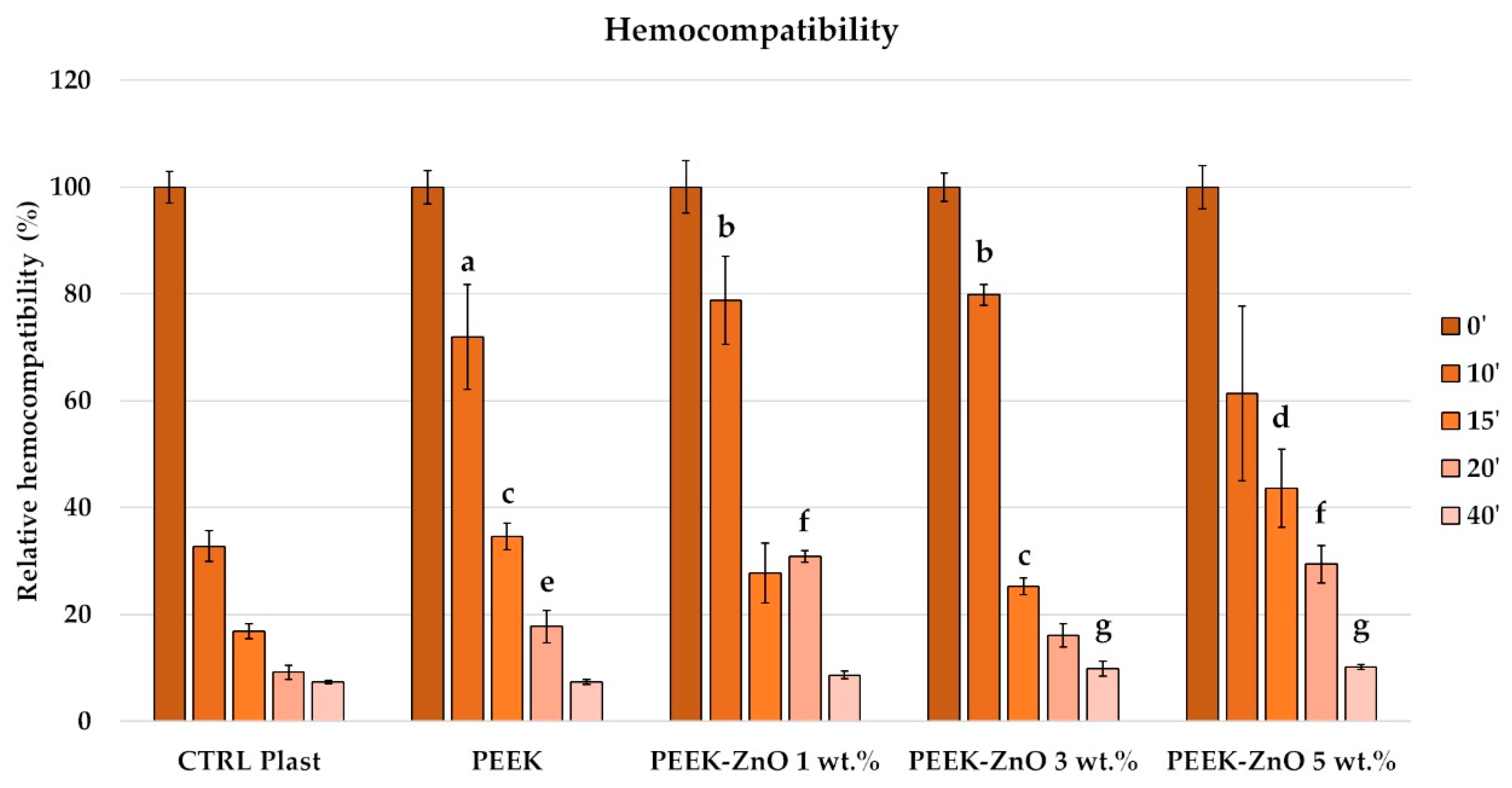
2.3.7. Clot Formation
2.3.8. Statistical Analysis
3. Results
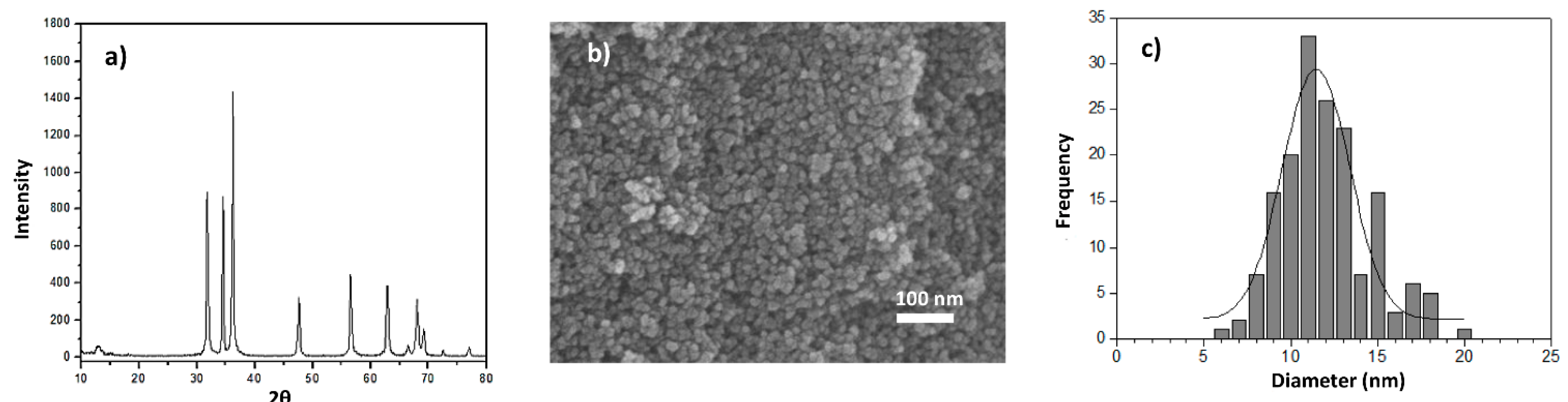
3.1. ZnO Synthesis
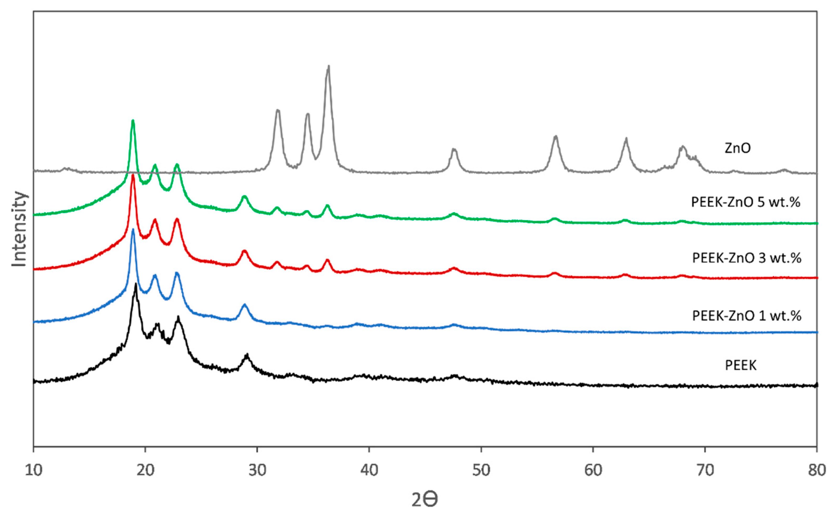
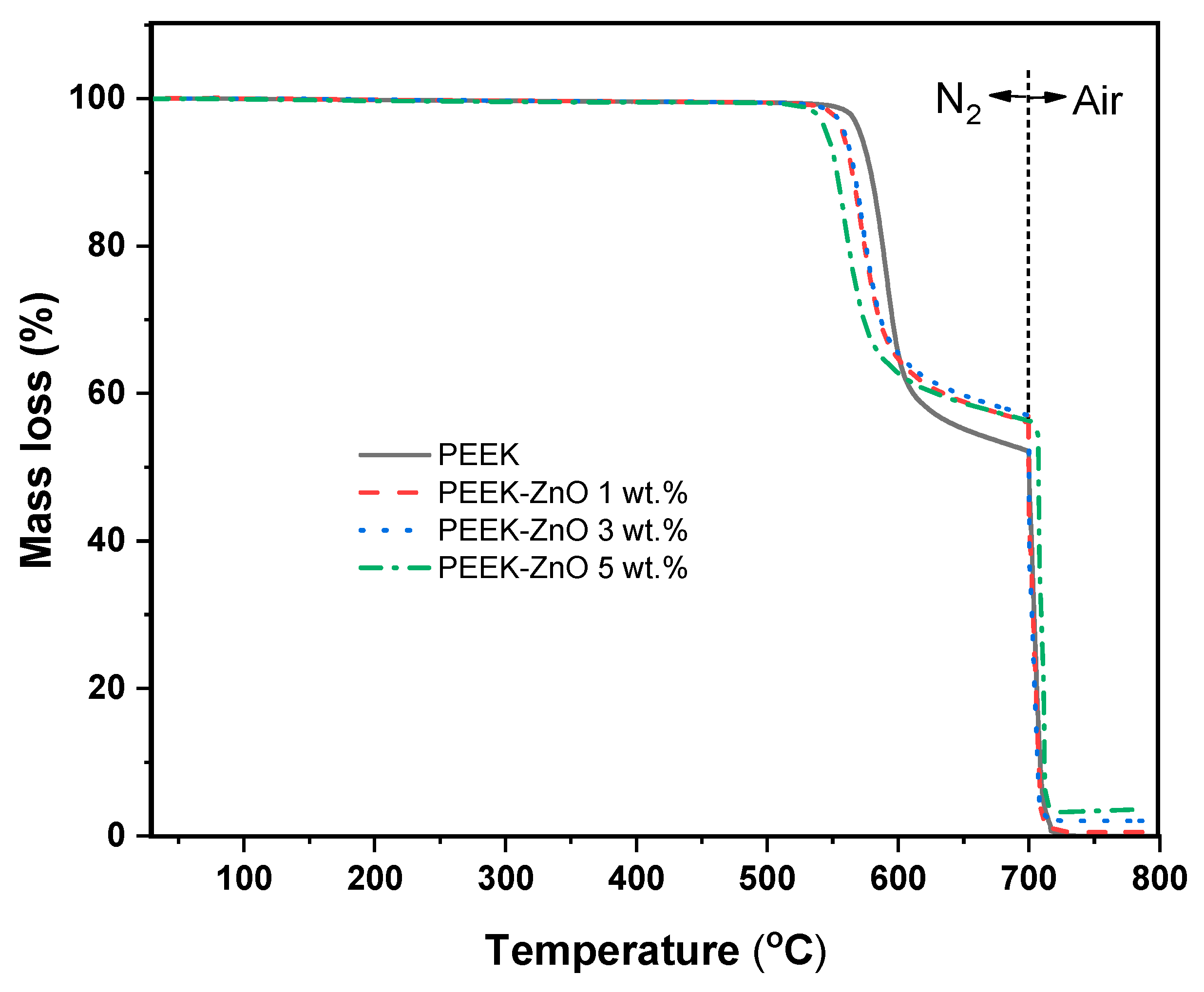
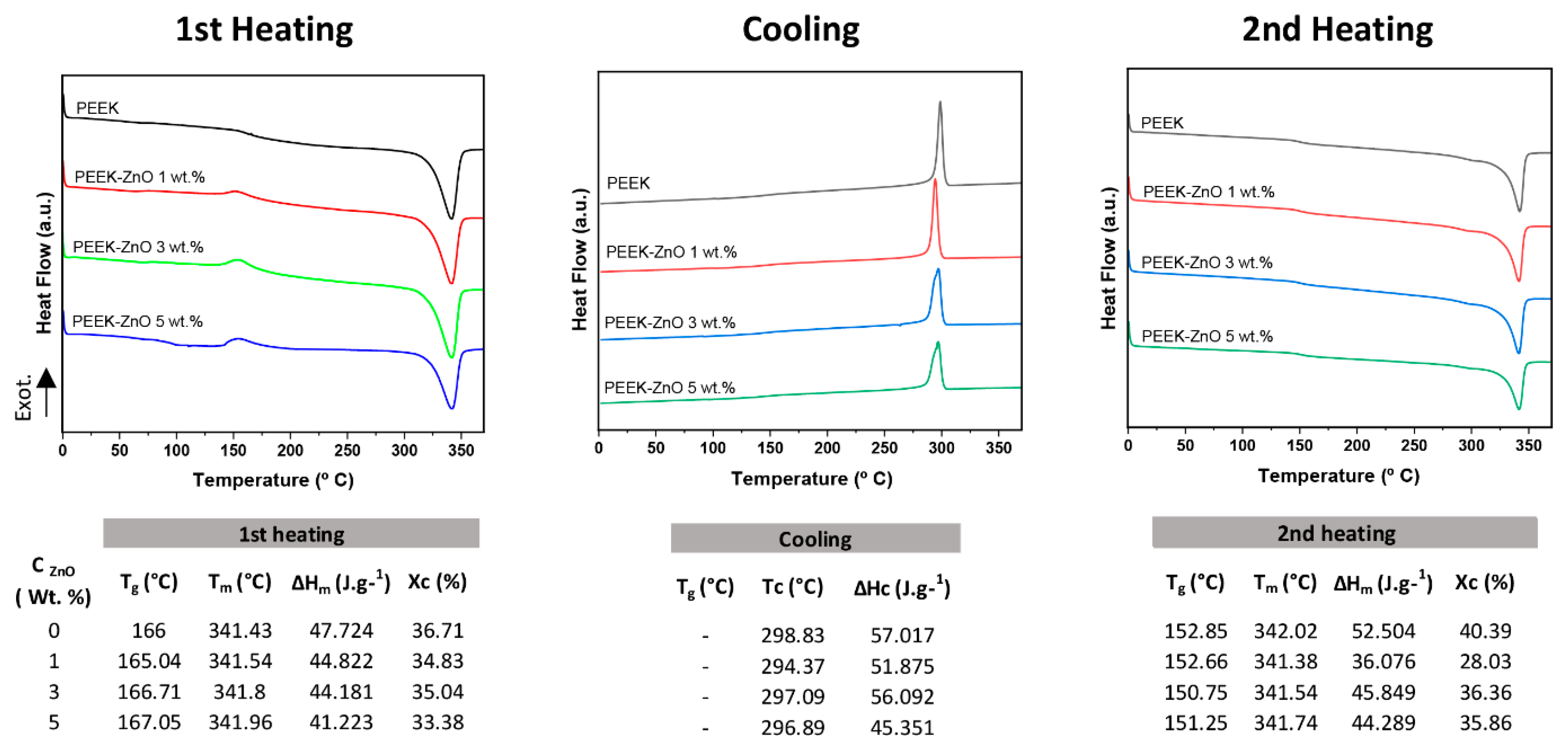
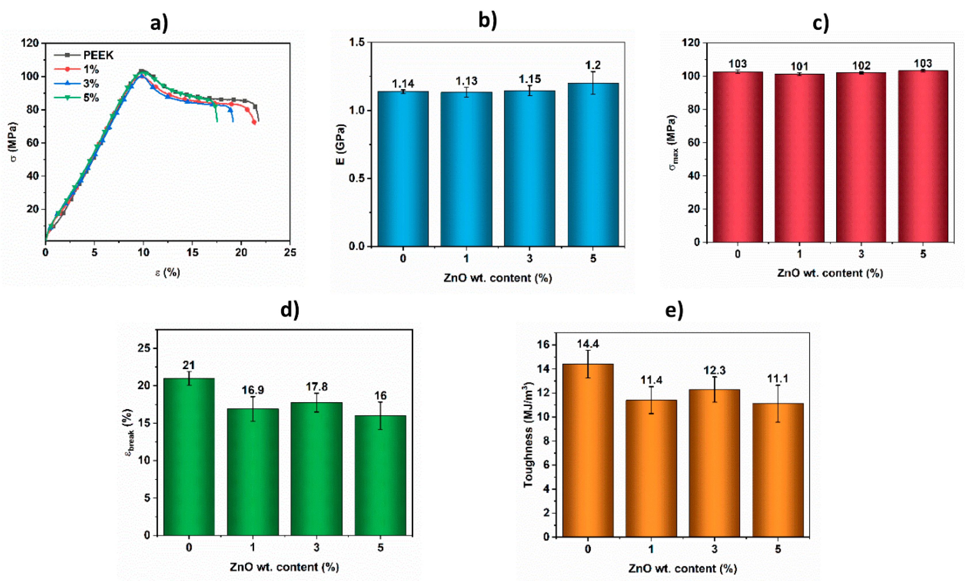
3.2. Characterization of PEEK-ZnO Matrices
3.3. Biological Performances
4. Discussion
5. Conclusions
Author Contributions
Funding
Institutional Review Board Statement
Informed Consent Statement
Data Availability Statement
Acknowledgments
Conflicts of Interest
References
- Duraccio, D.; Mussano, F.; Giulia, M. Biomaterials for dental implants: Current and future trends. J. Mater. Sci. 2015, 50, 4779–4812. [Google Scholar] [CrossRef]
- Osman, R.B.; Swain, M.V. A Critical Review of Dental Implant Materials with an Emphasis on Titanium versus Zirconia. Materials 2015, 8, 932–958. [Google Scholar] [CrossRef] [Green Version]
- Özkurt, Z.; Kazazog, E. Zirconia Dental Implants: A Literature Review. J. Oral Implant. 2011, 37, 367–376. [Google Scholar] [CrossRef]
- Skalak, R. Biomechanical considerations in osseointegrated prostheses. J. Prosthet. Dent. 1983, 49, 843–848. [Google Scholar] [CrossRef]
- Parmigiani-Izquierdo, J.M.; Cabaña-Muñoz, M.E.; Merino, J.J.; Sánchez-Pérez, A. Zirconia implants and peek restorations for the replacement of upper molars. Int. J. Implant Dent. 2017, 3, 1–5. [Google Scholar] [CrossRef] [PubMed] [Green Version]
- Soumeire, J.; Dejou, J. Shock absorbability of various restorative materials used on implants. J. Oral Rehabil. 1999, 26, 394–401. [Google Scholar] [CrossRef]
- Kurtz, S.M. (Ed.) PEEK Biomaterials Handbook; Elsevier: Amsterdam, The Netherlands, 2019; ISBN 9780128125243. [Google Scholar]
- Sagomonyants, K.B.; Jarman-Smith, M.L.; Devine, J.N.; Aronow, M.S.; Gronowicz, G.A. The in vitro response of human osteoblasts to polyetheretherketone (PEEK) substrates compared to commercially pure titanium. Biomaterials 2008, 29, 1563–1572. [Google Scholar] [CrossRef]
- Andreiotelli, M.; Wenz, H.J.; Kohal, R.J. Are ceramic implants a viable alternative to titanium implants? A systematic literature review. Clin. Oral Implants Res. 2009, 20, 32–47. [Google Scholar] [CrossRef]
- Godara, A.; Raabe, D.; Green, S. The influence of sterilization processes on the micromechanical properties of carbon fiber-reinforced PEEK composites for bone implant applications. Acta Biomater. 2007, 3, 209–220. [Google Scholar] [CrossRef]
- Schwitalla, A.; Müller, W.-D. PEEK Dental Implants: A Review of the Literature. J. Oral Implantol. 2013, 39, 743–749. [Google Scholar] [CrossRef] [PubMed]
- Rochford, E.T.J.; Poulsson, A.H.C.; Varela, J.S.; Lezuo, P.; Richards, R.G.; Moriarty, T.F. Bacterial adhesion to orthopaedic implant materials and a novel oxygen plasma modified PEEK surface. Colloids Surf. B Biointerfaces 2014, 113, 213–222. [Google Scholar] [CrossRef] [PubMed]
- Webster, T.J.; Patel, A.A.; Rahaman, M.N.; Bal, B.S. Acta Biomaterialia Anti-infective and osteointegration properties of silicon nitride, poly (ether ether ketone), and titanium implants. Acta Biomater. 2012, 8, 4447–4454. [Google Scholar] [CrossRef]
- Dong, T.; Duan, C.; Wang, S.; Gao, X.; Yang, Q.; Yang, W.; Deng, Y. Multifunctional Surface with Enhanced Angiogenesis for Improving Long-Term Osteogenic Fixation of Poly (ether ether ketone) Implants. ACS Appl. Mater. Interfaces 2020, 12, 14971–14982. [Google Scholar] [CrossRef]
- Ma, R.; Tang, T. Current strategies to improve the bioactivity of PEEK. Int. J. Mol. Sci. 2014, 15, 5426–5445. [Google Scholar] [CrossRef] [PubMed] [Green Version]
- Rosi, N.L.; Mirkin, C.A. Nanostructures in biodiagnostics. Chem. Rev. 2005, 105, 1547–1562. [Google Scholar] [CrossRef] [PubMed]
- Reddy, K.M.; Feris, K.; Bell, J.; Wingett, D.G.; Hanley, C.; Punnoose, A. Selective toxicity of zinc oxide nanoparticles to prokaryotic and eukaryotic systems. Appl. Phys. Lett. 2007, 90, 2139021–2139023. [Google Scholar] [CrossRef] [Green Version]
- Rodríguez-Tobías, H.; Ledezma, A.; Romero, J.; Grande, D. Novel antibacterial electrospun mats based on poly(d,l-lactide) nanofibers and zinc oxide nanoparticles. J. Mater. Sci. 2014, 49, 8373–8385. [Google Scholar] [CrossRef]
- Morales, G.; Ledezma, A.; Romero, J.; Rodríguez-Tobías, H.; Langlois, V.; Renard, E.; Grande, D. Electrospinning and electrospraying techniques for designing novel antibacterial poly(3-hydroxybutyrate)/zinc oxide nanofibrous composites. J. Mater. Sci. 2016, 51, 8593–8609. [Google Scholar]
- Khan, S.T.; Al-Khedhairy, A.A.; Musarrat, J. ZnO and TiO2 nanoparticles as novel antimicrobial agents for oral hygiene: A review. J. Nanoparticle Res. 2015, 17, 1–16. [Google Scholar] [CrossRef]
- Shen, M.; Liang, G.; Gu, A.; Yuan, L. Development of high performance dental resin composites with outstanding antibacterial activity, high mechanical properties and low polymerization shrinkage based on a SiO2 hybridized tetrapod-like zinc oxide whisker with C-C bonds. RSC Adv. 2016, 6, 56353–56364. [Google Scholar] [CrossRef]
- Yusa, K.; Yamamoto, O.; Takano, H.; Fukuda, M.; Iino, M. Zinc-modified titanium surface enhances osteoblast differentiation of dental pulp stem cells in vitro. Sci. Rep. 2016, 6, 29462. [Google Scholar] [CrossRef] [Green Version]
- Augustine, R.; Malik, H.N.; Singhal, D.K.; Mukherjee, A.; Malakar, D.; Kalarikkal, N.; Thomas, S. Electrospun polycaprolactone/ZnO nanocomposite membranes as biomaterials with antibacterial and cell adhesion properties. J. Polym. Res. 2014, 21, 1–17. [Google Scholar] [CrossRef]
- Díez-Pascual, A.M.; Díez-Vicente, A.L. Development of nanocomposites reinforced with carboxylated poly(ether ether ketone) grafted to zinc oxide with superior antibacterial properties. ACS Appl. Mater. Interfaces 2014, 6, 3729–3741. [Google Scholar] [CrossRef] [Green Version]
- Díez-Pascual, A.M.; Xu, C.; Luque, R. Development and characterization of novel poly(ether ether ketone)/ZnO bionanocomposites. J. Mater. Chem. B 2014, 2, 3065–3078. [Google Scholar] [CrossRef] [PubMed] [Green Version]
- Deng, Y.; Yang, L.; Huang, X.; Chen, J.; Shi, X.; Yang, W.; Hong, M.; Wang, Y.; Dargusch, M.S.; Chen, Z.G. Dual Ag/ZnO-Decorated Micro-/Nanoporous Sulfonated Polyetheretherketone with Superior Antibacterial Capability and Biocompatibility via Layer-by-Layer Self-Assembly Strategy. Macromol. Biosci. 2018, 18, 1–12. [Google Scholar] [CrossRef] [PubMed]
- Rodríguez Tobías, H. Nuevos Materiales Híbridos Basados en Poliésteres Biodegradables y Nanopartículas de ZnO: Propiedades Antibacterianas y Protección UV; Centro de Investigación en Química Aplicada (CIQA): Saltillo, Mexico, 2015. [Google Scholar]
- Victrex Peek Polymer. Available online: https://www.victrex.com/en/products/polymers/peek-polymers (accessed on 18 May 2019).
- Lopresti, F.; Carfì Pavia, F.; Vitrano, I.; Kersaudy-Kerhoas, M.; Brucato, V.; La Carrubba, V. Effect of hydroxyapatite concentration and size on morpho-mechanical properties of PLA-based randomly oriented and aligned electrospun nanofibrous mats. J. Mech. Behav. Biomed. Mater. 2020, 101, 103449. [Google Scholar] [CrossRef]
- Yang, Z.; Peng, H.; Wang, W.; Liu, T. Crystallization behavior of poly(ε-caprolactone)/layered double hydroxide nanocomposites. J. Appl. Polym. Sci. 2010, 116, 2658–2667. [Google Scholar] [CrossRef]
- Barreto, G.P.; Morales, G.; Quintanilla, M.L.L. Microwave Assisted Synthesis of ZnO Nanoparticles: Effect of Precursor Reagents, Temperature, Irradiation Time, and Additives on Nano-ZnO Morphology Development. J. Mater. 2013, 2013, 1–11. [Google Scholar] [CrossRef]
- Hu, Z.; Oskam, G.; Penn, R.L.; Pesika, N.; Searson, P.C. The Influence of Anion on the Coarsening Kinetics of ZnO Nanoparticles. J. Phys. Chem. B 2003, 107, 3124–3130. [Google Scholar] [CrossRef]
- Khoza, P.B.; Moloto, M.J.; Sikhwivhilu, L.M. The effect of solvents, acetone, water, and ethanol, on the morphological and optical properties of ZnO nanoparticles prepared by microwave. J. Nanotechnol. 2012, 2012, 1–6. [Google Scholar] [CrossRef] [Green Version]
- Kunjara Na Ayudhya, S.; Tonto, P.; Mekasuwandumrong, O.; Pavarajarn, V.; Praserthdam, P. Solvothermal Synthesis of ZnO with Various Aspect Ratios Using Organic Solvents. Cryst. Growth Des. 2006, 6, 2446–2450. [Google Scholar] [CrossRef]
- Sirelkhatim, A.; Mahmud, S.; Seeni, A.; Kaus, N.H.M.; Ann, L.C.; Bakhori, S.K.M.; Hasan, H.; Mohamad, D. Review on zinc oxide nanoparticles: Antibacterial activity and toxicity mechanism. Nano-Micro Lett. 2015, 7, 219–242. [Google Scholar] [CrossRef] [Green Version]
- Díez-Pascual, A.M.; Naffakh, M.; Gómez, M.A.; Marco, C.; Ellis, G.; Gonzlez-Domínguez, J.M.; Ansón, A.; Martínez, M.T.; Martínez-Rubi, Y.; Simard, B.; et al. The influence of a compatibilizer on the thermal and dynamic mechanical properties of PEEK/carbon nanotube composites. Nanotechnology 2009, 20, 315707. [Google Scholar] [CrossRef] [PubMed]
- Hao, L.; Hu, Y.; Zhang, Y.; Wei, W.; Hou, X.; Guo, Y.; Hu, X.; Jiang, D. Enhancing the mechanical performance of poly(ether ether ketone)/zinc oxide nanocomposites to provide promising biomaterials for trauma and orthopedic implants. RSC Adv. 2018, 8, 27304–27317. [Google Scholar] [CrossRef] [Green Version]
- Van Ngo, G.; Margaillan, A.; Villain, S.; Leroux, C.; Bressy, C. Synthesis of ZnO nanoparticles with tunable size and surface hydroxylation. J. Nanoparticle Res. 2013, 15, 1–15. [Google Scholar] [CrossRef]
- Morimoto, T.; Nagao, M.; Tokuda, F. Desorbability desorption of Chemisorbed Water on Metal Oxide Temperature of Chemisorbed Water on Rutile and Zinc Oxide. Bull. Chem. Soc. Jpn. 1968, 41, 1533–1537. [Google Scholar] [CrossRef]
- Ponsonnet, L.; Reybier, K.; Jaffrezic, N.; Comte, V.; Lagneau, C.; Lissac, M.; Martelet, C. Relationship between surface properties (roughness, wettability) of titanium and titanium alloys and cell behaviour. Mater. Sci. Eng. C 2003, 23, 551–560. [Google Scholar] [CrossRef]
- Tateishi, T.; Kyomoto, M.; Kakinoki, S.; Yamaoka, T.; Ishihara, K. Reduced platelets and bacteria adhesion on poly (ether ether ketone) by photoinduced and self-initiated graft polymerization of 2-methacryloyloxyethyl phosphorylcholine. J. Biomed. Mater. Res. Part A 2013, 102, 1342–1349. [Google Scholar] [CrossRef]
- Kawasaki, Y.; Iwasaki, Y. Surface modification of poly (ether ether ketone) with methacryloyl-functionalized phospholipid polymers via self-initiation graft polymerization. J. Biomater. Sci. Polym. Ed. 2014, 25, 895–906. [Google Scholar] [CrossRef]
- Hui, S.U.N.; Rui-Chao, C.; Shu, L.I.U.; Guo-Zhi, X.U. Surface Heparinization of Poly (ether ether ketone). Chem. Res. Chin. Univ. 2012, 28, 542–545. [Google Scholar]
- Bartolo, L.D.E.; Gugliuzza, A.; Morelli, S.; Cirillo, B.; Gordano, A.; Drioli, E. Novel PEEK-WC membranes with low plasma protein affnity related to surface free energy parameters. J. Mater. Sci. Mater. Med. 2004, 5, 877–883. [Google Scholar] [CrossRef] [PubMed]


| Intervals of Time | 1° | 2° | 3° | 4° | Cooling Time (s) |
|---|---|---|---|---|---|
| Temperature (°C) | 370 | 370 | 370 | 370 | * |
| Pressure (Ton) | 0 | 1 | 10 | 20 | 20 |
| Compression time (s) | 360 | 120 | 60 | 60 | 900 |
| System | WCA (°) | Roughness (rms, nm) | |
|---|---|---|---|
| Advancing | Receding | ||
| PEEK | 78 ± 3 | 33 ± 9 | 98 ± 13 |
| PEEK-ZnO 1 wt.% | 85 ± 1 | 39 ± 5 | 130 ± 8 |
| PEEK-ZnO 3 wt.% | 72 ± 5 | 25 ± 4 | 119 ± 23 |
| PEEK-ZnO 5 wt.% | 63 ± 7 | 16 ± 7 | 98 ± 11 |
Publisher’s Note: MDPI stays neutral with regard to jurisdictional claims in published maps and institutional affiliations. |
© 2021 by the authors. Licensee MDPI, Basel, Switzerland. This article is an open access article distributed under the terms and conditions of the Creative Commons Attribution (CC BY) license (http://creativecommons.org/licenses/by/4.0/).
Share and Cite
Montaño-Machado, V.; Chevallier, P.; Bonilla-Gameros, L.; Copes, F.; Quarta, C.; Kú-Herrera, J.d.J.; Soriano, F.; Padilla-Gainza, V.; Morales, G.; Mantovani, D. Development of Multifunctional Materials Based on Poly(ether ether ketone) with Improved Biological Performances for Dental Applications. Materials 2021, 14, 1047. https://0-doi-org.brum.beds.ac.uk/10.3390/ma14041047
Montaño-Machado V, Chevallier P, Bonilla-Gameros L, Copes F, Quarta C, Kú-Herrera JdJ, Soriano F, Padilla-Gainza V, Morales G, Mantovani D. Development of Multifunctional Materials Based on Poly(ether ether ketone) with Improved Biological Performances for Dental Applications. Materials. 2021; 14(4):1047. https://0-doi-org.brum.beds.ac.uk/10.3390/ma14041047
Chicago/Turabian StyleMontaño-Machado, Vanessa, Pascale Chevallier, Linda Bonilla-Gameros, Francesco Copes, Chiara Quarta, José de Jesús Kú-Herrera, Florentino Soriano, Victoria Padilla-Gainza, Graciela Morales, and Diego Mantovani. 2021. "Development of Multifunctional Materials Based on Poly(ether ether ketone) with Improved Biological Performances for Dental Applications" Materials 14, no. 4: 1047. https://0-doi-org.brum.beds.ac.uk/10.3390/ma14041047