
Antibacterial Effect of Amino Acid–Silver Complex Loaded Montmorillonite Incorporated in Dental Acrylic Resin

 , , , and
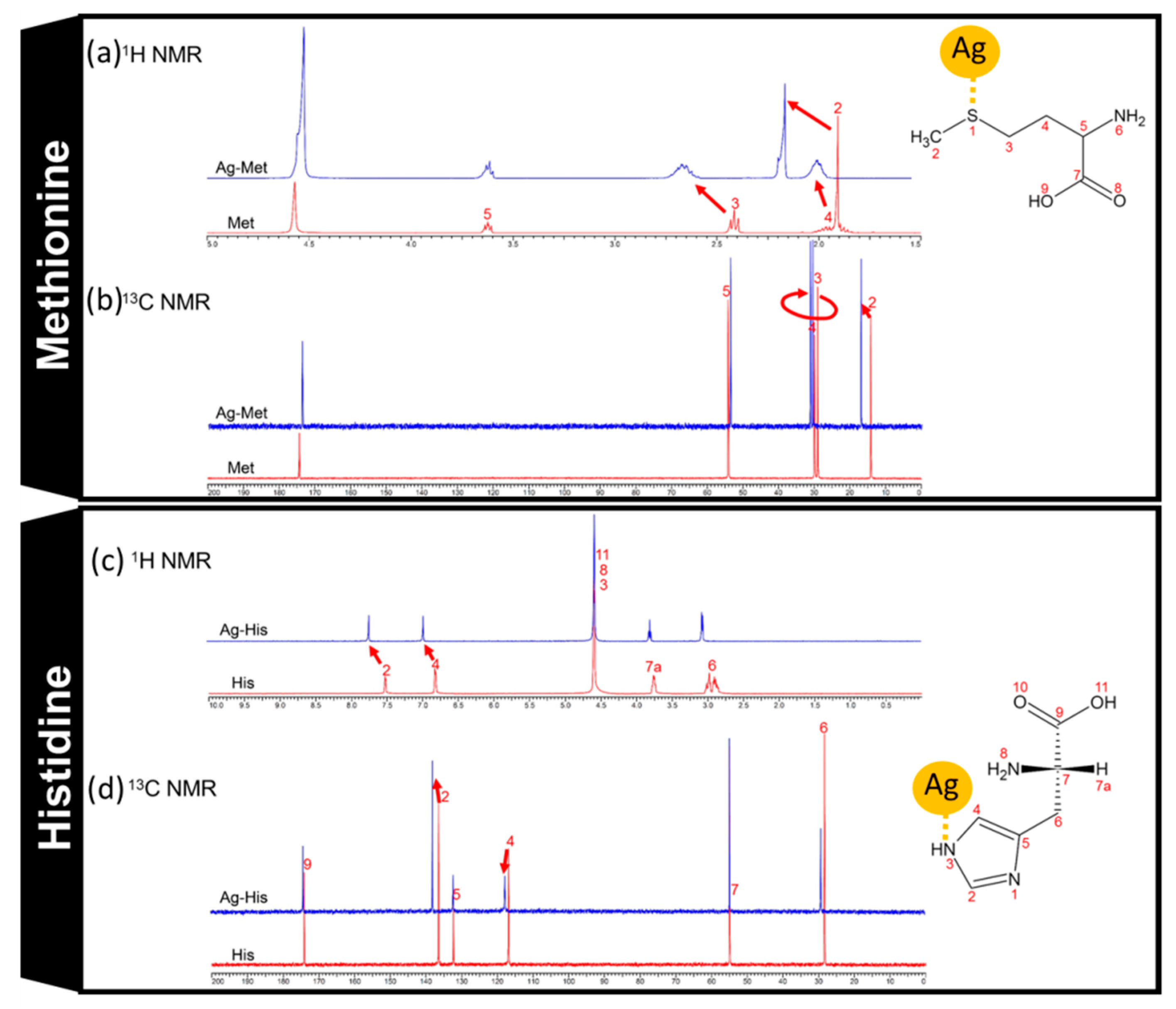
Abstract
:1. Introduction
2. Materials and Methods
2.1. Synthesis of Amino Acid–Silver Complexes Loaded onto Montmorillonite
2.2. Resin Sample Preparation
2.3. Nuclear Magnetic Resonance (NMR) Measurement
2.4. Scanning Electron Microscopy (SEM)
2.5. X-Ray Diffraction (XRD)
2.6. Bacteria
2.7. Bacterial Growth Spectrophotometry
2.8. Silver Ion Measurement in Different Concentrations of Chloride Ion Solution
3. Results and Discussion
3.1. Montmorillonite-Loaded Amino Acid–Silver Complex Preparation
3.2. NMR Measurement
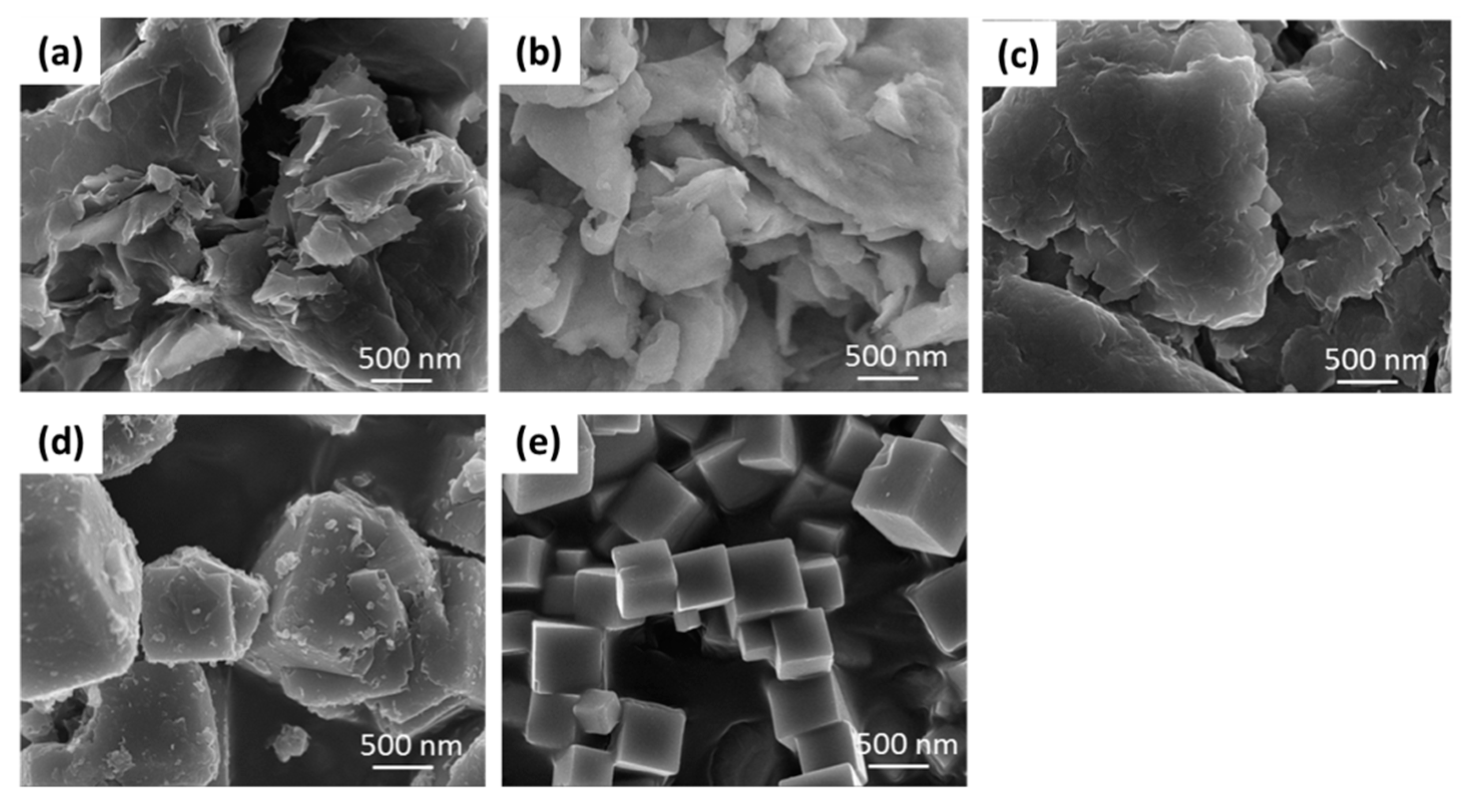
3.3. Scanning Electron Microscope (SEM) Observation and X-ray Diffraction (XRD) Analysis
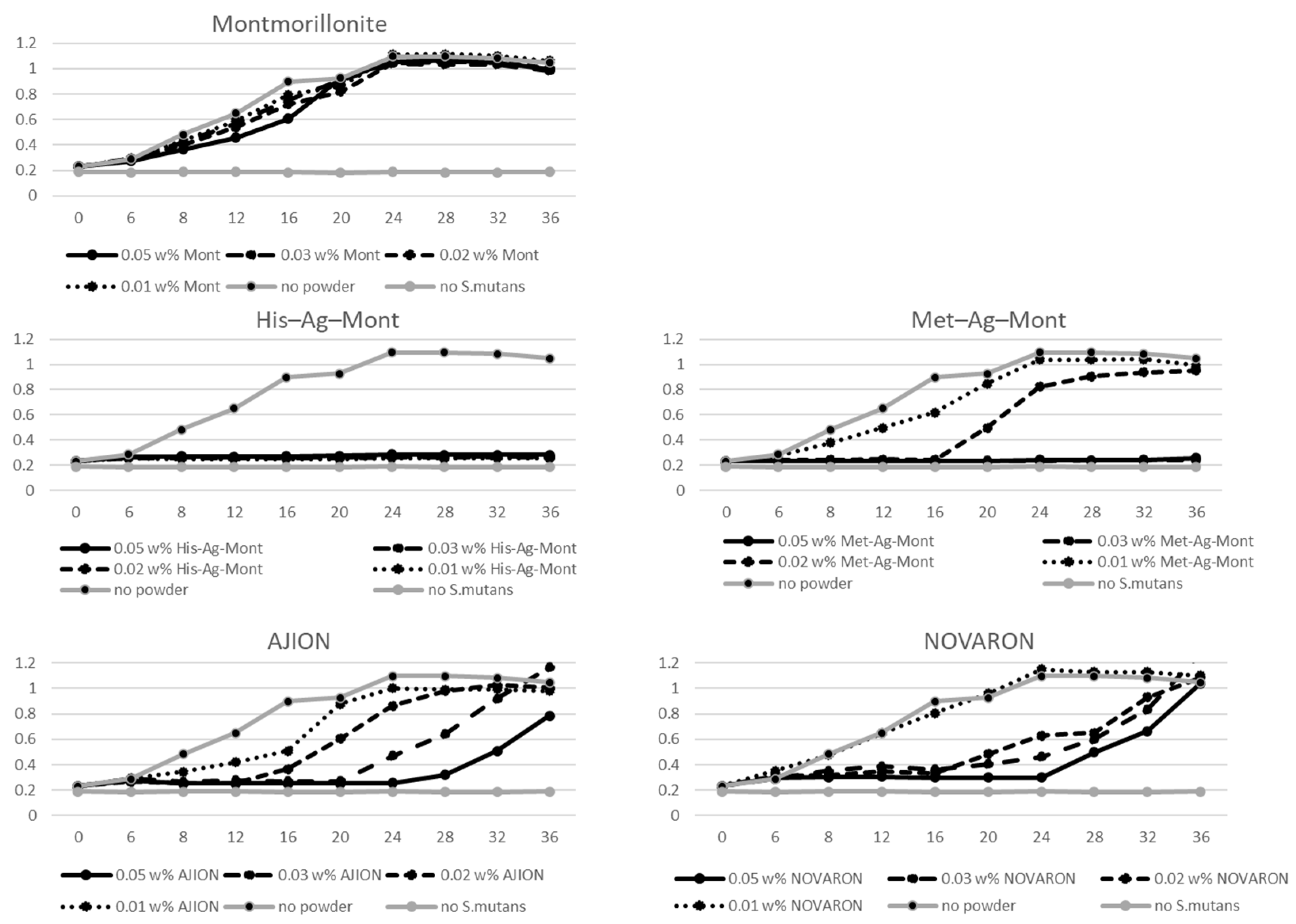
3.4. Bacterial Growth
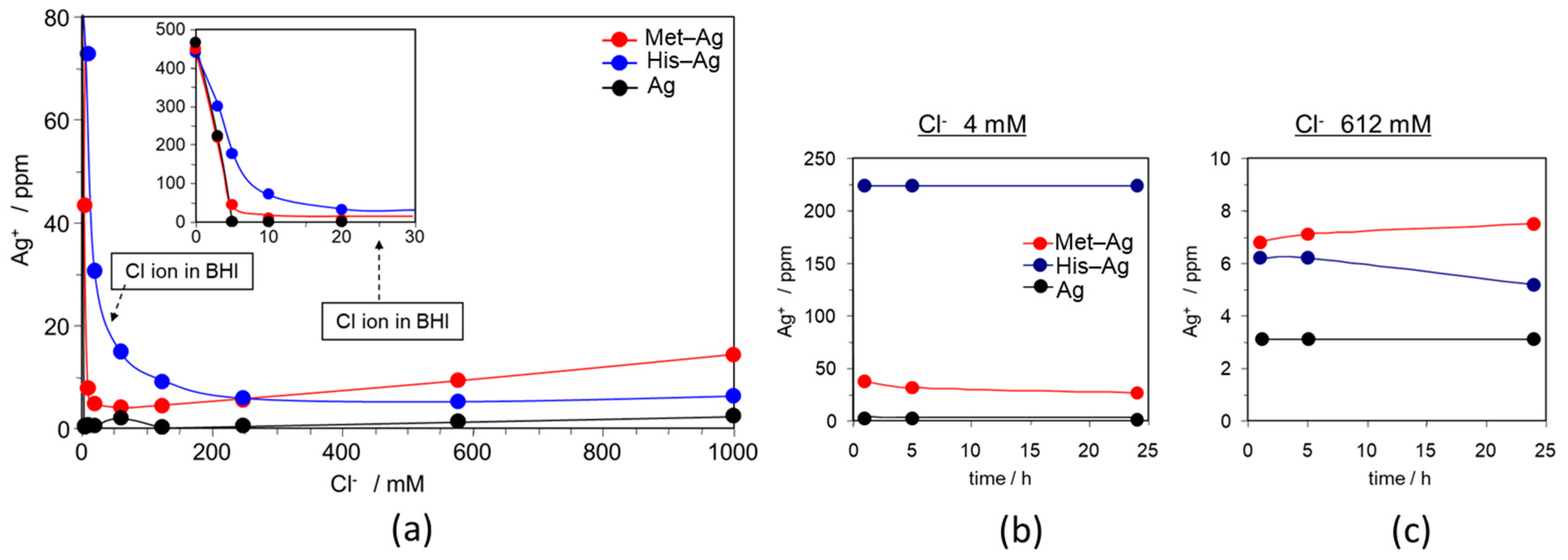
3.5. Silver Ion Measurement in Different Concentrations of Chloride Ion Solution
4. Conclusions
Author Contributions
Funding
Institutional Review Board Statement
Informed Consent Statement
Data Availability Statement
Conflicts of Interest
References
- Jung, W.K.; Koo, H.C.; Kim, K.W.; Shin, S.; Kim, S.H.; Park, Y.H. Antibacterial Activity and Mechanism of Action of the Silver Ion in Staphylococcus aureus and Escherichia coli. Appl. Environ. Microbiol. 2008, 74, 2171–2178. [Google Scholar] [CrossRef] [Green Version]
- Marambio-Jones, C.; Hoek, E.M. A review of the antibacterial effects of silver nanomaterials and potential implications for human health and the environment. J. Nanopart. Res. 2010, 12, 1531–1551. [Google Scholar] [CrossRef]
- Hendre, A.D.; Taylor, G.W.; Chávez, E.M.; Hyde, S. A systematic review of silver diamine fluoride: Effectiveness and application in older adults. Gerodontology 2017, 34, 411–419. [Google Scholar] [CrossRef]
- Zhao, I.S.; Gao, S.S.; Hiraishi, N.; Burrow, M.F.; Duangthip, D.; Mei, M.L.; Lo, E.C.; Chu, C.H. Mechanisms of silver diamine fluoride on arresting caries: A literature review. Int. Dent. J. 2018, 68, 67–76. [Google Scholar] [CrossRef] [PubMed] [Green Version]
- Gao, S.S.; Zhao, I.S.; Duffin, S.; Duangthip, D.; Lo, E.C.; Chu, C.H. Revitalizing Silver Nitrate for Caries Management. Int. J. Environ. Res. Public Health 2018, 15, 80. [Google Scholar] [CrossRef] [Green Version]
- Crystal, Y.O.; Niederman, R. Silver Diamine Fluoride Treatment Considerations in Children’s Caries Management. Pediatr. Dent. 2016, 38, 466–471. [Google Scholar]
- Swathy, J.R.; Sankar, M.U.; Chaudhary, A.; Aigal, S.; Pradeep, T. Antimicrobial silver: An unprecedented anion effect. Sci. Rep. 2014, 24, 7161. [Google Scholar] [CrossRef] [PubMed] [Green Version]
- Rai, M.; Yadav, A.; Gade, A. Silver nanoparticles as a new generation of antimicrobials. Biotechnol. Adv. 2009, 27, 76–83. [Google Scholar] [CrossRef]
- Noronha, V.T.; Paula, A.J.; Durán, G.; Galembeck, A.; Cogo-Müller, K.; Franz-Montan, M.; Durán, N. Silver nanoparticles in dentistry. Dent. Mater. 2017, 33, 1110–1126. [Google Scholar] [CrossRef] [PubMed]
- Besinis, A.; de Peralta, T.; Tredwin, C.J.; Handy, R.D. Review of nanomaterials in dentistry: Interactions with the oral microenvironment, clinical applications, hazards, and benefits. ACS Nano 2015, 9, 2255–2289. [Google Scholar] [CrossRef] [Green Version]
- Silver, S. Bacterial silver resistance: Molecular biology and uses and misuses of silver compounds. FEMS Microbiol. Rev. 2003, 27, 341–353. [Google Scholar] [CrossRef] [Green Version]
- Chernousova, S.; Epple, M. Silver as antibacterial agent: Ion, nanoparticle, and metal. Angew. Chem. Int. Ed. 2013, 52, 1636–1653. [Google Scholar] [CrossRef]
- Kasuga, N.C.; Yoshikawa, R.; Sakai, Y.; Nomiya, K. Syntheses, structures, and antimicrobial activities of remarkably light-stable and water-soluble silver complexes with amino acid derivatives, silver(I) N-acetylmethioninates. Inorg. Chem. 2012, 51, 1640–1647. [Google Scholar] [CrossRef] [PubMed]
- Nomiya, K.; Takahashi, S.; Noguchi, R.; Nemoto, S.; Takayama, T.; Oda, M. Synthesis and characterization of water-soluble silver(I) complexes with L-histidine (H2his) and (S)-(-)-2-pyrrolidone-5-carboxylic acid (H2pyrrld) showing a wide spectrum of effective antibacterial and antifungal activities. Crystal structures of chiral helical polymers [Ag(Hhis)]n and ([Ag(Hpyrrld)]2)n in the solid state. Inorg. Chem. 2000, 39, 3301–3311. [Google Scholar] [CrossRef] [PubMed]
- Legler, A.V.; Kazachenko, A.S.; Kazbanov, V.I.; Per’yanova, O.V.; Veselova, O.F. Synthesis and antimicrobial activity of silver complexes with arginine and glutamic acid. Pharm. Chem. J. 2001, 35, 501–503. [Google Scholar] [CrossRef]
- See, R.F.; Kruse, R.A.; Strub, W.M. Metal-ligand bond distances in first-row transition metal coordination compounds: Coordination number, oxidation state, and specific ligand effects. Inorg. Chem. 1998, 37, 5369–5375. [Google Scholar] [CrossRef]
- Nomiya, K.; Yokoyama, H. Syntheses, crystal structures and antimicrobial activities of polymeric silver(I) complexes with three amino-acids [aspartic acid (H2asp), glycine (Hgly) and asparagine (Hasn)]. J. Coord. Chem. 2000, 59, 1089–1099. [Google Scholar] [CrossRef]
- Hui, Y.; Qizhuang, H.; Meifeng, Z.; Yanming, X.; Jingyi, S. Synthesis, characterization and biological activity of rare earth complexes with L-aspartic acid and o-phenanthroline. J. Chin. Rare Earth Soc. 2007, 2, 3–4. [Google Scholar] [CrossRef]
- Makita, Y.; Obika, H.; Oi, K.; Umeno, A. Antibacterial agent for adhesive marine bacterium. Japan Patent JP2005-145923A, 15 November 2003. [Google Scholar]
- Cao, G.F.; Sun, Y.; Chen, J.G.; Song, L.P.; Jiang, J.Q.; Liu, Z.T.; Liu, Z.W. Sutures modified by silver-loaded montmorillonite with antibacterial properties. Appl. Clay Sci. 2014, 93–94, 102–106. [Google Scholar] [CrossRef]
- Brigatti, M.F.; Galan, E.; Theng, B.K.G. Chapter 2—Structure and mineralogy of clay minerals. In Handbook of Clay Science; Elsevier: Amsterdam, The Netherlands, 2013; Volume 5, pp. 21–81. ISBN 9780080993645. [Google Scholar]
- Bagchi, B.; Kar, S.; Dey, S.K.; Bhandary, S.; Roy, D.; Mukhopadhyay, T.K.; Das, S.; Nandy, P. In situ synthesis and antibacterial activity of copper nanoparticle loaded natural montmorillonite clay based on contact inhibition and ion release. Colloids Surf. B Biointerfaces 2013, 108, 358–365. [Google Scholar] [CrossRef]
- Yoshihara, K.; Nagaoka, N.; Maruo, Y.; Sano, H.; Yoshida, Y.; van Meerbeek, B. Bacterial adhesion not inhibited by ion-releasing bioactive glass filler. Dent. Mater. 2017, 33, 723–734. [Google Scholar] [CrossRef]
- Namba, N.; Yoshida, Y.; Nagaoka, N.; Takashima, S.; Matsuura-Yoshimoto, K.; Maeda, H.; van Meerbeek, B.; Suzuki, K.; Takashiba, S. Antibacterial effect of bactericide immobilized in resin matrix. Dent. Mater. 2009, 25, 424–430. [Google Scholar] [CrossRef]
- Jena, K.K.; Raju, K.; Narayan, R. Sodium montmorillonite clay loaded novel organic–inorganic hybrid composites: Synthesis and characterization. Prog. Org. Coat. 2012, 75, 33–37. [Google Scholar] [CrossRef]
- Xiao, J.; Hu, Y.; Wang, Z.; Tang, Y.; Chen, Z.; Fan, W. Preparation and characterization of poly (butylene terephthalate) nanocomposites from thermally stable organic-modified montmorillonite. Eur. Polym. J. 2005, 41, 1030–1035. [Google Scholar] [CrossRef]
- Kawahara, K.; Tsuruda, K.; Morishita, M.; Uchida, M. Antibacterial effect of silver-zeolite on oral bacteria under anaerobic conditions. Dent. Mater. 2000, 16, 452–455. [Google Scholar] [CrossRef]
- Kiriyama, T.; Kuroki, K.; Sasaki, K.; Tomino, M.; Asakura, M.; Kominami, Y.; Takahashi, Y.; Kawai, T. Antibacterial properties of a self-cured acrylic resin composed of a polymer coated with a silver-containing organic composite antibacterial agent. Dent. Mater. J. 2013, 32, 679–687. [Google Scholar] [CrossRef] [PubMed] [Green Version]
- Dawes, C. The effects of flow rate and duration of stimulation on the concentrations of protein and the main electrolytes in human submandibular saliva. Arch. Oral Biol. 1974, 19, 887–895. [Google Scholar] [CrossRef]
- Holla, G.; Yeluri, R.; Munshi, A.K. Evaluation of minimum inhibitory and minimum bactericidal concentration of nano-silver base inorganic anti-microbial agent (Novaron®) against Streptococcus mutans. Contemp. Clin. Dent. 2012, 3, 288–293. [Google Scholar] [CrossRef] [PubMed]
- Staff, J. Silver to Black-and Back. J. Chem. Educ. 2000, 77, 328. [Google Scholar] [CrossRef]
- Addy, M.; Moran, J. Mechanisms of stain formation on teeth, in particular associated with metal ions and antiseptics. Adv. Dent. Res. 1995, 9, 450–456. [Google Scholar] [CrossRef]
- Galui, S.; Pal, S.; Pabale, S.L.; Saha, S.; Sarkar, S. Stretching new boundaries of caries prevention with silver diamine fluoride: A review of literature. Int. J. Pedod. Rehabil. 2018, 3, 1–4. [Google Scholar] [CrossRef]
- Liu, G.; Haiqi, G.; Li, K.; Xiang, J.; Lan, T.; Zhang, Z. Fabrication of silver nanoparticle sponge leather with durable antibacterial property. J. Colloid Interface Sci. 2018, 15, 338–348. [Google Scholar] [CrossRef] [PubMed]
- Lansdown, A.B.G. A Pharmacological and Toxicological Profile of Silver as an Antimicrobial Agent in Medical Devices. Adv. Pharmacol. Sci. 2010, 2010, 910686. [Google Scholar] [CrossRef] [PubMed] [Green Version]
- Lin, C.X.; Yang, S.Y.; Gu, J.L.; Meng, J.; Xu, H.Y.; Cao, J.M. The acute toxic effects of silver nanoparticles on myocardial transmembrane potential, INa and IK1 channels and heart rhythm in mice. Nanotoxicology 2017, 11, 827–837. [Google Scholar] [CrossRef] [PubMed]
- Lee, V.W.M.; Li, H.; Lau, T.C.; Guevremont, R.; Siu, K.M. Relative silver (I) ion binding energies of α-amino acids: A determination by means of the kinetic method. J. Am. Soc. Mass Spectrom. 1998, 9, 760–766. [Google Scholar] [CrossRef]
- Durán, N.; Durán, M.; de Jesus, M.B.; Seabra, A.B.; Fávaro, W.J.; Nakazato, G. Silver nanoparticles: A new view on mechanistic aspects on antimicrobial activity. Nanomedicine 2016, 12, 789–799. [Google Scholar] [CrossRef]
- Asiani, K.R.; Williams, H.; Bird, L.; Jenner, M.; Searle, M.S.; Hobman, J.L.; Scott, D.J.; Soultanas, P. SilE is an intrinsically disordered periplasmic “molecular sponge” involved in bacterial silver resistance. Mol. Microbiol. 2016, 101, 731–742. [Google Scholar] [CrossRef]
- Chabert, V.; Hologne, M.; Sénèque, O.; Crochet, A.; Walker, O.; Fromm, K.M. Model peptide studies of Ag+ binding sites from the silver resistance protein SilE. Chem. Commun. 2017, 53, 6105–6108. [Google Scholar] [CrossRef] [Green Version]
- Mirolo, L.; Schmidt, T.; Eckhardt, S.; Meuwly, M.; Fromm, K.M. pH-Dependent coordination of AgI ions by histidine: Experiment, theory and a model for SilE. Chem. Eur. J. 2013, 19, 1754–1761. [Google Scholar] [CrossRef] [Green Version]


Publisher’s Note: MDPI stays neutral with regard to jurisdictional claims in published maps and institutional affiliations. |
© 2021 by the authors. Licensee MDPI, Basel, Switzerland. This article is an open access article distributed under the terms and conditions of the Creative Commons Attribution (CC BY) license (http://creativecommons.org/licenses/by/4.0/).
Share and Cite
Yoshihara, K.; Nagaoka, N.; Umeno, A.; Sonoda, A.; Obika, H.; Yoshida, Y.; Van Meerbeek, B.; Makita, Y. Antibacterial Effect of Amino Acid–Silver Complex Loaded Montmorillonite Incorporated in Dental Acrylic Resin. Materials 2021, 14, 1442. https://0-doi-org.brum.beds.ac.uk/10.3390/ma14061442
Yoshihara K, Nagaoka N, Umeno A, Sonoda A, Obika H, Yoshida Y, Van Meerbeek B, Makita Y. Antibacterial Effect of Amino Acid–Silver Complex Loaded Montmorillonite Incorporated in Dental Acrylic Resin. Materials. 2021; 14(6):1442. https://0-doi-org.brum.beds.ac.uk/10.3390/ma14061442
Chicago/Turabian StyleYoshihara, Kumiko, Noriyuki Nagaoka, Aya Umeno, Akinari Sonoda, Hideki Obika, Yasuhiro Yoshida, Bart Van Meerbeek, and Yoji Makita. 2021. "Antibacterial Effect of Amino Acid–Silver Complex Loaded Montmorillonite Incorporated in Dental Acrylic Resin" Materials 14, no. 6: 1442. https://0-doi-org.brum.beds.ac.uk/10.3390/ma14061442