Article

Micropowder Ca2YMgScSi3O12:Ce Silicate Garnet as an Efficient Light Converter for White LEDs

1
Institute of Physics, Kazimierz Wielki University in Bydgoszcz, 85090 Bydgoszcz, Poland
2
Institute of Materials for Electronics and Energy Technology (i-MEET), Department of Materials Science and Engineering VI, University of Erlangen-Nürnberg, 91058 Erlangen, Germany
*
Authors to whom correspondence should be addressed.
Submission received: 10 April 2022 / Revised: 23 May 2022 / Accepted: 24 May 2022 / Published: 1 June 2022

Abstract

:
This work is dedicated to the crystallization and luminescent properties of a prospective Ca2YMgScSi3O12:Ce (CYMSSG:Ce) micropowder (MP) phosphor converter (pc) for a white light–emitting LED (WLED). The set of MP samples was obtained by conventional solid-phase synthesis using different amounts of B2O3 flux in the 1–5 mole percentage range. The luminescent properties of the CYMSSG:Ce MPs were investigated at different Ce3+ concentrations in the 1–5 atomic percentage range. The formation of several Ce3+ multicenters in the CYMSSG:Ce MPs was detected in the emission and excitation spectra as well as the decay kinetics of the Ce3+ luminescence. The creation of the Ce3+ multicenters in CYMSSG:Ce garnet results from: (i) the substitution by the Ce3+ ions of the heterovalent Ca2+ and Y3+ cations in the dodecahedral position of the garnet host; (ii) the inhomogeneous local environment of the Ce3+ ions when the octahedral positions of the garnet are replaced by heterovalent Mg2+ and Sc3+ cations and the tetrahedral positions are replaced by Si4+ cations. The presence of Ce3+ multicenters significantly enhances the Ce3+ emission band in the red range in comparison with conventional YAG:Ce phosphor. Prototypes of the WLEDs were also created in this work by using CYMSSG:Ce MP films as phosphor converters. Furthermore, the dependence of the photoconversion properties on the layer thickness of the CYMSSG:Ce MP was studied as well. The changes in the MP layer thickness enable the tuning of the white light thons from cold white/daylight to neutral white. The obtained results are encouraging and can be useful for the development of a novel generation of pcs for WLEDs.

1. Introduction

With development of blue-emitting light-emitting diodes (LEDs) based on AlInGaN semiconductor chips, the lighting industry is in a technical breakthrough. In the past few years, these LEDs, in combination with blue-to-yellow light converters emitting white light (WLEDs), have caused quite a stir in the lighting industry. With their long service life, low production costs, and high efficiency, WLEDs are a suitable environmentally friendly alternative to conventional light bulbs and fluorescent lamps. They are also competitive thanks to their low energy consumption and resource-saving production. As a result, these diodes are significantly more ecological in terms of production and application than conventional light sources. They also have a useful color rendering index (CRI) and an easily adjustable correlated color temperature (CCT) [1,2,3,4,5]. LED technology can be found in the industry in many application areas, for example, in the automotive industry, in medical technology, and in lighting and sensor technology [6,7,8,9,10].
Development and spectroscopic investigation of Ca2+-Si4+-based garnets are very conducive for designing novel generations of phosphor converters (pc) in so-called planar-type technology for high-power WLED [11,12] and new materials for luminescent thermometry as well [13]. Nowadays, the development of such types of phosphors is a prospective direction in semiconductor lighting technology [12,13,14,15,16,17,18,19]. Currently, for the production of high-power WLEDs, YAG:Ce garnets in crystal or ceramic form are mainly used in combination with blue LED [11,20,21,22,23].
Of all the Ca-Si-based garnet phosphors, the most well-known is Ca3Sc2Si3O12 (CSSG) garnet, doped with different types of rare-earth or/and transition metal ions [11,20,24]. The simultaneous localization of Ce3+ ions and other rare-earth and transition metal ions in the different valence states in the dodecahedral sites enables the creation of a wide class of novel silicate-based garnet phosphors. Furthermore, additional Y3+-Mg2+ pair-alloying into the Ca3Sc2Si3O12:Ce garnet host opens additional possibilities for the modification of luminescent properties and the development of more efficient types of pc for WLED [13,25]. Namely, such an approach allows achieving the more suitable redshift of the emission spectra and better color properties of pc-WLEDs in comparison with conventional YAG:Ce pc [13,25].
The current work aims to study the characteristics of the luminescent properties of Ce3+-doped Ca2YMgScSi3O12 (CYMSSG:Ce) microcrystalline powder (MP) phosphors prepared using conventional solid-state synthesis, which can be used as an efficient pc for the creation of high-power WLEDs. Prototypes of pc-WLEDs based on CYMSSG:Ce MP planar layers of different thicknesses were fabricated in this work and their pc properties were investigated as well. The development of this type of phosphor is nowadays a hot topic in lighting technology, especially given that currently only phosphors based on YAG:Ce crystal or ceramic garnet are available for the production of power WLEDs with the excitation of blue LEDs.

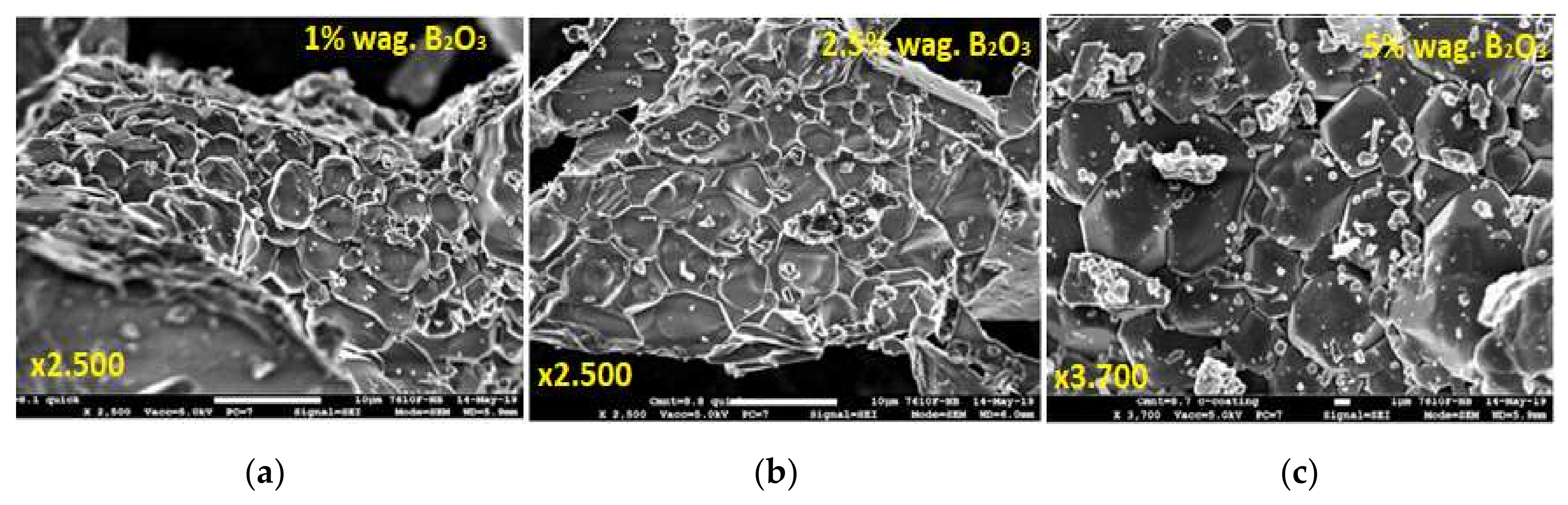
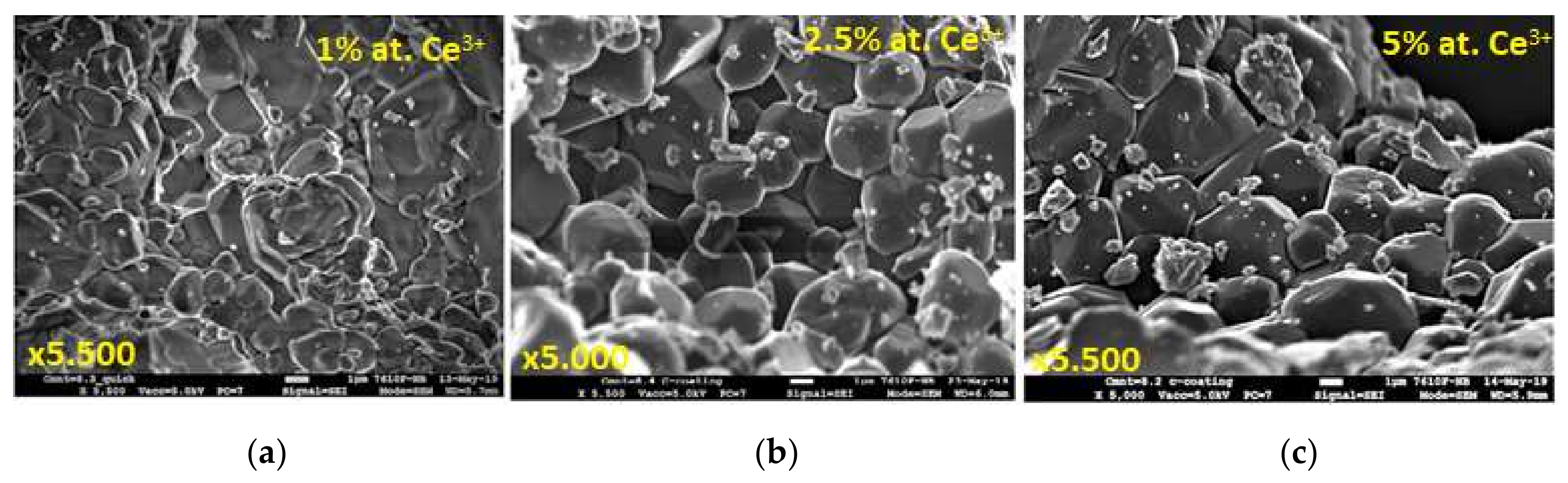
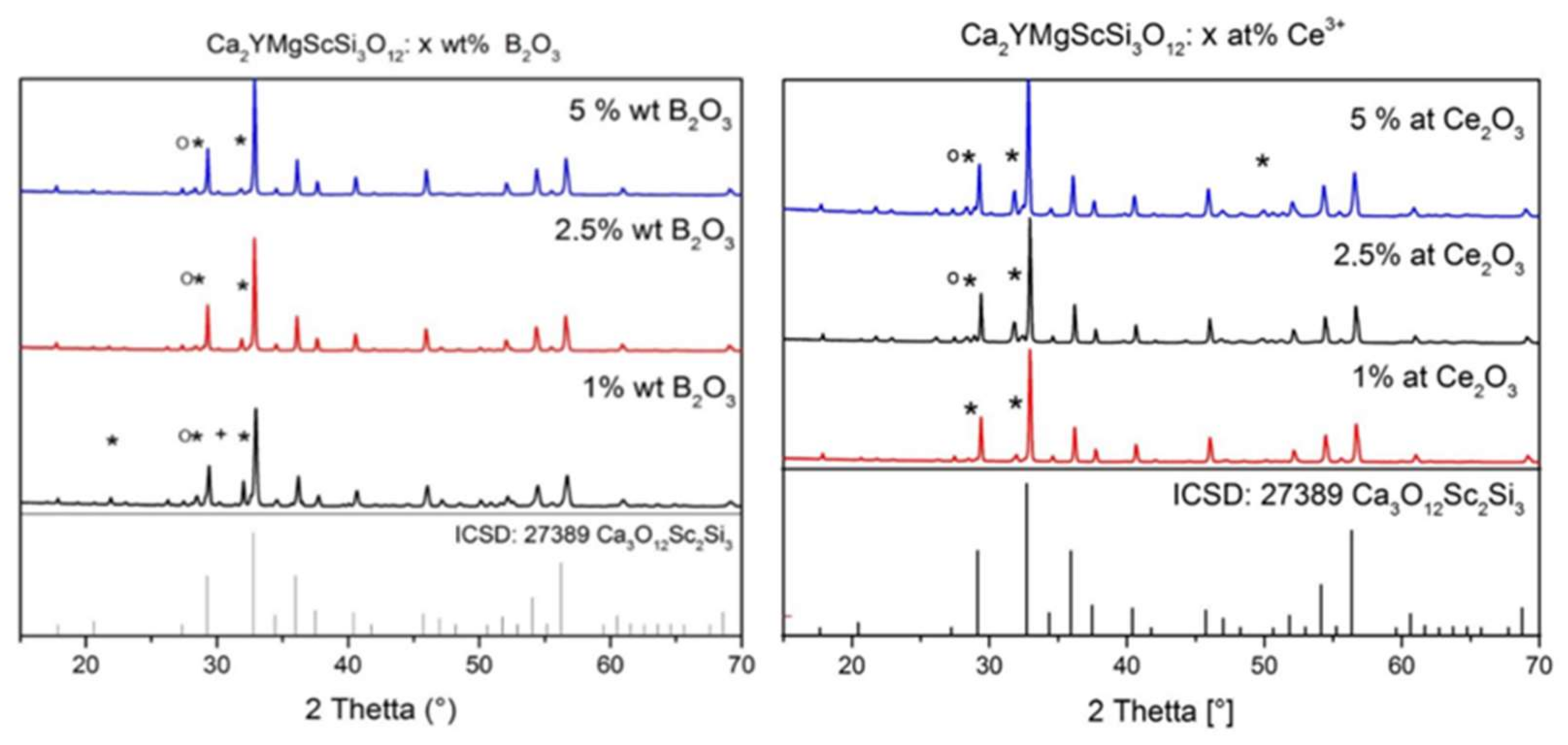
2. Synthesis of CYMSSG:Ce Micropowder Samples and Their Structural Qualities

CYMSSG:Ce MP were synthesized by conventional solid synthesis as an effective production method based on a solid–solid reactions, and in this case, between the microcrystalline grains of the raw components. During the synthesis of MPs from CaO, Y2O3, MgO, Sc2O3, and SiO2 raw oxides with 4N purity, they were first weighed and then ground in agate solution for 20 min to obtain the greatest homogeneity of the powder. It is necessary to note here that the homogeneity of the samples significantly enhances the luminescent properties of the phosphors. The final phosphor mass was baked in an Al2O3 crucible at a heating rate of 20 °C/min to 1300 °C for 10 h in a reducing atmosphere (95% N2 +5% H2).
In the solid-state method, the preparation of phosphors using flux can substantially support the formation of the garnet phase and enables the obtaining of microparticles with high quantum yield. The application of flux can also significantly improve the morphology of the grains and, in this way, additionally enhance the material PL intensity. For this reason, the synthesis of CYMSSG:Ce MP samples was performed using B2O3 flux with concentration in the 1–5 wt.% range with respect to the total weight of the garnet charge. By using B2O3 flux to improve the condition of the solid-state reaction, the Ca2YMgScSi3O12 garnet phase in the MP samples under study was successfully obtained (Figure 1 and Figure 2).
Figure 1 demonstrates the SEM images of the CYMSSG:Ce MP samples synthesized from the charge containing 1 wt.%, 2.5 wt.%, and 5 wt.% B2O3 flux agent. Overall, the structure of the garnet is visible in all MP samples under study. From Figure 1, the influence of the flux and its concentration on the formation of the cubic particles with a garnet structure can also be observed. Cubic grains corresponding to the garnet structure are present in small amounts in the MP sample synthesized at a concentration of 1 wt.% B2O3 (Figure 1a). Furthermore, strong grain agglomeration is visible for this MP sample. On the contrary, the MP samples synthesized from compounds with a higher B2O3 content (Figure 1b,c) have a more homogeneous garnet structures, especially for the sample with the highest (5 wt.%) flux concentration (Figure 1c). That means that increasing the flux content leads to a uniformity of the grain distribution and some increase in the average grain size, up to 5–8 μm for a Ba2O3 concentration of 5 wt.%.
SEM images of CYMSSG:Ce MPs synthesized with an activator concentration in the 1–5% range are shown in Figure 2. Generally, garnet grains are visible in all samples. However, the strong grain agglomeration is observed for samples with the lowest Ce3+ content of 1%. Furthermore, the quantity of agglomerates is significantly lower in the samples with Ce3+ contents of 2.5 and 5 at.%, especially in the last one (Figure 2c). For the last sample, the most uniform distributions of the grain are observed, with their average size in the 3–5 μm range.
The X-ray diffraction (XRD) of MP samples sintered with different B2O3 flux amounts (Figure 3a) and different Ce3+ concentrations shows almost pure phases of Ca3Sc2Si3O12 garnet (CSSG) in both cases and excellent correspondence with the respective ICSD-27389 pattern. However, some minor amounts of unreacted starting materials, (SiO2) as well as the secondary phases (Ca2Ce8O26Si6, YBO3), are also observed in the XRD patterns (Figure 3).
From the XRD data, a garnet phase with maxima corresponding mostly to Ca3Sc2Si3O12 was detected. The main peak of the garnet at about 32.7° (2θ), which belongs to the {024} family of lattice planes, is very well defined in these compositions. However, additional peaks, which cannot be precisely identified, can be seen in Figure 3.
It is noticeable here that the peaks are shifted by several 2θ. The incorporation of Mg2+ ions into the crystal lattice is a probable reason for the discernible shift to the right. The Mg2+ ions take the lattice sites of the Sc3+ ions. Due to the smaller ionic radius, the lattice spacing increases, which means that 2θ increases accordingly. As a result of the substitution with Y3+ and Mg2+, a newly modified silicate garnet structure that is not yet known in the database has probably emerged.
Furthermore, the increase in B2O3 achieves better single-phase character. This is illustrated by the peaks at around 32° (2θ) and between 51 and 52° (2θ) (Figure 3a). The proportion of the Ca2Ce8O26Si6 foreign phase and the intensity of the corresponding peak decreases with the increasing B2O3 concentration.
As expected, a garnet phase similar to that of Ca3Sc2Si3O12 could also be detected in a series of measurements with different concentrations of Ce ions using XRD analysis (Figure 3b). The main peaks of the silicate garnet phase are consistent with those of synthesized samples. However, as already mentioned, these are shifted. By increasing the CeO2 concentration, the intensity of the peak at 33° increases, and the proportion of the Ca2Ce8O26Si6 foreign phase increases as well.

3. Photoluminescence Quantum Yield of CYMSSG:Ce MPs

The photoluminescence quantum yield (PLQY) of the CYMSSG:Ce MPs depending on the synthesis conditions is given in Table 1. The PLOY values ranges lie between 42.1 and 63.6%, depending mainly on the content of the garnet phase in the MP samples under study. The highest quantum efficiency has the MP sample with an activator concentration of 2.5 at.% and a B2O3 flux agent content of 2.5 wt.%. Correspondingly, the garnet content in these MP samples is highest as well and equal to 82% (Table 1). It is necessary to note that second phases do not influence the emission properties in the Ce3+ emission spectral region and serve as light scattering centers and, probably, as emission centers in the UV region in the samples investigated in this work (see results below).
The largest garnet phase content, in the 80–82% range, is observed for the MP samples sintered with a Ba2O3 flux content in the 2.5–5 at.% range and the activator concentration in the 1–2.5% range. The most optimal condition is the sintering with a flux content of 2.5 wt.% B2O3 and a Ce3+ concentration of 2.5 at.%, enabling 82% garnet content in the MP sample (Table 1).
Some ways of improving the PLQY were investigated. For instance, an efficiency of more than 71% was achieved by the synthesis of CYMSSG:Ce in pellets by a two-step synthesis process. This was a case in which only 68% of the main phase was detected by XRD analysis.

4. Luminescent and Photoconversion Properties of CYMSSG:Ce MP Samples

For characterization of the optical properties of the CYMSSG:Ce MPs under study, the cathodoluminescence (CL), photoluminescence emission (PE) and excitation spectra (PLE), PL decay kinetics, and PLQY and photoconversion spectra (PC) measurements were used. The CL spectra were investigated using the e-beam from a scanning electron microscope SEM JEOL JSM-820 (JEOL, Warsaw, Poland) additionally equipped with a Stellar Net spectrometer with a TE-cooled CCD detector working in the 200–1200 nm range. PL and PLE spectra and PL decay kinetics were measured using an FS-5 spectrometer (Edinburgh Instruments, Livingston, UK). An EPL-450 picosecond pulsed diode laser was used to measure of the decay kinetics, and the typical average power of this laser is 0.15 mW (Edinburgh Instruments). The PC spectra and PLQY measurements were performed using a fiber-optic spectrophotometer AvaSpec-ULS 2048-LTEC (Avantes, Apeldoorn, The Netherlands), and an integrating sphere AvaSphere-50-IRRAD. The photoconverter (pc) prepared from CYMSSG:Ce MP films of different thicknesses was excited by the blue LED at a wavelength of λ = 450 nm. All luminescence measurements were performed at room temperature (RT).

4.1. Cathodoluminescence Spectra

The normalized CL spectra of the CYMSSG:Ce MPs samples sintered with different amounts of B2O3 flux and different concentrations of Ce3+ ions are shown in Figure 4a,b, respectively. The dominant luminescence band with a peak in the 550–555 nm range in all MP CYMSSG:Ce samples corresponds to the 5d1 → 4f (2F5/2,7/2) transitions of Ce3+ in these garnet compounds. The position of the Ce3+ band is slightly red-shifted from 551 to 555 nm in the MP samples with an increase of the flux contents from 1 wt.% to 5 wt.% (Figure 4a) and Ce3+ content from 1 to 5% (Figure 4b).
Apart from the luminescence of the Ce3+ ions in the garnet structure in the visible range, the other emission bands in the UV range peaked in the 380–392 nm range are also observed in the CL spectra for the CYMSSG:Ce MPs. Furthermore, the lowest UV luminescence efficiency is observed in the CL spectra of the CYMSSG:Ce MP sample sintered with flux and activator contents of 2.5 wt.% and 5 at.%, respectively (Figure 4b).
It is important to note here that the low intensity bands in the UV range of Ca2+-Mg2+-Si4+ -based garnets usually are assigned to defect luminescence [26,27]. According to the authors [26,27], the presence of a high concentration of oxygen vacancies in these gannets is expected due to possible deviations in the concentration of heterovalent Ca2+, Mg2+, and Si4+ cations and the demands for local charge compensation. For this reason, the bands peaked at 317 nm and the 380–392 nm range may correspond to the luminescence of the F+ and F centers (single- and double-charged oxygen vacancies, respectively) in the CYMSSG host [27,28]. However, it is also worth mentioning here that, due to the relatively large contents of the secondary phases in the tested MP samples, the luminescence spectra in the UV range may also partly correspond to the Ce3+ luminescence in these compounds. However, a visible correlation between the contents of the secondary phases and the intensity of the UV luminescence was not observed for the tested CYMSSG:Ce MP samples.

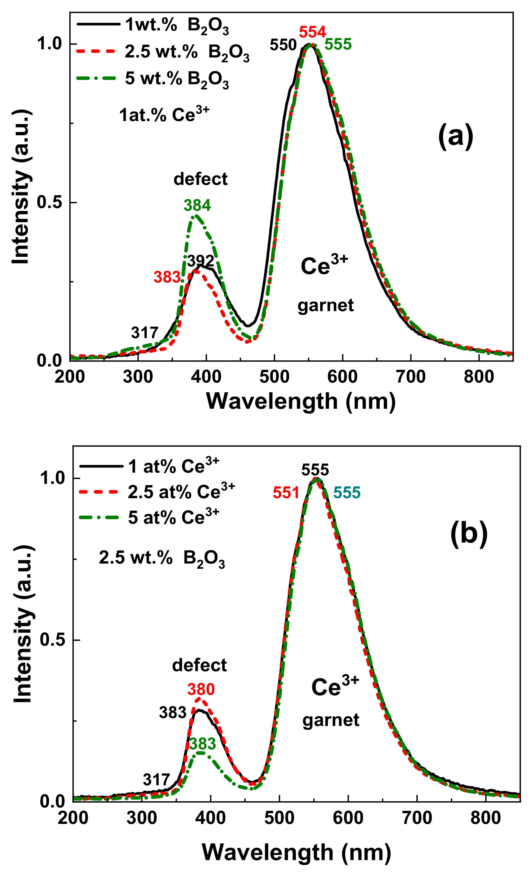
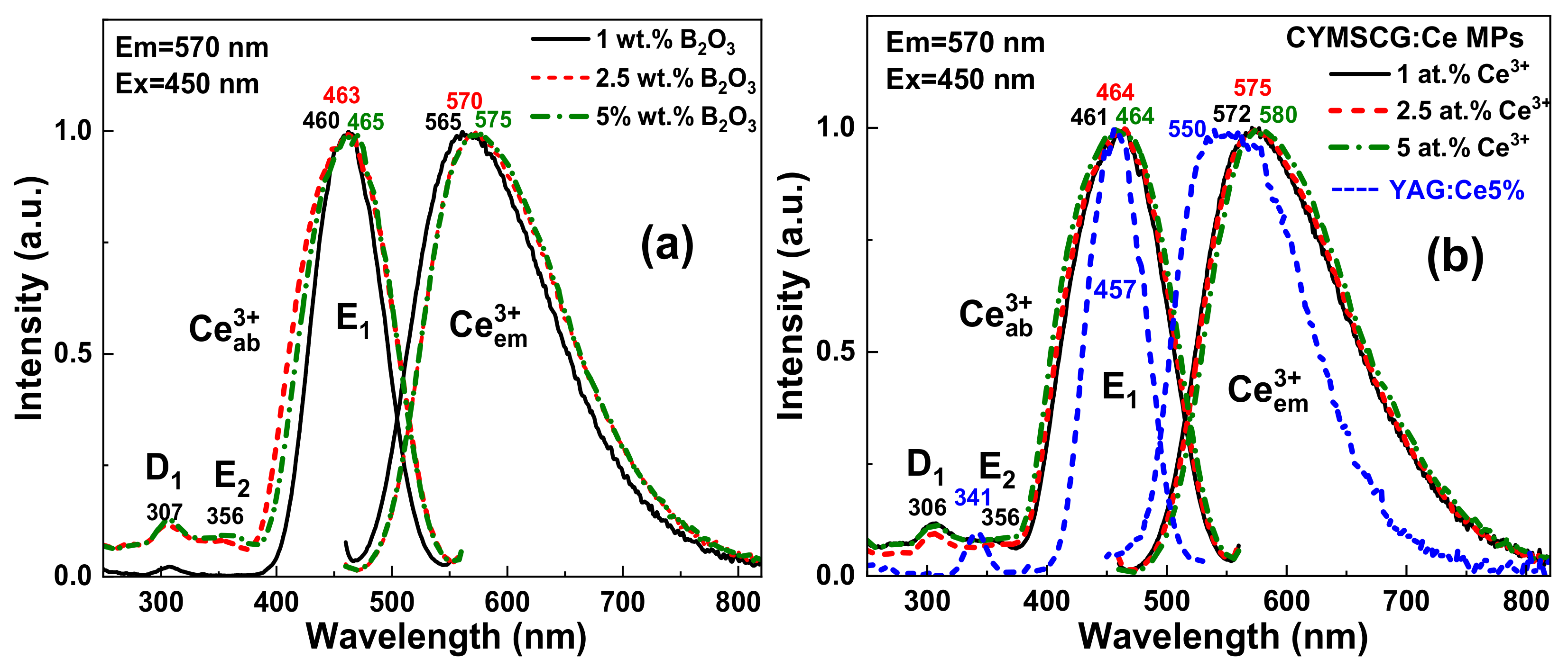
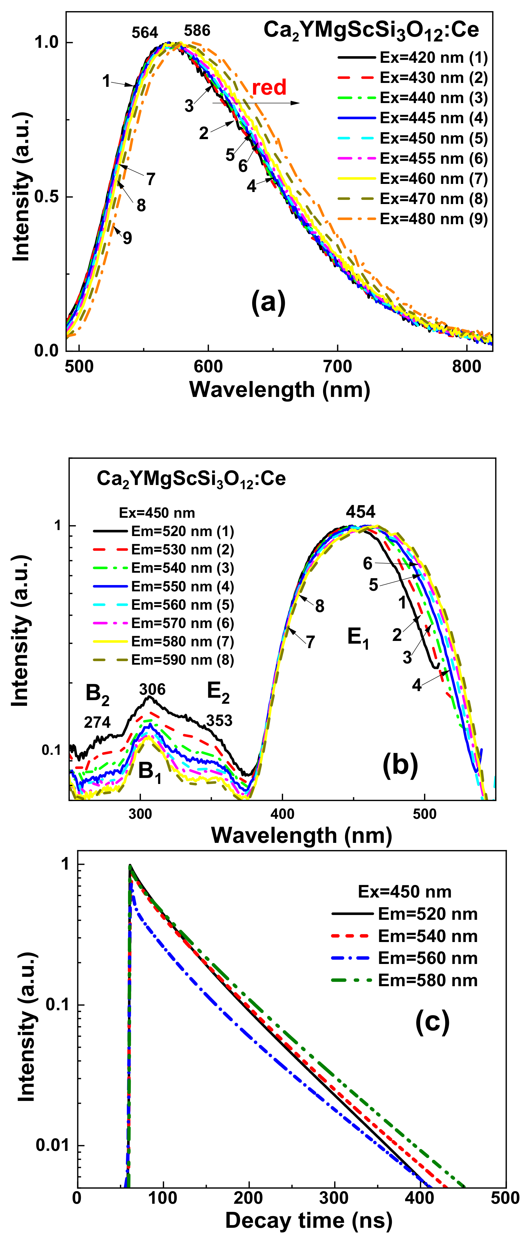
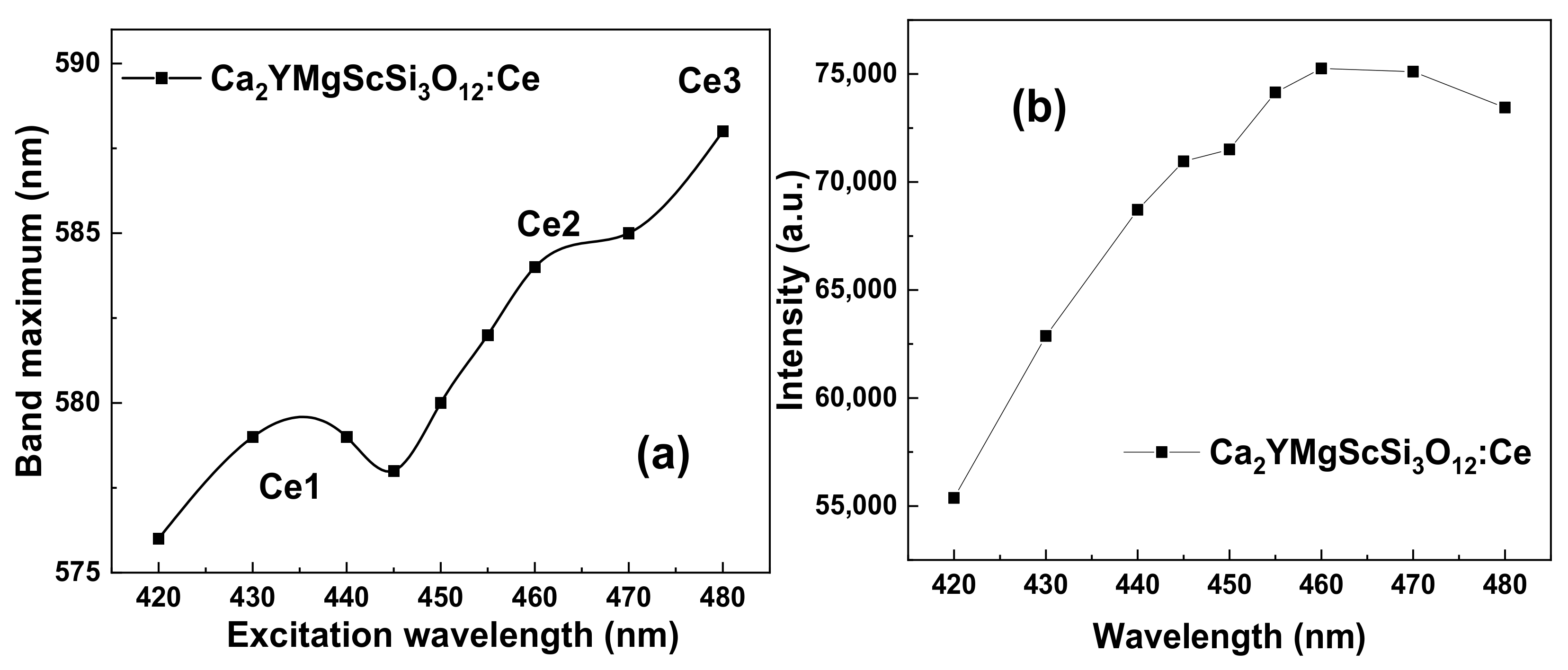
4.2. PL and PLE Spectra

The PL spectra of the CYMSSG:Ce (1 at.%) MPs (Figure 5a) sintered with different B2O3 flux amounts show wide bands peaked in the 565–575 nm range, which corresponds to the Ce3+ radiation transitions from the lowest 5d1 level of the excited state to the 4f(2F5/2,7/2) levels of the ground state. Increasing the concentration of B2O3 leads to a shift in the emission spectra of the Ce3+ ions to the long-wavelength spectral range (Figure 5a) [11,27,28,29].
The PLE spectra of these MP samples (Figure 5b) show several bands in the 250–550 nm region. The main band E1 peaked in the 460–465 nm range is explained by the absorption of the allowed transitions from the 4f ground state to the lowest 5d1 levels of the Ce3+ ions. Moreover, two MP samples, synthesized with a higher concentration of boron oxide, have a E2 band peaked at 356 nm, which also corresponds to the 4f (5d2) excitation band of the Ce3+ ions. The low intensity of the E2 band in comparison with conventional YAG:Ce garnets (Figure 4b) is characteristic of Ca2+ (or Mg2+) -Si4+-based garnets (see also [27,30,31]).
Figure 5b demonstrates the PL and PLE spectra of the CYMSSG:Ce MPs with variable Ce3+ ion concentrations in the range 1–5 at.% synthesized with a flux B2O3 concentration of 2.5 wt.%. Increasing the Ce3+ concentration in these MP samples leads to the increases of the crystal field strength in the dodecahedron position of the garnet compounds, which results in a long-wavelength shift in the Ce3+ emission spectrum (Figure 5b). Moreover, the PL emission and excitation spectra of all CYMSSG:Ce MP samples is considerably broadened in comparison with the conventional CSSG:Ce and YAG:Ce phosphors (Figure 5b). Namely, the PL emission spectrum in CYMSSG:Ce MPs shifted with respect to YAG:Ce spectrum by more than 40 nm into the red range, extending up to even 800 nm. The respective FWHM of the Ce3+ emission bands increases from 124.5 nm in the YAG:Ce 5% to 139.5 nm in the CYMSSG:Ce5% MPs. The FWHM of the main Ce3+ excitation band in the PLE excitation spectrum of the CYMSSG:Ce MPs is also significantly larger in the CYMSSG:Ce5% MPs (104 nm) in comparison with the YAG:Ce5% sample (56 nm) (Figure 5b). Such results, without any doubt, indicate the existence of different Ce3+ centers in the CYMSSG garnets (see also [27,30,31,32,33] for details).
It is also worth noting that the PLE spectra of these MP samples (Figure 5a) also contain a band in the UV range peaked at 308 nm, which can correspond to the excitation of the defect luminescence in the CYMSSG host in the bands peaked in the 380–393 nm range (Figure 2). Such emission bands are overlapped with the E1 and E2 excitation bands of the Ce3+ luminescence in the garnets. That results in the excitation of the Ce3+ luminescence via the luminescence of the defects centers. Thus, energy transfer between the defect and Ce3+ centers is observed in all CYMSSG:Ce MP samples under study.
Figure 6 shows the PL decay kinetics of the MP CSSG: Ce samples with different concentrations of B2O3 flux (a) and Ce3+ activator (b). The approximation parameters of the respective decay curves are given in Table 2 and Table 3, respectively.
Similarly to other Ca2+-Si4+-based garnets [26,32,33], the decay kinetics of the Ce3+ emission in Ca2YMgScSi3O12:Ce MPs is non-exponential. For this reason, the three-exponential fit of the decay curves was used for the quantitative description of the luminescence timing properties (Table 1 and Table 2). Although such a three-exponential approximation does not describe the luminescence decay behavior correctly at quenching due to the energy transfer, the decay time values can be considered as estimations of the luminescence decay times of the respective Ce3+ multicenters.

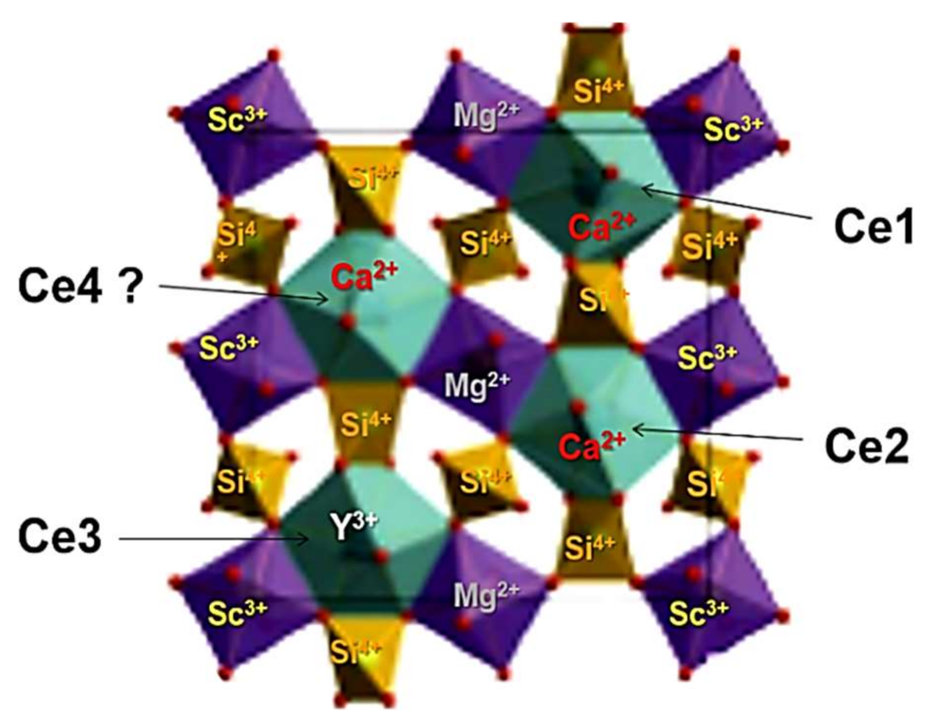
5. Ce3+ Multicenter Formation in CYMSSG:Ce Phosphor

The RT PL emission spectra of the CYMSSG:5%Ce MPs are shown in Figure 7a under excitation in the characteristic PLE bands (Figure 7b). The PL spectra of these samples show the intensive luminescence in the form of wide bands peaked in the green range related to the 5d1→4f(2F5/2,7/2) transitions of the Ce3+ ions. Moreover, by increasing the excitation wavelength from 420 to 480 nm, the PL spectra of the CYMSSG:Ce MPs are significantly red-shifted from 564 to 587 nm and slightly narrowed (Figure 7a). (Figure 7a, curves 1–9, respectively). Furthermore, the shift in the main maxima of the PL spectra of the CYMSSG:Ce MPs and the intensity in the peak positions occurs non-monotonically with the increasing excitation wavelength (Figure 8a,b) in comparison with the similar dependencies for the YAG:Ce SCF sample (not shown in Figure 8). Behavior such as that shown in the PL emission spectra indicates the Ce3+ multicenter formation in the CYMSSG:Ce garnet.
The normalized excitation spectra of the Ce3+ luminescence in the Ca2YMgScSi3O12:Ce MP samples are shown in Figure 7b. In these spectra, the most intensive excitation bands, E1 and E2, peaked in the 454 nm (E1) and 353 nm (E2) ranges, are attributed to the intrinsic transitions from the 4f(2F5/2) level of the ground state to the 5d (E2) excited level of the Ce3+ ions (Table 4). The excitation bands peaked at 306 nm (B1 band) and 274 nm (B2 band) are also observed in the excitation spectra of the Ca2YMgScSi3O12:Ce MPs (Figure 7a,b). These excitation bands are not related to the intrinsic transitions of the Ce3+ ions and probably correspond to the Ce3+ luminescence excitation by the emission of the defect centers or the emission of flux-related impurities [30,31].
The decay kinetics of the Ce3+ luminescence in the Ca2YMgScSi3O12:Ce MPs registered at 520–580 nm under excitation at 450 nm are shown in Figure 7c. Due to the strong non-exponentiality of the decay kinetics, the three-exponential fit of the decay curves was used for the quantitative description of the luminescence timing properties (Table 5).
Behavior such as that shown in the decay curves of the Ca2YMgScSi3O12:Ce MPs can also indicate possible energy transfer from high-energy to low-energy Ce3+ emitting centers in these garnets [32,33]. The different decay times in the Ca3Sc2Si3O12:Ce SCF can correspond to the different Ce3+ centers in the dodecahedral positions of the garnet lattice with various local surroundings by oxygen and cations (Sc3+ and Si4+ ions in the octahedral and tetrahedral positions, respectively, and Ca2+ and Y3+ cations in the dodecahedral positions of the garnet host; Figure 9).

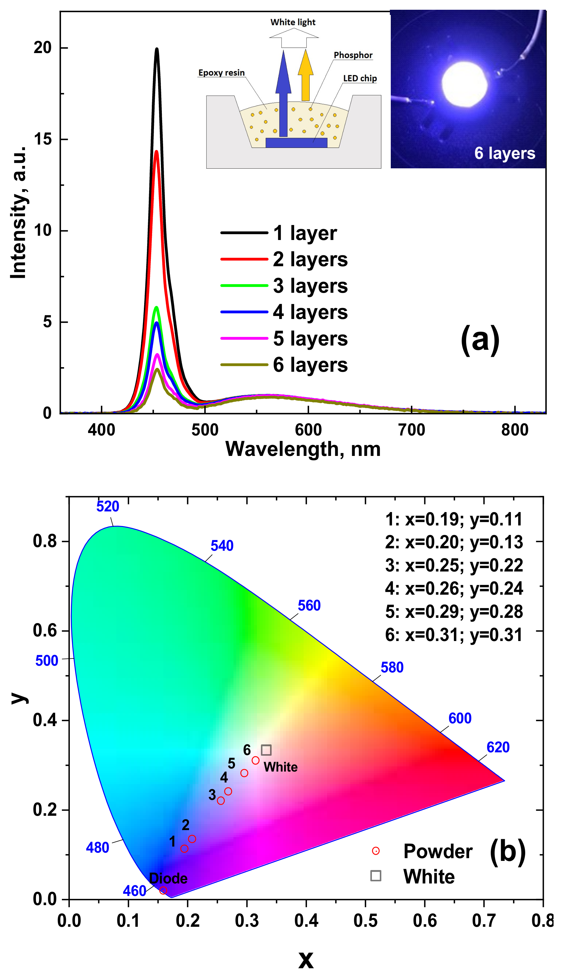
6. WLED Prototype Creation

To demonstrate the application possibility of the development of CYMSSG:Ce phosphor, WLED prototypes were fabricated by coating a blue LED with an emission wavelength of 450 nm with several films containing CYMSSG:Ce phosphor embedded in epoxy resin (Figure 10a). PC measurements of the WLED prototypes were performed after each successive PC layer of a thickness of approximately 100–120 µm. The emission spectra of these WLEDs are quite broad and cover the entire visible range from 400 to 780 nm. The emission spectra of the WLED prototypes also show a significant decrease in the intensity of the blue component and a respective continuous increase in the intensity of the yellow emission by increasing the total film thickness (Figure 10a). As can be seen from Figure 10b, the color coordinates move to the diagram’s center by increasing the total thickness of the PC film. Finally, the WLED prototypes, based on six films of CYMSSG:Ce MP PC, give quite cold white emissions with the correlated color coordinates (CCC) x = 0.315; y = 0.31 and a correlated color temperature (CCT) of 6930 K. Furthermore, as the thickness of the photoconverter film increases, the color coordinates move to the center of the diagram, including the warm white color pattern. However, these trends of the photoconverting properties of the developed WLED prototypes are quite limited by the conversion efficiency of the CYMSSG:Ce MP phosphor with a Ce3+ concentration of 1 at.%. To obtain a precise tuning of the photoconversion properties (CCC and CCT), the CYMSSG:Ce MP phosphor with Ce3+ concentration above 1% can be used as well.
The fabricated device with bright-light characteristics demonstrates that the WLEDs based on the CYMSSG:Ce MPs are very promising and can be applied to lighting systems (Figure 10a, insert). The CIE chromaticity coordinates of the prototype WLEDs are shown in Table 6.

7. Conclusions

Microcrystalline powder (MP) samples of Ca2YMgScSi3O12:Ce garnet with Ce3+ concentrations in the range of 1–5 at.% were obtained using solid-state synthesis by adding B2O3 flux in the 1–5 wt. % concentration of the total charge content. To determine the luminescent properties of the CYMSSG:Ce MPs, the cathodoluminescence (CL) spectra, photoluminescence (PL) emission and excitation spectra, PL decay kinetics, and the PL quantum yields (PL QY) were measured.
The obtained results confirm the Ce3+ multicenter formation in the MP of the CYMSSG:Ce garnet. It is mainly caused by the location of the Ce3+ ions in the dodecahedral positions of the Ca2+ and Y3+ cations, and the different local surroundings of these centers. Namely, Ce3+ multicenters such as those in the mentioned dodecahedral positions of the garnet host possess additional local asymmetry and crystal field strength due to inhomogeneity in the local environment of these positions during the substitutions at the octahedral sites by heterovalent Mg2+ and Sc3+ ions and the at the tetrahedral sites by Si4+ ions. Based on the results of the optical investigations, the luminescent characteristics of different Ce3+-based multicenters were estimated.
The application potential of the developed CYMSSG:Ce MP phosphor was demonstrated. Planar WLED prototypes were fabricated by coating GaN 450 nm blue LED chips with several 100–120 μm thick layers of CYMSSG:Ce MP phosphor mixed with epoxy resin. Furthermore, the WLED prototypes, based on five CYMSSG:Ce MP photoconverters, give emissions close to white light with the coordinates x = 0.315; y = 0.31 and a color temperature of 6930 K.

Author Contributions

A.S. collected and analyzed the structural and optical properties of MPs and participated in writing and preparation of the paper; A.M. participated in preparation of the prototypes of WLED and measured their photoconversion properties; S.W.-Ł. performed and analyzed the cathodoluminescence measurements; Y.V. and A.O. performed the synthesis of MPs and taking part in XRD investigations; T.Z. participated in measurements of PL of PL decay ki-netics of MP samples and analyzed them; J.E. performed and analyzed the XRD measurements; C.J.B. and M.B. managed the MP preparation and XRD investigations; Y.Z. analyzed the whole experimental materials and participated in writing and preparation of the paper. All authors have read and agreed to the published version of the manuscript.

Funding

The work was performed in the framework of the Polish NCN 2017/25/B/ST8/02932 project and partly in the frame of NCN 2019/33/B/ST3/00406 project.

Institutional Review Board Statement

Not applicable.

Informed Consent Statement

Not applicable.

Data Availability Statement

Not applicable.

Conflicts of Interest

The authors declare no conflict of interest.

References

  1. Schubert, E.F. Light-Emitting Diodes, 2nd ed.; Cambridge University Press: New York, NY, USA, 2006. [Google Scholar]
  2. Withnall, R.; Silver, J. Luminescence of phosphors. In Handbook of Visual Display Technology; Chen, J., Cranton, W., Fihn, M., Eds.; Springer: Berlin/Heidelberg, Germany, 2012. [Google Scholar]
  3. CIE. Commission Internationale de l’Eclairage Proceedings; Cambridge University Press: Cambridge, UK, 1995. [Google Scholar]
  4. Bass, M. Handbook of Optics; R.R. Donnelly & Sons Company: Chicago, IL, USA, 1995. [Google Scholar]
  5. Jargus, J.; Vitasek, J.; Nedoma, J.; Vasinek, V.; Martinek, R. Effect of Selected Luminescent Layers on CCT, CRI, and Response Times. Materials 2019, 12, 2095. [Google Scholar] [CrossRef] [PubMed] [Green Version]
  6. Li, X.; Jiang, Y.; Li, J.; Shi, Z.; Zhu, G.; Wang, Y. Integrated photonics chip with InGaN/GaN light-emitting diode and bended waveguide for visible-light communications. Opt. Laser Technol. 2019, 114, 103–109. [Google Scholar] [CrossRef]
  7. Xu, Y.; Chen, Z.; Gong, Z.; Xia, Z.; Yuan, T.; Gu, Z.; Zhao, W.; Chen, J. Hybrid modulation scheme for Visible Light Communication using CMOS camera. Opt. Commun. 2019, 440, 89–94. [Google Scholar] [CrossRef]
  8. Yan, D.; Mao, X.; Xie, S.; Cong, J.; Chen, H. Design Fully Integrated Driver Circuit for Phosphorescent White Light-Emitting-Diode High Speed Real-Time Wireless Communication. IEEE Photonics J. 2019, 11, 1–10. [Google Scholar] [CrossRef]
  9. Song, Y.H.; Ji, E.K.; Jeong, B.W.; Jung, M.K.; Kim, E.Y.; Yoon, D.H. High power laser-driven ceramic phosphor plate for outstanding efficient white light conversion in application of automotive lighting. Sci. Rep. 2016, 6, 31206. [Google Scholar] [CrossRef]
  10. Steigerwald, D.A.; Bhat, J.C.; Collins, D.; Fletcher, R.M.; Holcomb, M.O.; Ludowise, M.J.; Martin, P.S.; Rudaz, S.L. Illumination with solid state lighting technology. IEEE J. Sel. Top. Quantum Electron. 2002, 8, 310–320. [Google Scholar] [CrossRef]
  11. Levchuk, I.; Osvet, A.; Brabec, C.J.; Batentschuk, M.; Shakhno, A.; Zorenko, T.; Zorenko, Y. Micro-powder Ca3Sc2Si3O12:Ce silicate garnets as efficient light converters for WLEDs. Opt. Mater. 2020, 107, 109978. [Google Scholar] [CrossRef]
  12. Shimomura, Y.; Honma, T.; Shigeiwa, M.; Akai, T.; Okamoto, K.; Kijima, N. Photoluminescence and Crystal Structure of Green-Emitting Ca3Sc2Si3O12:Ce3+ Phosphor for White Light Emitting Diodes. J. Electrochem. Soc. 2007, 154, J35. [Google Scholar] [CrossRef]
  13. Katelnikovas, A.; Bareika, T.; Vitta, P.; Jüstel, T.; Winkler, H.; Kareiva, A.; Žukauskas, A.; Tamulaitis, G. Y3−xMg2AlSi2O12: Phosphors—Prospective for warm-white light emitting diodes. Opt. Mater. 2010, 32, 1261–1265. [Google Scholar] [CrossRef]
  14. Zhong, J.; Zhuang, W.; Xing, X.; Liu, R.; Li, Y.; Liu, Y.; Hu, Y. Synthesis, Crystal Structures, and Photoluminescence Properties of Ce3+-Doped Ca2LaZr2Ga3O12: New Garnet Green-Emitting Phosphors for White LEDs. J. Phys. Chem. C 2015, 119, 5562–5569. [Google Scholar] [CrossRef]
  15. Li, G.; Tian, Y.; Zhao, Y.; Lin, J. Recent progress in luminescence tuning of Ce3+and Eu2+-activated phosphors for pc-WLEDs. Chem. Soc. Rev. 2015, 44, 8688–8713. [Google Scholar] [CrossRef] [PubMed]
  16. Shang, M.; Fan, J.; Lian, H.; Zhang, Y.; Geng, D.; Lin, J. A Double Substitution of Mg2+–Si4+/Ge4+ for Al(1)3+–Al(2)3+ in Ce3+-Doped Garnet Phosphor for White LEDs. Inorg. Chem. 2014, 53, 7748–7755. [Google Scholar] [CrossRef]
  17. Katelnikovas, A.; Bettentrup, H.; Uhlich, D.; Sakirzanovas, S.; Jüstel, T.; Kareiva, A. Synthesis and optical properties of Ce3+-doped Y3Mg2AlSi2O12 phosphors. J. Lumin. 2009, 129, 1356–1361. [Google Scholar] [CrossRef]
  18. Kishore, M.S.; Kumar, N.P.; Chandran, R.G.; Setlur, A.A. Solid Solution Formation and Ce3+ Luminescence in Silicate Garnets. Electrochem. Solid-State Lett. 2010, 13, J77. [Google Scholar] [CrossRef]
  19. Feng, G.; Jiang, W.; Liu, J.; Li, C.; Zhang, Q.; Miao, L.; Wu, Q. Synthesis and luminescence properties of Al2O3@YAG: Ce core–shell yellow phosphor for white LED application. Ceram. Int. 2018, 44, 8435–8439. [Google Scholar] [CrossRef]
  20. Chen, L.-C.; Tseng, Z.-L.; Chang, W.-W.; Lin, Y.W. Warm white light-emitting diodes using organic–inorganic halide perovskite materials coated YAG:Ce3+ phosphors. Ceram. Int. 2018, 44, 3868–3872. [Google Scholar] [CrossRef]
  21. Gu, G.; Xiang, W.; Yang, C.; Fan, W.; Lv, Y.; Zhang, Z.; Liang, X. A Novel Single-Component White-Emitting Tb and Mn Co-Doped Large-Sized Y3Al5O12:Ce3+ Single Crystal for White LED. Sci. Adv. Mater. 2016, 8, 1354–1360. [Google Scholar] [CrossRef]
  22. Du, Y.; Shao, C.; Dong, Y.; Yang, Q. Electroluminescent properties of WLEDs with the structures of Ce:YAG single crystal/blue chip and Sr2Si5N8:Eu2+/Ce:YAG single crystal/blue chip. J. Disp. Technol. 2016, 12, 323–327. [Google Scholar] [CrossRef]
  23. Zhao, B.Y.; Liang, X.; Chen, Z.; Xie, C.; Luo, L.; Zhang, Z.; Xiang, W. Studies on optical properties and Ce concentration of Ce: YAG single crystal for WLEDs. Chem. J. Chin. Univ. 2014, 25, 230–236. [Google Scholar] [CrossRef]
  24. Pan, Z.; Xu, Y.; Hu, Q.; Li, W.; Zhou, H.; Zheng, Y. Combination cation substitution tuning of yellow-orange emitting phosphor Mg2Y2Al2Si2O12:Ce3+. RSC Adv. 2015, 5, 9489–9496. [Google Scholar] [CrossRef]
  25. Tyagi, M.; Meng, F.; Koschan, M.; Donnald, S.B.; Rothfuss, H.; Melcher, C.L. Effect of codoping on scintillation and optical properties of a Ce-doped Gd3Ga3Al2O12scintillator. J. Phys. D Appl. Phys. 2013, 46, 475302. [Google Scholar] [CrossRef]
  26. Melcher, C.L.; Koschan, M.; Zhuravleva, M.; Wu, Y.; Rothfuss, H.; Meng, F.; Tyagi, M.; Donnald, S.; Yang, K.; Hayward, J.P.; et al. Scintillator Design via Codoping. In Proceedings of International Symposium on Radiation Detectors and Their Uses (ISRD2016). J. Phys. Soc. Jpn. 2016, 11, 020001. [Google Scholar] [CrossRef]
  27. Gorbenko, V.; Zorenko, T.; Paprocki, K.; Iskaliyeva, A.; Fedorov, A.; Schröppel, F.; Levchuk, I.; Osvet, A.; Batentschuk, M.; Zorenko, Y. Epitaxial growth of single crystalline film phosphors based on the Ce3+-doped Ca2YMgScSi3O12garnet. CrystEngComm 2017, 19, 3689–3697. [Google Scholar] [CrossRef]
  28. Gorbenko, V.; Zorenko, T.; Witkiewicz, S.; Paprocki, K.; Iskaliyeva, A.; Kaczmarek, A.M.; Van Deun, R.; Khaidukov, M.N.; Batentschuk, M.; Zorenko, Y. Luminescence of Ce3+ multicenters in Ca2+-Mg2+-Si4+ based garnet phosphors. J. Lumin. 2018, 199, 245–250. [Google Scholar] [CrossRef]
  29. Gorbenko, V.; Zorenko, T.; Pawlowski, P.; Iskaliyeva, A.; Paprocki, K.; Suchocki, A.; Zhydachevskii Ya Fedorov, A.; Khaidukov, N.; Van Deun, R.; Schröppel, F.; et al. Luminescent and scintillation properties of Ce 3+ doped Ca2RMgScSi3O12 (R = Y, Lu) single crystalline films. J. Lumin. 2018, 195, 362–370. [Google Scholar] [CrossRef]
  30. Gorbenko, V.; Zorenko, T.; Witkiewicz-Łukaszek, S.; Shakhno, A.; Osvet, A.; Batentschuk, M.; Fedorov, A.; Zorenko, Y. Crystallization and Investigation of the Structural and Optical Properties of Ce3+-Doped Y3−xCaxAl5−ySiyO12 Single Crystalline Film Phosphors. Crystals 2021, 11, 788. [Google Scholar] [CrossRef]
  31. Khaidukov, N.M.; Makhov, V.N.; Zhang, Q.; Shi, R.; Liang, H. Extended broadband luminescence of dodecahedral multisite Ce 3+ ions in garnets {Y3}[MgA](BAlSi)O 12 (A = Sc, Ga, Al; B = Ga, Al). Dye Pigment. 2017, 142, 524–529. [Google Scholar] [CrossRef]
  32. Khaidukov, N.M.; Zhidkova, I.A.; Kirikova, N.Y.; Makhov, V.N.; Zhang, Q.; Shi, R.; Liang, H. Mechanism for bifurcation of broadband luminescence spectra from Ce 3+ ions at dodecahedral sites in garnets {CaZY2}[M2](Al2Si)O12 (M = Al, Ga, Sc). Dye Pigment. 2018, 148, 189–195. [Google Scholar] [CrossRef]
  33. Setlur, A.A.; Heward, W.J.; Gao, Y.; Srivastava, A.M.; Chandran, R.G.; Shankar, M.V. Crystal Chemistry and Luminescence of Ce3+-Doped Lu2CaMg2(Si,Ge)3O12 and Its Use in LED Based Lighting. Chem. Mater. 2006, 18, 3314–3322. [Google Scholar] [CrossRef]
Figure 1. SEM images of CYMSSG: 2.5%Ce MPs, prepared using different amounts of B2O3 flux agent: (a) 1 wt.%, (b) 2.5 wt.%, and (c) 5 wt.%.
Materials 15 03942 g001
Figure 2. SEM images of CYMSSG:Ce MPs samples with different Ce3+ concentrations, prepared using 2.5 wt.% B2O3 flux agent: (a) 1 at.%, (b) 2.5 at.%, and (c) 5 at.%.
Materials 15 03942 g002
Figure 3. XRD patterns of CYMSSG:Ce MPs with different flux and activator amounts: (a) x wt.% B2O3 and (b) x at.% CeO2, where * is Ca2Ce8O26Si6, + is SiO2, and o is YBO3.
Materials 15 03942 g003
Figure 4. Normalized CL spectra of CYMSSG:Ce MPs sintered with different amounts of B2O3 flux (a) and different concentrations of Ce3+ ions (b).
Materials 15 03942 g004
Figure 5. PL spectra and PLE spectra of CYMSSG:Ce (1 at.%) MPs sintered with different amounts of B2O3 flux (a) and PL spectra and PLE spectra of CYMSSG:Ce MPs samples with different concentrations of Ce3+ ions sintered with a B2O3 flux content of 2.5 wt.% (b).
Materials 15 03942 g005
Figure 6. PL decay kinetics at RT recorded at 570 nm under blue light excitation (450 nm) in MPs sintered with different amounts of B2O3 flux (a) and different concentrations of Ce3+ ions (b).
Materials 15 03942 g006
Figure 7. Detailed luminescent properties of Ca2YMgScSi3O12:Ce5% MPs at RT. Emission (a) and excitation (b) recorded in different parts of the respective spectra. PL decay kinetics (c) at RT recorded under blue light (450 nm) excitation in different parts of the Ce3+ emission band.
Materials 15 03942 g007
Figure 8. Dependencies of the maxima (a) and intensities (b) in the peak positions of Ce3+ emission bands on excitation wavelength in Ca2YMgScSi3O12:Ce MPs (see Figure 7a,b).
Materials 15 03942 g008
Figure 9. Visualization of possible Ce3+ multicenter formation in the structure of Ca2YMgScSi3O12:Ce garnet.
Materials 15 03942 g009
Figure 10. Emission spectrum (a) and chromaticity diagram (b) of a WLED lamp fabricated on the base of 450 nm LED chip and CYMSSG:1%Ce phosphor.
Materials 15 03942 g010
Table 1. The garnet/secondary phase proportions and PLQY of the CYMSSG:Ce MPs sintered with different flux and activator contents.
Nominal Chemical Composition Ca2MgYScSi3O12:CeGarnet Content, %Secondary Phases Content, (%)PLQY, %
1 at.% Ce3+ + 1 wt.% B2O349.5CaO (10.9); SiO2 (21.8)
YBO3 (5); Ce2O3 (12.9)
42.1
1 at.% Ce3+ + 2.5 wt.% B2O380Ca2Ce8O26Si6 (11); SiO2 (7); CaO2 (2)54.5
1 at.% Ce3+ + 5 wt.% B2O380Ca2Ce8O26Si6 (9) SiO2(4); Ce2O3 (5); YBO3 (2)47.9
1 at.% Ce3+ + 2.5 wt.% B2O381Ca2Ce8O26Si6 (9); SiO2 (1); Ce2O3 (2); MgO (2); Ca (5)48.5
2.5 at.% Ce3++ 2.5 wt.% B2O382Ca2Ce8Si6O26 (14); Ce2O3 (2); SiO2 (2);63.6
5 at.% Ce3+ + 2.5 wt.% B2O362Ca2Ce8O26Si6 (13); SiO2 (17) CaO2 (2); MgO (6)44.3
Table 2. Parameters of three exponential approximations of the decay curves presented in Figure 6a (Em = 570 nm, Ex = 450 nm).
x% wag. B2O3t1, nsA1t2, nsA2t3, nsA3
14.4421.5942.121.5866.8821.78
2.55.318.1254.529.4668.1729.75
53.9353.1747.948.6458.6255.74
Table 3. Parameters of three exponential approximations of the decay curves presented in Figure 6b (Em = 570 nm, Ex = 450 nm).
x% at. Ce3+t1, nsA1t2, nsA2t3, nsA3
18.4926.7951.6565.9380.1857.11
2.56.9026.8772.0334.5973.1332.18
511.5429.0572.7933.9979.7442.03
Table 4. Spectral characteristics of the different Ce3+ multicenters in Ca2YMgScSi3O12:Ce garnet.
Type of CentersMaximum of Dominant Emission Band, nmPosition of E2 and E1 Excitation Bands, nmΔE = E2 − E1, eVStokes Shift, eV
Ce1569349;4460.7730.601
Ce2573358;4580.7560.542
Ce3586354;4610.8130.574
Table 5. Parameters of three exponential approximations of the decay curves presented in Figure 7c.
Emission Wavelengtht1, nsA1t2, nsA2t3, nsA3
520 nm6.380.0536.320.3977.640.48
540 nm15.470.1266.760.6179.040.51
560 nm2.540.8537.340.2990.710.25
580 nm5.310.1444.720.3485.190.49
Table 6. CIE chromaticity coordinates of a WLED lamp fabricated on the base of 450 nm LED chip and CYMSSG:1%Ce phosphor.
SamplesUnpolished Samples (h = 1 mm)
CIE Coordinates
xy
1 layer0.1950.112
2 layers0.2080.134
3 layers0.2570.220
4 layers0.2790.241
5 layers0.2960.282
6 layers0.3150.31
Publisher’s Note: MDPI stays neutral with regard to jurisdictional claims in published maps and institutional affiliations.

