

Highlighting Bacteria with Calcifying Abilities Suitable to Improve Mortar Properties
 , , , , , and
Abstract
:1. Introduction
2. Materials and Methods
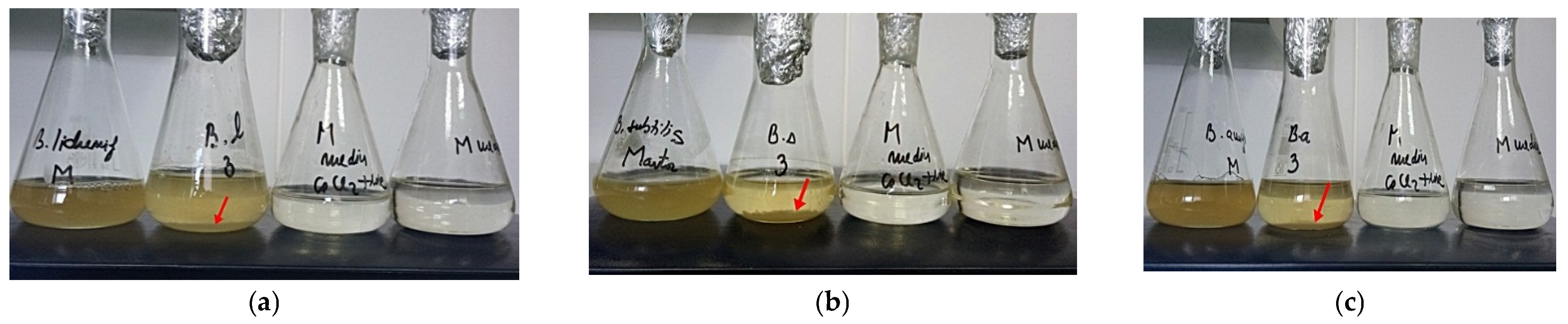
2.1. Microorganisms and Culture Conditions for Testing Carbonate Precipitation with Alizarine Red S
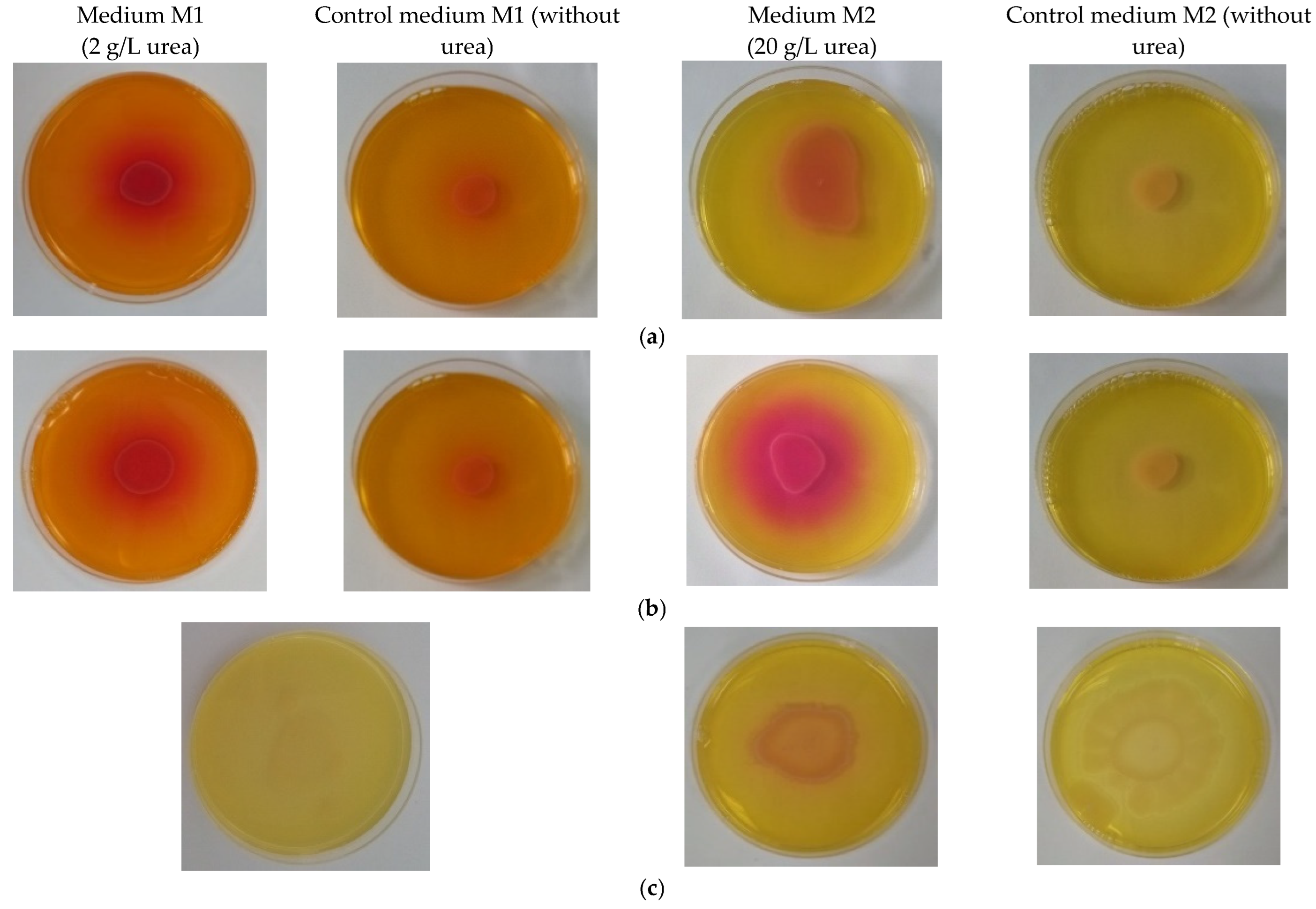
2.2. Qualitative Test for Ureolytic Activity
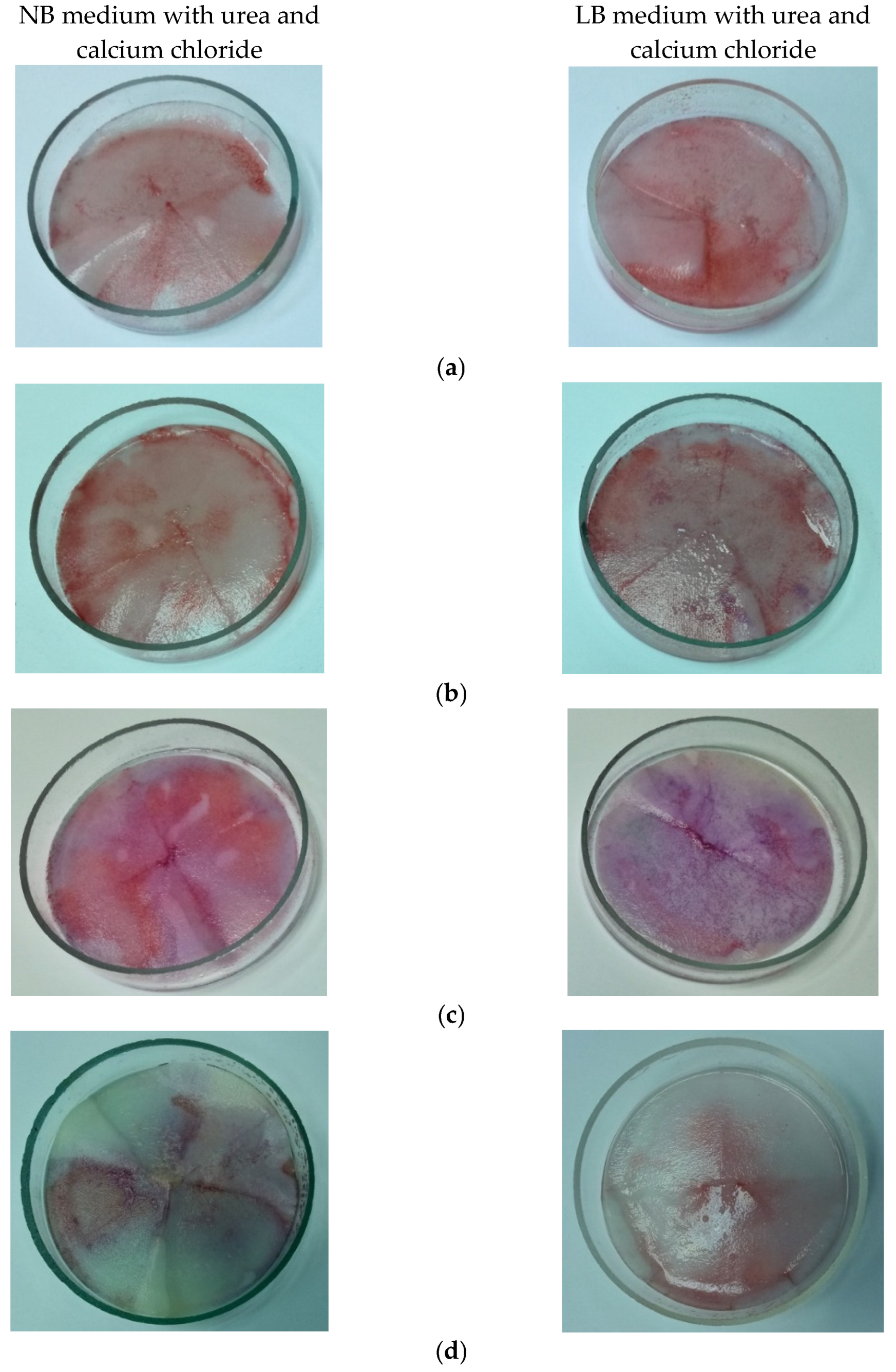
2.3. Cultivation of Bacterial Strains for Calcium Carbonate Precipitate
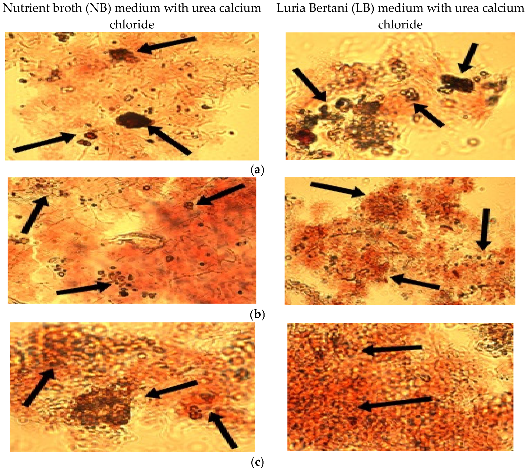
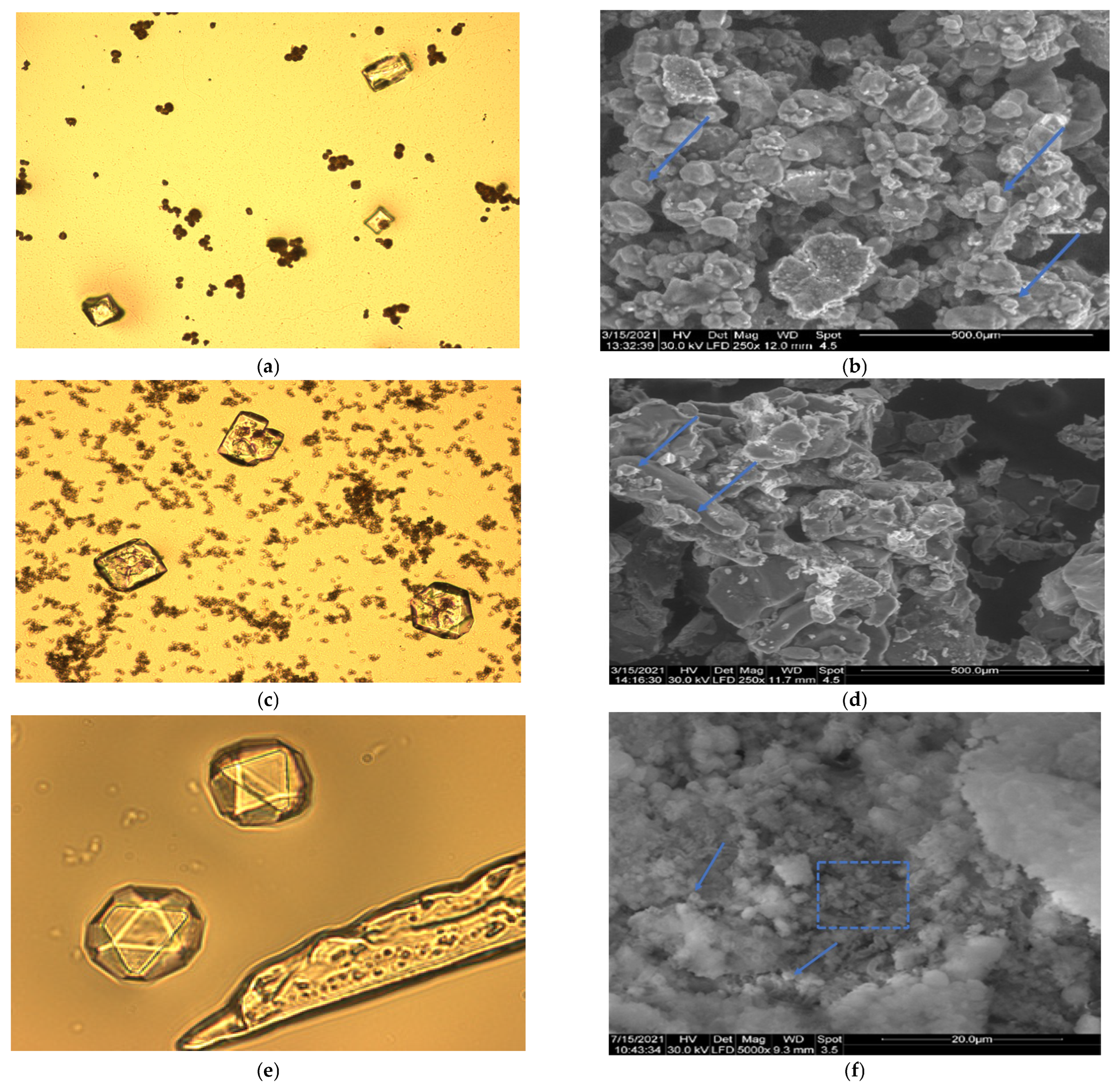
2.4. Microscopical Observations
2.5. Analysis of Precipitates from Bacterial Cultures
2.6. Preparation of Bacterial Mortars Prisms
2.7. Investigations of Mortar Samples
- W1 = mass of oven dried sample in air, g
- W2 = saturated mass of sample after immersion, g
3. Results
3.1. Screening of Microorganisms and Culture Conditions
3.2. Qualitative Test for Ureolytic Activity
3.3. Cultivation of Bacterial Strains for Calcium Carbonate Precipitate
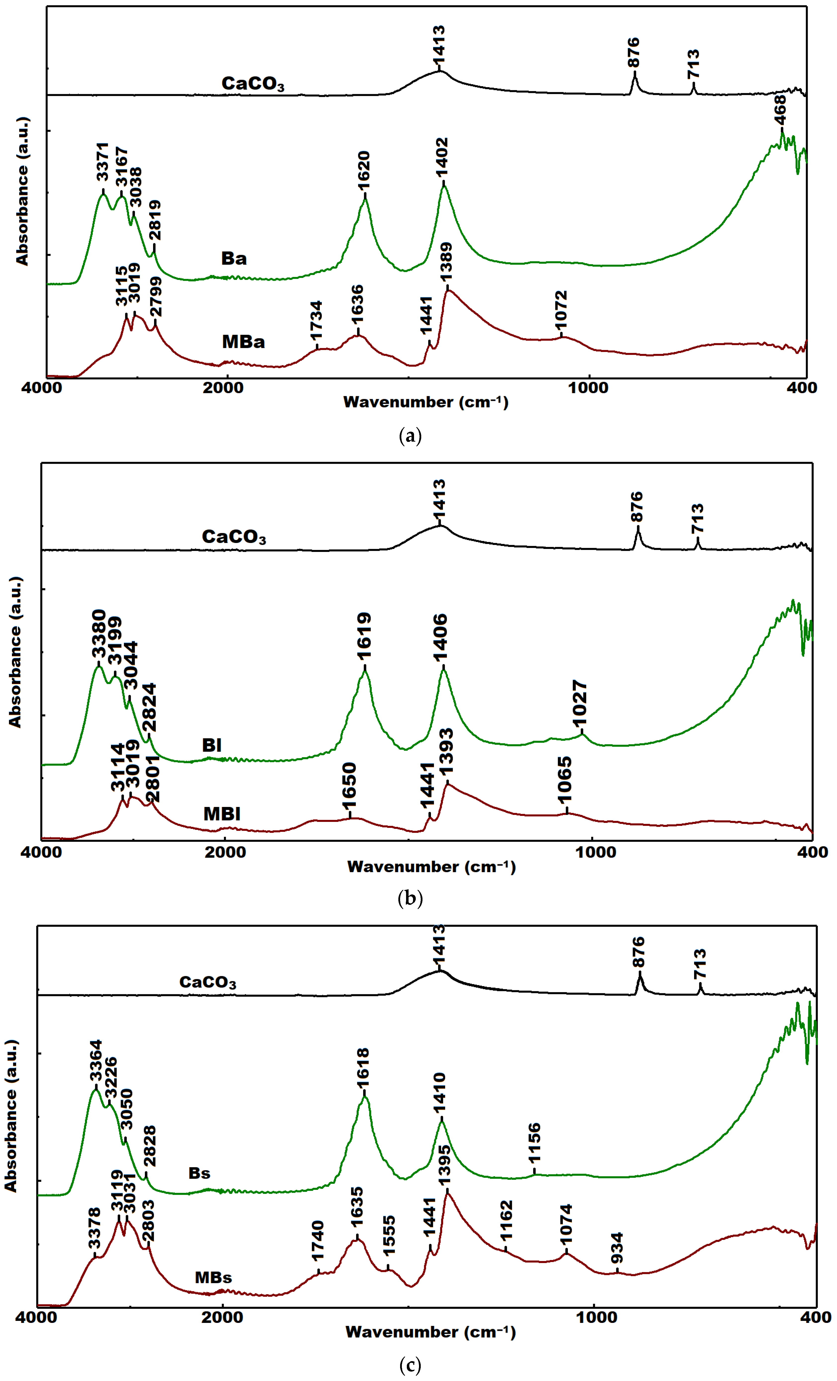
3.4. FTIR Analysis
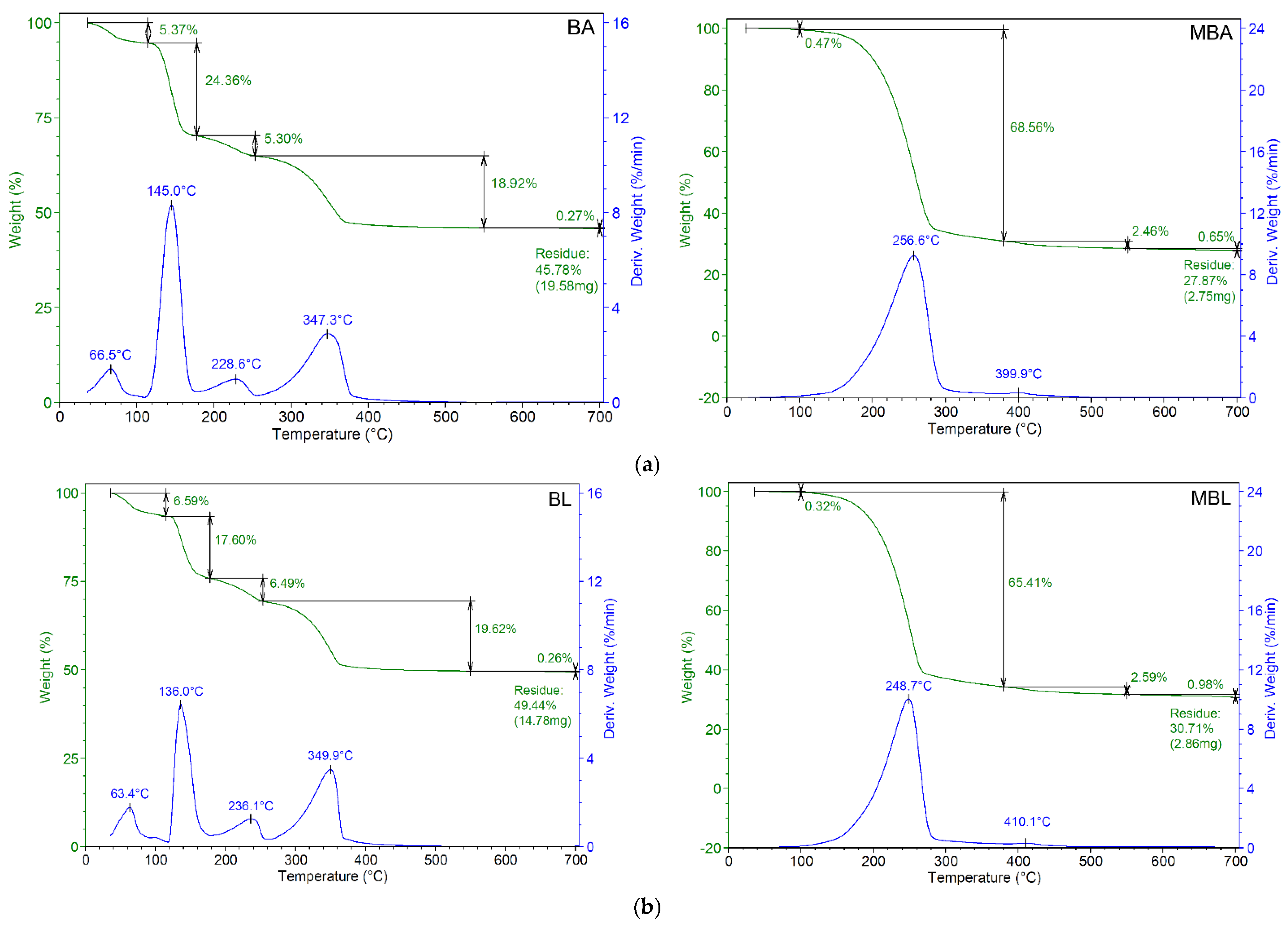
3.5. TGA Analysis
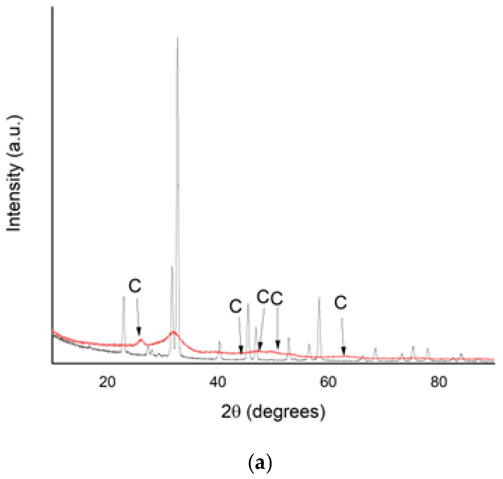
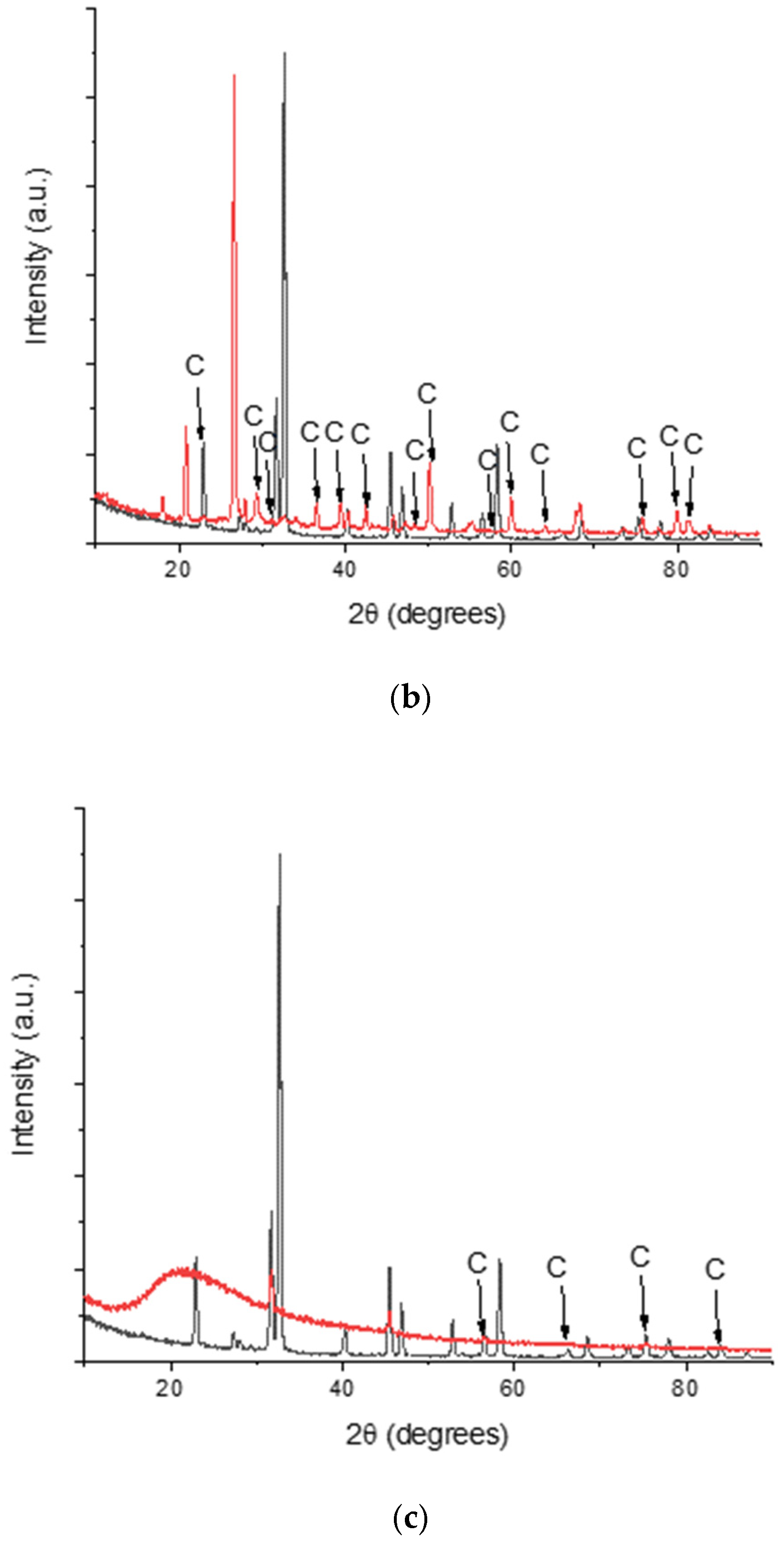
3.6. XRD Analysis
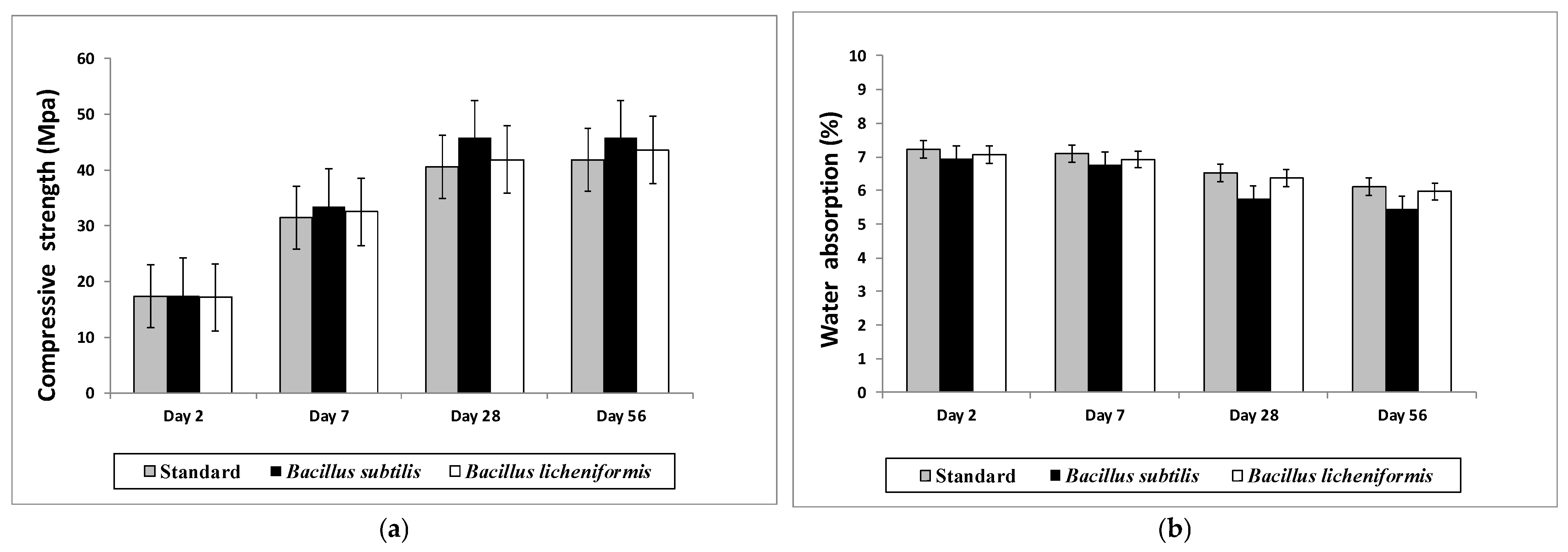
3.7. Compressive Strength and Water Absorption
4. Discussion
5. Conclusions
Author Contributions
Funding
Institutional Review Board Statement
Informed Consent Statement
Data Availability Statement
Acknowledgments
Conflicts of Interest
References
- Han, P.; Geng, W.J.; Li, M.N.; Jia, S.R.; Yin, J.L.; Xu, X.R. Improvement of Biomineralization of Sporosarcina pasteurii as Biocementing Material for Concrete Repair by Atmospheric and Room Temperature Plasma Mutagenesis and Response Surface Methodology. J. Microbiol. Biotechnol. 2021, 31, 1311–1322. [Google Scholar] [CrossRef] [PubMed]
- Li, M.; Zhu, X.; Mukherjee, A.; Huang, M.; Achal, V. Biomineralization in metakaolin modified cement mortar to improve its strength with lowered cement content. J. Hazard. Mater. 2017, 329, 178–184. [Google Scholar] [CrossRef] [PubMed]
- Seifan, M.; Berenjian, A. Application of microbially induced calcium carbonate precipitation in designing bio self-healing concrete. World J. Microbiol. Biotechnol. 2018, 34, 168. [Google Scholar] [CrossRef] [PubMed]
- Shaheen, N.; Jalil, A.; Adnan, F.; Khushnood, R.A. Isolation of alkaliphilic calcifying bacteria and their feasibility for enhanced CaCO3 precipitation in bio-based cementitious composites. Microb. Biotechnol. 2021, 14, 1044–1059. [Google Scholar] [CrossRef]
- Lee, Y.S.; Park, W. Current challenges and future directions for bacterial self-healing concrete. Appl. Microbiol. Biotechnol. 2018, 102, 3059–3070. [Google Scholar] [CrossRef]
- Zhu, T.; Dittrich, M. Carbonate precipitation through microbial activities in natural environment, and their potential in biotechnology: A review. Front. Bioeng. Biotechnol. 2016, 4, 4. [Google Scholar] [CrossRef] [Green Version]
- Wu, Y.; Li, H.; Li, Y. Biomineralization Induced by Cells of Sporosarcina pasteurii: Mechanisms, Applications and Challenges. Microorganisms 2021, 9, 2396. [Google Scholar] [CrossRef]
- Görgen, S.; Benzerara, K.; Skouri-Panet, F.; Gugger, M.; Chauvat, F.; Cassier-Chauvat, C. The diversity of molecular mechanisms of carbonate biomineralization by bacteria. Discover. Mater. 2021, 1, 1–21. [Google Scholar] [CrossRef]
- Dhami, N.K.; Reddy, M.S.; Mukherjee, A. Biomineralization of calcium carbonates and their reengineered applications: A review. Front. Microbiol. 2013, 4, 314. [Google Scholar] [CrossRef] [Green Version]
- De Muynck, W.; De Beliea, N.; Verstraete, W. Microbial carbonate precipitation in construction materials: A review. Ecol. Eng. 2010, 36, 118–136. [Google Scholar] [CrossRef]
- Al-Thawadi, S. Ureolytic Bacteria and Calcium Carbonate Formation as a Mechanism of Strength Enhancement of Sand. J. Adv. Sci. Eng. Res. 2011, 1, 98–114. [Google Scholar]
- Achal, V.; Mengmeng, L.; Zhang, Q. BioCement, recent research in construction engineering: Status of China against rest of World. Adv. Cem. Res. 2013, 26, 281–291. [Google Scholar] [CrossRef]
- Achal, V.; Mukherjee, A.; Kumari, D.; Zhang, Q. Biomineralization for sustainable construction—A review of processes and applications. Earth Sci. Rev 2015, 148, 1–17. [Google Scholar] [CrossRef]
- Ma, L.; Pang, A.P.; Luo, Y.; Lu, X.; Lin, F. Beneficial factors for biomineralization by ureolytic bacterium Sporosarcina pasteurii. Microb. Cell Fact. 2020, 19, 12. [Google Scholar] [CrossRef]
- Achal, V.; Pan, X.; Zhang, D. Bioremdiation of strontium (Sr) contaminated aquifer quartz sand based on carbonate precipitation induced by Sr resistant Halomonas sp. Chemosphere 2012, 89, 764–768. [Google Scholar] [CrossRef]
- Li, M.; Cheng, X.; Guo, H. Heavy metal removal by biomineralization of urease producing bacteria isolated from soil. Internat. Biodeter. Biodegrad. 2013, 76, 81–85. [Google Scholar] [CrossRef]
- Chen, L.; Shen, Y.; Xie, A.; Huang, B.; Jia, R.; Guo, R.; Tang, W. Bacteria-Mediated Synthesis of Metal Carbonate Minerals with Unusual Morphologies and Structures. Cryst. Growth Des. 2009, 9, 743–754. [Google Scholar] [CrossRef]
- Gwenzi, W. Carbon Sequestration via biomineralization: Processes, applications and future directions. In Sustainable Agriculture Reviews 37; Inamuddin, Asiri, A., Lichtfouse, E., Eds.; Springer: Cham, Switzerland, 2019; Volume 37, pp. 93–106. [Google Scholar] [CrossRef]
- Cheng, L.; Cord-Ruwisch, R. Upscaling effects of soil improvement by microbially induced calcite precipitation by surface percolation. Geomicrobiol. J. 2014, 31, 396–406. [Google Scholar] [CrossRef] [Green Version]
- Bagriacik, B.; Sani, Z.K.; Uslu, F.M.; Yigittekin, E.S.; Dince, S. An experimental approach to microbial carbonate precipitation in improving the engineering properties of sandy soils. Ann. Microbiol. 2021, 71, 37. [Google Scholar] [CrossRef]
- De Jong, J.T.; Soga, K.; Kavazanjian, E.; Burns, S.; van Paassen, L.A.; Al Qabany, A.; Aydilek, A.; Bang, S.S.; Burbank, M.; Caslake, L.F.; et al. Biogeochemical processes and geotechnical applications: Progress, opportunities and challenges. Geotech 2013, 63, 287–301. [Google Scholar] [CrossRef]
- Yoshida, N.; Higashimura, E.; Saeki, Y. Catalytic biomineralization of fluorescent calcite by the thermophilic bacterium Geobacillus thermoglucosidasius. Appl. Environ. Microbiol. 2010, 76, 7322–7327. [Google Scholar] [CrossRef] [PubMed] [Green Version]
- Portugal, C.R.M.e.; Fonyo, C.; Machado, C.C.; Meganck, R.; Jarvis, T. Microbiologically induced calcite precipitation biocementation, green alternative for roads—Is this the breakthrough? A critical review. J. Clean. Prod. 2020, 262, 121–372. [Google Scholar] [CrossRef]
- Bhutange, S.P.; Latkar, M.V. Microbially induced calcium carbonate precipitation in construction materials. J. Mater. Civ. Eng. 2020, 32, 03120001. [Google Scholar] [CrossRef]
- Qin, W.; Wang, C.Y.; Ma, Y.X.; Shen, M.J.; Li, J.; Jiao, K.; Tay, F.R.; Niu, L.N. Microbe-mediated extracellular and intracellular mineralization: Environmental, industrial, and biotechnological applications. Adv. Mater. 2020, 32, 1907833. [Google Scholar] [CrossRef] [PubMed]
- Osinubi, K.J.; Eberemu, A.O.; Ijimdiya, T.S.; Yakubu, S.E.; Gadzama, E.W.; Sani, J.E.; Yohanna, P. Review of the use of microorganisms in geotechnical engineering applications. SN Appl. Sci. 2020, 2, 207. [Google Scholar] [CrossRef] [Green Version]
- Jimenez-Martinez, J.; Nguyen, J.; Or, D. Controlling pore-scale processes to tame subsurface biomineralization. Rev. Environ. Sci. Biotechnol. 2022, 21, 27–52. [Google Scholar] [CrossRef] [PubMed]
- Omoregie, A.I.; Palombo, E.A.; Nissom, P.M. Experimental optimisation of various cultural conditions on urease activity for isolated Sporosarcina pasteurii strains and evaluation of their biocement potentials. Ecol. Eng. 2017, 109, 65–75. [Google Scholar] [CrossRef] [Green Version]
- Murugan, R.; Suraishkumar, G.K.; Mukherjee, A.; Dhami, N.K. Insights into the influence of cell concentration in design and development of microbially induced calcium carbonate precipitation (MICP) process. PLoS ONE 2021, 16, e0254536. [Google Scholar] [CrossRef] [PubMed]
- Wen, K.; Li, Y.; Amini, F.; Li, L. Impact of bacteria and urease concentration on precipitation kinetics and crystal morphology of calcium carbonate. Acta Geotech. 2020, 15, 17–27. [Google Scholar] [CrossRef]
- Krajewska, B. Urease-aided calcium carbonate mineralization for engineering applications: A review. J. Adv. Res. 2018, 13, 59–67. [Google Scholar] [CrossRef]
- Khanjani, M.; Westenberg, D.J.; Kumar, A.; Ma, H. Tuning Polymorphs and Morphology of Microbially Induced Calcium Carbonate: Controlling Factors and Underlying Mechanisms. ACS Omega 2021, 6, 11988–12003. [Google Scholar] [CrossRef] [PubMed]
- Chuo, S.C.; Mohamed, S.F.; Setapar, S.H.M.; Ahmad, A.; Jawaid, M.; Wani, W.A.; Yaqoob, A.A.; Ibrahim, M.N.M. Insights into the Current Trends in the Utilization of Bacteria for Microbially Induced Calcium Carbonate Precipitation. Materials 2020, 13, 4993. [Google Scholar] [CrossRef] [PubMed]
- Zhang, J.; Shi, X.; Chen, X.; Huoa, X.; Yu, Z. Microbial-Induced Carbonate Precipitation: A Review on Influencing Factors and Applications. Adv. Civ. Eng. 2021, 2021, 1–16. [Google Scholar] [CrossRef]
- Xu, G.; Li, D.; Jiao, B.; Li, D.; Yin, Y.; Lun, L.; Zhao, Z.; Li, S. Biomineralization of a calcifying ureolytic bacterium Microbacterium sp. GM-1. Electron. J. Biotechnol. 2017, 25, 21–27. [Google Scholar] [CrossRef] [Green Version]
- Anitha, V.; Abinaya, S.; Prakash, A.; Seshagiri, R.; Vanavil, B. Bacillus cereus KLUVAA Mediated Biocement Production Using Hard Water and Urea. Chem. Biochem. Eng. Q. 2018, 32, 257–266. [Google Scholar] [CrossRef]
- Reeksting, B.; Hoffmann, T.; Tan, L.; Paine, K.; Gebhard, S. In-Depth Profiling of Calcite Precipitation by Environmental Bacteria Reveals Fundamental Mechanistic Differences with Relevance to Application. Appl. Environ. Microbiol. 2020, 86, e02739-19. [Google Scholar] [CrossRef] [Green Version]
- Tepe, M.; Arslan, S.; Koralay, T.; Mercan Doğan, N. Precipitation and characterization of CaCO3 of Bacillus amyloliquefaciens U17 strain producing urease and carbonic anhydrase. Turk. J. Biol. 2019, 43, 198–208. [Google Scholar] [CrossRef]
- Kim, G.; Kim, J.; Youn, H. Effect of Temperature, pH and Reaction Duration on Microbially Induced Calcite Precipitation. Appl. Sci. 2018, 8, 1277. [Google Scholar] [CrossRef] [Green Version]
- Kim, G.; Youn, H. Microbially induced calcite precipitation employing environmental isolates. Materials 2016, 9, 468. [Google Scholar] [CrossRef] [Green Version]
- Helmi, F.M.; Elmitwwalli, H.R.; Elnagdy, S.M.; El-Hagrassy, A.F. Calcium carbonate precipitation induced by ureolytic bacteria Bacillus licheniformis. Ecol. Eng. 2016, 90, 367–371. [Google Scholar] [CrossRef]
- Lian, B.; Hu, Q.; Chen, J.; Junfeng, J.; Teng, H.H. Carbonate biomineralization induced by soil bacterium Bacillus megaterium. Geochim. Cosmochim. Acta 2006, 70, 5522–5535. [Google Scholar] [CrossRef]
- Radu, N.; Constantin, M.; Raut, I.; Panea, G.V.; Gurban, A.M.; Doni, M.; Jecu, L. Culture Medium Composition for Sporulation. Patent A/00663, 5 November 2021. [Google Scholar]
- ASTM C 642–97; Standard Test Method for Density, Absorption, and Voids in Hardened Concrete. American Society for Testing and Materials: West Conshohocken, PA, USA. Available online: https://www.astm.org/c0642-97.html (accessed on 19 October 2020).
- SR EN 196-1:2016; Methods of testing cement—Part 1: Determination of Strength. ICS Code (Cement. Gypsum. Lyme, Mortar) 91.100.10. European Committee for Standardization: Brussels, Belgium, 1 April 2016; 38p. Available online: https://standards.iteh.ai/catalog/standards/cen/37b8816e-4085-4dcc-a642-a383d9bddd6c/en-196-1-2016 (accessed on 19 October 2020).
- Castro-Alonso, M.J.; Montañez-Hernandez, L.E.; Sanchez-Muñoz, M.A.; Macias Franco, M.R.; Narayanasamy, R.; Balagurusamy, N. Microbially Induced Calcium Carbonate Precipitation (MICP) and Its Potential in Bioconcrete: Microbiological and Molecular Concepts. Front. Mater. 2019, 6, 126. [Google Scholar] [CrossRef]
- Cacchio, P.; Ercole, C.; Capuccio, G.; Lepidi, A. Calcium Carbonate Precipitation by Bacterial Strains Isolated from a Limestone Cave and from a Loamy Soil. Geomicrobiol. J. 2003, 2, 85–98. [Google Scholar] [CrossRef]
- Zdenek, F.; Herrmann, S.; Kubat, J. FT-IR spectroscopic characteristics of differently cultivated Bacillus subtilis. Microbiol. Res. 2004, 159, 257–260. [Google Scholar] [CrossRef]
- Šovljanski, O.; Pezo, L.; Stanoje, J.; Bajac, B.; Kovac, S.; Tóth, E.; Ristic, I.; Tomic, A.; Ranitovic, A.; Cvetkovic, D.; et al. Comprehensive Profiling of Microbiologically Induced CaCO3 Precipitation by Ureolytic Bacillus Isolates from Alkaline Soils. Microorganisms 2021, 9, 1691. [Google Scholar] [CrossRef]
- Iqbal, D.M.; Wong, L.S.; Kong, S.Y. Bio-Cementation in Construction Materials: A Review. Materials 2021, 14, 2175. [Google Scholar] [CrossRef]
- Feng, Z.; Zhao, Y.; Zeng, W.; Lu, Z.; Shah, S.P. Using microbial carbonate precipitation to improve the properties of recycled fine aggregate and mortar. Constr. Build. Mater. 2020, 230, 116949. [Google Scholar] [CrossRef]
- Salmasi, F.; Mostofinejad, D. Investigating the effects of bacterial activity on compressive strength and durability of natural lightweight aggregate concrete reinforced with steel fibers. Constr. Build. Mater. 2020, 251, 119032. [Google Scholar] [CrossRef]
- Andalib, R.; Abd Majid, M.Z.; Hussin, M.W.; Ponraj, M.; Keyvanfar, M.; Mirza, J.; Lee, H.S. Optimum concentration of Bacillus megaterium for strengthening structural concrete. Constr. Build. Mater. 2016, 118, 180–193. [Google Scholar] [CrossRef]
- Nain, N.; Surabhi, R.; Yathish, N.V.; Krishnamurthy, V.; Deepa, T.; Tharannum, S. Enhancement in strength parameters of concrete by application of Bacillus bacteria. Constr. Build. Mater. 2019, 202, 904–908. [Google Scholar] [CrossRef]
- Liu, R.; Huang, S.; Zhang, X.; Song, Y.; He, G.; Wang, Z.; Lian, B. Bio-mineralisation, characterization, and stability of calcium carbonate containing organic matter. RSC Adv. 2021, 11, 14415. [Google Scholar] [CrossRef] [PubMed]
- Schwantes-Cezario, N.; Medeiros, L.P.; De Oliveira, A.G., Jr.; Nakazato, G.; Kobayashi, R.K.T.; Toralles, B.N.M. Bioprecipitation of calcium carbonate induced by Bacillus subtilis isolated in Brazil. Intern. Biodeter. Biodegrad. 2017, 123, 200–205. [Google Scholar] [CrossRef]
- Henry, D.; Watson, J.S.; John, C.M. Assessing and calibrating the ATR-FTIR approach as a carbonate rock characterization tool. Sediment Geol. 2017, 347, 36–52. [Google Scholar] [CrossRef] [Green Version]
- Saracho, A.C.; Haigh, S.K.; Hata, T.; Soga, K.; Farsang, S.; Redfern, S.T.; Marek, E. Characterisation of CaCO3 phases during strain-specifc ureolytic precipitation. Sci. Rep. 2020, 10, 10168. [Google Scholar] [CrossRef] [PubMed]
- Tourney, J.; Ngwenya, B.T. Bacterial extracellular polymeric substances (EPS) mediate CaCO 3 morphology and polymorph. Chem. Geol. 2009, 262, 138–146. [Google Scholar] [CrossRef]
- Li, W.; Liu, L.; Chen, W.; Yu, L.; Li, W.; Yu, H. Calcium carbonate precipitationand crystal morphology induced by microbial carbonic anhydrase and otherbiological factors. Process Biochem. 2010, 45, 1017–1021. [Google Scholar] [CrossRef]
- Dhami, N.K.; Mukherjee, A.; Reddy, S.M. Micrographical, minerological and nano-mechanical characterisation of microbial carbonates from urease and carbonic anhydrase producing bacteria. Ecol. Eng. 2016, 94, 443–454. [Google Scholar] [CrossRef]
- Wang, Y.; Chen, Y.; Guo, B.; Zhang, S.; Tong, Y.; Niu, D. Study on the Strength and Hydration Behaviour of Sulphate-Resistant Cement in High Geothermal Environment. Materials 2022, 15, 2790. [Google Scholar] [CrossRef]
- Abo-El-Enein, S.; Ali, A.; Talkhan, F.N.; Abdel-Gawwad, H. Utilization of microbial induced calcite precipitation for sand consolidation and mortar crack remediation. HBRC J. 2012, 8, 185–192. [Google Scholar] [CrossRef] [Green Version]
- Kim, H.; Son, H.M.; Park, S.; Lee, H.K. Effects of biological admixtures on hydration and mechanical properties of Portland cement paste. Constr. Build. Mater. 2020, 235, 117461. [Google Scholar] [CrossRef]
- Pei, R.; Liu, J.; Wang, S.; Yang, M. Use of bacterial cell walls to improve the mechanical performance of concrete. Cem. Concr. Compos. 2013, 39, 122–130. [Google Scholar] [CrossRef]
- Huang, Y.H.; Chen, H.J.; Maity, J.P.; Chen, C.C.; Sun, C.A.; Chen, C.Y. Efficient option of industrial wastewater resources in cement mortar application with rivers and by microbial induced calcium carbonate precipitation. Sci. Rep. 2020, 10, 6742. [Google Scholar] [CrossRef] [PubMed] [Green Version]
- Sujatha, S.; Sarayu, K.; Annaselvi, M.; Murthy, R.A.; Kumar, R.V.; Iyer, N.R. Soil Bacteria for the Strength Enhancement of Cement Mortar. J. Civ. Eng. Res. 2014, 4, 51–54. [Google Scholar] [CrossRef]
- Achal, V.; Mukerjee, A.; Basu, P.C.; Reddy, M.S. Strain improvement of Sporosarcina pasteurii for enhanced urease and calcite production. J. Ind. Microbiol. Biotechnol. 2009, 36, 981–988. [Google Scholar] [CrossRef]


| Culture Medium | Composition (g/L) |
|---|---|
| Nutrient Broth (NB) medium | 1, meat extract; 2, yeast extract; 5, peptone; 5, NaCl; pH = 7.4. |
| Luria Bertani (LB) medium | 10, tryptone; 5, yeast extract; 10, NaCl; pH = 7.0 |
| Tryptic Soy Broth (TSB) medium | 30, Scharlau dehydrated powder; 20, peptone; 5, NaCl; 2.5, K2HPO4; 2.5, dextrose; pH = 7.3 ± 0.2 |
| M1 medium for urease activity | 1, peptone; 5, NaCl; 2, KH2PO4; 0.1, glucose; 2, red phenol; 15, agar; 2, urea |
| M2 medium for urease activity [35] | 1, peptone; 5, NaCl; 2, KH2PO4; 1, glucose; 0.12, red phenol; 15, agar; 20, urea [35] |
| M3 medium for urease activity [36] | 4, glucose; 1, NH4Cl; 3, NaHPO4; 0.5, NaCl; 1, KH2PO4; 44.16 µg NiCl2; 20, urea |
| M4 medium for urease activity | 4, glucose; 1, NH4Cl; 3, NaHPO4; 0.5, NaCl; 1, KH2PO4. |
| Medium for carbonate precipitation | 3, NB (Nutrient Broth—Scharlau dehydrated power); 3, vinasse; 10, NH4Cl; 2.12, NaHCO3; pH = 7.0 ± 0.2 |
| Sample | RT–115 °C | 115–178 °C | 178–253.5 °C | 253.5–550 °C | 550–700 °C | Residue | ||||
|---|---|---|---|---|---|---|---|---|---|---|
| Wt. Loss (%) | Tmax (°C) | Wt. Loss (%) | Tmax (°C) | Wt. Loss (%) | Tmax (°C) | Wt. Loss (%) | Tmax (°C) | Wt. Loss (%) | 700 °C (%) | |
| B amyloliquefaciens (BA) | 5.37 | 66.5 | 24.36 | 145.0 | 5.30 | 228.6 | 18.92 | 347.3 | 0.27 | 45.78 |
| B. licheniformis (BL) | 6.59 | 63.4 | 17.60 | 136.0 | 6.49 | 236.1 | 19.62 | 349.9 | 0.26 | 49.44 |
| B. subtilis (BS) | 4.73 | 55.3 | 23.20 | 138.1 | 3.01 | 214.9 | 20.16 | 343.9 | 0.34 | 48.56 |
| Sample | RT–100 °C | 100–380 °C | 380–550 °C | 550–700 °C | Residue | ||
|---|---|---|---|---|---|---|---|
| Wt. Loss (%) | Wt. Loss (%) | Tmax (°C) | Wt. Loss (%) | Tmax (°C) | Wt. Loss (%) | 700 °C (%) | |
| B amyloliquefaciens (MBA) | 0.47 | 68.56 | 256.6 | 2.46 | 399.9 | 0.65 | 27.87 |
| B. licheniformis (MBL) | 0.32 | 65.41 | 248.7 | 2.59 | 410.1 | 0.98 | 30.71 |
| B. subtilis (MBS) | 0.57 | 68.59 | 281.2 | 2.40 | 398.1 | 0.77 | 27.67 |
Publisher’s Note: MDPI stays neutral with regard to jurisdictional claims in published maps and institutional affiliations. |
© 2022 by the authors. Licensee MDPI, Basel, Switzerland. This article is an open access article distributed under the terms and conditions of the Creative Commons Attribution (CC BY) license (https://creativecommons.org/licenses/by/4.0/).
Share and Cite
Răut, I.; Constantin, M.; Petre, I.; Raduly, M.; Radu, N.; Gurban, A.-M.; Doni, M.; Alexandrescu, E.; Nicolae, C.-A.; Jecu, L. Highlighting Bacteria with Calcifying Abilities Suitable to Improve Mortar Properties. Materials 2022, 15, 7259. https://0-doi-org.brum.beds.ac.uk/10.3390/ma15207259
Răut I, Constantin M, Petre I, Raduly M, Radu N, Gurban A-M, Doni M, Alexandrescu E, Nicolae C-A, Jecu L. Highlighting Bacteria with Calcifying Abilities Suitable to Improve Mortar Properties. Materials. 2022; 15(20):7259. https://0-doi-org.brum.beds.ac.uk/10.3390/ma15207259
Chicago/Turabian StyleRăut, Iuliana, Mariana Constantin, Ionela Petre, Monica Raduly, Nicoleta Radu, Ana-Maria Gurban, Mihaela Doni, Elvira Alexandrescu, Cristi-Andi Nicolae, and Luiza Jecu. 2022. "Highlighting Bacteria with Calcifying Abilities Suitable to Improve Mortar Properties" Materials 15, no. 20: 7259. https://0-doi-org.brum.beds.ac.uk/10.3390/ma15207259