
A Novel Porcine Graft for Regeneration of Bone Defects

 ,
,
Abstract
:1. Introduction
2. Results
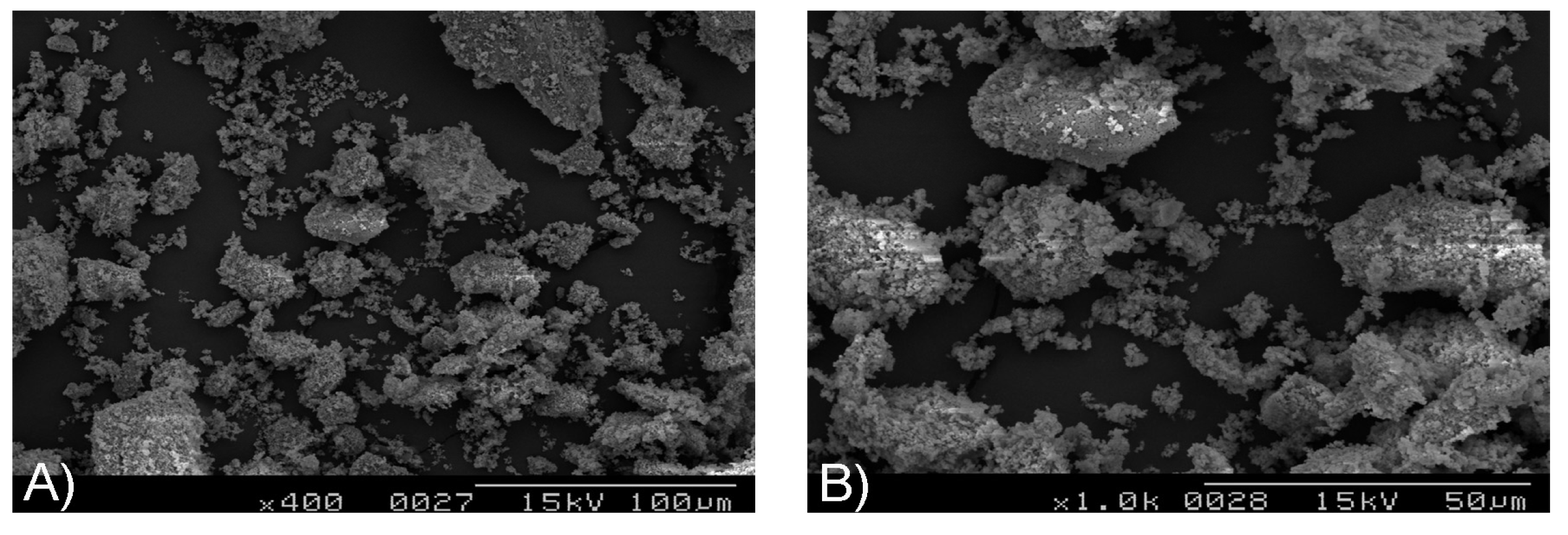
2.1. Scanning Electron Microscopy

2.2. Energy Dispersive Spectrometry

| Element | Weight (%) | Atomic (%) |
|---|---|---|
| C | 8.01 | 13.90 |
| O | 44.32 | 57.77 |
| Na | 0.86 | 0.78 |
| Mg | 1.78 | 1.53 |
| P | 16.88 | 11.37 |
| Ca | 28.16 | 14.65 |
| Total | 100.00 | 100.00 |
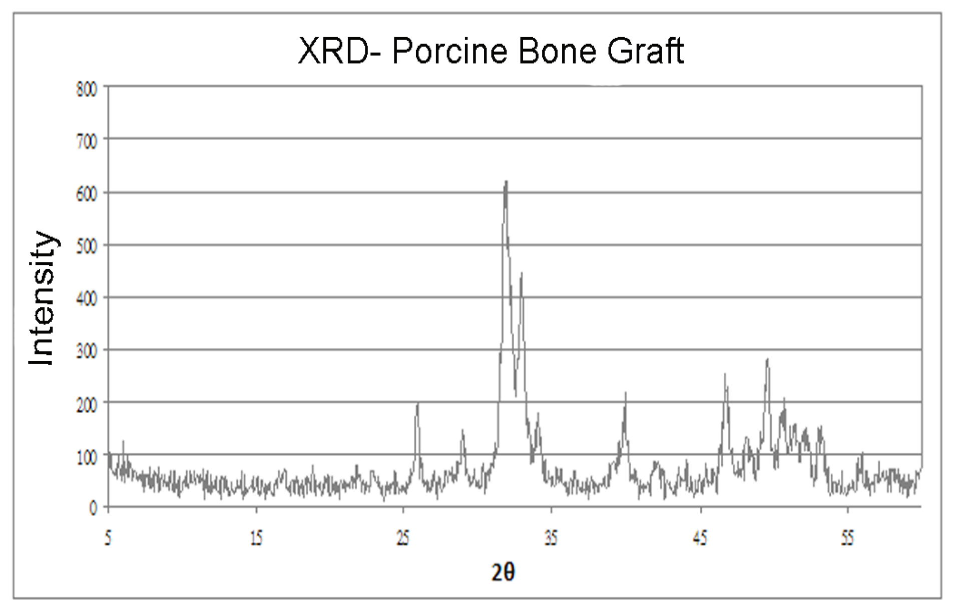
2.3. X-ray Diffraction

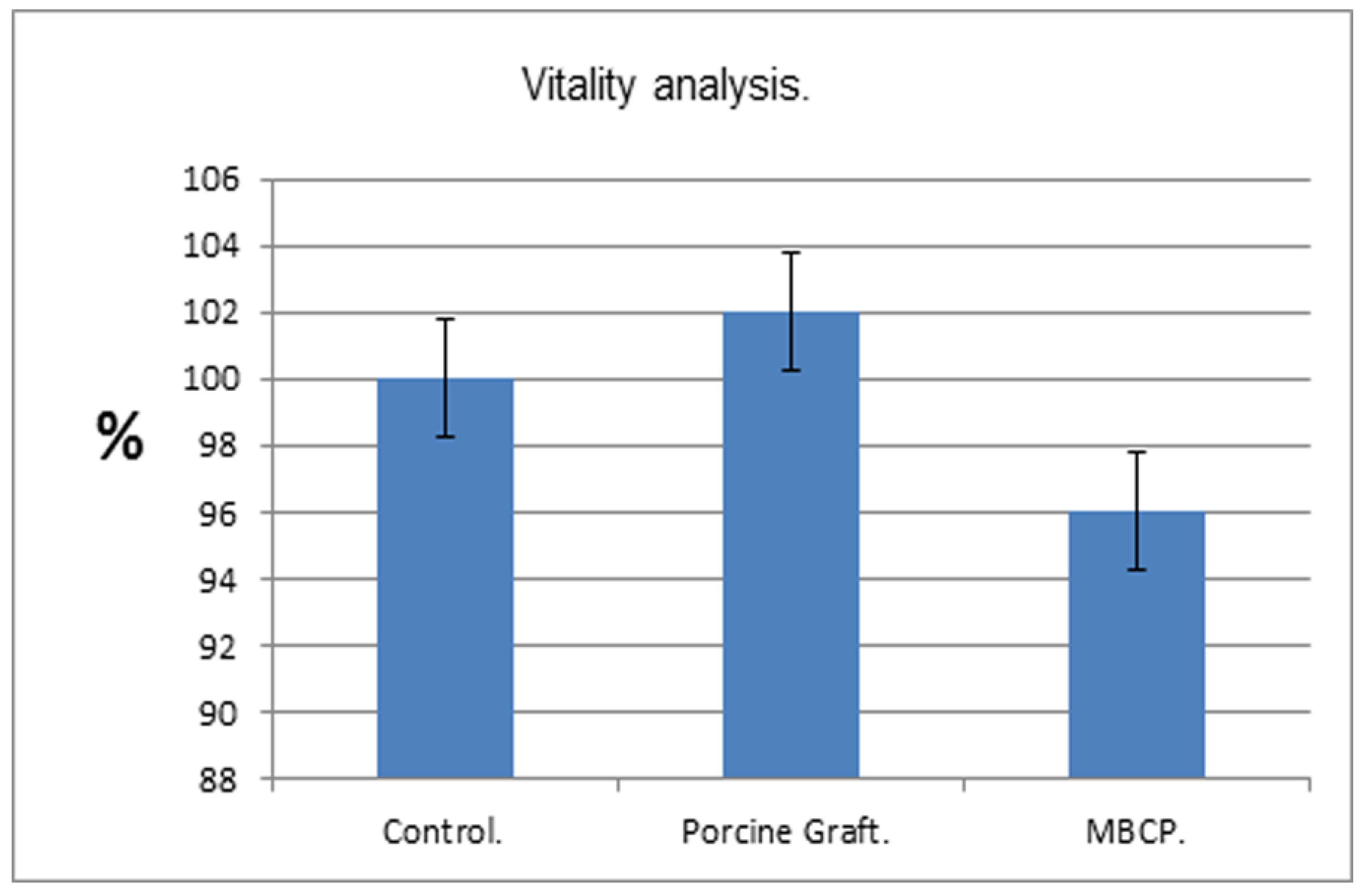
2.4. MTT Proliferation Assay

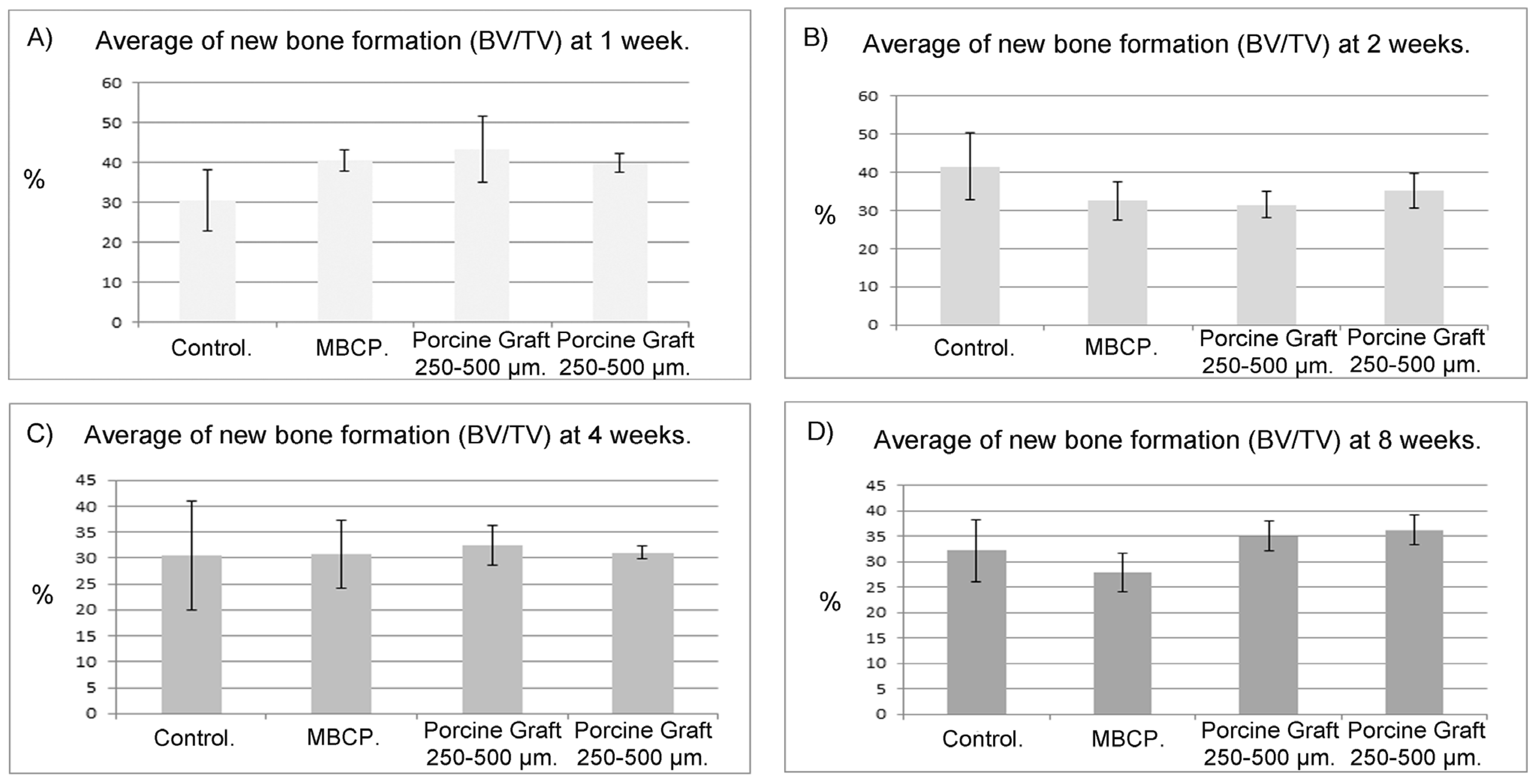
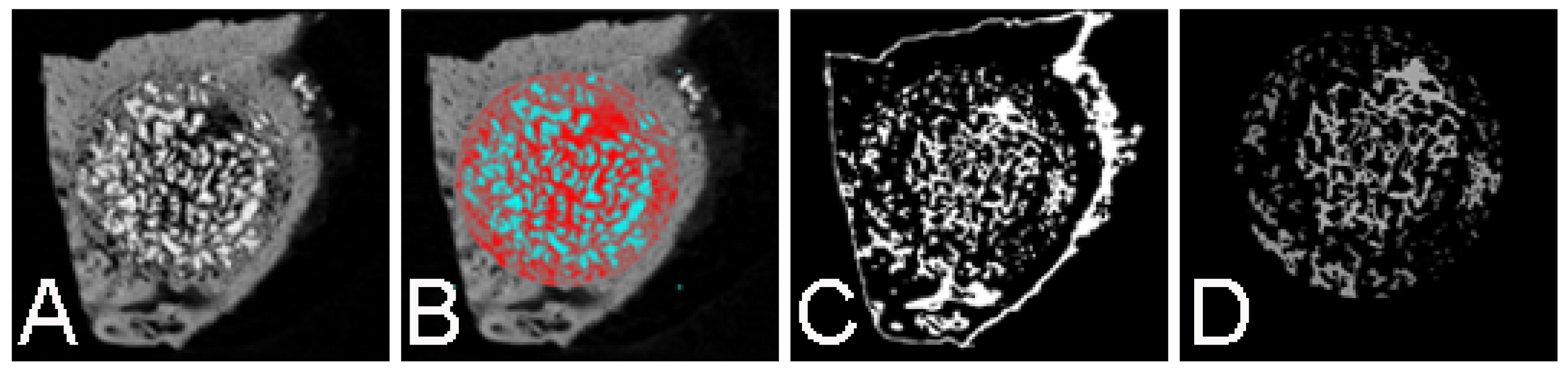
2.5. Micro-CT Scanning
| Micro CT Average-SD Values for Newly Formed Bone | ||||
|---|---|---|---|---|
| Time | Control | MBCP | Porcine Graft 250–500 μm | Porcine Graft 500–1000 μm |
| Week 1 | 30.57 ± 7.57 | 40.42 ± 2.64 | 43.29 ± 8.37 | 40 ± 2.33 |
| Week 2 | 41.57 ± 8.71 | 32.59 ± 5.05 | 31.52 ± 3.51 | 35.24 ± 2.33 |
| Week 4 | 30.38 ± 10.54 | 30.7 ± 6.5 | 32.47 ± 3.83 | 31.05 ± 1.3 |
| Week 8 | 31.55 ± 7.25 | 27.81 ± 3.86 | 35.02 ± 2.92 | 36.19 ± 2.85 |

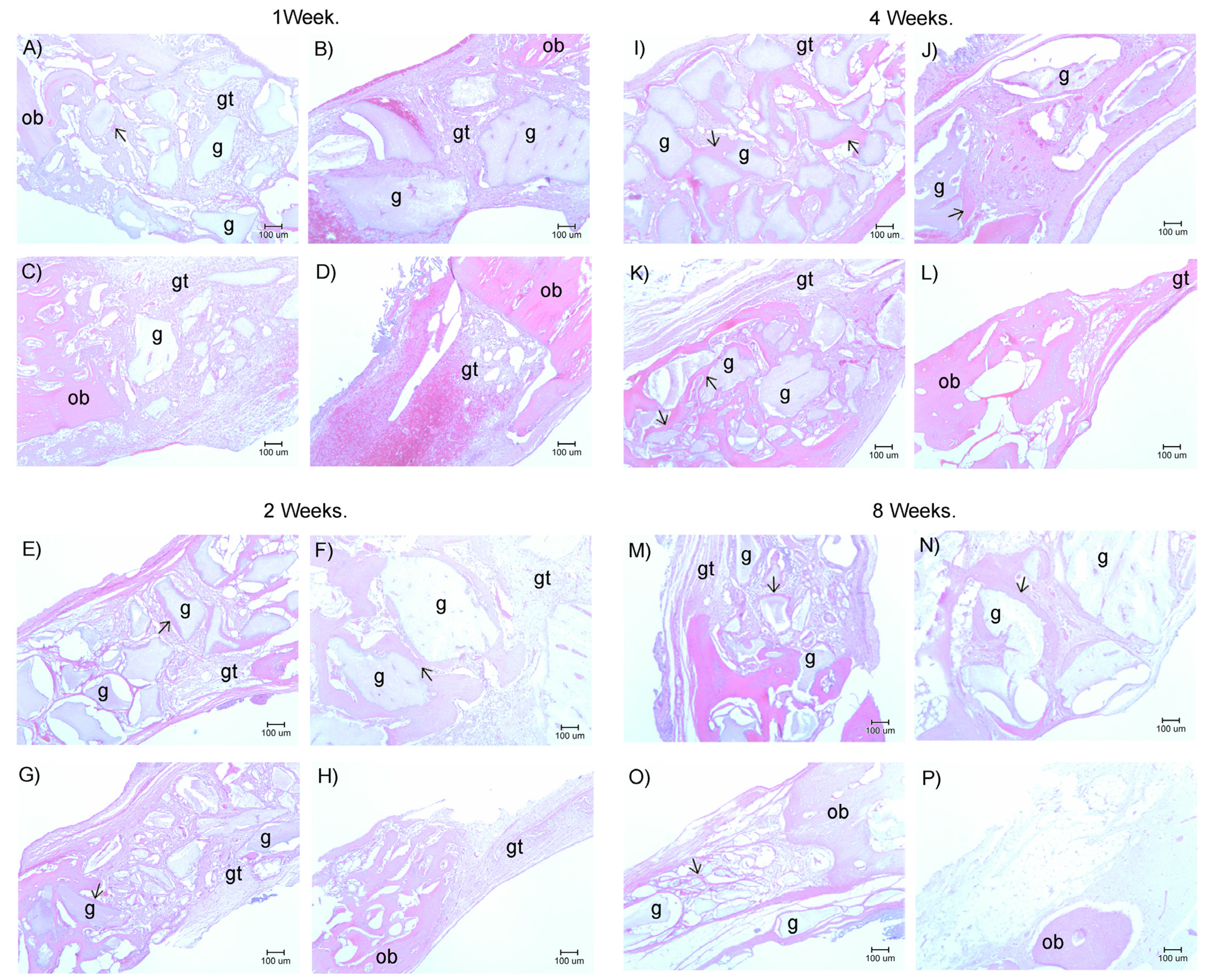
2.6. Histological Examination

3. Discussion
4. Materials and methods
4.1. Biomaterials
- 5 °C/min heated to 100 °C and then maintained for 30 min;
- 5 °C/min heated to 300 °C and then maintained for 60 min;
- 10 °C/minute heated to 800 °C and then maintained for 120 min.
4.2. Porcine Graft Analysis
4.2.1. Scanning Electron Microscopy and Energy Dispersive Spectrometry
4.2.2. X-ray Diffraction
4.2.3. Cell Proliferation (MTT Assay)
4.2.4. Statistical Analysis
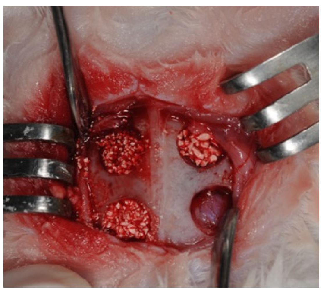
5. Surgical Procedure


- Decalcification: The bone specimens were trimmed to a thickness of less than 0.5 cm (to accelerate decalcification) and were decalcified in fresh fluid. Decalcification times, specimen thicknesses, temperature, decalcification solution freshness, and block samples’ decalcification conditions were recorded.
- Washing with water: After decalcification, the specimens were washed in running water for several hours to neutralize the strongly acidic decalcifying solution.
- Dehydration and embedding: The bone tissues were dehydrated in alcohol and were then embedded in liquid paraffin.
- Sectioning: Slice thicknesses were set at 5–7 μm.
- Staining: Hematoxylin and Eosin (H&E) staining was performed on all paraffin embedded tissues.
- Visualization: The histological slides were viewed using an optical microscope (Olympus BH-2, Tokyo, Japan), and the images were captured at 40× magnification using a camera SPOT idea tm Camera that was connected to the microscope and were analyzed using the corresponding software (SPOT imaging software, Sterling Heights, MI, USA).
6. Conclusions
Acknowledgments
Author Contributions
Conflicts of Interest
References
- Trombelli, L. Which reconstructive procedures are effective for treating the periodontal intraosseous defect? Periodontology 2000 2005, 37, 88–105. [Google Scholar] [CrossRef] [PubMed]
- Susin, C.; Wikesjö, U.M.E. Regenerative periodontal therapy: 30 years of lessons learned and unlearned. Periodontology 2000 2013, 62, 232–242. [Google Scholar] [CrossRef] [PubMed]
- Ramseier, C.A.; Rasperini, G.; Batia, S.; Giannobile, W.V. Advanced reconstructive technologies for periodontal tissue repair. Periodontology 2000 2012, 59, 185–202. [Google Scholar] [CrossRef] [PubMed]
- Weiss, P.; Layrolle, P.; Clergeau, L.P.; Enckel, B.; Pilet, P.; Amouriq, Y.; Daculsi, G.; Giumelli, B. The safety and efficacy of an injectable bone substitute in dental sockets demonstrated in a human clinical trial. Biomaterials 2007, 28, 3295–3305. [Google Scholar] [CrossRef] [PubMed] [Green Version]
- Scarano, A.; Piattelli, A.; Perrotti, V.; Manzon, L.; Iezzi, G. Maxillary sinus augmentation in humans using cortical porcine bone: A histological and histomorphometrical evaluation after 4 and 6 months. Clin. Implant Dent. Relat. Res. 2011, 13, 13–18. [Google Scholar] [CrossRef] [PubMed]
- Orsini, G.; Scarano, A.; Piattelli, M.; Piccirilli, M.; Caputi, S.; Piattelli, A. Histologic and ultrastructural analysis of regenerated bone in maxillary sinus augmentation using a porcine bone—Derived biomaterial. J. Periodontol. 2006, 77, 1984–1990. [Google Scholar] [CrossRef] [PubMed]
- Trombelli, L.; Heitz-Mayfield, L.J.A.; Needleman, I.; Moles, D.; Scabbia, A. A systematic review of graft materials and biological agents for periodontal intraosseous defects. J. Clin. Periodontol. 2002, 29, 117–135. [Google Scholar] [CrossRef] [PubMed]
- Ramírez-Fernández, M.; Calvo-Guirado, J.L.; Delgado-Ruiz, R.A.; Maté-Sánchez del Val, J.E.; Vicente-Ortega, V.; Meseguer-Olmos, L. Bone response to hydroxyapatites with open porosity of animal origin (porcine [osteobiol®mp3] and bovine [endobon®]): A radiological and histomorphometric study. Clin. Oral Implants Res. 2011, 22, 767–773. [Google Scholar] [CrossRef] [PubMed]
- Tamimi, F.M.; Torres, J.; Tresguerres, I.; Clemente, C.; López-Cabarcos, E.; Blanco, L.J. Bone augmentation in rabbit calvariae: Comparative study between Bio-Oss® and a novel β-TCP/DCPD granulate. J. Clin. Periodontol. 2006, 33, 922–928. [Google Scholar] [CrossRef] [PubMed]
- Nery, E.B.; LeGeros, R.Z.; Lynch, K.L.; Lee, K. Tissue response to biphasic calcium phosphate ceramic with different ratios of HA/βTCP in periodontal osseous defects. J. Periodontol. 1992, 63, 729–735. [Google Scholar] [CrossRef] [PubMed]
- Garrido, C.A.; Lobo, S.E.; Turíbio, F.M.; LeGeros, R.Z. Biphasic calcium phosphate bioceramics for orthopaedic reconstructions: Clinical outcomes. Int. J. Biomater. 2011, 2011. [Google Scholar] [CrossRef]
- Blokhuis, T.J.; Termaat, M.F.; den Boer, F.C.; Patka, P.; Bakker, F.C.; Haarman, H.J. Properties of calcium phosphate ceramics in relation to their in vivo behavior. J. Trauma Inj. Infect. Crit. Care 2000, 48, 179–186. [Google Scholar] [CrossRef]
- Huffer, W.E.; Benedict, J.J.; Turner, A.S.; Briest, A.; Rettenmaier, R.; Springer, M.; Walboomers, X.F. Repair of sheep long bone cortical defects filled with COLLOSS®, COLLOSS® E, OSSAPLAST®, and fresh iliac crest autograft. J. Biomed. Mater. Res. B Appl. Biomater. 2007, 82B, 460–470. [Google Scholar] [CrossRef] [PubMed]
- Habibovic, P.; Kruyt, M.C.; Juhl, M.V.; Clyens, S.; Martinetti, R.; Dolcini, L.; Theilgaard, N.; van Blitterswijk, C.A. Comparative in vivo study of six hydroxyapatite-based bone graft substitutes. J. Orthop. Res. 2008, 26, 1363–1370. [Google Scholar] [CrossRef] [PubMed]
- Maté-Sánchez de Val, J.E.; Calvo-Guirado, J.L.; Delgado-Ruiz, R.A.; Ramírez-Fernández, M.P.; Negri, B.; Abboud, M.; Martínez, I.M.; de Aza, P.N. Physical properties, mechanical behavior, and electron microscopy study of a new α-TCP block graft with silicon in an animal model. J. Biomed. Mater. Res. A 2012, 100, 3446–3454. [Google Scholar] [CrossRef] [PubMed]
- Calvo-Guirado, J.L.; Ramírez-Fernández, M.P.; Delgado-Ruíz, R.A.; Maté-Sánchez, J.E.; Velasquez, P.; de Aza, P.N. Influence of biphasic β-TCP with and without the use of collagen membranes on bone healing of surgically critical size defects. A radiological, histological, and histomorphometric study. Clin. Oral Implants Res. 2014, 25, 1228–1238. [Google Scholar] [CrossRef] [PubMed]
- Magre, S.; Takeuchi, Y.; Bartosch, B. Xenotransplantation and pig endogenous retroviruses. Rev. Med. Virol. 2003, 13, 311–329. [Google Scholar] [CrossRef] [PubMed]
- Calvo Guirado, J.L.; Ramírez Fernández, M.P.; Negri, B.; Delgado Ruiz, R.A.; Maté Sánchez de-Val, J.E.; Gómez-Moreno, G. Experimental model of bone response to collagenized xenografts of porcine origin (OsteoBiol® mp3): A radiological and histomorphometric study. Clin. Implant Dent. Relat. Res. 2013, 15, 143–151. [Google Scholar]
- Pagliani, L.; Andersson, P.; Lanza, M.; Nappo, A.; Verrocchi, D.; Volpe, S.; Sennerby, L. A collagenated porcine bone substitute for augmentation at neoss implant sites: A prospective 1-year multicenter case series study with histology. Clin. Implant Dent. Relat. Res. 2012, 14, 746–758. [Google Scholar] [CrossRef] [PubMed]
- Barone, A.; Ricci, M.; Calvo-Guirado, J.L.; Covani, U. Bone remodelling after regenerative procedures around implants placed in fresh extraction sockets: An experimental study in beagle dogs. Clin. Oral Implants Res. 2011, 22, 1131–1137. [Google Scholar] [CrossRef] [PubMed]
- Kim, Y.; Nowzari, H.; Rich, S.K. Risk of prion disease transmission through bovine-derived bone substitutes: A systematic review. Clin. Implant Dent. Relat. Res. 2013, 15, 645–653. [Google Scholar] [PubMed]
- Calvo-Guirado, J.L.; Gómez-Moreno, G.; López-Marí, L.; Guardia, J.; Marínez-González, J.M.; Barone, A.; Tresguerres, I.F.; Paredes, S.D.; Fuentes-Breto, L. Actions of melatonin mixed with collagenized porcine bone versus porcine bone only on osteointegration of dental implants. J. Pineal Res. 2010, 48, 194–203. [Google Scholar] [CrossRef] [PubMed]
- Calvo-Guirado, J.L.; Gómez-Moreno, G.; Guardia, J.; Ortiz-Ruiz, A.; Piatelli, A.; Barone, A.; Martínez-González, J.M.; Meseguer-Olmo, L.; López-Marí, L.; Dorado, C.B. Biological response to porcine xenograft implants: An experimental study in rabbits. Implant Dent. 2012, 21, 112–117. [Google Scholar] [CrossRef] [PubMed]
- Zhu, H.; Li, J.; Wang, S.; Liu, K.; Wang, L.; Huang, L. Hmgb1-TLR4-IL-23-IL-17A axis promote ischemia-reperfusion injury in a cardiac transplantation model. Transplantation 2013, 95, 1448–1454. [Google Scholar] [CrossRef] [PubMed]
- Donos, N.; Lang, N.P.; Karoussis, I.K.; Bosshardt, D.; Tonetti, M.; Kostopoulos, L. Effect of GBR in combination with deproteinized bovine bone mineral and/or enamel matrix proteins on the healing of critical-size defects. Clin. Oral Implants Res. 2004, 15, 101–111. [Google Scholar] [CrossRef] [PubMed]
- Sawyer, A.A.; Song, S.J.; Susanto, E.; Chuan, P.; Lam, C.X.F.; Woodruff, M.A.; Hutmacher, D.W.; Cool, S.M. The stimulation of healing within a rat calvarial defect by mPCL–TCP/collagen scaffolds loaded with rhBMP-2. Biomaterials 2009, 30, 2479–2488. [Google Scholar] [CrossRef] [PubMed]
- Yeom, H.; Blanchard, S.; Kim, S.; Zunt, S.; Chu, T.-M.G. Correlation between micro-computed tomography and histomorphometry for assessment of new bone formation in a calvarial experimental model. J. Craniofac. Surg. 2008, 19, 446–452. [Google Scholar] [CrossRef] [PubMed]
- Albrektsson, T.; Johansson, C. Osteoinduction, osteoconduction and osseointegration. Eur. Spine J. 2001, 10, S96–S101. [Google Scholar] [CrossRef] [PubMed]
- Traini, T.; Piattelli, A.; Caputi, S.; Degidi, M.; Mangano, C.; Scarano, A.; Perrotti, V.; Iezzi, G. Regeneration of human bone using different bone substitute biomaterials. Clin. Implant Dent. Relat. Res. 2013, 17, 150–162. [Google Scholar] [CrossRef] [PubMed]
- Barone, A.; Ricci, M.; Covani, U.; Nannmark, U.; Azarmehr, I.; Calvo-Guirado, J.L. Maxillary sinus augmentation using prehydrated corticocancellous porcine bone: Hystomorphometric evaluation after 6 months. Clin. Implant Dent. Relat. Res. 2012, 14, 373–379. [Google Scholar] [CrossRef] [PubMed]
- Slotte, C.; Lindfors, N.; Nannmark, U. Surgical reconstruction of peri-implant bone defects with prehydrated and collagenated porcine bone and collagen barriers: Case presentations. Clin. Implant Dent. Relat. Res. 2013, 15, 714–723. [Google Scholar] [PubMed]
- Go, A.; Kim, S.E.; Shim, K.M.; Lee, S.-M.; Choi, S.H.; Son, J.S.; Kang, S.S. Osteogenic effect of low-temperature-heated porcine bone particles in a rat calvarial defect model. J. Biomed. Mater. Res. A 2014, 102, 3609–3617. [Google Scholar] [CrossRef] [PubMed]
- Le Guehennec, L.; Goyenvalle, E.; Aguado, E.; Pilet, P.; Bagot D’Arc, M.; Bilban, M.; Spaethe, R.; Daculsi, G. MBCP® biphasic calcium phosphate granules and Tissucol® fibrin sealant in rabbit femoral defects: The effect of fibrin on bone ingrowth. J. Mater. Sci. Mater. Med. 2005, 16, 29–35. [Google Scholar] [CrossRef] [PubMed]
- Mosmann, T. Rapid colorimetric assay for cellular growth and survival: Application to proliferation and cytotoxicity assays. J. Immunol. Methods 1983, 65, 55–63. [Google Scholar] [CrossRef] [PubMed]
- Messora, M.R.; Nagata, M.J.; Mariano, R.C.; Dornelles, R.C.; Bomfim, S.R.; Fucini, S.E.; Garcia, V.G.; Bosco, A.F. Bone healing in critical-size defects treated with platelet-rich plasma: A histologic and histometric study in rat calvaria. J. Periodontal Res. 2008, 43, 217–223. [Google Scholar] [CrossRef] [PubMed]
© 2015 by the authors; licensee MDPI, Basel, Switzerland. This article is an open access article distributed under the terms and conditions of the Creative Commons Attribution license (http://creativecommons.org/licenses/by/4.0/).
Share and Cite
Salamanca, E.; Lee, W.-F.; Lin, C.-Y.; Huang, H.-M.; Lin, C.-T.; Feng, S.-W.; Chang, W.-J. A Novel Porcine Graft for Regeneration of Bone Defects. Materials 2015, 8, 2523-2536. https://0-doi-org.brum.beds.ac.uk/10.3390/ma8052523
Salamanca E, Lee W-F, Lin C-Y, Huang H-M, Lin C-T, Feng S-W, Chang W-J. A Novel Porcine Graft for Regeneration of Bone Defects. Materials. 2015; 8(5):2523-2536. https://0-doi-org.brum.beds.ac.uk/10.3390/ma8052523
Chicago/Turabian StyleSalamanca, Eisner, Wei-Fang Lee, Chin-Yi Lin, Haw-Ming Huang, Che-Tong Lin, Sheng-Wei Feng, and Wei-Jen Chang. 2015. "A Novel Porcine Graft for Regeneration of Bone Defects" Materials 8, no. 5: 2523-2536. https://0-doi-org.brum.beds.ac.uk/10.3390/ma8052523