
Wear Characteristics of Metallic Biomaterials: A Review

Abstract
:1. Introduction
2. Desired Properties of Biomaterials
- Mechanical properties: Stress shielding can be prevented by matching the modulus of elasticity of biomaterials to that of bone, which varies from 4 to 30 GPa [12,13]. Additionally, the material should have a low modulus combined with high strength to prolong the service period of the implant and prevent loosening, thereby preventing the need for revision surgery.
- Biocompatibility: The developed material should be compatible with living systems and not cause any bodily harm, which includes all of the negative effects a material can have on the components of a biological system (bone, extra- and intracellular tissues, and ionic composition of plasma) [11,12,13].
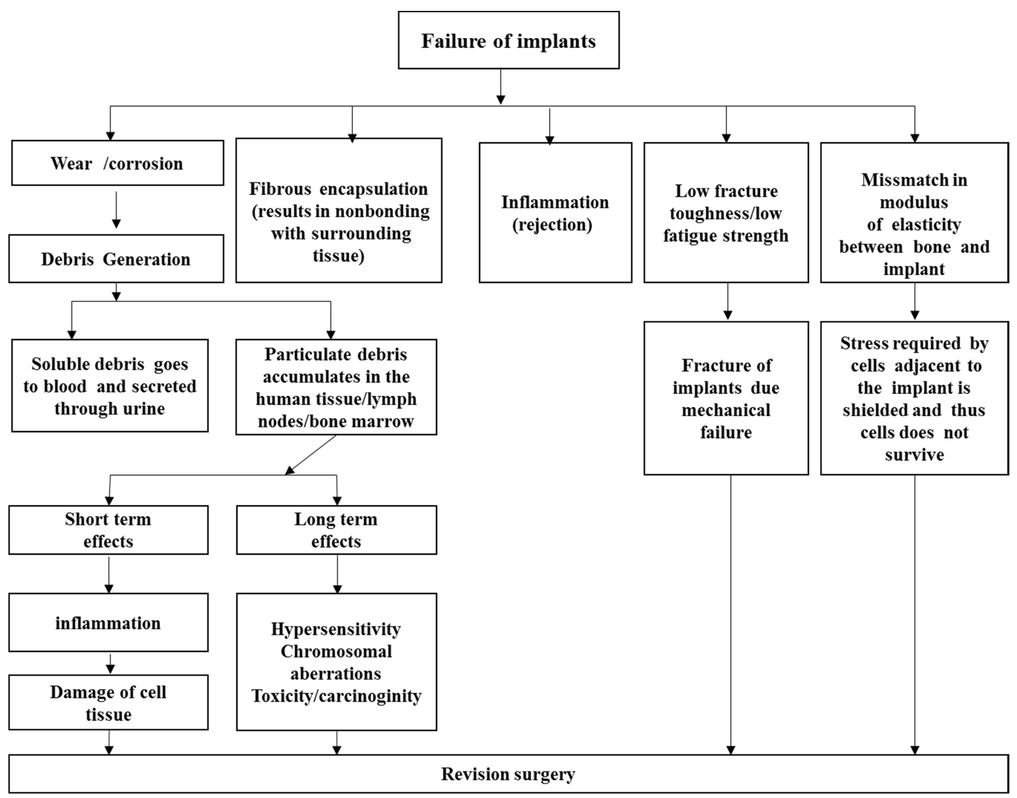
- High wear resistance: The material should have a high wear resistance and exhibit a low friction coefficient when sliding against body tissues. An increase in the friction coefficient or a decrease in the wear resistance can cause the implant to loosen [14,15]. Moreover, the wear debris generated can cause inflammation that is destructive to the bone supporting the implant.
- High corrosion resistance: An implant that is made of a biomaterial with a low corrosion resistance can release metal ions into the body, which in turn produces toxic reactions [16].
- Osseointegration: Osseointegration was first defined as “a direct structural and functional connection between ordered, living bone and the surface of a load-carrying implant” [17]. The roughness, chemistry, and topography of the surface play a major role in good osseointegration [18]. Implant loosening results from the non-integration of the implant surface into the adjacent bone [19]. Few researchers mention that osseiontegration is undesirable due to the risk of not being able to remove the implant after use [20]. However, a few of them have also demonstrated that the implant could be removed safely [20]. Thus osseointegration is a desirable property for a biomaterial in some applications such as in implant where it is to be made sure that the implant will integrate properly with the bone and other tissues [21].
- Non-toxic: The material should be neither genotoxic (which can alter the DNA of the genome) nor cytotoxic (causes damage to individual cells).
- Long fatigue life: The material should exhibit a high resistance to failure by fatigue to prevent implant failure and stress shielding from fatigue fracture. The failure of implants by fatigue has been reported for hip prostheses [22].
3. Types of Biomaterials
3.1. Metallic Alloys for Biomaterials

| Alloy | Microstructure |
|---|---|
| 1. Pure Ti | (ASTM F67-89) |
| 2. Ti-6Al-4V ELI (ASTM F136-84, F620-87) | α+β type |
| 3. Ti-6Al-4V (ASTM F1108-88) | α+β type |
| 4. Ti-6Al-7Nb (ASTM F1295-92, ISO5832-11) | α+β type(Swiss) |
| 5. Ti-5Al-2.5Fe (ISO5832-10) | α+β type (Germany) |
| 6. Ti-5Al-3Mo-4Zr | α+β type (Japan) |
| 7. Ti-15Sn-4Nb-2Ta-0.2Pd | α+β type (Japan) |
| 8. Ti-15Zr-4Nb-2Ta-0.2Pd | α+β type (Japan) |
| 9. Ti-13Nb-13Zr (ASTM F1713-96) | near β type (U.S.A.), Low modulus |
| 10. Ti-12Mo-6Zr-2Fe (ASTM F1813-97) | near β type (U.S.A.), Low modulus |
| 11. Ti-15Mo | β type (U.S.A.), Low modulus |
| 12. Ti-16Nb-10Hf | β type (U.S.A.), Low modulus |
| 13. Ti-15Mo-5Zr-3Al | β type (Japan), Low modulus |
| 14. Ti-15Mo-2.8Nb-0.2Si-0.26O | β type (U.S.A.), Low modulus |
| 15. Ti-35Nb-7Zr-5Ta | β type (U.S.A.), Low modulus |
| 16. Ti-29Nb-13Ta-4.6Zr | β type (Japan), Low modulus |
| 17. Ti-40Ta, Ti-50Ta | β type (U.S.A), High corrosion resistance |
| ASTM designation | Alloy | Cr | Ni | Mo | N | Mn | C | P | S | Si | Cu | Fe |
|---|---|---|---|---|---|---|---|---|---|---|---|---|
| (F138-92) | Bar and Wire | |||||||||||
| Grade 1 | 17.00–19.00 | 13.00–15.50 | 2.00–3.00 | −0.1 | −2.0 | −0.08 | −0.025 | −0.01 | −0.75 | −0.5 | balance | |
| Grade 2 | 17.00–19.00 | 13.00–15.50 | 2.00–3.00 | −0.1 | −2.0 | −0.03 | −0.025 | −0.01 | −0.75 | −0.5 | balance | |
| (F139-96) | 18Cr-14Ni-2.5Mo | 17.00–19.00 | 13.00–15.00 | 2.25–3.00 | −0.1 | −2.0 | −0.03 | −0.025 | −0.01 | −0.75 | −0.5 | balance |
| (F621-92) | Sheet and Strip | Same chemical composition as specified in Specification F138, grade 1 and 2 | ||||||||||
| (F1314-95) | Forgings Nitrogen strengthened | 20.5–23.5 | 11.5–13.5 | 2.0–3.0 | 0.2–0.4 | 4.0–6.0 | −0.03 | −0.025 | −0.01 | −0.75 | −0.5 | balance |
| 22Cr-12.5Ni-5Mn-2.5Mo | (0.10 < Nb < 0.30, 0.10 < V < 0.30) | |||||||||||
| (F1586-95) | Bar and Wire | |||||||||||
| Nitrogen strengthened | ||||||||||||
| 21Cr-10Ni-3Mn-2.5Mo | ||||||||||||
| 0.25 < Nb < 0.80 | ||||||||||||
| ASTM designation | Alloy | Cr | Mo | Ni | W | Fe | Ti | C | Si | P | S | Mn | Co |
|---|---|---|---|---|---|---|---|---|---|---|---|---|---|
| (F75-92) | Co-Cr-Mo Cast alloy | 27.0–30.0 | 5.0–7.0 | −1.0 | −0.75 | −0.35 | −1.0 | −1.0 | balance | ||||
| (F90-96) | Co-20Cr-15W-10Ni Wrought alloy | 19.0–21.0 | 9.0–11.0 | 14.0–16.0 | −3.0 | 0.05–0.15 | −0.4 | −0.03 | −0.03 | 1–2 | balance | ||
| (F562-95) | Co-35Ni-20Cr-10Mo | 19.0–21.0 | 9–10.5 | 33.0–37 | −1.0 | −1.0 | 0.025 | −0.15 | −0.015 | −0.01 | −0.15 | balance | |
| Wrought alloy | (B < 0.0015) | ||||||||||||
| (F563-95) | Co-Ni-Cr-Mo-W-Fe Wrought alloy | 18–22 | 3–4 | 15–25 | 3–4 | 4–6 | 0.5–3.5 | 0.05 | 0.5 | 0.01 | 1.0 | balance | |
| (F799-96) | Co-28Cr-6Mo forgings | 26.0–30.0 | 5–7 | −1.0 | −0.75 | −0.35 | -1.0 | −1.0 | balance | ||||
| (F1058-91) | Co-Cr-Ni-Mo-Fe Wrought alloy | ||||||||||||
| Grade 1 | 19.0–21.0 | 6.0–8.0 | 14.0–16.0 | balance | 0.15 | −1.2 | −0.015 | −0.015 | 1.5–2.5 | 39.0–41 | |||
| (Be < 0.01) | |||||||||||||
| Grade 2 | 18.5–21.5 | 6.5–7.5 | 15.0–18.0 | balance | 0.15 | −1.2 | −0.015 | −0.015 | 1.0–2.0 | 39.0–42 | |||
| (Be < 0.001) | |||||||||||||
| (F1537-94) | Co-28Cr-6Mo Wrought alloy | 26.0–30.0 | 5.0–7.0 | −1.0 | −0.75 | 0.35 | −1.0 | 1.0 | balance | ||||
| (N < 0.25) | |||||||||||||
3.1.1. Ti Alloys
3.1.2. Stainless Steels
3.1.3. Co Alloys
| Material | Young’s Modulus, E (GPa) | Yield Strength, (MPa) | Tensile Strength (MPa) | Fatigue Limit, (MPa) |
|---|---|---|---|---|
| Stainless steel | 190 | 221–1213 | 586–1351 | 241–820 |
| Co-Cr alloys | 210–253 | 448–1606 | 655–1896 | 207–950 |
| Titanium (Ti) | 110 | 485 | 760 | 300 |
| Ti-6Al-4V | 116 | 896–1034 | 965–1103 | 620 |
| Cortical bone | 15–30 | 30–70 | 70–150 |
| Metals and alloys | Selected examples | Advantages | Disadvantages | Principal applications [29] |
|---|---|---|---|---|
| Titanium-based Alloys | CP-Ti, Ti-Al-V, Ti-Al-Nb, Ti- 13Nb-13Zr, Ti-Mo-Zr-Fe | High biocompatibility [24,25,26]. Low Young’s modulus excellent corrosion resistance, low density | Poor tribological properties [27], Toxic effect of Al and V on long term | Bone and joint replacement, fracture fixation, dental implants, pacemaker encapsulation |
| Cobalt and Cr alloys | Co-Cr-Mo, Cr-Ni-Cr-Mo | High wear resistance [20] | Allergy consideration with Ni, Cr and Co [2] much higher modulus than bone | Bone and joint replacement, dental implants, dental restorations, heart valves |
| Stainless steels | 316L stainless steel | High wear resistance [23] | Allergy consideration with Ni, Cr and Co [2] much higher modulus than bone | Fracture fixation, stents, surgical instruments |
| Others | Ni-Ti | Low Young’s modulus | Ni cause allergy [2] | Bone plates, stents, orthodontic wires |
| Platinum and Pt-Ir | High corrosion resistant under extreme voltage potential and charge transfer conditions [30] | Electrodes | ||
| Hg-Ag-Sn amalgam | Easy in situ formability to a desired shape susceptible to corrosion in the oral environment [30] | Concerns related to Hg toxicity [30] | Dental restorations |
3.1.4. Limitations of Current Metallic Biomaterials

4. Wear of Metallic Biomaterials
4.1. Wear Testing Methods

| Test | Advantages | Disadvantages |
|---|---|---|
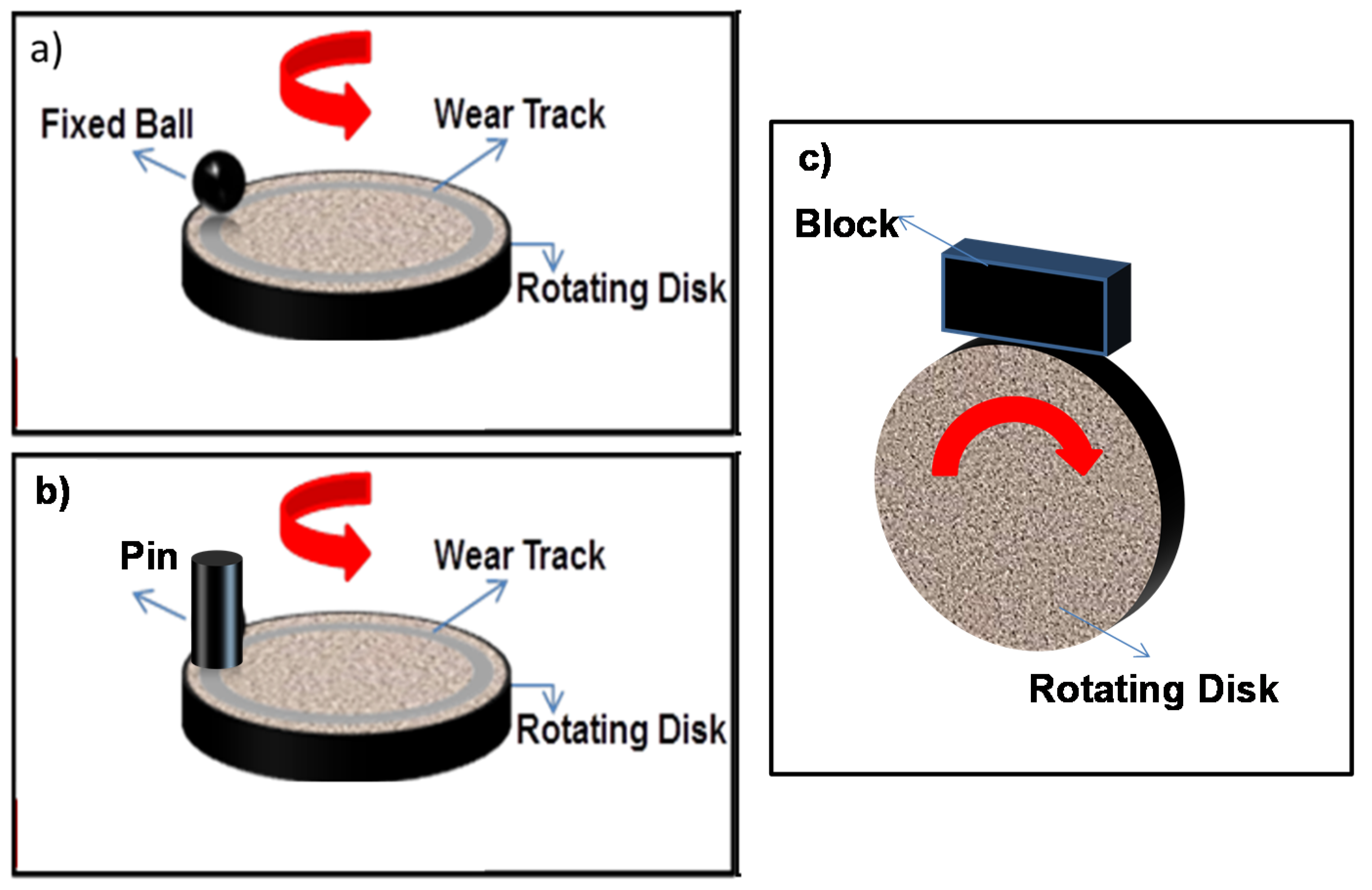
| Pin-on-Disk | After run-in, surface pressure remains constant. Easy to determine wear volume and wear rate. The model closely simulates a linear friction bearing. | Difficult to align pin. If the pin does not stand perfectly vertical on the plate, edge contact results. A very long run-in time is therefore necessary. The front edge of the pin can skim off lubricant. This makes a defined lubrication state impossible. |
| Ball-on-Disk | High surface pressures are possible. The ball skims off lubricant less than a pin does. The model is similar to a linear friction bearing and a radial friction bearing. | Very small contact ratio: The contact surface of the ball is small compared to the sliding track on the disk. The contact area is enlarged by wear. Difficult to determine the wear volume of the ball. |
| Block-on-disc | The model is capable of simulating a variety of harsh field conditions, e.g., high temperature, high speed, and high loading pressure. |
4.2. Characterization Techniques for the Wear of Biomaterials
4.3. Wear Performance of Different Biomaterials
| First author, year | Material & fabrication processes | Experimental test techniques & parameters | Main results |
|---|---|---|---|
| Cvijovic’-Alagic et al. [36] | Ti-13Nb-13Zr Ti-6Al-4V Arc melting | A block-on-disc tribometer was used to conduct wear and friction tests in a simulated body fluid (Ringer’s solution). Temperature: ambient Normal load: 20–60 N Sliding speed: 0.26–1.0 m/s | The Ti-6Al-4V alloy showed a higher wear resistance than the Ti-13Nb-13Zr alloy. Abrasion was the primary wear mechanism. |
| Stefano Gialanella et al. [37] | NiTi Commercial alloy | A block-on-disc was used to measure dry sliding wear. A profilometer was used to quantify wear. Sliding speed: 0.837 ms−1 Sliding distance: 1004 m Loads: 50 to 200 N | A NiTi/WC-Co coupling exhibited a high wear rate. Wear mechanism: a transition from delamination wear to a regime featuring a mixture of delamination and oxidation wear. |
| K.S. Suresh et al. [38] | Ti-13Nb-13Zr Equal channel angular pressing (ECAE) | A tribometer was used as a lubricity fretting test system for texture and wear behavior; fretting wear and3D surface texture measurements were performed. Normal loads: 6 N Frequency: 20 Hz Temperature: 37 ± 0.1 °C | The grain size and the texture of material affected the wear of the surface. There was no difference in the friction coefficient between the ECAE processed and as-received samples. |
| Li-juan Xu et al. [39] | β-type Ti-15Mo-xNb arc-melting vacuum-pressure casting system | A ball-on-disc was used for dry wear tests. Normal load: 1 N and 2 N Test-disc rate: 100 r/min | The lowest friction coefficient was obtained for a Ti-15Mo-5Nb alloy under a 1-N load. Adhesion was the primary wear mechanism. |
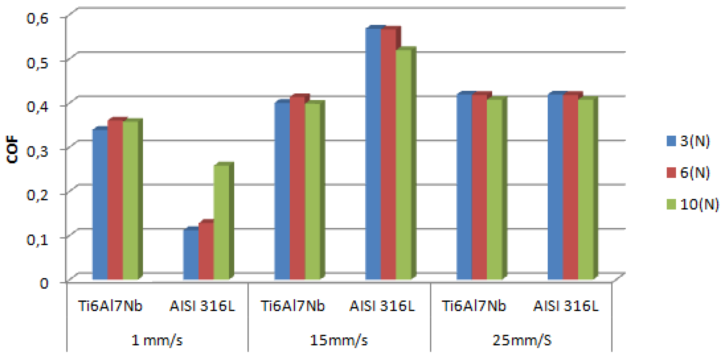
| M. Fellah et al. [41] | Ti-6Al-7Nb and AISI 316L stainless steel | Ball-on–disc and sphere-on-plane Load: 3 N, 6 N and 10 N Sliding speed: 1 mm/s, 15 mm/s and 25 mm/s | The same mechanisms of wear and friction were found for all of the tested samples. |
| S.J. Li et al. [42] | Ti-Nb-Ta-Zr and Ti-6Al-4V induction skull melting method | Reciprocal pin-on-disc in a 0.9% NaCl solution Reciprocating velocity: 45 rpm Sliding distance: 30 km | The wear resistance of Ti-29Nb-13Ta-4.6Zr was enhanced by incorporating Nb2O5 oxide particles into the diffusion-hardened surface of the alloy. |
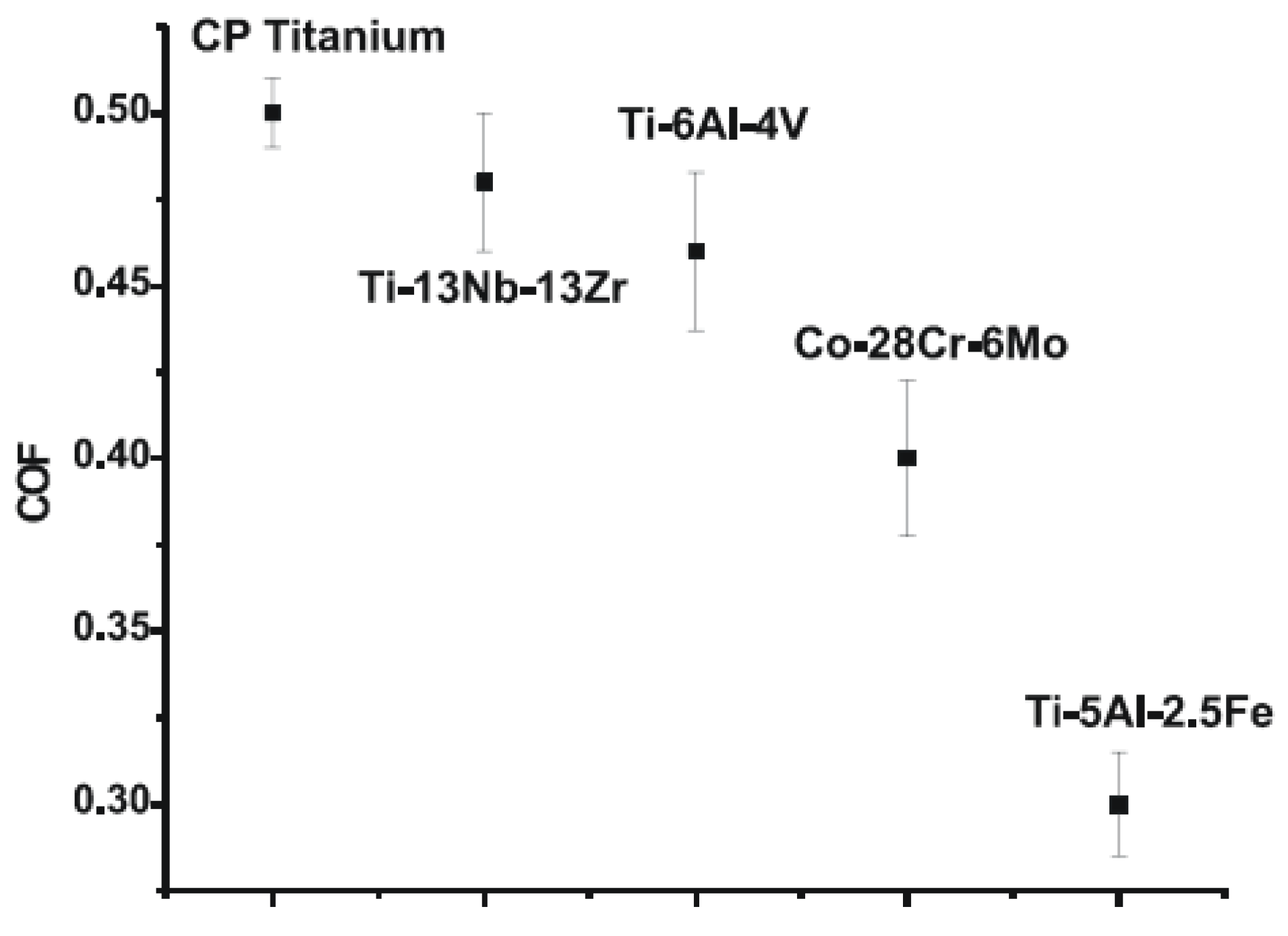
| Animesh Choubey et al. [45] | CP Titanium, Ti-6Al-4V, Ti-5Al-2.5Fe, Ti-13Nb-13Zr and Co-28Cr-6Mo | Ball on flat fretting wear tester: Hanks’ balanced salt solution Normal load: 10 N for 10,000 cycles Frequency: 10 Hz | The primary wear mechanisms of Ti alloys were tribomechanical abrasion, transfer layer formation and cracking. |
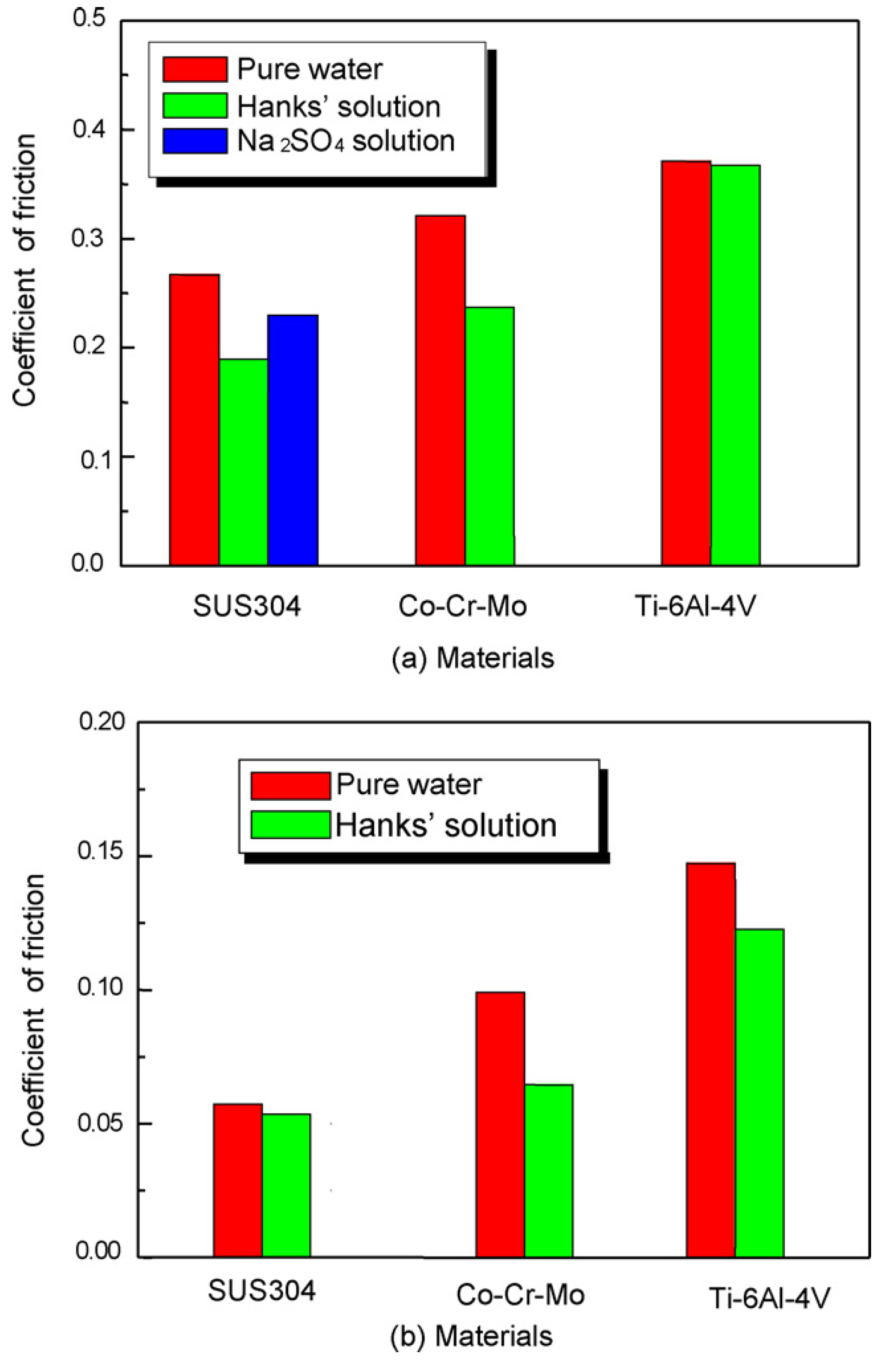
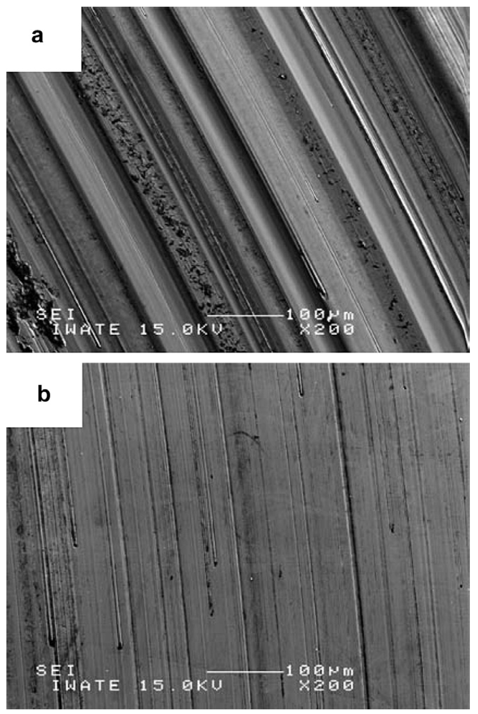
| A. Iwabuchi et al. [46] | Co-29Cr-6Mo alloy and Ti-6Al-4V | Fretting apparatus and a reciprocating sliding tribometer: Quasi-body fluid, Hanks’s solution. Normal load: 5 N; frequency: 10 Hz Temperature in the solutions: 37 ± 2 °C | Co alloy exhibited good wear resistance; Ti alloy exhibited good fretting resistance. |
| X. Luo et al. [47] | ASTM F1537 Co-Cr alloy | Pin-on-disc tribometer Load: 20 N Rotation speed: 60 (rpm) | The tribocorrosion properties of the Co-Cr alloy were enhanced by a layer of the S-phase. |
| Akihiko Chiba et al. [48] | Co-Cr-Mo forged | Pin-on-disc Load: 9.8 N 24 rpm | Forged CoCr exhibited a lower wear loss than a cast CoCr alloy. |
| S. M. T. Chan et al. [49] | (CoCr), stainless steel (SS) | Pin-on-disc sliding speed: 0.5 mm/s, 5 mm wear track radius Normal load: 1.8 N | |
| Alfons Fischer et al. [50] | AISI 316L CoCr29Mo6 | Pin-on-disc for dry sliding wear tests Load: 5 N Relative velocity: 0.1 m/s Ambient temperature: 25 °C | Ni-free high-nitrogen steel and LC-CoCrMo alloy exhibited higher wear resistance and dry friction than Ni-containing austenitic steels. |
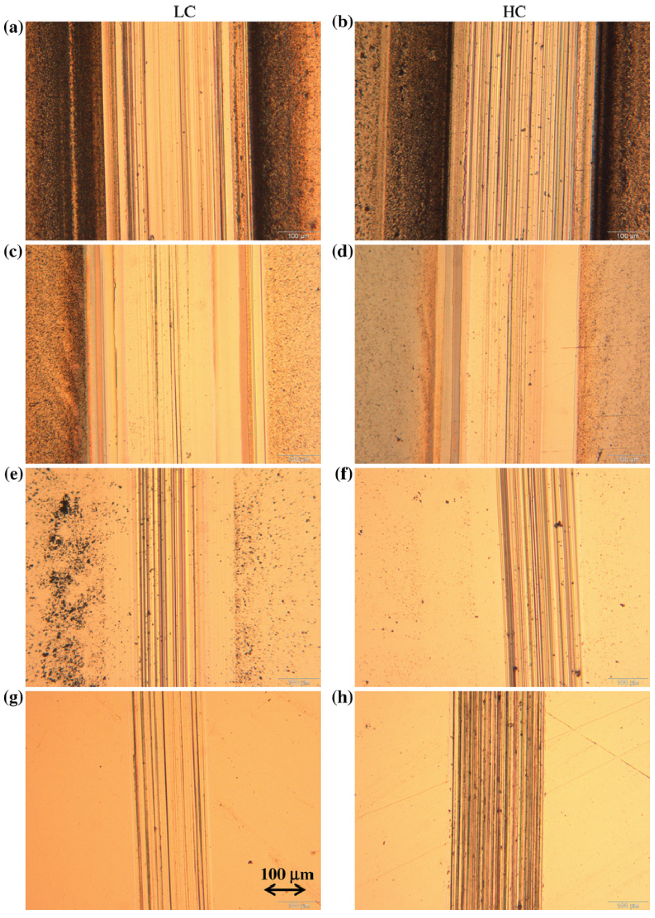
| A. Igual Muñoz et al. [40] | Co-Cr-Mo Low and high carbon | Tribocorrosion techniques Load: 1.2 N; frequency: 1 Hz Temperature: 37 ± 0.1 °C Simulated body fluids [NaCl and phosphate-buffered solutions (PBS) with and without albumin] | LC CoCrMo had a higher wear resistance in NaCl and PBS albumin than HC. No differences were observed for the alloys in the other solutions. |
| M. Alvarez-Vera, et al. [51] | Co-Cr alloy with boron additions (0, 0.3, 0.6 and 1 B wt%) by casting method | three-axial hip joint simulator | Wear resistance as the boron increased. |
| L. Mohan, et al. [52] | Commercial Ti-13Nb-3Zr alloy oxygen implanted | Reciprocating type wear tester normal forces: 3, 5 and 10 N. The stroke length: 10 mm and an alumina ball of 6 mm diameter was used as the counter surface | The implanted samples display a lower friction coefficient as compared to the substrate one. |
| H. Attar et al. [53] | Commercially pure titanium (CP-Ti) parts produced using selective laser melting (SLM) and casting | a pin-on-disc at room temperature A stainless steel disc of 45 mm diameter loads: (15 N, 20 N,25 N and 30 N) sliding speed: 0.5 m/s for 15 min. | SLM CP-Ti showed better wear resistance compared to casting as a result of fine grains and higher microhardness. |


4.4. Techniques to Improve Wear Resistance of Metallic Biomaterials
- Ion implantation (physical deposition) is considered a simple technique for significantly modifying the physical and/or chemical properties of the near surface of a material in which suitable ions are embedded into the surface of a material from a beam of ionized particles. This technique has been reported to improve the wear performance of Ti6Al4V and Co28Cr6Mo alloys [62].
- Carburization and boriding techniques are used to enhance surface hardness, which in turn improves the wear resistance.
- Plasma spray coating has also been used to enhance the wear resistance of few biomaterials.
5. Summary
- Ti alloys
- In general, adding Nb to Ti alloys enhances the wear resistance of these alloys and slightly increases the friction coefficient primarily because of the increase in the hardness of the alloy. The heat treatment of these alloys has been observed to further increase the wear resistance because of the formation of Nb2O5 particles.
- Abrasive wear has been observed to be the predominant wear mechanism.
- Hence, surface treatments and coating are necessary to enhance the resistance of alloys to wear and friction.
- Co alloys
- Forged CoCr alloys exhibit higher wear resistance than cast CoCr alloys. However, the friction coefficient of the forged CoCr alloys has been observed to be higher than that of the cast alloys.
- The wear behavior of LC and HC CoCrMo alloys depends on the surrounding environment.
- Ni-free CrMnMo exhibits improved wear behavior compared to CrNiMo steels.
Acknowledgments
Author Contributions
Conflicts of Interest
References
- Park, J.; Lakes, R.S. Biomaterials an Introduction, 3rd ed.; Springer: Berlin/Heidelberg, Germany, 2007. [Google Scholar]
- Niinomi, M. Metallic biomaterials. J. Artif. Organs 2008, 11, 105–110. [Google Scholar] [CrossRef] [PubMed]
- Karanjai, M.; Sundaresan, G.V.N.; Rao, T.R.; Mohan, R.; Kashyap, B.P. Development of titanium based biocomposite by powder metallurgy processing with in situ forming of Ca–P phases. Mat. Sci. Eng. A 2007, 447, 19–26. [Google Scholar] [CrossRef]
- Patel, N.R.; Gohil, P.P. A review on biomaterials: Scope. Application s & human anatomy significance. Int. J. Emerg. Technol. Adv. Eng. 2012, 2, 91–101. [Google Scholar]
- Litonjua, L.A.; Andreana, S.; Bush, P.J.; Cohen, R.E. Tooth wear: Attrition, erosion, and abrasion. Quintessence Int. 2003, 34, 435–446. [Google Scholar] [PubMed]
- Turssi, C.P.; Purquerio, B.; Serra, M.C. Wear of dental resin composites: Insights into underlying processes and assessment methods—A review. J. Biomed. Mater. Res. 2003, 65B, 280–285. [Google Scholar] [CrossRef]
- Reul, H.; Schmitz, C.; Pfaff, E.M.; Hohlstein, C.; Schmidt, P.A.; Rau, G.; Arru, P. In-vitro assessment of the wear development mechanism and stabilization of wear in the Edwards MIRA/Sorin Bicarbon mechanical heart valve orifice ring. J. Heart Valve D 2002, 11, 409–418. [Google Scholar]
- Shahgaldi, B.F.; Compson, J. Wear and corrosion of sliding counterparts of stainless-steel hip screw-plates. Injury 2000, 31, 85–92. [Google Scholar] [CrossRef] [PubMed]
- Dowson, D. Tribology and the skin structure. In Bioengineering of the Skin: Methods and Instrumentation, Part III: General Aspects; Berardesca, E., Elsner, P., Wilhelm, K.P., Maibach, H.I., Eds.; CRC Press: Boca Raton, FL, USA, 1995; p. 159. [Google Scholar]
- Sivamani, R.K.; Goodman, J.; Gitis, N.V.; Maibach, H.I. Coefficient of friction: Tribological studies in man—An overview. Skin Res. Technol. 2003, 9, 227–234. [Google Scholar] [CrossRef] [PubMed]
- Walowit, J.A. The analysis, design, and testing of a blood lubricated hydrodynamic journal bearing. ASAIO J. 1997, 43, M556. [Google Scholar] [CrossRef] [PubMed]
- Lawrence, K.J. Anisotropy of Young’s modulus of bone. Nature 1980, 283, 106–107. [Google Scholar] [CrossRef] [PubMed]
- Black, J.; Hastings, G.W. Handbook of Biomaterials Properties; Chapman and Hall: London, UK, 1998. [Google Scholar]
- Alvarado, J.; Maldonado, R.; Marxuach, J.; Otero, R. Biomechanics of hip and knee prostheses. Appl. Eng. Mechan. Med. GED–Univ. Puerto Rico Mayaguez 2003, 6, 22. [Google Scholar]
- Ramsden, J.J. The design and manufacture of biomedical surfaces. CIRP Ann.-Manuf. Technol. 2007, 56, 687–711. [Google Scholar] [CrossRef]
- Hallab, N.J.; Anderson, S.; Stafford, T.; Glant, T.; Jacobs, J.J. Lymphocyte responses in patients with total hip arthroplasty. J. Orthop. Res. 2005, 23, 384–391. [Google Scholar] [CrossRef] [PubMed]
- Branemark, P.I. Osseointegration and its experimental background. J. Pros. Dent. 1983, 50, 399–410. [Google Scholar] [CrossRef]
- Geetha, M.; Singh, A.K.; Asokamani, R.; Gogia, A.K. Ti based biomaterials, the ultimate choice for orthopaedic implants—A review. Progr. Mater. Sci. 2009, 54, 397–425. [Google Scholar] [CrossRef]
- Viceconti, M.; Muccini, R.; Bernakiewicz, M.; Baleani, M.; Cristofolini, L. Large-sliding contact elements accurately predict levels of bone–implant micromotion relevant to osseointegration. J. Biomech. 2000, 33, 1611–1618. [Google Scholar] [CrossRef] [PubMed]
- Wennerberg, A.; Albrektsson, T.; Jimbo, R. Implant Surfaces and Their Biological and Clinical Impact, 1st ed.; Springer-Verlag: Berlin/Heidelberg, Germany, 2015; p. 168. [Google Scholar]
- Barfeie, A.; Wilson, J.; Rees, J. Implant surface characteristics and their effect on osseointegration. Br. Dental J. 2015, 218, 1–9. [Google Scholar] [CrossRef]
- Teoh, S.H. Fatigue of biomaterials: A review. Int. J. Fatigue 2000, 22, 825–837. [Google Scholar] [CrossRef]
- Niinomi, M. Recent metallic materials for biomedical applications. Metall. Mater. Trans. A 2002, 33, 477–486. [Google Scholar] [CrossRef]
- Ribeiro, A.L.R.; Fla, R.C.J.; Cardoso, F.; Belon, R.; Vaz, F.F.L.G. Mechanical, physical, and chemical characterization of Ti–35Nb–5Zr and Ti–35Nb–10Zr casting alloys. J. Mater. Sci.: Mater. Med. 2009, 20, 1629–1636. [Google Scholar] [CrossRef]
- Niespodziana, K.; Jurczk, K.; Jurczk, M. The synthesis of titanium alloys for biomedical applications. Rev. Adv. Mater. Sci. 2008, 18, 236–240. [Google Scholar]
- Henriques, V.A.R.; Galvani, E.T.; Petroni, S.L.G.; Paula, M.S.M.; Lemos, T.G. Production of Ti–13Nb–13Zr alloy for surgical implants by powder metallurgy. J. Mater. Sci. 2010, 45, 5844–5850. [Google Scholar] [CrossRef]
- Ohidul Alam, M.; Haseeb, A.S.M.A. Response of Ti–6Al–4V and Ti–24Al–11Nb alloys to dry sliding wear against hardened steel. Tribol. Int. 2002, 35, 357–362. [Google Scholar] [CrossRef]
- Ratner, J.B.B.D.; Hoffman, A.S.; Shoen, F.J.; Lemons, J.E. Biomaterials Science: An Introduction to Materials in Medicine; Academic Press: Waltham, MA, USA, 1996; pp. 37–50. [Google Scholar]
- Williams, D. An Introduction to Medical and Dental Materials, Concise Encyclopedia of Medical & Dental Materials; Williams, D., Ed.; Pergamon Press: Oxford, UK; MIT Press: Cambridge, MA, USA, 1990; pp. xvii–xx. [Google Scholar]
- Pilliar, R.M. Metallic Biomaterials; Springer Science + Business Media: New York, NY, USA, 2009. [Google Scholar]
- Yoshimitsu, O.; Emiko, G. Comparison of metal release from various metallic biomaterials in vitro. Biomaterials 2005, 26, 11–21. [Google Scholar] [CrossRef] [PubMed]
- Nag, S.; Banerjee, R.; Fraser, H.L. Microstructural evolution and strengthening mechanisms in Ti-Nb-Zr-Ta, Ti-Mo-Zr-Fe and Ti-15Mo biocompatible alloys. Mater. Sci. Eng. C 2005, 25, 357–362. [Google Scholar] [CrossRef]
- McGregor, D.B.; Baan, R.A.; Partensky, C.; Rice, J.M.; Wibourn, J.D. Evaluation of the carcinogenic risks to humans associated with surgical implants and other foreign bodies-a report of an IARC monographs programmed meeting. Eur. J. Cancer 2000, 36, 307–313. [Google Scholar] [CrossRef] [PubMed]
- Yang, Y.; Yang, C.; Zhao, H.; Qu, S.; Li, X.; Li, Y. New developments of Ti-based alloys for biomedical applications. Materials 2014, 7, 1709–1800. [Google Scholar] [CrossRef]
- Liang, P.G.; Ferguson, A.; Hodge, E.S. Tissue reaction in rabbit muscle exposed to metallic implants. J. Biomed. Mater. Res. 1967, 1, 135–149. [Google Scholar] [CrossRef] [PubMed]
- Cvijovic, A.I.; Cvijovic, Z.; Mitrovic, S.; Rakin, M.; Veljovic, D.; Babic, M. Tribological behaviour of orthopaedic Ti-13Nb-13Zr and Ti-6Al-4V alloys. Tribol. Lett. 2010, 40, 59–70. [Google Scholar] [CrossRef]
- Gialanella, S.; Ischia, G.; Straffelini, G. Phase composition and wear behavior of NiTi alloys. J. Mater. Sci. 2008, 43, 1701–1710. [Google Scholar] [CrossRef]
- Suresh, K.S.; Geetha, M.; Richard, C.; Landoulsi, J.; Ramasawmy, H.; Suwas, S.; Asokamani, R. Effect of equal channel angular extrusion on wear and corrosion behavior of the orthopedic Ti-13Nb-13Zr alloy in simulated body fluid. Mater. Sci. Eng. C 2012, 32, 763–771. [Google Scholar] [CrossRef]
- Xu, L.; Xiao, S.; Tian, J.; Chen, Y. Microstructure, mechanical properties and dry wear resistance of β-type Ti–15Mo–xNb alloys for biomedical applications. Trans. Nonferrous Met. Soc. China 2013, 23, 692–698. [Google Scholar] [CrossRef]
- Muñoz, A.I. Effect of the environment on wear ranking and corrosion of biomedical CoCrMo alloys. J. Mater. Sci. Mater. Med. 2011, 22, 437–450. [Google Scholar] [CrossRef] [PubMed]
- Fellah, M.; Labaïz, M.; Assala, O.; Iost, A. Comparative Tribological study of biomaterials AISI 316L and Ti-6Al-7Nb. TMS 2014, 237, 237–246. [Google Scholar]
- Li, S.J.; Yang, R.; Li, S.; Hao, Y.L.; Cui, Y.Y.; Niinomi, M.; Guo, Z.X. Wear characteristics of Ti-Nb-Ta-Zr and Ti-6Al-4V alloys for biomedical applications. Wear 2004, 257, 869–876. [Google Scholar] [CrossRef]
- Bhushan, B. Introduction to Tribology, 2nd ed.; John Wiley & Sons, Ltd.: New York, NY, USA, 2013; p. 621. [Google Scholar]
- Bhushan, B. Nanotribology, nanomechanics and nanomaterials characterization. Philos. Trans. R. Soc. A 2008, 366, 1351–1381. [Google Scholar] [CrossRef]
- Choubey, A.; Basu, B.; Balasubramaniam, R. Tribological behaviour of Ti-based alloys in simulated body fluid solution at fretting contacts. Trends Biomater. Artif. Organs 2005, 18, 141–147. [Google Scholar]
- Iwabuchi, A.; Lee, J.W.; Uchidate, M. Synergistic effect of fretting wear and sliding wear of Co-alloy and Ti-alloy in Hankssolution. Wear 2007, 263, 492–500. [Google Scholar] [CrossRef]
- Luo, X.; Li, X.; Sun, Y.; Dong, H. Tribocorrosion behavior of S-phase surface engineered medical grade Co-Cr alloy. Wear 2013, 302, 1615–1623. [Google Scholar] [CrossRef]
- Chiba, A. Pin-on-disk wear behavior in a like-on-like configuration in a biological environment of high carbon cast and low carbon forged Co-29Cr-6Mo alloys. Acta Mater. 2007, 55, 1309–1318. [Google Scholar] [CrossRef]
- Chan, S.M.T.; Neu, C.P.; Komvopoulos, K.; Reddi, A.H.; Di Cesare, P. Friction and wear of hemiarthroplasty biomaterials in reciprocating sliding contact with articular cartilage. J. Tribol. 2011, 133, 1–7. [Google Scholar] [CrossRef]
- Fischer, A.; Weiß, S.; Wimmer, M.A. The tribological difference between biomedical steels and CoCrMo-alloys. J. Mech. Behav. Biomed. Mater. 2012, 9, 50–62. [Google Scholar] [CrossRef] [PubMed]
- Alvarez-Vera, M.; Ortega-Saenz, J.A.; Hernandez-Rodríguez, M.A.L. A study of the wear performance in a hip simulator of a metal-metal Co-Cr alloy with different boron additions. Wear 2013, 301, 175–181. [Google Scholar] [CrossRef]
- Mohan, L.; Anandan, C. Wear and corrosion behavior of oxygen implanted biomedical titanium alloy Ti-13Nb-13Zr. Appl. Surf. Sci. 2013, 282, 281–290. [Google Scholar] [CrossRef]
- Attar, H.; Prashanth, G.; Chaubey, A.K.; Calin, M.; Zhang, S.L.C.; Eckert, J. Comparison of wear properties of commercially pure titanium prepared by selective laser melting and casting processes. Mater. Lett. 2015, 142, 38–41. [Google Scholar] [CrossRef]
- Rack, H.J.; Qazi, J.I. Titanium alloys for biomedical applications. Mater. Sci. Eng. C 2006, 26, 1269–1277. [Google Scholar] [CrossRef]
- Latysh, V.; Krallics, G.; Alexandrov, I.; Fodor, A. Application of bulk nanostructured materials in medicine. Curr. Appl. Phys. 2006, 6, 262–266. [Google Scholar] [CrossRef]
- Stolyarov, V.V.; Shuster, L.S.; Migranov, M.S.; Valiev, R.Z.; Zhu, Y.T. Reduction of friction coefficient of ultrafine-grained CP titanium. Mater. Sci. Eng.: A 2004, 371, 313–317. [Google Scholar] [CrossRef]
- Spary, I.D.; Bushby, A.J.; Jennet, N.M. On the indentation size effect in spherical indentation. Philos. Mag. 2006, 86, 5581–5593. [Google Scholar] [CrossRef]
- Hou, X.D.; Bushby, A.J.; Jennett, N.M. Study of the interaction between indentation size effect and Hall–Petch effect with spherical indenters on annealed polycrystalline copper. J. Phys. D: Appl. Phys. 2008, 41, 074006–074007. [Google Scholar] [CrossRef]
- Hou, X.D.; Jennett, N.M. Application of a modified slip-distance theory to the indentation of single-crystal and polycrystalline copper to model the interactions between indentation size and structure size effects. Acta Mater. 2012, 60, 4128–4135. [Google Scholar] [CrossRef]
- Beake, B.D.; Liskiewicz, T.W. Comparison of nano-fretting and nano-scratch tests on biomedical materials. Tribol. Int. 2013, 63, 123–131. [Google Scholar] [CrossRef]
- Sun, D.; Wharton, J.A.; Wood, R.J.K. Micro-abrasion mechanisms of cast CoCrMo in simulated body fluids. Wear 2009, 267, 1845–1855. [Google Scholar] [CrossRef]
- Díaz, C.; Lutz, J.; Mändl, S.; García, J.A.; Martínez, R.; Rodríguez, R.J. Improved bio-tribology of biomedical alloys by ion implantation techniques. Nucl. Instrum. Methods Phys. Res. 2009, 267, 1630–1633. [Google Scholar] [CrossRef]
- Manhabosco, T.M.; Tamborim, S.M.; Dos Santos, C.B.; Müller, I.L. Tribological, electrochemical and tribo-electrochemical characterization of bare and nitrided Ti6Al4V in simulated body fluid solution. Corros. Sci. 2011, 53, 1786–1793. [Google Scholar] [CrossRef]
- Sathish, S.; Geetha, M.; Pandey, N.D.; Richard, C.; Asokamani, R. Studies on the corrosion and wear behavior of the laser nitrided biomedical titanium and its alloys. Mater. Sci. Eng. C 2010, 30, 376–382. [Google Scholar] [CrossRef]
© 2015 by the authors; licensee MDPI, Basel, Switzerland. This article is an open access article distributed under the terms and conditions of the Creative Commons Attribution license (http://creativecommons.org/licenses/by/4.0/).
Share and Cite
Hussein, M.A.; Mohammed, A.S.; Al-Aqeeli, N. Wear Characteristics of Metallic Biomaterials: A Review. Materials 2015, 8, 2749-2768. https://0-doi-org.brum.beds.ac.uk/10.3390/ma8052749
Hussein MA, Mohammed AS, Al-Aqeeli N. Wear Characteristics of Metallic Biomaterials: A Review. Materials. 2015; 8(5):2749-2768. https://0-doi-org.brum.beds.ac.uk/10.3390/ma8052749
Chicago/Turabian StyleHussein, Mohamed A., Abdul Samad Mohammed, and Naser Al-Aqeeli. 2015. "Wear Characteristics of Metallic Biomaterials: A Review" Materials 8, no. 5: 2749-2768. https://0-doi-org.brum.beds.ac.uk/10.3390/ma8052749