Article

Molecular Characterization of Small Ruminant Lentiviruses in Polish Mixed Flocks Supports Evidence of Cross Species Transmission, Dual Infection, a Recombination Event, and Reveals the Existence of New Subtypes within Group A

1
Department of Swine Diseases, National Veterinary Research Institute, 24-100 Pulawy, Poland
2
Department of Biochemistry, National Veterinary Research Institute, 24-100 Pulawy, Poland
*
Author to whom correspondence should be addressed.
Submission received: 12 November 2021 / Revised: 10 December 2021 / Accepted: 14 December 2021 / Published: 16 December 2021
(This article belongs to the Special Issue Non-Primate Lentiviruses 2021)

Abstract

:
Small ruminant lentiviruses (SRLVs) are a group of highly divergent viruses responsible for global infection in sheep and goats. In a previous study we showed that SRLV strains found in mixed flocks in Poland belonged to subtype A13 and A18, but this study was restricted only to the few flocks from Małopolska region. The present work aimed at extending earlier findings with the analysis of SRLVs in mixed flocks including larger numbers of animals and flocks from different part of Poland. On the basis of gag and env sequences, Polish SRLVs were assigned to the subtypes B2, A5, A12, and A17. Furthermore, the existence of a new subtypes, tentatively designed as A23 and A24, were described for the first time. Subtypes A5 and A17 were only found in goats, subtype A24 has been detected only in sheep while subtypes A12, A23, and B2 have been found in both sheep and goats. Co-infection with strains belonging to different subtypes was evidenced in three sheep and two goats originating from two flocks. Furthermore, three putative recombination events were identified within gag and env SRLVs sequences derived from three sheep. Amino acid (aa) sequences of immunodominant epitopes in CA protein were well conserved while Major Homology Region (MHR) had more alteration showing unique mutations in sequences of subtypes A5 and A17. In contrast, aa sequences of surface glycoprotein exhibited higher variability confirming type-specific variation in the SU5 epitope. The number of potential N-linked glycosylation sites (PNGS) ranged from 3 to 6 in respective sequences and were located in different positions. The analysis of LTR sequences revealed that sequences corresponding to the TATA box, AP-4, AML-vis, and polyadenylation signal (poly A) were quite conserved, while considerable alteration was observed in AP-1 sites. Interestingly, our results revealed that all sequences belonging to subtype A17 had unique substitution T to A in the fifth position of TATA box and did not have a 11 nt deletion in the R region which was noted in other sequences from Poland. These data revealed a complex picture of SRLVs population with ovine and caprine strains belonging to group A and B. We present strong and multiple evidence of dually infected sheep and goats in mixed flocks and present evidence that these viruses can recombine in vivo.

1. Introduction

Small ruminant lentiviruses (SRLVs) include two retroviruses, Caprine arthritis encephalitis virus (CAEV), and Maedi-visna virus (MVV), members of the genus Lentivirus of the Retroviridae family. Originally, MVV and CAEV were considered as distinct viral species restricted to sheep and goats, respectively, but several reports indicated that there are different lentiviral subtypes able to infect both sheep and goats [1]. SRLVs induce a multisystem disease with progressive and debilitating inflammatory lesions in the mammary gland, joints, lungs, and the brain. Diseases caused by SRLVs may take severe clinical course; however, clinical signs develop after a several-year-long period and only in one third of the infected animals. On rare occasions, young children may develop a leukoencephalomyelitis with CNS signs [2]. Both asymptomatic and symptomatic animals can transmit the virus. Infection takes place through the ingestion of infected colostrum/milk and/or by direct contact through respiratory exudates from infected animals [3,4]. The incidence of SRLV infections causes economic losses and welfare problems in small ruminant production, since no therapy or vaccine is currently available.
The genome of SRLVs, which is integrated into host cells in the form of a provirus, is composes of three gene’s coding for structural proteins, gag, pol, and env, and additional open reading frames (vpr-like, rev, and vif), which encode for nonstructural proteins with regulatory functions in virus replication [5]. The provirus genome is flanked by repeated sequences known as long terminal repeats (LTRs) composed of three regions, U3, R, and U5. U3 region contains promoter sequence and different transcription factor binding sites, like AP-1, AP-4, AML (vis), and GAS, and play a regulatory role in transcription, integration, and polyadenylation of viral RNA [6,7]. The gag gene encodes the internal structural proteins, capsid (CA) protein (p25), the nucleocapsid (NC) protein (p14), and the matrix (MA) protein (p17). The capsid protein contains linear epitopes that induce antibody production and for this reason it is used for serological diagnostic tests. In addition, because it is most conserved and is also commonly used as the target fragment for phylogenetic analysis and genomic characteristics of SRLV strains. The env gene encodes two proteins inserted in an envelope, the surface (SU) protein (gp135) and the transmembrane TM protein (gp46). SU also stimulates the production of antibodies but is genetically variable and determines the antigenic variability of the strains [8].
SRLVs are characterized by a high degree of genetic variability leading to the variety of divergent strains and quasispecies. SRLV isolates have been classified into five genetic groups, A-E, and further divided into several subtypes. Group A is the most heterogenous group and has been subdivided into 22 subtypes (A1-A22), so far. Group B contains five subtypes (B1-B5) while group E contain to subtypes (E1 and E2) [9,10]. Group A and B refer to MVV-like and CAEV-like viruses, respectively. Viruses belonging to these two groups are the most predominant strains around the world while isolates belonging to groups C, D and E are geographically restricted to limited areas. Groups C and E refer to Norwegian and Italian strains, respectively, while group D was found in a few isolates from Switzerland and Spain, but they are now re-classified as group A [11,12,13,14,15].
Many studies have documented the circulation of different subtypes of SRLVs in both sheep and goats due to interspecies transmission. In fact, the majority of subtypes can cross the species barrier between sheep and goats under field conditions [1,16,17]. Early and accurate diagnosis of SRLVs is crucial for prevention of infection and disease control. However, antigenic variation of SRLVs poses a relevant problem for SRLVs testing and diagnosis since heterogeneity of circulating strains may be wider than those covered by available ELISA tests making serological response not always detectable. Therefore, genotype and subtype surveys of the circulating SRLV strains in each country should be constantly updated to ensure reliability of diagnostic tests. In particular, mixed flocks where sheep and goats live in close contact, represent a suitable environment to evaluate the degree of SRLVs heterogeneity due to the growing evidence of increased cross-species transmission in mixed flocks and possible recombination events [18,19,20].
In Poland, a seroprevalence study confirmed the presence of SRLVs in 33.3% and 71.9% of sheep and goat flocks, respectively [20,21,22]. SRLVs isolated so far from sheep and goats in Poland belonged to the well-known subtypes B1, B2, A1, A5, and A16, as well as subtypes A12, A13, A17, and A18 detected only in Poland [23,24,25,26]. In a previous study we showed that SRLV strains found in mixed flocks in Poland belonged to subtype A13 and A18, but this study was restricted to only the few flocks from the Małopolska region. In the present study, we aimed at extending the previous investigation carrying out genetic characterization and phylogenetic analysis of SRLVs from mixed flocks including a larger numbers of animals, and flocks from different parts of Poland. Genetic analysis of SRLVs from mixed flocks may help to understand the genetic and antigenic make-up of these viruses, phylogenetic relationship and their allocation into the recently established groups. Moreover, genetic studies may also be useful for the development of regionally-tailored diagnostic tests.

2. Materials and Methods

2.1. Animals and Samples

A total of 263 samples were investigated in this study, 163 from sheep and 100 from goats, and originating from 17 mixed flocks and different geographic regions of Poland. Sheep and goats were housed in the same barn with the possibility of direct contact and via water and feed troughs. Animals were randomly chosen from the flocks for serological study and were clinically healthy, without any clinical signs. Blood was taken in EDTA and serum tubes for serology and molecular analysis. Sera samples were tested for MVV/CAEV antibodies using the commercially test ID Screen MVV/CAEV Indirect Screening (IDvet, Grabels, France). EDTA-anticoagulated blood was used as a source of PBLs, which were isolated according to standard protocols [24]. The genomic DNA was extracted using a NucleoSpin Blood Quick Pure Kit (Macherey-Nagel GmbH & Co. KG, Dueren, Germany), according to the manufacturer’s recommendation. The quality and quantity of DNA was evaluated in a Nanophotometer (Implen GmbH, Munich, Germany). All methods were performed in accordance with the relevant guidelines and regulations. Specifically, blood collection was approved (no. 37/2016) by the Local Ethical Committee on Animal Testing at the University of Life Sciences in Lublin (Lublin, Poland).

2.2. PCR Technique

The CA (625 bp) fragment of the gag gene fragment, the V4V5 fragment of env gene, and the U3-R fragment of the LTR region were amplified by nested PCR. Env fragments were amplified with the combination of 423/564 primers, followed by nested PCRs using either 423/425, 563/564, 563/425, or 567/564. The PCR reactions were performed as previously described [26,27,28] (Table 1).

2.3. DNA Sequencing and Analysis

PCR products were purified using NucleoSpin Gel and PCR Clean-up (Marcherey-Nagel, GmbH 7 Co, Hamburg, Germany) and cloned into the pDRIVE vector (Qiagen, GmbH, Hilden, Germany). Ligation products were used to transform EZ Competent Cells (Qiagen, GmbH, Hilden, Germany) and plasmid DNA was extracted using the NucleoSpin Plasmid kit (Marcherey-Nagel, GmbH 7 Co., Hamburg, Germany). A minimum of five clones derived from each DNA sample were sequenced on a 3730 xl DNA Analyzer (Applied Biosystems, Foster City, CA, USA) using BigDye Terminator v3.1 Cycle Sequencing kit. The obtained SRLV sequences were trimmed and analyzed using the Geneious Pro 5.3 software (Biomatters Ltd., Auckland, New Zealand). All novel sequences reported in this study were submitted to the Gen-Bank database under accession numbers: OL348000- OL348058 for gag sequences and OL436259- OL436303 for env sequences. The evolutionary relationships of analyzed strains with other published sequences were investigated by constructing the phylogenetic trees from multiple alignments. The available sequences of the reference SRLV strains of genotypes A-C and E, represented isolates from a wide range of countries, were included in the analysis. In the present study, the SRLVs found by Colitti et al. [13] were renamed from A18 to A19 and from A19 to A20. All sequences were aligned using MUSCLE. Model testing was performed to select the best evolutionary model based on the Bayesian information criterion (BIC) and Akaike information criterion (AIC). According to the results General Time Reversible (GTR) model with the gamma distribution (+G) with 5 rate categories and by assuming that a certain fraction of sites are evolutionarily invariable (+I) was applied to infer a phylogenetic tree using maximum likelihood (ML) and neighbor-joining method. The reliability of the phylogenetic relationships was evaluated by nonparametric bootstrap analysis with 1000 iterations. Alignment, model testing, and tree building were performed using MEGA 6 application [29]. The tree topology was confirmed using the Bayesian method with the GTR model implemented in Genious software. Nucleotide and amino acid sequence percent identity (percentage of bases/residues which are identical) was estimated using Geneious software while pairwise genetic distances were calculated with the MEGA 6 software. The nonsynonymous (dn) and synonymous (ds) substitution rate was calculated using SNAP (Synonymous No-synonymous Analysis Program) v 2.1.1 [30]. Potential N-linked glycosylation sites were identified using the N-GlycoSite tool [31].

2.4. Analysis of Recombination

To detect possible recombination events, the Recombination Detection Program version 4 (RDP4) with default setting was used [32]. The software used seven primary exploratory recombination signal detection methods, RDP [33], GENECONV [34], BootScan [35], MaxChi [36], Chimaera [37], SiScan [38], and 3Seq [39]. The beginning and end breakpoints of the potential recombinant sequences were also defined by the RDP4 software. Putative recombinant events were considered significant when p ≤ 0.01 was observed for the same event using four or more algorithms.

3. Results

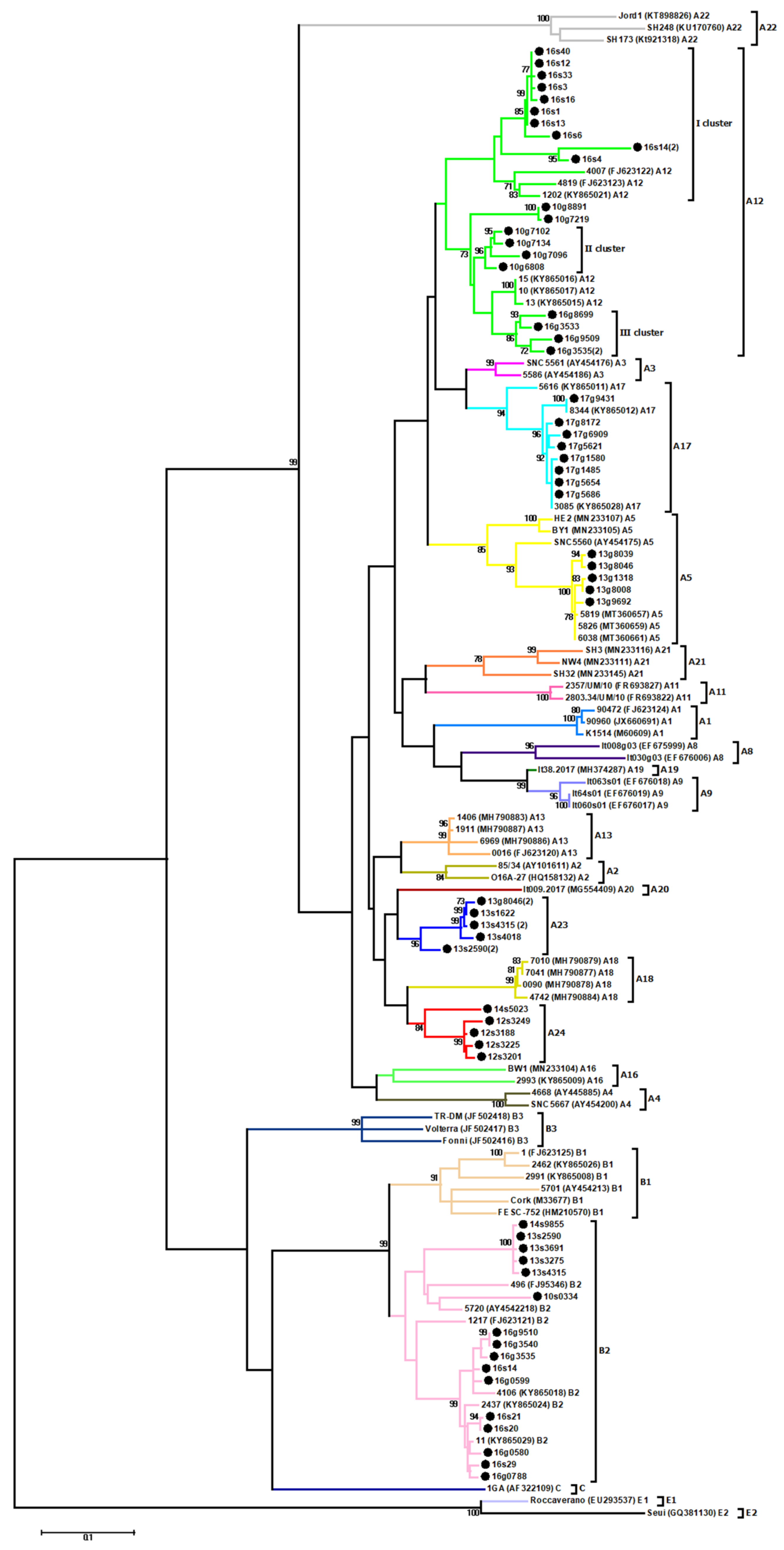
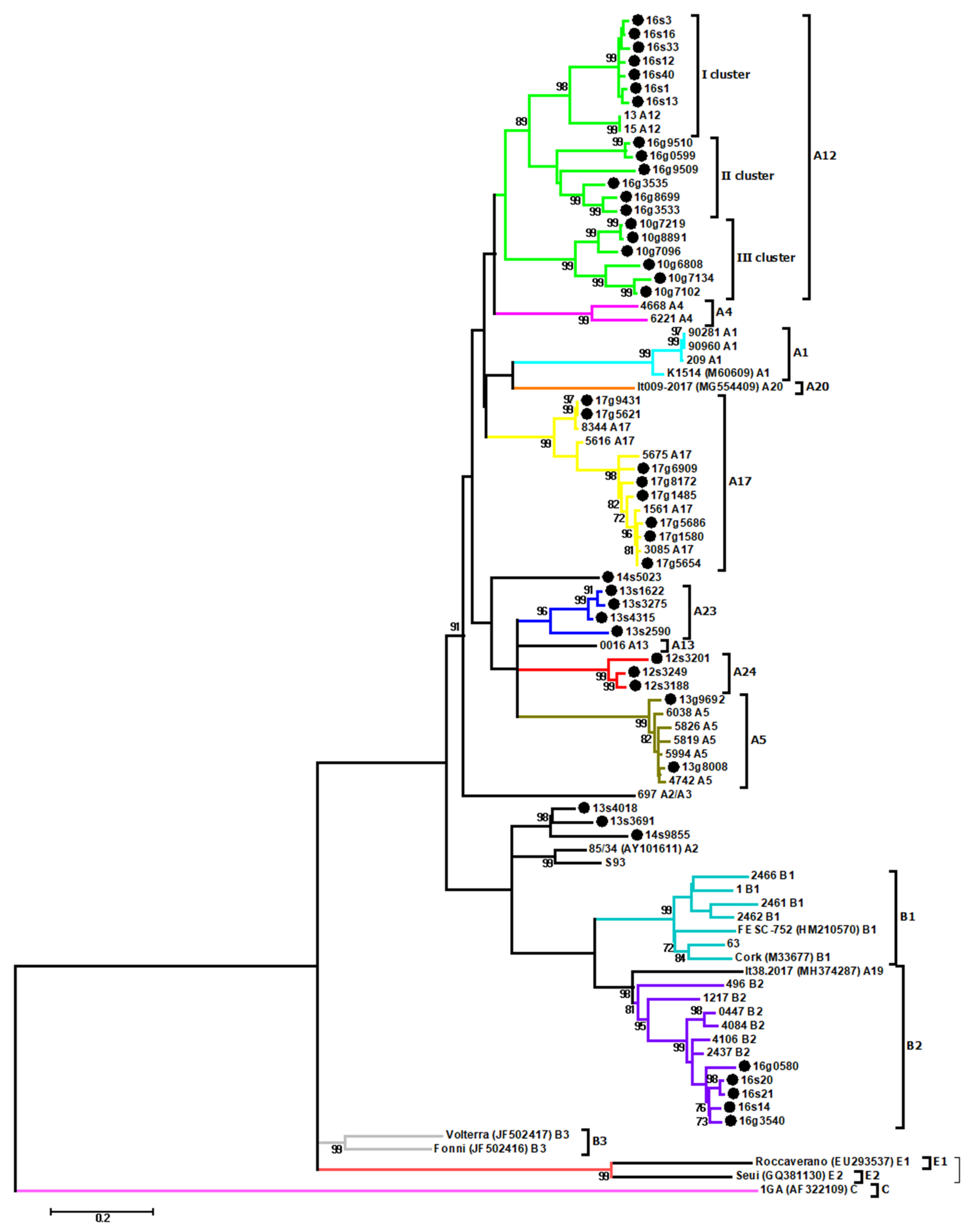
3.1. Phylogenetic Analysis

Out of 263 serum samples, 84 (32.0%) (53 from goats and 31 from sheep) originating from eight flocks were positive in the ELISA test. DNA extracted from the blood of serologically positive animals was used to amplify the CA fragment of the gag gene for phylogenetic analysis. Proviral DNA of 54 samples originated from 26 sheep and 28 goats from six different flocks was successfully amplified and sequenced (Table 2).
Obtained sequences were aligned with reference sequences representing the genotypes of SRLVs described to data, however, we included only sequences of appropriate length matching to data obtained in this study. An unrooted phylogenetic tree is shown in Figure 1.
Sequences of Polish strains analyzed in this study were widely distributed on the tree, clustering in subtype B2, A5, A12, and A17. In particular, sequences of sample s#21, s#20, g#9510, g#3540, s#14, g#0599, g#3535, g#0788, g#0580, and s#29 from flock 16 clustered within subtype B2 were closely related with Polish strains #11, #2437 and #4106, which were previously detected in the same Polish region (mean nt distance 2.6% ± 0.5%). Sequences originated from sheep #2590, #3691, #3275, #4315 from flock 13, from sheep #9855 from flock 14 and from sheep #0334 from flock 10 also clustered within subtype B2 but were more closely related with sequences of Spanish strain #496 and Swiss strain #5720 (mean nt distance 7.3% ± 1.8%). Sequences originating from goat #8039, #8046, #9692, #1318, and #8008 from flock 13 were closely related with Polish strains #6038, #5819, and #5826 (mean nt distance 1.3% ± 0.4%), representing subtype A5, while sequences originated from goat #9431, #8172, #6909, #5621, #1580, #1485, #5654, and #5686 from flock 17 were more closely related with Polish strains #5616, #8344, and #3085 representing subtype A17 (mean nt distance 2.6% ± 0.6%). Twenty sequences had been assigned to the A12 subtype and phylogenetic analysis clearly showed the existence of three separated subgroups within this cluster. Sequences from sheep #40, #12, #33, #3, #16, #1, #13, #6, #4 and #14 from flock 16 (I cluster) were closely related with sequences of Polish strains #4007, #4819 and #1202 with mean nt distance of 4.0% ± 1.4%. Sequences from goat #7219, #8891, #7102, #7134, #7096 and #6808 from flock 10 (II cluster) and sequences from goat #8699, #3533, #9509 and #3535, from flock 16 (III cluster) were closely related with sequences of Polish strains #15, #10 and #13 but they formed separated clusters (mean nt distance 7.9% ± 1.2%). Additionally, sequences from sheep #1622, #4315, #4018, #2590, and goat #8046, from flock 13 as well as sequences from sheep #5023, #3249, #3188, #3225, and #3201 formed new clusters within group A, which could be tentatively named as A23 and A24, respectively. Affiliation of these new clusters were supported with high bootstrap values ≥84. Sequences of the proposed subtype A23 and A24 had a mean sequence similarity (intra-subtype similarity) of 4.3% and 3.4%, respectively. The mean genetic distances between new subtypes and other subtypes representative for genotype A varied from 13.1% to 24.6% for subtype A23, and from 12.5% to 22.3% for subtype A24 (Table 3). To evaluate the robustness of our analysis, we also performed phylogenetic analysis using neighbor-joining and the Bayesian inference method (Supplementary Materials Figures S1 and S2), which resulted the same classification of all strains, whereby supporting the existence of new subtypes A23 and A24. Subtypes A5 and A17 have been found only in goats while subtype A24 has only been detected in sheep. In contrast, subtypes A12, A23 and B2 have been found in both sheep and goats. Dual infection with B2 and A12 was found in sheep #14 and goat #3535 from flock 16. Co-infection with B2/A23 was detected in sheep #2590 and sheep #4315 from flock 13 while co-infection with subtypes A5/A23 was detected in goat #8046, also originating from flock 13. Only in two flocks detected SRLV sequences representing one genotype. In flock 12 circulated only subtype A24 while in flock 17, subtype A17. This was confirmed by pairwise nucleotide comparison, in which distances estimated among sequences derived from flocks 12 and 17 varied from 1% to 2.9% and from 0% to 4.3%, respectively. On the other hand, in four flocks highly divergent SRLV subtypes were found: subtypes B2/A12 in flock 10, subtypes A5/A23/B2 in flock 13, subtypes A24/B2 in flock 14, and subtypes B2/A12 in flock 16.
All samples were also used to amplify 608 bp fragment of env gene. Out of 54 tested samples, 45 (20 from sheep and 25 from goats) were successfully amplified, and after sequencing were subjected to phylogenetic analysis. The phylogenetic tree (Figure 2) confirmed that Polish sequences belonged to subtype B2, A5, A12, and A17, as well as to new identified subtypes A23 and A24. Similarly to the gag phylogenetic assignment, env sequences from sheep #2590, #4315, #3275, and #1622 from flock 13 formed the new subtype A23, while sequences from sheep #3188, #3249 and #3201 from flock 12 formed subtype A24. The mean nucleotide distance between sequences belonging to the A23 subtype and those belonging to A24 subtype was 22.0%, while the mean distance between these subtypes and other subtypes within group A ranged from 18.5% to 27.8% and from 20.4% to 27.6% for A23 and A24, respectively. Sequences from sheep #9855, #3691 and #4018 previously located in subtypes B2 and A23, respectively, now created a separate branch clustered closely with North American MVV strains 85/34 and S93. In addition, sequences from sheep #5023, which on the basis of gag fragment was affiliated to subtype A23, now formed a separate cluster.

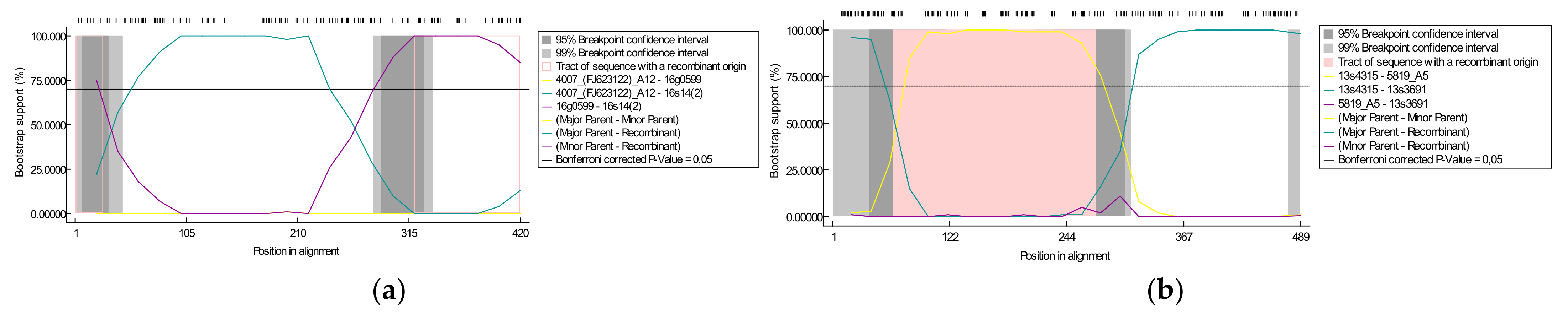
3.2. Identification of Putative Recombination

A recombination analysis of sequences used for phylogenetic analysis was performed to verify if sequences obtained in this study resulted from a recombination of already known sequences. On the basis of gag alignment, one putative recombination event was detected by five statistical methods with high significance and reliability. The recombinant sequence #14(2) was detected in sheep co-infected with strains A12/B2 from flock 16. In this recombination event, the beginning and ending breakpoints were located at 27 and 320 nucleotides in alignments and the major and minor parents were #4007, representing subtype A12 (90.9% similarity) and #0599 representing subtype B2 (100% similarity), respectively (Figure 3a). On the basis of env alignment, two putative recombination events were detected. Four methods detected a recombination event in #13s4018 between positions 64 and 276 in alignment with #13s4315 (subtype A23) as the minor parent and unknown, suggesting #5819 (subtype A5), as the major parent. The results also indicated that #13s3691 arose from recombination events between the same breakpoints position, 64 and 276 in alignment, but with #13s4315 (subtype A23) and unknown, (suggesting #5819 A5), as the major and minor parents, respectively (Figure 3b).

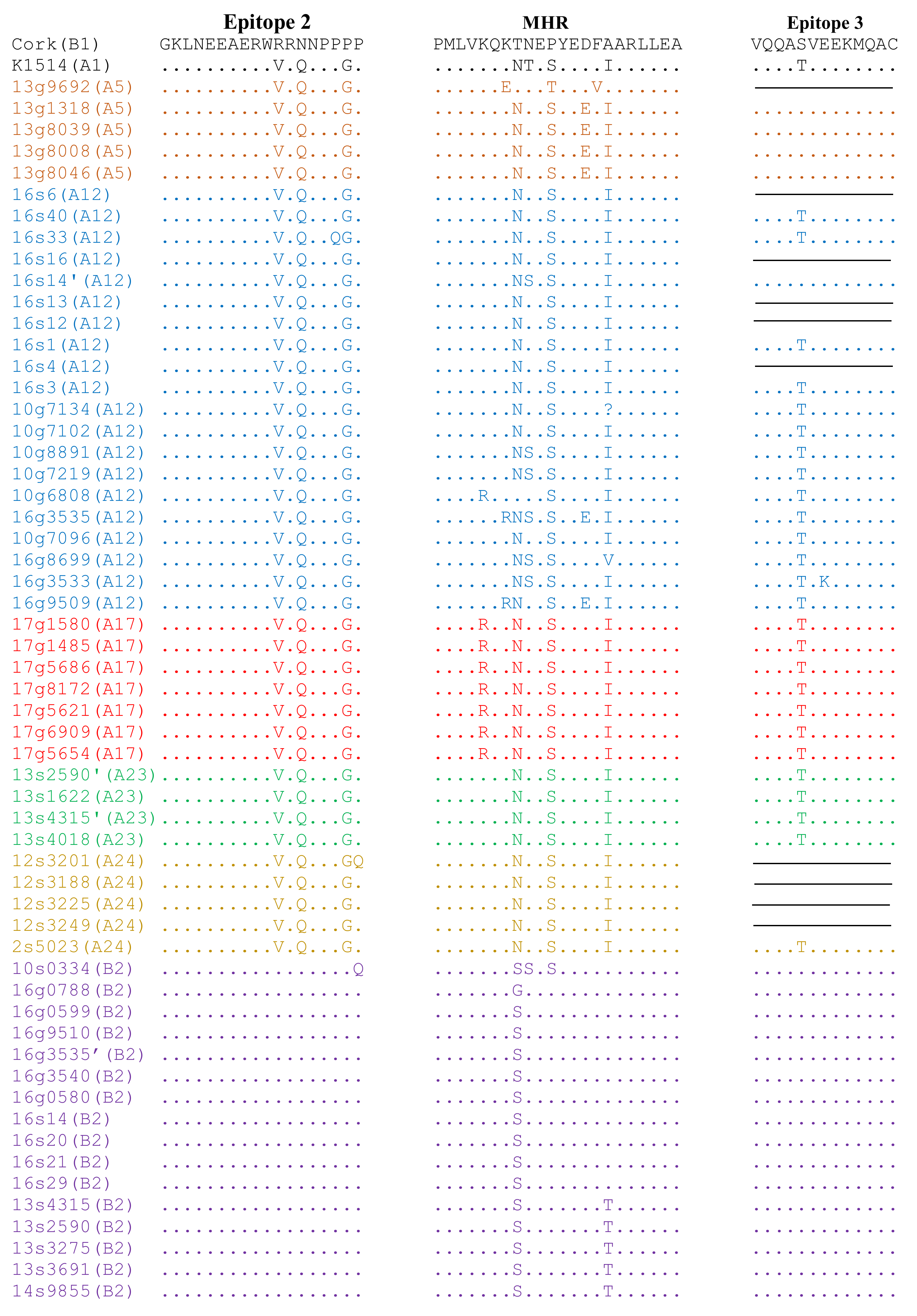
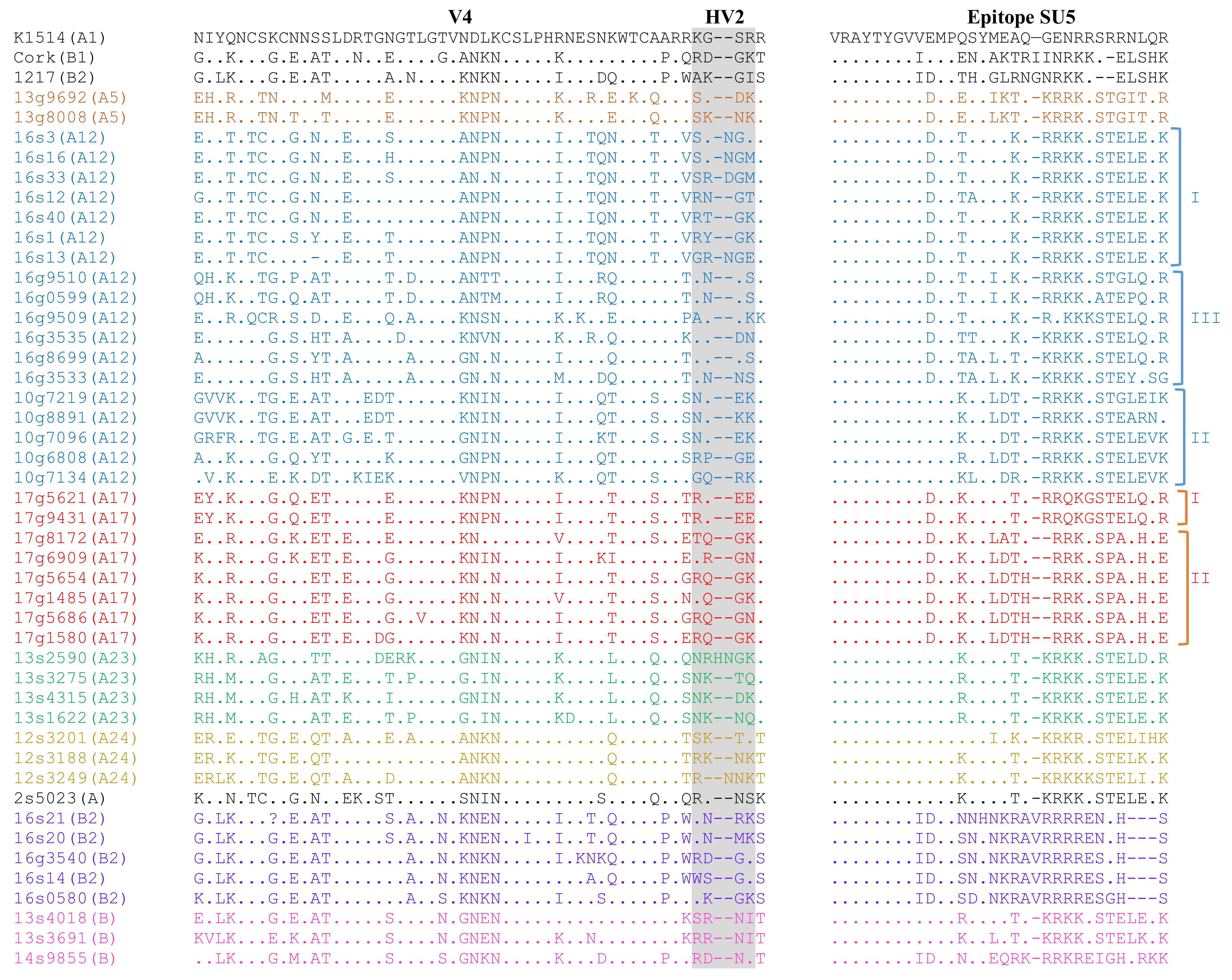
3.3. Analysis of Immunodominant Regions

To analyze the conservation of immunodominant regions of sequences of Polish strains analyzed in this study, their deduced amino acid (aa) sequences of capsid and surface proteins were aligned with the aa sequences of reference parental strains, Cork and K1514. Pairwise percent identity of the gag amino acid sequences of Polish SRLVs was high and ranged from 81.3% to 100%. Furthermore, Polish sequences shared 82.1–96.3% and 92.4–97.7% amino acid sequence identity with strains Cork and K1514, respectively. Analysis revealed that all gag sequences belonging to group B had glycine-glycine (GG) motifs and all MVV-like sequences had asparagine-valine (NV) motif, typical for strains belonging to group A (Supplementary Materials Figure S3). Immunodominant epitope 3, situated at the C-terminal end of the capsid protein, was highly conserved between sequences belonging to group A and B, while the analysis of sequences in epitope 2 revealed a perfectly conserved region (GKLNEEAERW) located at the N-terminal part of epitope and distinct region at the C-part of the epitope specific for MVV-like (VRQNPPGP) and CAEV-like (RRNNPPPP) strains. In the Major Homology Region (MHR), which is usually highly conserved in the gag gene of all retroviruses, some alterations were present within group A (Figure 4). In particular, sequence from goat #9692 had isoleucine (I) instead of valine (V) at the fourth position compared to strain #K1514, while sequences from goat #6808 and #8699 had threonine (T) instead of asparagine (N), and valine (V) instead of isoleucine (I) at the eighth and 16th positions, respectively. All sequences representing subtype A17 and sequence from goat #6808 representing subtype A12 had substitutions arginine (R) instead of lysine (K) at the fifth position, while sequences from goat #3535 and #9509, representing subtype A12, had substitution arginine (R) instead of lysine (K) in the seventh position of MHR. All goat-derived sequences representing subtype A5 and sequences from goats #3535 and #9509 had substitution glutamic acid (E) instead of aspartic acid (D) at the 14th position. Six sequences had substitution serine (S) instead of threonine (T) and 35 sequences had substitution asparagine (N) instead of threonine (T) at the ninth position compared to the sequences of strain #K1514. Type B sequences had more conservative MHR sequences. Compared to sequences of strain Cork, all sequences had substitution at the eighth position (T/S or T/G). Sequences from sheep #0334 had substitution serine (S) instead of asparagine (N) and serine (S) instead of proline (P) at the ninth and eleventh positions. Five sequences representing subtype B2 which formed subclusters on the phylogenetic tree had unique substitution threonine (T) instead of alanine (A) at the 16th position of MHR.
The env aa sequences of Polish strains were more heterogenous than gag sequences showing 54.1–100% similarity to each other and 57.1–73.1% and 59.4–73.7% to the strains K1514 and Cork, respectively. Sequences of the variable region (V4) of analyzed strains differed significantly. Comparison of aa sequences in epitope SU5 revealed the conserved region (VRAYTYGV) located at the N-terminal part of the epitope. The variable region was conserved among the sequences of strains belonging to subtypes A5, A23, A24, and B2 showing type-specific variation. Sequences belonging to subtypes A12 and A17 showed intra-subtype variability (Figure 5). Sequences could be divided into groups corresponding groups formed on the phylogenetic tree.
To determine if the nucleotide sequences encoding Gag and Env proteins evolved under positive selective pressure, the ratio of nonsynonymous to synonymous base substitutions were estimated. The results showed that for both fragments, the dN/dS ratio for caprine and ovine sequences was below 1, showing a negative selection. Additionally, the number of potential N-linked glycosylation sites (PNGS) was estimated and ranged from 3 to 6 in respective sequences. In all caprine sequences of subtype A17 from flock 17, and in all caprine sequences of subtype A12 from flock 10, four potential N-linked glycosylation sites were detected, in positions 8, 14, 32, and 39 in the alignment. These four PNGS were also detected in ovine sequence #5023 from flock 14, which formed a new cluster within group A on the phylogenetic tree. In two out of three ovine sequences, representing subtype A24 from flock 14, and in three sequences (14s9855, 13s4018, and 13s3691) which formed a cluster distinct from known A and B subtypes additional glycosylation site, at position 53, was detected. In sequences from flocks 13 and 16, 3-6 N-linked glycosylation sites were observed, but with some heterogeneity in the positions between different isolates. In most ovine sequences representing subtype A23 from flock 13, four PNGS were detected (at positions 8, 14, 32, and 39), while in caprine sequences representing subtype A5, the number of PNGS ranged from 3 to 5 and were located at different positions. In most of the A12 sequences originating from the sheep from flock 16, four PNGS were detected (at positions 14, 32, 39, and 43), while in caprine sequences representing subtype A12, five PNGS were detected (at positions 8, 14, 32, 39, 43, and 52). B2 sequences derived from this flock had four PNGS at positions 8, 14, 32, and 39.

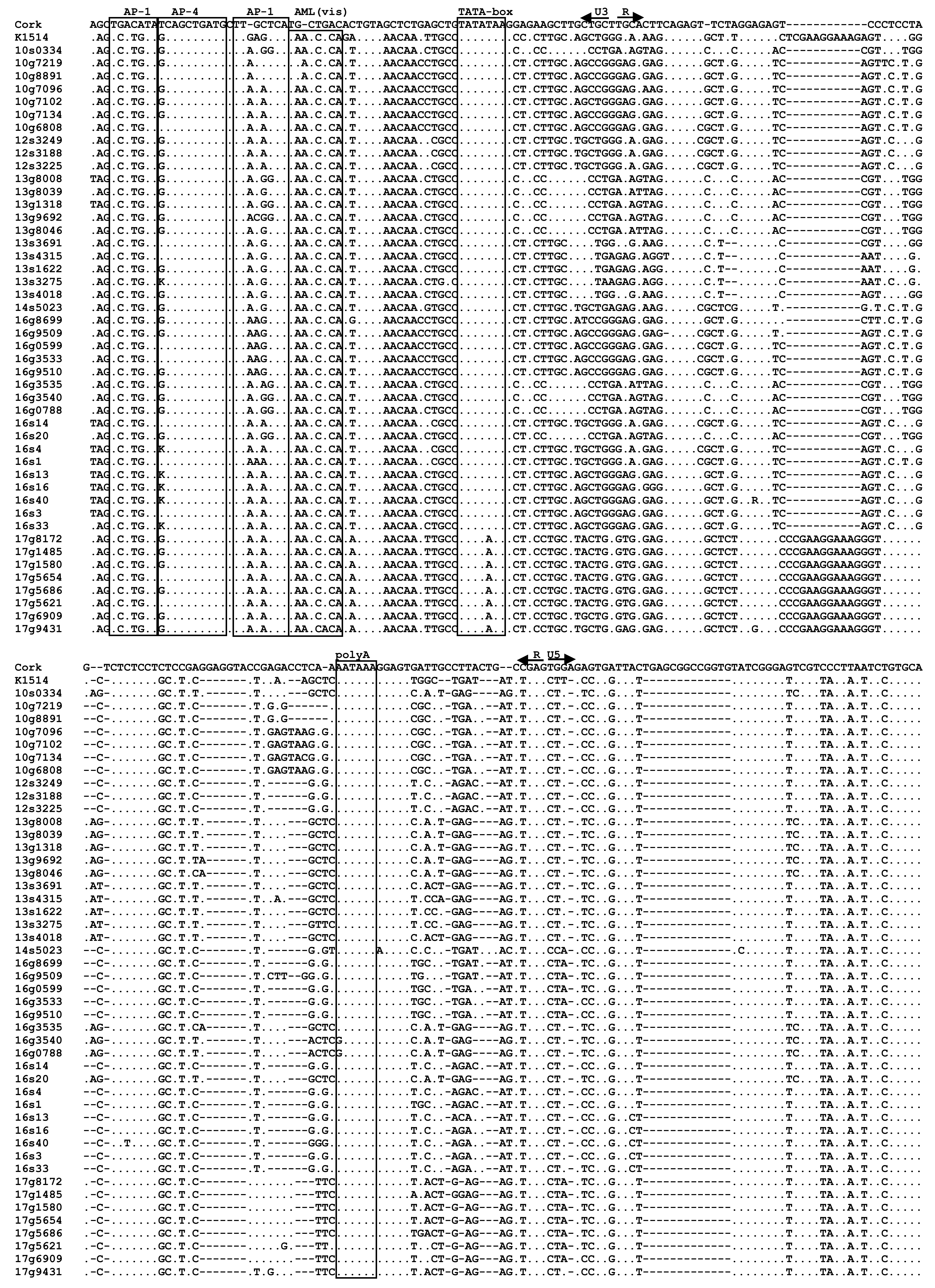
3.4. Analysis of LTR Sequences

LTR sequences from 47 out of 54 samples (87%), which were successfully amplified, were aligned with sequences of prototype strains K1514 and CAEV-Cork representative for SRLVs groups A and B, respectively. The nucleotide pairwise percent identity of Polish sequences ranged from 72.3% to 100%. Furthermore, Polish sequences shared 48.4–53.8% and 74.0–85.8% sequence identity with strains Cork and K1514, respectively. The LTR regions analyzed in this study contained two AP-1, one AML (vis) and one AP-4 putative motifs without any duplication or deletion in their U3 regions compared with reference sequences (Figure 6). Sequences corresponding to the TATA box, AP-4, and polyadenylation signal (poly A) were quite conserved. All sequences belonging to subtype A17 had unique substitution T to A in the fifth position of TATA box. Sequence of AML(vis) motif was present in all Polish samples and was identical with the sequence of K1514 strain, except for sequences originating from goats #8891 and #7219 which had substitution A to G in the second position of AML-vis. The AP-1 sites were less conserved. Sequences of the first AP-1 site were identical in all Polish sequences analyzed and the sequence of K1514 strain (TCATGTA), but differed from the sequence of Cork strain (TGACATA), while in the second AP-1 site considerable nucleotide changes were observed. All Polish sequences analyzed, except sequences representing subtype A17, had the specific 11 nt deletion in the R region. However, A17 sequences had the CCGAAGGAAAG insertion almost identical, like in the K1514 strain. Furthermore, all Polish sequences had 13 nt deletion in the U5 region.

4. Discussion

Previous studies revealed that the SRLVs population in Poland is highly heterogeneous. SRLVs isolated so far from sheep and goats in Poland belonged to the well-known subtypes B1, B2, A1, A5, and A16, as well as subtypes A12, A13, A17, and A18—detected only in Poland. Since mixed flocks promote interspecies transmission and the emergence of new variants [18,19,20], the aim of this study was to perform genetic characterization of field SRLV strains present in Polish mixed flocks to get a better insight into their heterogeneity.
The current SRLVs phylogeny consisting of five main groups, which are divided in multiple subtypes, emphasizes the high genetic diversity among SRLV strains. In 2004, Shah et al. proposed a classification of SRLVs based on sequences of the gag-pol (1.8 kb) [40]. However, due to low proviral load and high genetic variability of SRLV strains, in many cases this fragment could not be obtained [12,18,41,42]. As a result, classification of SRLVs is more often performed on a very conservative ~0.4 kb gag fragment for which sequences representing most of subtypes are available. Using this fragment, we confirmed circulation of subtypes B2, A5, A12, and A17 in analyzed flocks and revealed circulation of new subtypes A23 and A24. These new subtypes were closely related to subtypes A13 and A18 which were previously detected only in Poland, suggesting their common origin. Clearly separation of these subtypes from other Polish subtypes, located in distinct clusters on the phylogenetic tree, suggests the presence of at least three SRLV genetic lines circulating in Poland which are independently evolving. As was previously reported, some strains can cluster to different subtypes depending on the fragment that is analyzed. Such an observation was noted for the subtype A19, A20, and B5 strains ([10], this report). This suggests that these strains could be generated by recombination. Thus, to confirm the existence of our putative new subtypes, we decided to perform phylogenetic analysis using variable env sequences, which confirmed that they formed a separate clusters with no clear relation to any of the previously described group A subtypes.
Lentiviral genomes are among the most rapidly evolving known. Most of the mutations are introduced during the reverse transcription stage of the viral life cycle as a consequence of low fidelity of reverse transcriptase, which has no proofreading activity. However, interspecies transmission, co-infections, and recombinations are the main mechanisms which contribute significantly to genetic variability and accelerate viral evolution [17,43,44,45]. Direct evidence of interspecies transmission of SRLVs from sheep to goats and vice versa has been documented by the detection of most subtypes in both sheep and goats [16,18,24,46]. It is also evidenced that mixed flocks, where sheep and goats are kept together, is the factor promoting cross-species infections which can result in the emergence of new variants [18,19,20]. In our study, subtypes A5 and A17 have been found only in goats, subtype A24 has been detected only in sheep, while subtypes A12, A23, and B2 have been found in both sheep and goats. This confirms the ability of SRLVs to frequently cross the species barrier under natural conditions. Furthermore, co-infection with strains belonging to different subtypes was evidenced in three sheep and two goats which originated from two flocks. Sheep #14 and goat #3535 from flock 16 were co-infected with B2/A12, while two sheep (#2590 and #4315) and one goat (#8046) originated from flock 13 were co-infected with B2/A23 and A5/A23, respectively. Existence of co-infected animals can be explained by circulation of more than one subtype in these flocks. Our results revealed that in four out of six flocks, highly divergent SRLV subtypes were found (subtypes B2/A12, A5/A23/B2, A24/B2, and B2/A12). In flock 13, circulation of sequences belonging to subtypes A5, A23 and B2 were found. All goats were infected with strains representing subtype A5, while in sheep, only sequences belonging to subtypes A23 and B2 were found. Only one goat was co-infected with A5/A23 which strongly indicates that the direction of transmission of subtype A23 in this case was from sheep to goat. Mean gag nucleotide distance of A23 sequences found in this co-infected goat and A23 sequences found in sheep was 3.5%—strongly confirming their common origin. Furthermore, two sheep from this flock were co-infected with subtypes A23 and B2. However, it is difficult to explain which subtype was first introduced to the flock, because both subtypes were detected at the same frequency. The gag mean nucleotide distance of B2 and A23 sequences was 0.5% and 3.9%, respectively, while the mean nucleotide distance between these subtypes was 32.9%. This confirms the circulation of two distinct subtypes in the sheep from this flock which do not represent the evolution of the homologous strains. In flock 16, circulation of two subtypes, A12 and B2, was detected in both, sheep and goats. Because the most of sheep were infected with subtype A12 and most of goats were infected with subtype B2, the transmission of these subtypes is suggested to be from goats to sheep and from sheep to goats, respectively. Interspecies transmission was also observed in flock 10 and flock 14, where subtypes A12/B2 and B3/A24 were found. The high genetic distance of sequences found in these flocks strongly suggests that they originated from different sources. The introduction of different subtypes of SRLVs in analyzed flocks could not be tracked back because we don’t know the history of animal movements, but they most likely resulted from the purchase of infected animals from other flocks. The introduction of new animals to a flock for genetic improvement is common practice in Poland and undoubtedly represents a high risk factor for the spread of SRLVs, which may result in higher diversity of SRLVs [40,47,48]. In Poland, it is also facilitated by the lack of SRLVs eradication programs and any veterinary controls.
Co-infection with at least two subtypes offers opportunities for viral recombination which is believed to be a powerful source of genetic variability of lentiviruses leading to the emergence of new strains. Previous studies have provided clear evidence of recombination between SRLVs belonging to groups A and B, as well as between subtypes belonging to the same group [18,20,24,45,46,49,50]. In the present study, three examples of recombinations have been found. One putative recombination event was detected in the gag fragment in sheep #14 co-infected with strains A12/B2, while on the basis of env fragments, two putative recombination events were detected in two sheep from flock 13. Recombination is a frequent event in the envelope gene which could lead to the generation of chimerical SRLVs with altered cell tropism, pathogenicity and transmission efficiency which may endanger not only domestic ruminants but also other animal species [51,52,53].
Analysis of sequences is important, not only for evaluating the spread of SRLV subtypes but also for gaining knowledge of antigenic variability. Alterations in the amino acid sequences of immunodominant epitopes determine their antigenicity and may impact on sensitivity of serological tests. The primers used in this study allowed the sequencing of two immunodominant epitopes of capsid protein—epitopes 2 and 3, which were identified by Rosati et al. [54] and which are used in SRLVs ELISA tests. The CA is the major viral core protein and antibodies against this antigen are usually first generated in sheep and goats infected with SRLVs and remain detectable for a long time [55]. Our results, which are consistent with other reports, showed conservation of epitope 3 and the amino-terminal part of epitope 2 (GKLNEEAERW), as well as the group-specific part located at the C-termini of epitope 2 (group A, VRQNPPGP, group B, RRNNPPPP) [24,25,56,57]. More variability was found in the MHR, which is usually highly conserved in many retroviruses [12,58,59]. MHR of Polish MVV-like sequences had more alteration and revealed unique mutations in sequences of subtypes A5 and A17. All Polish sequences representing subtype A17 had unique substitution lysine (K) to arginine (R) at the fifth position of MHR, while substitution aspartic acid (D) to glutamic acid (E) at the 14th position of MHR was exclusively found in Polish sequences representing subtype A5. Because these subtypes were found in goats, we hypothesize that these changes may have resulted from cross-species transmission, as many genetic changes after SRLVs cross-species transmission were reported in many publications [1,16,20,40,51]. However, subtypes A17 and A5 were detected only in goats of Polish White Improved/Polish Fawn Improved and Carpathian breeds, respectively. This may also suggest that mentioned changes could have arisen as a result of long host-virus adaptation and evolution.
As expected, the SU sequences showed more extensive variations in comparison to CA sequences. Previous sequence analysis of SRLV strains defined five major variable regions of SU [60], but sequences obtained in this study allowed for the analysis of only the V4 and V5 regions. Although the dN/dS ratio for V4V5 sequences was below 1, indicating the existence of a purifying selection, we noticed that some regions may be under positive selection. Our results revealed that the V4 region of Polish strains differed significantly, and that the differences mainly occurred within a region previously proposed to be part of a variable, conformational neutralization epitope [61,62]. Moreover, insertion and deletion only occurred in highly variable region (HV2) [61], confirming that this region underwent rapid sequence evolution during SRLVs infection. Mutation in this region resulted in escape from neutralization and the creation of a new type of neutralization specificity [62]. Thus, it is suggested that the V4 region may have an analogous function to V3 in HIV-1 since the V3 loop of HIV-1 is the major target for neutralizing antibodies [45,63]. Interestingly, in the V4 region, a ’signature pattern’ related to different clinical status in sheep and goats has been found [64]. Comparison of aa sequences in epitope SU5 revealed conserved region (VRAYTYGV) located at the N-terminal part of the epitope and highly variable motif at the C- terminal which was well-conserved among strains belonging to the same subtypes confirming that the SU5 epitope is responsible for type-specific immune response allowing strain-specific diagnosis. Moreover, this observation appears to support the hypothesis that this region may function as a decoy antigen and, therefore, in a given subtype, evolves under negative selection [65]. Additionally, the V4V5 regions contain the majority of the conserved N-linked glycosylation sites and cysteine residues, suggesting that they form a highly constrained and surface-exposed domain [60]. These sugars, or “glycans,” play several important roles in the infective cycle of the viruses. In HIV-1, effects of glycosylation on viral replication, glycoprotein cleavage, CD4 binding activity, and coreceptor usage have been documented. Removal of the glycan may indirectly increase viral activity by causing a shift in the V3 loop, leading to an increase in co-receptor binding while high density of glycans protect the virus against neutralizing antibodies as a “glycan shield” [66,67]. In this study, we observed differences in the number of potential N-linked glycosylation sites (PNGS) which ranged from 3 to 6 in respective sequences. Most of the sequences had four PNGS which were detected, at positions 8, 14, 32, and 39 in the alignment, suggesting that these PNGSs may be evolutionarily conserved. Although some glycans are evolutionarily conserved, the number of others may vary extensively within infected individuals, since they may appear or disappear over the course of an infection in a single host [66,67]. Thus, variation in the number and position of PNGSs in Polish sequences may result from a host-species adaptation of SRLVs, as was evidenced for HIV-1. In general, the average numbers of predicted glycosylated positions were relatively conserved between the different subtypes of SRLVs.
U3-R regions contain elements described as important for the regulation of SRLVs transcription and replication. Therefore, sequence variation in LTRs might affect the interactions with cellular factors and alter viral expression and replication. Furthermore, it was also demonstrated that LTR sequence variability may affect tissue tropism and disease outcome [63]. The analysis of Polish LTR sequences revealed that sequences corresponding to the TATA box, AP-4, AML-vis, and polyadenylation signal (poly A) were quite conserved which was previously reported in strains from different geographic areas. This sequence conservation argues in favor of their importance in the replication strategies of SRLVs [12,26,42,68,69,70]. On the other hand, considerable alteration was observed in AP-1 sites, which confirmed previous findings suggesting that AP-1 sites may by functional even despite some changes [70,71]. AP-1 binding sites are important for regulation of SRLVs expression in macrophages and are required for phorbol ester-inducible gene expression of SRLV. Multiple copies of AP_1 sites presumably allow transcription regardless of mutations [69,70,71]. The TATA box is present and highly conserved in all retroviruses [70]. What is interesting is that our results revealed that all sequences belonging to subtype A17 had unique substitution T to A in the fifth position of the TATA box. Mutation in the TATA box of HIV-1 sequence changes the binding of the TATA-binding protein (TBP) which leads to a decrease in transcription [72,73]. The meaning of mutation detected in TATA box of subtype A17 SRLVs is unknown and warrants further study. Additionally, our results revealed that all sequences representing subtype A17 did not have a 11 nt deletion in the R region, which was noted for other sequences from Poland. Correlation between this deletion and the occurrence of clinical signs in infected animals has been suggested [51,69,74], but our results did not confirm these assumptions. All animals infected with both virus carrying the deletion and without deletions, were without clinical signs.
In conclusion, the results of this work extend the current knowledge on the distribution of SRLV subtypes in sheep and goats from Poland. Our results showed that SRLVs circulating in Poland are highly heterogeneous with ovine and caprine strains belonging to group A and B. We present strong and multiple evidence of dually infected sheep and goats in mixed flocks, and present evidence that these viruses can recombine in vivo. The results of the phylogenetic analysis revealed the existence of putative new subtypes which should lead to the consideration of an update of current SRLVs classification. Furthermore, genetic analysis of Polish SRLV sequences revealed some specific alteration present in gag and LTR gene fragments in some subtypes. Thus, the isolation and characterization of biological properties of these viruses should be performed to evaluate their pathogenic potential.

Supplementary Materials

The following are available online at https://0-www-mdpi-com.brum.beds.ac.uk/article/10.3390/v13122529/s1, Figure S1: Neighbor-joining phylogenetic tree based on alignment of CA fragment of gag gene. Figure S2: Bayesian phylogenetic tree based on alignment of CA fragment of gag gene. Figure S3: Alignment of deduced amino acid sequences of immunodominant epitopes of capsid protein and major homology region (MHR) of the SRLVs obtained in this study and reference strains.

Author Contributions

Conceptualization, M.O. and J.K.; methodology, M.O.; software, M.O.; formal analysis, M.O.; investigation, M.O.; resources, M.O. and J.K.; data curation, M.O.; writing—original draft preparation, M.O.; writing—review and editing, M.O. and J.K.; visualization, M.O.; funding acquisition, M.O. All authors have read and agreed to the published version of the manuscript.

Funding

The work was supported by the National Science Center (NCN) project no. 2016/21/D/NZ7/00626.

Institutional Review Board Statement

The study was conducted in accordance with the relevant guidelines and regulations. Specifically, blood collection was approved (no. 37/2016) by the Local Ethical Committee on Animal Testing at the University of Life Sciences in Lublin (Poland).

Informed Consent Statement

Not applicable.

Data Availability Statement

All data generated and analyzed in this study are included in this article.

Conflicts of Interest

The authors declare no conflict of interest.

References

  1. Minardi da Cruz, J.C.; Singh, D.K.; Lamara, A.; Chebloune, Y. Small ruminant lentiviruses (SRLVs) break the species barrier to acquire new host range. Viruses 2013, 5, 1867–1884. [Google Scholar] [CrossRef] [PubMed] [Green Version]
  2. Narayan, O.; Clements, J.E.; Strandberg, J.D.; Cork, L.C.; Griffin, D.E. Biological characterization of the virus causing leukoencephalitis and arthritis in goats. J. Gen. Virol. 1980, 50, 69–79. [Google Scholar] [CrossRef] [PubMed]
  3. Peterhans, E.; Greenland, T.; Badiola, J.; Harkiss, G.; Bertoni, G.; Amorena, B.; Eliaszewicz, M.; Juste, R.; Kraßnig, R.; Lafont, J.-P.; et al. Routes of transmission and consequences of small ruminant lentiviruses (SRLVs) infection and eradication schemes. Vet. Res. 2004, 35, 257–274. [Google Scholar] [CrossRef] [Green Version]
  4. Minguijón, E.; Reina, R.; Pérez, M.; Polledo, L.; Villoria, M.; Ramírez, H.; Leginagoikoa, I.; Badiola, J.J.; García-Marín, J.F.; de Andrés, D.; et al. Small ruminant lentivirus infections and diseases. Vet. Microbiol. 2015, 181, 75–89. [Google Scholar] [CrossRef] [PubMed]
  5. Villet, S.; Bouzar, B.A.; Morin, T.; Verdier, G.; Legras, C.; Chebloune, Y. Maedi-visna virus and caprine arthritis encephalitis virus genomes encode a Vpr-like but no Tat protein. J. Virol. 2003, 77, 9632–9638. [Google Scholar] [CrossRef] [Green Version]
  6. Pepin, M.; Vitu, C.; Russo, P.; Mornex, J.F.; Peterhans, E. Meadi-Visna virus infection in sheep: A review. Vet. Res. 1998, 29, 341–367. [Google Scholar]
  7. Saltarelli, M.; Querat, G.; Konings, D.A.M.; Vigne, R.; Clements, J.E. Nucleotide sequence and transcriptional analysis of molecular clones of CAEV which generate infectious virus. Virology 1990, 179, 347–364. [Google Scholar] [CrossRef]
  8. Gomez-Lucia, E.; Barquero, N.; Domenech, A. Maedi-Visna virus: Current perspectives. Vet. Med. 2018, 9, 11–21. [Google Scholar] [CrossRef] [PubMed] [Green Version]
  9. Molaee, V.; Bazzucchi, M.; De Mia, G.M.; Otarod, V.; Abdollahi, D.; Rosati, S.; Lühken, G. Phylogenetic analysis of small ruminant lentiviruses in Germany and Iran suggests their expansion with domestic sheep. Sci. Rep. 2020, 10, 2243. [Google Scholar] [CrossRef] [PubMed] [Green Version]
  10. Michiels, R.; Adjadj, N.R.; De Regge, N. Phylogenetic Analysis of Belgian Small Ruminant Lentiviruses Supports Cross Species Virus Transmission and Identifies New Subtype B5 Strains. Pathogens 2020, 9, 183. [Google Scholar] [CrossRef] [Green Version]
  11. Reina, R.; Bertolotti, L.; Giudici, S.D.; Puggioni, G.; Ponti, N.; Profiti, M.; Patta, C.; Rosati, S. Small Ruminant Lentivirus Genotype E Is Widespread in Sarda Goat. Vet. Microbiol. 2010, 144, 24–31. [Google Scholar] [CrossRef] [PubMed] [Green Version]
  12. Reina, R.; Mora, M.I.; Glaria, I.; García, I.; Solano, C.; Luján, L.; Badiola, J.J.; Contreras, A.; Berriatua, E.; Juste, R.; et al. Molecular characterization and phylogenetic study of Maedi Visna and Caprine Arthritis Encephalitis viral sequences in sheep and goats from Spain. Virus Res. 2006, 121, 189–198. [Google Scholar] [CrossRef] [PubMed]
  13. Colitti, B.; Coradduzza, E.; Puggioni, G.; Capucchio, M.T.; Reina, R.; Bertolotti, L.; Rosati, S. A new approach for Small Ruminant Lentivirus full genome characterization revealed the circulation of divergent strains. PLoS ONE 2019, 14, e0212585. [Google Scholar] [CrossRef] [PubMed]
  14. Gjerset, B.; Storset, A.K.; Rimstad, E. Genetic diversity of small-ruminant lentiviruses: Characterization of Norwegian isolates of Caprine arthritis encephalitis virus. J. Gen. Virol. 2006, 87, 573–580. [Google Scholar] [CrossRef] [PubMed]
  15. Gjerset, B.; Rimstad, E.; Teige, J.; Soetaert, K.; Jonassen, C.M. Impact of natural sheep-goat transmission on detection and control of small ruminant lentivirus group C infections. Vet. Microbiol. 2009, 135, 231–238. [Google Scholar] [CrossRef] [PubMed]
  16. Pisoni, G.; Quasso, A.; Moroni, P. Phylogenetic analysis of small-ruminant lentivirus subtype B1 in mixed flocks: Evidence for natural transmission from goats to sheep. Virology 2005, 339, 147–152. [Google Scholar] [CrossRef] [Green Version]
  17. Leroux, C.; Cruz, J.C.; Mornex, J.F. SRLVs: A genetic continuum of lentiviral species in sheep and goats with cumulative evidence of cross species transmission. Curr. HIV Res. 2010, 8, 94–100. [Google Scholar]
  18. Fras, M.; Leboeuf, A.; Labrie, F.M.; Laurin, M.A.; Singh Sohal, J.; L’Homme, Y. Phylogenetic analysis of small ruminant lentiviruses in mixed flocks: Multiple evidence of dual infection and natural transmission of types A2 and B1 between sheep and goats. Infect. Genet. Evol. 2013, 19, 97–104. [Google Scholar] [CrossRef] [PubMed]
  19. Grego, E.; Bertolotti, L.; Quasso, A.; Profiti, M.; Lacerenza, D.; Muz, D.; Rosati, S. Genetic characterization of small ruminant lentivirus in Italian mixed flocks: Evidence for a novel genotype circulating in a local goat population. J. Gen. Virol. 2007, 88, 3423–3427. [Google Scholar] [CrossRef] [PubMed]
  20. Santry, L.A.; de Jong, J.; Gold, A.C.; Walsh, S.R.; Menzies, P.I.; Wootton, S.K. Genetic characterization of small ruminant lentiviruses circulating in naturally infected sheep and goats in Ontario, Canada. Virus Res. 2013, 175, 30–44. [Google Scholar] [CrossRef] [PubMed]
  21. Olech, M.; Osiński, Z.; Kuźmak, J. Bayesian estimation of seroprevalence of small ruminant lentiviruses in sheep from Poland. Prev. Veter Med. 2017, 147, 66–78. [Google Scholar] [CrossRef] [PubMed]
  22. Kaba, J.; Czopowicz, M.; Ganter, M.; Nowicki, M.; Witkowski, L.; Nowicka, D.; Szaluś-Jordanow, O. Risk factors associated with seropositivity to small ruminant lentiviruses in goat herds. Res. Vet. Sci. 2013, 94, 225–227. [Google Scholar] [CrossRef]
  23. Olech, M.; Rachid, A.; Croisé, B.; Kuźmak, J.; Valas, S. Genetic and antigenic characterization of small ruminant lentiviruses circulating in Poland. Virus Res. 2012, 163, 528–536. [Google Scholar] [CrossRef]
  24. Olech, M.; Valas, S.; Kuźmak, J. Epidemiological survey in single-species flocks from Poland reveals expanded genetic and antigenic diversity of small ruminant lentiviruses. PLoS ONE 2018, 13, e193892. [Google Scholar] [CrossRef] [Green Version]
  25. Olech, M.; Murawski, M.; Kuźmak, J. Molecular analysis of small-ruminant lentiviruses in Polish flocks reveals the existence of a novel subtype in sheep. Arch. Virol. 2019, 164, 1193–1198. [Google Scholar] [CrossRef] [PubMed] [Green Version]
  26. Olech, M.; Kuźmak, J. Molecular Characterization of Small Ruminant Lentiviruses of Subtype A5 Detected in Naturally Infected but Clinically Healthy Goats of Carpathian Breed. Pathogens 2020, 9, 992. [Google Scholar] [CrossRef] [PubMed]
  27. Ryan, S.; Tiley, L.; McConnell, I.; Blacklaws, B. Infection of dendritic cells by the Maedi-Visna lentivirus. J. Virol. 2000, 74, 10096–10103. [Google Scholar] [CrossRef] [PubMed] [Green Version]
  28. Mordasini, F.; Vogt, H.R.; Zahno, M.L.; Maeschli, A.; Nenci, C.; Zanoni, R.; Peterhans, E.; Bertoni, G. Analysis of the antibody response to an immunodominant epitope of the envelope glycoprotein of a lentivirus and its diagnostic potential. J. Clin. Microbiol. 2006, 44, 981–991. [Google Scholar] [CrossRef] [Green Version]
  29. Tamura, K.; Stecher, G.; Peterson, D.; Filipski, A.; Kumar, S. MEGA6: Molecular Evolutionary Genetics Analysis version 6.0. Mol. Biol. Evol. 2013, 30, 2725–2729. [Google Scholar] [CrossRef] [Green Version]
  30. Korber, B.; Rodrigo, A.G.; Learn, G.H. HIV Signature and Sequence Variation Analysis, Computational Analysis of HIV Molecular Sequences; Kluwer Academic Publishers: Dordrecht, The Netherlands, 2000; pp. 55–72. [Google Scholar]
  31. Zhang, M.; Gaschen, B.; Blay, W.; Foley, B.; Haigwood, N.; Kuiken, C.; Korber, B. Tracking global patterns of N-linked glycosylation site variation in highly variable viral glycoproteins: HIV, SIV, and HCV envelopes and influenza hemagglutinin. Glycobiology 2004, 14, 1229–1246. [Google Scholar] [CrossRef] [PubMed] [Green Version]
  32. Martin, D.P.; Murrell, B.; Golden, M.; Khoosal, A.; Muhire, B. RDP4: Detection and analysis of recombination patterns in virus genomes. Virus Evol. 2015, 1, vev003. [Google Scholar] [CrossRef] [PubMed] [Green Version]
  33. Martin, D.; Rybicki, E. RDP: Detection of recombination amongst aligned sequences. Bioinformatics 2000, 16, 562–563. [Google Scholar] [CrossRef] [PubMed]
  34. Padidam, M.; Sawyer, S.; Fauquet, C.M. Possible emergence of new geminiviruses by frequent recombination. Virology 1999, 265, 218–225. [Google Scholar] [CrossRef] [Green Version]
  35. Martin, D.P.; Posada, D.; Crandall, K.A.; Williamson, C. A modified bootscan algorithm for automated identification of recombinant sequences and recombination breakpoints. AIDS Res. Hum. Retroviruses 2005, 21, 98–102. [Google Scholar] [CrossRef] [Green Version]
  36. Smith, J.S.; Joshi, S.B. Reusable software concepts applied to the development of fms control software. Int. J. Comput. Integr. Manuf. 1992, 5, 182–196. [Google Scholar] [CrossRef]
  37. Posada, D.; Crandall, K.A. Evaluation of methods for detecting recombination from DNA sequences: Computer simulations. Proc. Natl. Acad. Sci. USA 2001, 98, 13757–13762. [Google Scholar] [CrossRef] [PubMed] [Green Version]
  38. Gibbs, M.J.; Armstrong, J.S.; Gibbs, A.J. Sister-scanning: A Monte Carlo procedure for assessing signals in rebombinant sequences. Bioinformatics 2000, 16, 573–582. [Google Scholar] [CrossRef]
  39. Boni, M.F.; Posada, D.; Feldman, M.W. An exact nonparametric method for inferring mosaic structure in sequence triplets. Genetics 2007, 176, 1035–1047. [Google Scholar] [CrossRef] [PubMed] [Green Version]
  40. Shah, C.; Böni, J.; Huder, J.B.; Vogt, H.-R.; Mühlherr, J.; Zanoni, R.; Miserez, R.; Lutz, H.; Schüpbach, J. Phylogenetic analysis and reclassification of caprine and ovine lentiviruses based on 104 new isolates: Evidence for regular sheep-to-goat transmission and worldwide propagation through livestock trade. Virology 2004, 319, 12–26. [Google Scholar] [CrossRef] [Green Version]
  41. Muz, D.; Oğuzoğlu, T.C.; Rosati, S.; Reina, R.; Bertolotti, L.; Burgu, I. First molecular characterization of visna/maedi viruses from naturally infected sheep in Turkey. Arch. Virol. 2013, 158, 559–570. [Google Scholar] [CrossRef] [PubMed]
  42. Kokawa, S.; Oba, M.; Hirata, T.; Tamaki, S.; Omura, M.; Tsuchiaka, S.; Nagai, M.; Omatsu, T.; Mizutani, T. Molecular characteristics and prevalence of small ruminant lentiviruses in goats in Japan. Arch Virol. 2017, 162, 3007–3015. [Google Scholar] [CrossRef]
  43. Smyth, R.P.; Davenport, M.P.; Mak, J. The origin of genetic diversity in HIV-1. Virus Res. 2012, 169, 415–429. [Google Scholar] [CrossRef] [PubMed]
  44. Pecon-Slattery, J.; Troyer, J.L.; Johnson, W.E.; O’Brien, S.J. Evolution of feline immunodeficiency virus in Felidae: Implications for human health and wildlife ecology. Vet. Immunol. Immunopathol. 2008, 123, 32–44. [Google Scholar] [CrossRef] [Green Version]
  45. Pisoni, G.; Bertoni, G.; Puricelli, M.; Maccalli, M.; Moroni, P. Demonstration of coinfection with and recombination by caprine arthritis-encephalitis virus and maedi-visna virus in naturally infected goats. J. Virol. 2007, 81, 4948–4955. [Google Scholar] [CrossRef] [PubMed] [Green Version]
  46. Ramírez, H.; Reina, R.; Bertolotti, L.; Cenoz, A.; Hernández, M.M.; San Román, B.; Glaria, I.; de Andrés, X.; Crespo, H.; Jáuregui, P.; et al. Study of compartmentalization in the visna clinical form of small ruminant lentivirus infection in sheep. BMC Vet. Res. 2012, 8, 8. [Google Scholar] [CrossRef] [PubMed] [Green Version]
  47. Pisoni, G.; Bertoni, G.; Boettcher, P.; Ponti, W.; Moroni, P. Phylogenetic analysis of the gag region encoding the matrix protein of small ruminant lentiviruses: Comparative analysis and molecular epidemiological applications. Virus Res. 2006, 116, 159–167. [Google Scholar] [CrossRef] [PubMed]
  48. Bertolotti, L.; Mazzei, M.; Puggioni, G.; Carrozza, M.L.; Dei Giudici, S.; Muz, D.; Juganaru, M.; Patta, C.; Tolari, F.; Rosati, S. Characterization of new small ruminant lentivirus subtype B3 suggests animal trade within the Mediterranean Basin. J. Gen. Virol. 2011, 92, 1923–1929. [Google Scholar] [CrossRef]
  49. Pisoni, G.; Bertoni, G.; Manarolla, G.; Vogt, H.R.; Scaccabarozzi, L.; Locatelli, C.; Moroni, P. Genetic analysis of small ruminant lentiviruses following lactogenic transmission. Virology 2010, 407, 91–99. [Google Scholar] [CrossRef] [Green Version]
  50. Dickey, A.M.; Smith, T.P.L.; Clawson, M.L.; Heaton, M.P.; Workman, A.M. Classification of small ruminant lentivirus subtype A2, subgroups 1 and 2 based on whole genome comparisons and complex recombination patterns [version 2; peer review: 1 approved, 1 approved with reservations]. F1000Research 2021, 9, 1449. [Google Scholar] [CrossRef]
  51. Glaria, I.; Reina, R.; Crespo, H.; de Andres, X.; Ramirez, H.; Biescas, E.; Perez, M.M.; Badiola, J.; Lujan, L.; Amorena, B.; et al. Phylogenetic analysis of SRLV sequences from an arthritic sheep outbreak demonstrates the introduction of CAEV-like viruses among Spanish sheep. Vet. Microbiol. 2009, 138, 156–162. [Google Scholar] [CrossRef] [PubMed] [Green Version]
  52. Patton, K.M.; Bildfell, R.J.; Anderson, M.L.; Cebra, C.K.; Valentine, B.A. Fatal Caprine arthritis encephalitis virus-like infection in 4 Rocky Mountain goats (Oreamnos americanus). J. Vet. Diagn. Invest. 2012, 24, 392–396. [Google Scholar] [CrossRef] [PubMed] [Green Version]
  53. Morin, T.; Guiguen, F.; Bouzar, B.A.; Villet, S.; Greenland, T.; Grezel, D.; Gounel, F.; Gallay, K.; Garnier, C.; Durand, J.; et al. Clearance of a productive lentivirus infection in calves experimentally inoculated with caprine arthritis-encephalitis virus. J. Virol. 2003, 77, 6430–6437. [Google Scholar] [CrossRef] [PubMed] [Green Version]
  54. Rosati, S.; Mannelli, A.; Merlo, T.; Ponti, N. Characterization of the immunodominant cross-reacting epitope of visna maedi virus and caprine arthritis-encephalitis virus capsid antigen. Virus Res. 1999, 61, 177–183. [Google Scholar] [CrossRef]
  55. Kalogianni, A.I.; Stavropoulos, I.; Chaintoutis, S.C.; Bossis, I.; Gelasakis, A.I. Serological, Molecular and Culture-Based Diagnosis of Lentiviral Infections in Small Ruminants. Viruses 2021, 13, 1711. [Google Scholar] [CrossRef]
  56. Grego, E.; Profiti, M.; Giammarioli, M.; Giannino, L.; Rutili, D.; Woodall, C.; Rosati, S. Genetic heterogeneity of small ruminant lentiviruses involves immunodominant epitope of capsid antigen and affects sensitivity of single-strain-based immunoassay. Clin. Diagn. Lab. Immunol. 2002, 9, 828–832. [Google Scholar] [CrossRef] [PubMed] [Green Version]
  57. Rosati, S.; Profiti, M.; Grego, E.; Carrozza, M.L.; Mazzei, M.; Bandecchi, P. Antigenic variability of ovine lentivirus isolated in Italy. Vet. Res. Commun. 2004, 28, 319–322. [Google Scholar] [CrossRef]
  58. Purdy, J.B.; Freeman, A.F.; Martin, S.C.; Ryder, C.; Elliott-DeSorbo, D.K.; Zeichner, S.; Hazra, R. Virologic response using directly observed therapy in adolescents with HIV: An adherence tool. J. Assoc. Nurses AIDS Care 2008, 19, 158–165. [Google Scholar] [CrossRef] [Green Version]
  59. Chu, H.H.; Chang, Y.F.; Wang, C.T. Mutations in the alpha-helix directly C-terminal to the major homology region of human immunodeficiency virus type 1 capsid protein disrupt Gag multimerization and markedly impair virus particle production. J. Biomed. Sci. 2006, 13, 645–656. [Google Scholar] [CrossRef] [Green Version]
  60. Valas, S.; Benoit, C.; Baudry, C.; Perrin, G.; Mamoun, R.Z. Variability and immunogenicity of caprine arthritis encephalitis virus surface glycoprotein. J. Virol. 2000, 74, 6178–6185. [Google Scholar] [CrossRef] [Green Version]
  61. Hötzel, I.; Kumpula-McWhirter, N.; Cheevers, W.P. Rapid evolution of two discrete regions of the caprine arthritis-encephalitis virus envelope surface glycoprotein during persistent infection. Virus Res. 2002, 84, 17–25. [Google Scholar] [CrossRef]
  62. Skraban, R.; Matthíasdóttir, S.; Torsteinsdóttir, S.; Agnarsdóttir, G.; Gudmundsson, B.; Georgsson, G.; Meloen, R.H.; Andrésson, Ó.S.; Staskus, K.A.; Thormar, H.; et al. Naturally occurring mutations within 39 amino acids in the envelope glycoprotein of maedi-visna virus alter the neutralization phenotype. J. Virol. 1999, 73, 8064–8072. [Google Scholar] [CrossRef] [PubMed] [Green Version]
  63. Ramírez, H.; Reina, R.; Amorena, B.; de Andrés, D.; Martínez, H.A. Small ruminant lentiviruses: Genetic variability, tropism and diagnosis. Viruses 2013, 5, 1175–1207. [Google Scholar] [CrossRef] [PubMed] [Green Version]
  64. González Méndez, A.S.; Cerón Téllez, F.; Tórtora Pérez, J.L.; Martínez Rodríguez, H.A.; García Flores, M.M.; Ramírez Álvarez, H. Signature patterns in region V4 of small ruminant lentivirus surface protein in sheep and goats. Virus Res. 2020, 280, 197900. [Google Scholar] [CrossRef] [PubMed]
  65. Zahno, M.L.; Bertoni, G. An Immunodominant Region of the Envelope Glycoprotein of Small Ruminant Lentiviruses May Function as Decoy Antigen. Viruses 2018, 10, 231. [Google Scholar] [CrossRef] [PubMed] [Green Version]
  66. Wang, W.; Nie, J.; Prochnow, C.; Truong, C.; Jia, Z.; Wang, S.; Chen, X.S.; Wang, Y. A systematic study of the N-glycosylation sites of HIV-1 envelope protein on infectivity and antibody-mediated neutralization. Retrovirology 2013, 10, 14. [Google Scholar] [CrossRef] [PubMed] [Green Version]
  67. Poon, A.F.; Lewis, F.I.; Pond, S.L.; Frost, S.D. Evolutionary interactions between N-linked glycosylation sites in the HIV-1 envelope. PLoS Comput. Biol. 2007, 3, e11. [Google Scholar] [CrossRef]
  68. Gayo, E.; Cuteri, V.; Polledo, L.; Rossi, G.; García Marín, J.F.; Preziuso, S. Genetic Characterization and Phylogenetic Analysis of Small Ruminant Lentiviruses Detected in Spanish Assaf Sheep with Different Mammary Lesions. Viruses 2018, 10, 315. [Google Scholar] [CrossRef] [Green Version]
  69. Angelopoulou, K.; Brellou, G.D.; Greenland, T.; Vlemmas, I. A novel deletion in the LTR region of a Greek small ruminant lentivirus may be associated with low pathogenicity. Virus Res. 2006, 118, 178–184. [Google Scholar] [CrossRef] [PubMed]
  70. Gomez-Lucia, E.; Rowe, J.; Collar, C.; Murphy, B. Diversity of caprine arthritis-encephalitis virus promoters isolated from goat milk and passaged in vitro. Vet. J. 2013, 196, 431–438. [Google Scholar] [CrossRef]
  71. Mendiola, W.P.S.; Tórtora, J.L.; Martínez, H.A.; García, M.M.; Cuevas-Romero, S.; Cerriteño, J.L.; Ramírez, H. Genotyping Based on the LTR Region of Small Ruminant Lentiviruses from Naturally Infected Sheep and Goats from Mexico. Biomed. Res. Int. 2019, 2019, 4279573. [Google Scholar] [CrossRef]
  72. van Opijnen, T.; Kamoschinski, J.; Jeeninga, R.E.; Berkhout, B. The human immunodeficiency virus type 1 promoter contains a CATA box instead of a TATA box for optimal transcription and replication. J. Virol. 2004, 78, 6883–6890. [Google Scholar] [CrossRef] [PubMed] [Green Version]
  73. Miller-Jensen, K.; Skupsky, R.; Shah, P.S.; Arkin, A.P.; Schaffer, D.V. Genetic selection for context-dependent stochastic phenotypes: Sp1 and TATA mutations increase phenotypic noise in HIV-1 gene expression. PLoS Comput. Biol. 2013, 9, e1003135. [Google Scholar] [CrossRef] [PubMed] [Green Version]
  74. Oskarsson, T.; Hreggvidsdóttir, H.S.; Agnarsdóttir, G.; Matthíasdóttir, S.; Ogmundsdóttir, M.H.; Jónsson, S.R.; Georgsson, G.; Ingvarsson, S.; Andrésson, O.S.; Andrésdóttir, V. Duplicated Sequence Motif in the Long Terminal Repeat of Maedi-Visna Virus Extends Cell Tropism and Is Associated with Neurovirulence. J. Virol. 2007, 81, 4052–4057. [Google Scholar] [CrossRef] [PubMed] [Green Version]
Figure 1. Maximum-likelihood phylogenetic tree based on alignment of CA fragment of gag gene. Sequences from this study are labeled by black circles and their names are proceeded by the flock origin and the animal species (s-sheep; g-goat). Subtypes are marked in different colors. Numbers at the branches indicate the percentage of bootstrap values obtained from 1000 replicates. Bootstrap values >70% are shown.
Viruses 13 02529 g001
Figure 2. Maximum-likelihood phylogenetic tree based on alignment of the fragment of env gene. Sequences from this study are labeled by black circles and their names are proceeded by the flock origin and the animal species (s-sheep; g-goat). Subtypes are marked using different colors. Numbers at the branches indicate the percentage of bootstrap values obtained from 1000 replicates. Bootstrap values >70% are shown.
Viruses 13 02529 g002
Figure 3. The BootScan analysis of recombination in the gag (a) and env (b) alignments. The analysis was performed with the pairwise distance model with a window size of 200, step size of 20, and 1000 bootstrap replicates by the RPD4 program.
Viruses 13 02529 g003
Figure 4. Alignment of deduced amino acid sequences of immunodominant epitopes of capsid protein and major homology region (MHR) of the SRLVs obtained in this study and K1514 (GenBank accession number M60609) and Cork (GenBank accession number M33677) reference strains, which are MVV (group A) and CAEV (group B) prototype strains, respectively. Identical residues are indicated by dots (.).
Viruses 13 02529 g004
Figure 5. Alignment of deduced amino acid sequences of immunodominant epitope of ENV protein and variable region V4 of the SRLVs obtained in this study and K1514 (GenBank accession number M60609) and Cork (GenBank accession number M33677) reference strains, which are MVV (group A) and CAEV (group B) prototype strains, respectively. Deletions are indicated by a dash (-) and identical residues are indicated by dots (.).
Viruses 13 02529 g005
Figure 6. Alignment of nucleotide sequences of the LTR region of the Polish SRLV strains. Sequences are aligned against prototype strains K1514 (GenBank accession number M60609) and Cork (GenBank accession number M33677) representative for SRLV group A and B, respectively. Dots indicate identity with Cork and dashes represent gaps. The boundaries between U3, R, and U5 are indicated by straight arrows. AP-1, AP-4, AML (vis) motifs, the TATA box, and polyadenylation signal (poly A) are marked by boxes.
Viruses 13 02529 g006
Table 1. Primers pair used for PCRs.
PairPrimersSequences (5′-3′)OrientationPCRProduct Length (bp)
LTR
ALTREFWACTGTCAGGRCAGAGAACARATGCCF1407
LTRERVCTCTCTTACCTTACTTCAGGR1
BLTRIFWAAGTCATGTAKCAGCTGATGCTTF2213
LTRIRVTTGCACGGAATTAGTAACGR2
Env
C423GGRGCAGARATMATHCCWGAARVYHTGAF11205
564GCYAYATGCTGIACCATGGCATAR1
D423GGRGCAGARATMATHCCWGAARVYHTGAF21076
425CCTGCRGCAGCYAYTATHGCCATR2
E563GAYATGRYRGARCAYATGACF2818
564GCYAYATGCTGIACCATGGCATAR2
F563GAYATGRYRGARCAYATGACF2689
425CCTGCRGCAGCYAYTATHGCCATR2
G567GGIACIAAIACWAATTGGACF2608
564GCYAYATGCTGIACCATGGCATAR2
Gag
HGAGf1TGGTGARKCTAGMTAGAGACATGGF11350
P15GTTATTCCATAGGAGGAGCGGACGGCACCAR1
ICAGAG5GCRGGRGGGAAGRAGYTGGAAF2625
CAGAG3ACATGCTTGCATTTTTTYTTCTACR2
Table 2. Information on SRLV sequences obtained from sheep/goats originated from Polish mixed flocks.
GAGENV
Sample No.FlockRegionHostStrainGenBank
Accession Number
Proposed SubtypeGenBank
Accession Number
Proposed Subtype
110Wielkopolskiegoat7134OL348032A12OL436271A12
2goat7102OL348031A12OL436303A12
3goat6808OL348029A12OL436270A12
4goat7219OL348023A12OL436269A12
5goat7096OL348030A12OL436268A12
6goat8891OL348024A12OL436297A12
7sheep0334OL348005B2N/AN/A
812Podkarpackiesheep3225OL348051A24N/AN/A
9sheep3201OL348052A24OL436300A24
10sheep3188OL348050A24OL436296A24
11sheep3249OL348049A24OL436302A24
1213Podkarpackiegoat1318OL348020A5N/AN/A
13goat8008OL348021A5OL436267A5
14goat9692OL348019A5OL436266A5
15goat8039OL348017A5N/AN/A
16goat8046OL348018, OL348056A5/A23N/AN/A
17sheep4315OL348001, OL348058A23/B2OL436285A23
18sheep1622OL348057A23OL436287A23
19sheep4018OL348055A23OL436260B
20sheep2590OL348004, OL348054A23/B2OL436284A23
21sheep3691OL348002B2OL436261B
22sheep3275OL348003B2OL436286A23
2314Podkarpackiesheep9855OL348000B2OL436298B
24sheep5023OL348053A24OL436272A
2516Lubelskiesheep40OL348037A12OL436276A12
26sheep33OL348035A12OL436299A12
27sheep3OL348036A12OL436259A12
28sheep12OL348038A12OL436275A12
29sheep16OL348039A12OL436277A12
30sheep1OL348034A12OL436273A12
31sheep13OL348040A12OL436274A12
32sheep6OL348033A12N/AN/A
33sheep4OL348022A12N/AN/A
34sheep14OL348006, OL348016A12/B2OL436264B2
35sheep21OL348014B2OL436301B2
36sheep20OL348015B2OL436263B2
37sheep29OL348011B2N/AN/A
38goat8699OL348025A12OL436282A12
39goat3533OL348026A12OL436283A12
40goat3535OL348010, OL348028A12/B2OL436281A12
41goat9509OL348027A12OL436280A12
42goat9510OL348008B2OL436278A12
43goat3540OL348009B2OL436265B2
44goat0599OL348007B2OL436279A
45goat0788OL348012B2N/AN/A
46goat0580OL348013B2OL436262B2
4717Mazowieckiegoat1485OL348044A17OL436290A17
48goat5654OL348045A17OL436292A17
49goat5686OL348046A17OL436291A17
50goat1580OL348043A17OL436293A17
51goat5621OL348048A17OL436295A17
52goat6909OL348047A17OL436288A17
53goat8172OL348042A17OL436289A17
54goat9431OL348041A17OL436294A17
N/A—not available.
Table 3. Estimated of mean evolutionary divergence between subtypes of genotype A (inter-genotype) based on the CA fragment of gag gene.
A1A2A3A4A5A8A9A11A12A13A16A17A18A19A20A21A22A23A24
A1-------------------
A218.6------------------
A317.914.5-----------------
A418.519.019.1----------------
A519.217.415.017.8---------------
A819.419.418.019.719.8--------------
A918.717.114.618.816.716.9-------------
A1119.017.317.622.219.219.016.4------------
A1220.415.013.019.515.919.018.316.7-----------
A1318.311.614.417.016.019.618.816.215.1----------
A1621.217.720.618.318.722.322.421.319.317.7---------
A1716.613.610.917.013.818.013.718.113.315.217.4--------
A1821.613.517.120.218.922.619.920.116.714.018.716.1-------
A1918.615.413.417.214.916.25.115.316.716.520.112.617.8------
A2021.916.317.219.316.920.618.917.817.116.918.618.018.516.8-----
A2118.516.316.121.917.021.519.017.016.515.419.717.417.218.219.3----
A2228.023.922.527.322.127.125.726.624.925.827.523.623.625.323.623.9---
A2318.313.115.417.816.718.115.918.614.913.517.515.013.914.713.617.724.6--
A2419.012.515.819.115.618.418.017.115.312.918.415.512.617.214.715.422.312.2-
Publisher’s Note: MDPI stays neutral with regard to jurisdictional claims in published maps and institutional affiliations.

