Brief Report

Induction of Plant Resistance in Tobacco (Nicotiana tabacum) against Tomato Spotted Wilt Orthotospovirus through Foliar Application of dsRNA

by
Naga Charan Konakalla
1,2,
Sudeep Bag
1,*,
Anushi Suwaneththiya Deraniyagala
1,
Albert K. Culbreath
1 and
Hanu R. Pappu
3
1
Department of Plant Pathology, University of Georgia, Tifton, GA 31793, USA
2
Department of Plant Protection Biology, Swedish University of Agricultural Sciences, 230 53 Alnarp, Sweden
3
Department of Plant Pathology, Washington State University, Pullman, WA 99163, USA
*
Author to whom correspondence should be addressed.
Submission received: 2 March 2021 / Revised: 9 April 2021 / Accepted: 9 April 2021 / Published: 12 April 2021
(This article belongs to the Special Issue Plant Immunity to Virus Infections 2021)

Abstract

:
Thrips-transmitted tomato spotted wilt orthotospovirus (TSWV) continues to be a constraint to peanut, pepper, tobacco, and tomato production in Georgia and elsewhere. TSWV is being managed by an integrated disease management strategy that includes a combination of cultural practices, vector management, and growing virus-resistant varieties where available. We used a non-transgenic strategy to induce RNA interference (RNAi)-mediated resistance in tobacco (Nicotiana tabacum) plants against TSWV. Double-stranded RNA (dsRNA) molecules for the NSs (silencing suppressor) and N (nucleoprotein) genes were produced by a two-step PCR approach followed by in vitro transcription. When topically applied to tobacco leaves, both molecules elicited a resistance response. Host response to the treatments was measured by determining the time to symptom expression, and the level of resistance by absolute quantification of the virus. We also show the systemic movement of dsRNA_N from the inoculated leaves to younger, non-inoculated leaves. Post-application, viral siRNAs were detected for up to nine days in inoculated leaves and up to six days in non-inoculated leaves. The topical application of dsRNAs to induce RNAi represents an environmentally safe and efficient way to manage TSWV in tobacco crops and could be applicable to other TSWV-susceptible crops.

1. Introduction

Tomato spotted wilt disease (TSWD) in the USA was detected in 1974 in peanut [1], and field epidemics were reported in Texas by 1985 [2]. The disease is caused by the tomato spotted wilt virus (TSWV; Tospoviridae; genus Orthotospovirus) and is vectored by several species of thrips. The two main thrips species that efficiently transmit TSWV are tobacco thrips (Frankliniella fusca (Hinds)) and western flower thrips (F. occidentalis (Pergande)), and both are common in the southeastern USA [3]. The broad host range of both TSWV and thrips vectors makes disease management difficult and requires the implementation of multiple tactics in an integrated fashion [4]. TSWV was first confirmed in flue-cured tobacco in Georgia in 1986, and since then the virus has become a major constraint to the production of several crops including pepper, tobacco, tomato, and peanut in Georgia [5,6].
The severity of tomato spotted wilt epidemics and resulting damage in tobacco varies from year to year. Tobacco remains an economically important crop in Georgia even though the acreage has decreased over the years, from 7300 hectares in 2010 to 2800 hectares in 2020. While the reduction is partly due to supply and demand, TSWV is also a major factor as a 10% stand loss early in the season is attributable to TSWV with 20–35% of final incidence of symptomatic plants each year [7]. That resulted in an 18 percent loss in yield with an economic loss of over $9.5 million in Georgia alone [7]. In Georgia and other tobacco-growing states in the southeastern US, growers are reducing the tobacco acreage due to spotted wilt disease and non-profitability.
Considerable progress was made toward developing an integrated disease management program for TSWV in peanut, where growing TSWV resistant or tolerant varieties is the central component. A Risk Index for TSWV based on production and pest management practices was developed which was adapted widely by Georgia growers resulting in the reduced impact of TSWV in peanut [7,8]. While the use of biologics that trigger systemic acquired resistance was shown to be effective against TSWV in tobacco [9,10], there are, however, no known tobacco varieties with resistance to TSWV, and current management practices almost exclusively depend on thrips management [11,12,13].
A relatively recent addition to the toolbox for developing resistant varieties includes RNAi-based virus resistance, which offers a promising approach as part of this general strategy [14]. RNAi was used to develop transgenic plants with desirable traits, particularly for the control of viral diseases [15,16]. However, the acceptance of the cultivation of transgenic crops has not been realized worldwide [17,18,19,20,21]. One approach to alleviate the biosafety issues of transgenic plants is the use of non-transgenic, RNAi-derived virus resistance. Resistance could be induced through topical/foliar application of dsRNA molecules, as it has been shown that dsRNA is the crucial molecule in the induction of RNAi-derived resistance, and a topical application in the form of a spray was found to be effective in triggering resistance against several viruses [22,23].
dsRNA molecules that are homologous to a particular mRNA sequence induce the endogenous RNA silencing or RNA interference (RNAi) mechanism via a sequence-specific degradation [24,25,26,27]. RNA silencing plays a significant role in plant defense against plant viruses [28,29,30,31,32,33,34]. Studies have shown that dsRNAs and small interfering RNAs (siRNAs) are the key players that initiate RNAi [35]. There are several reviews that summarized the state of the art of RNAi [36,37,38,39]. Previous studies have shown that exogenous application of dsRNA molecules produced either in vitro or in vivo methods by targeting the viral silencing suppressor sequences conferred resistance against several cognate plant viruses such as bean common mosaic virus (BCMV) [29], papaya ringspot virus (PRSV) [40], pigeonpea sterility mosaic virus (PPSMV) [41], sesbania mosaic virus (SeMV) [42], tobacco mosaic virus (TMV) [43,44], and zucchini yellow mosaic virus (ZYMV) [45].
Methods to produce long (up to 4 kb) dsRNA followed by large-scale production include in vitro hybridization of sense and antisense single RNA strands [46]. These were synthesized by T7 DNA-dependent RNA polymerase combined with either in vitro RNA transcription catalyzed by the RNA-dependent RNA polymerase or by in vivo RNA amplification employing phi6 polymerase complexes in a phi6 bacterial cell line carrier [47,48,49,50]. Other strategies include the use of bacterial expression systems such as bacteriophage λ dependent red recombination, which were effective for dsRNA production [43].
While plants use RNAi as a defense mechanism, plant viruses code for viral suppressors of RNA silencing (VSRs) to counter the host’s RNAi. VSRs of different viruses were shown to use other mechanisms to overcome the host’s defense [14]. TSWV genome consists of three single-stranded RNAs designated as L (8.9 kb), M (4.8 kb), and S (2.9 kb). The L RNA is completely in a negative sense and contains a gene encoding the L protein (RdRP). The ambisense M RNA encodes the NSm protein (movement protein) and the structural proteins, G1 and G2 [51]. The ambisense S RNA encodes N (nucleocapsid) protein and NSs protein (RNA silencing suppressor) [52]. The NSs protein of TSWV acts as a VSR and mainly localizes and aggregates in paracrystalline arrays in the cytoplasm in plant and insect cells [53].
The aim of this study was to develop a sprayable biological molecule to induce plant resistance against TSWV in tobacco. Toward that goal, we used a two-step PCR to incorporate a T7 RNA polymerase promoter sequence at the 5’ ends of NSs and N genes. The in vitro produced dsRNAs were exogenously applied to tobacco leaves and were tested for their efficacy to trigger resistance to TSWV. We also determined the accumulation of N gene-targeted dsRNA molecules, as well as siRNAs in the younger, non-inoculated leaves, using stem-loop PCR [54], and investigated the movement of dsRNAs and vsiRNAs from inoculated leaves (where dsRNA applied) to younger, non-inoculated leaves.

2. Materials and Methods

2.1. Nicotiana tabacum cv. NC 196 Growth Conditions

Nicotiana tabacum cv. NC 196 is commercially grown in Georgia and several other states in the southeastern USA and was used in this study. Plants were grown in a growth chamber with 25/22 °C day/night temperature and 16/8 h light/dark cycles. Ten tobacco plants, at the four-leaf stage, were used in each treatment, and each experiment was repeated three times. TSWV isolate used in this study were obtained from naturally infected tobacco and tomato crops grown in the UGA Farms and were maintained on N. tabacum plants in a growth chamber.

2.2. Targeting TSWV Genome Sequences for dsRNA Production

Sequences of N and NSs genes were obtained from NCBI GenBank, and a multiple alignment tool [55] was used to identify the most conserved regions within each gene. Consensus sequences of each gene were then used for designing primers using the Primer 3 selection tool (Table 1) [56].

2.3. Two-Step PCR Approach and In Vitro Transcription

For the production of dsRNA molecules targeting the N and NSs genes, an in vitro method based on a two-step PCR approach was used as previously described [34,37]. A T7 RNA polymerase promoter sequence was introduced at both 5’ and 3’ ends in the second PCR reaction using a specific T7 linker primer (Table 1). PCR conditions were 95 °C 2 min, 95 °C 30 s, 58 °C 30 s, 72 °C 30 s, and 72 °C 5 min for 35 cycles for the N-gene and 95 °C 2 min, 95 °C 30 s, 55 °C 30 s, 72 °C 30 s, and 72 °C 5 min for 35 cycles for the NSs-gene; each reaction mixture was comprised of 2 µL 10× Thermo Taq polymerase buffer, 2 µL of the 1st PCR product, 0.08 µL of Thermo Taq DNA polymerase, 0.4 µL dNTPs (10 mM), 1.6 µL volume of T7 linker primer (10 pm/µL), and the final volume was adjusted to 20 µL with water. The second PCR product was used as a template for in vitro transcription and production of the respective dsRNA.
The resulting construct/2nd PCR product was used in an in vitro transcription system, T7 RibomaxTM Express large-scale RNA production kit (Promega, Madison, WI, USA). The reaction mixture comprised of 10 µL RibomaxTM Express T7 2X buffer, 3 µL of N or NSs amplicons, 2 µL enzyme mix, and the final volume of 20 µL was adjusted with water. The reaction mixture was maintained at 37 °C for 4 h, 85 °C for 10 min, and 25 °C for 20 min. The concentration of total nucleic acid molecules was estimated by NanoDrop™ One/OneC Microvolume UV-Vis Spectrophotometer (Thermo Fisher Scientific, Waltham, MA, USA).

2.4. Topical Application of N and NSs dsRNA on Tobacco Leaves

Inoculum mixture was prepared by adding 4 µL of in vitro produced dsRNA (N or NSs) to 16 µL of TSWV-infected plant sap (4 × 10⁵ dilution) which was obtained by using inoculation buffer (0.1 M phosphate buffer (pH 7.0) and 0.01 M 2-mercaptoethanol) mixed with 1% of silicon carbide (320 grit). The sap was extracted from 20 days post-inoculation (dpi) TSWV-infected tobacco plants and per 300 mg of tissue, 600 µL of inoculation buffer was used. Each plant received 250.2 µg of dsRNA_N and 280.5 µg of dsRNA_NSs. The positive control group consisted of inoculating two leaves with TSWV sap at 4 × 10⁵ dilution, and the negative control group consisted of 0.1 M phosphate buffer (pH 7.0) and 0.01 M 2-mercaptoethanol-treated tobacco leaves. Ten plants per treatment were inoculated by gently rubbing 20 µL of the inoculation mixtures onto two fully expanded leaves dusted with silicon carbide. Inoculated leaves were thoroughly washed with 0.05% Triton X-100 in 10 min intervals for one-hour post-inoculation (hpi) and received a final wash with water. Inoculated plants were maintained in the conditions mentioned above, and symptom development was monitored until 20 dpi.

2.5. Stability of dsRNA and Transport in Nicotiana tabacum

To monitor the stability and transport of dsRNA in plants over time, 4 µL (250.2 µg) of dsRNA targeted to the N gene was mixed with 16 µL water, and the mixture was applied to two fully expanded silicon carbide-dusted N. tabacum leaves by gently rubbing. The dsRNA-treated tobacco leaves were washed with Triton X-100 (0.05%) and water, and treated plants were kept in the growth chamber.
Two leaf disks (~5 mm) from inoculated, as well as non-inoculated leaves, were collected from all plants at time points one hpi, three, six, and nine dpi. Leaf samples from 10 individual plants were pooled and total RNA was extracted using TRIzol (Invitrogen, Carlsbad, CA, USA) and RNA concentration was determined as mentioned above. cDNA was synthesized by using gene-specific TSWV N-R primer (2 pm/µL) and 50 ng total RNA. Primers TSWV N-F and TSWV N-R were used to detect the presence of dsRNA_N in the inoculated and non-inoculated leaves.

2.6. RT-qPCR Assay for the Detection of Viral RNA Copy Number in Inoculated and Non-Inoculated Leaves

Total plant RNA was extracted from inoculated plants using TRIzol at three, six, 12, and 20 dpi. cDNA was synthesized using 100 ng of total RNA in a 10 µL reaction volume employing SuperScript III reverse transcriptase (Invitrogen, Carlsbad, CA, US. cDNA was diluted two-fold with water before performing qPCR. Each qPCR reaction consisted of 2 µL of diluted cDNA, 6 µL of SSo Advanced Universal SYBR Green Supermix (Bio-Rad, Hercules, CA, USA), and 1 µL of each primer (10 µM) [45] (Table 1) and performed in a CFX 96 well qPCR thermal cycler (Bio-Rad, Hercules, CA, USA). The reaction was carried out under the following conditions: 95 °C for 3 min, followed by 35 cycles of 95 °C for 15 s, 55 °C for 45 s, and 72 °C for 15 s. At the end of each annealing step, the fluorescent signal of SYBR Green was measured. The cycle thresholds (Ct) were calculated by CFX Maestro Software (Bio-Rad, Hercules, CA, USA). For the purpose of absolute quantification, the TSWV partial N gene sequence was cloned into the pGEM-T Easy vector (Promega, Madison, WI, USA) used as a standard for quantitative analyses. Conversion of double-stranded DNA (dsDNA) to nanograms was performed considering the average molecular weight of a base pair of deoxynucleotide (650 Da) and the recombinant DNA base pair number. Avogadro’s constant (6.022 × 10²³ molecules/mol) was used to estimate the dsDNA copy number. To generate external standard curves for absolute quantification, 10-fold serial dilutions containing circular dsDNA at 109 to 100 molecules/µL of each plasmid were prepared and analyzed by qPCR with the two designed primers [57] (Table 1). Total cDNA from healthy plants of tobacco were run in parallel to ensure the absence of any TSWV amplification. Each sample collected at different time points as mentioned above was analyzed in triplicate, and the mean quantification cycle (Cq) and its SE were calculated. Every Cq mean value was extrapolated on the corresponding standard curve. TSWV copy number was calculated from the formula 10(Cq−b/slope of the standard curve), where b is the y-intercept.

2.7. Detection of TSWV siRNA by Stem-Loop RT-PCR

The 21-nt long TSWV siRNA hotspot in the N gene was used to identify a consensus vsiRNA of TSWV. In brief, the TSWV hotspot used in the study was located within the antisense strand of 717 bp fragment of the N gene of TSWV (used for the production of dsRNA) [47]. RT reaction was done with siRNA3S_RT primer (1 µM), and SuperScript III reverse transcriptase using the conditions of 16 °C 30 min, 30 °C 30 s, 42 °C 30 s, 50 °C 1 s for 60 cycles, and 85 °C 5 min according to a pulsed RT protocol [54]. The targeted vsiRNA was amplified using siRNA3S_F (10 µM) as the forward primer and universal reverse primer (10 µM), using the PCR conditions 94 °C 2 min, 94 °C 15 s, and 60 °C 1 min. Primer sequences are shown in Table 1.

2.8. Statistical Analysis

The data obtained were statistically analyzed by using Statistica v 8.1 (StatSoft, Round Rock, TX, USA) and MS office excel software. To investigate the effect of dsRNA and TMV on tobacco by using contrast statistics, a student’s t-test was performed and two-tailed p values are reported throughout. Differences at p ≤ 0.05 were considered significant and represented with different letters.

3. Results

3.1. Production for dsRNA for TSWV N and NSs Genes

A 717 bp fragment within the N gene and a 646 bp fragment within the NSs gene whose sequences were highly conserved among the known TSWV isolates were selected. Both fragments were amplified using primers that contained a linker sequence at their 5’ end. In the second PCR reaction, a T7-linker primer sequentially introduced a T7 promoter sequence at the 5’ ends of both sense and antisense strands of the targeted DNA fragments. The T7 RibomaxTM Express large-scale RNA production system was used to transcribe the second PCR product. The products of the first PCR, second PCR, and the in vitro transcription reaction for the N and NSs genes are shown in Figure 1.

3.2. dsRNA-Dedicated Resistance in Tobacco against TSWV

In order to test the efficacy of the dsRNA molecules in suppressing TSWV infection, ten N. tabacum plants were used for each of the following treatments: (a) TSWV plus dsRNA_N, (b) TSWV plus dsRNA_NSs, (c) TSWV (positive control), and (d) inoculation buffer (negative control). At the sap dilution of 5 × 10², TSWV-inoculated plants (positive control to ensure that the TSWV inoculum is infectious), exhibited systemic vein clearing and chlorotic lesions and necrosis at six dpi in inoculated leaves, and all plants exhibited severe mosaic symptoms at 12 dpi (Figure 2B). Symptom development was monitored in all treatments until 20 dpi. From the three experiments performed, based on symptom appearance and subsequent progression, 84% of the plants in the dsRNA_N + TSWV treatment had no visible symptoms, whereas 54% of dsRNA_NSs + TSWV-treated plants did not develop any symptoms (Figure 2A). Both treatments protected tobacco plants from TSWV infection, compared to 100% of the plants that were inoculated with TSWV only became symptomatic. Plants that received dsRNAs topically (either dsRNA_N and dsRNA_NSs) exhibited a delay in symptom expression since no symptoms were observed at six dpi, whereas at the same time point, 13% of TSWV-inoculated plants exhibited severe TSWV disease symptoms (Figure 2A,B).
The copy number of TSWV was determined by employing qRT-PCR and the presence of vsiRNAs was confirmed by using stem-loop RT-PCR, respectively [54,57]. In the positive control plants (those inoculated with TSWV only), a constant increase in TSWV copy number (=accumulation) was noticed from three dpi, which was reached its maximum at 12 dpi in the TSWV-inoculated plants (Figure 3A). In the dsRNA_N + TSWV treatment, the TSWV accumulation was significantly lower from six dpi in the inoculated leaves. Although the dsRNA_NSs + TSWV treatment showed significantly less accumulation at six dpi in the inoculated leaves, the virus copy number was increased at 12 dpi (Figure 3A) in this treatment. In the non-inoculated/young leaves of the dsRNA_N + TSWV treatment, the TSWV accumulation was significantly lower than the younger leaves of TSWV treatment (Figure 3B) from 3 dpi, whereas dsRNA_NSs + TSWV treatment showed a significant difference from 6 dpi onwards. (Figure 3B).

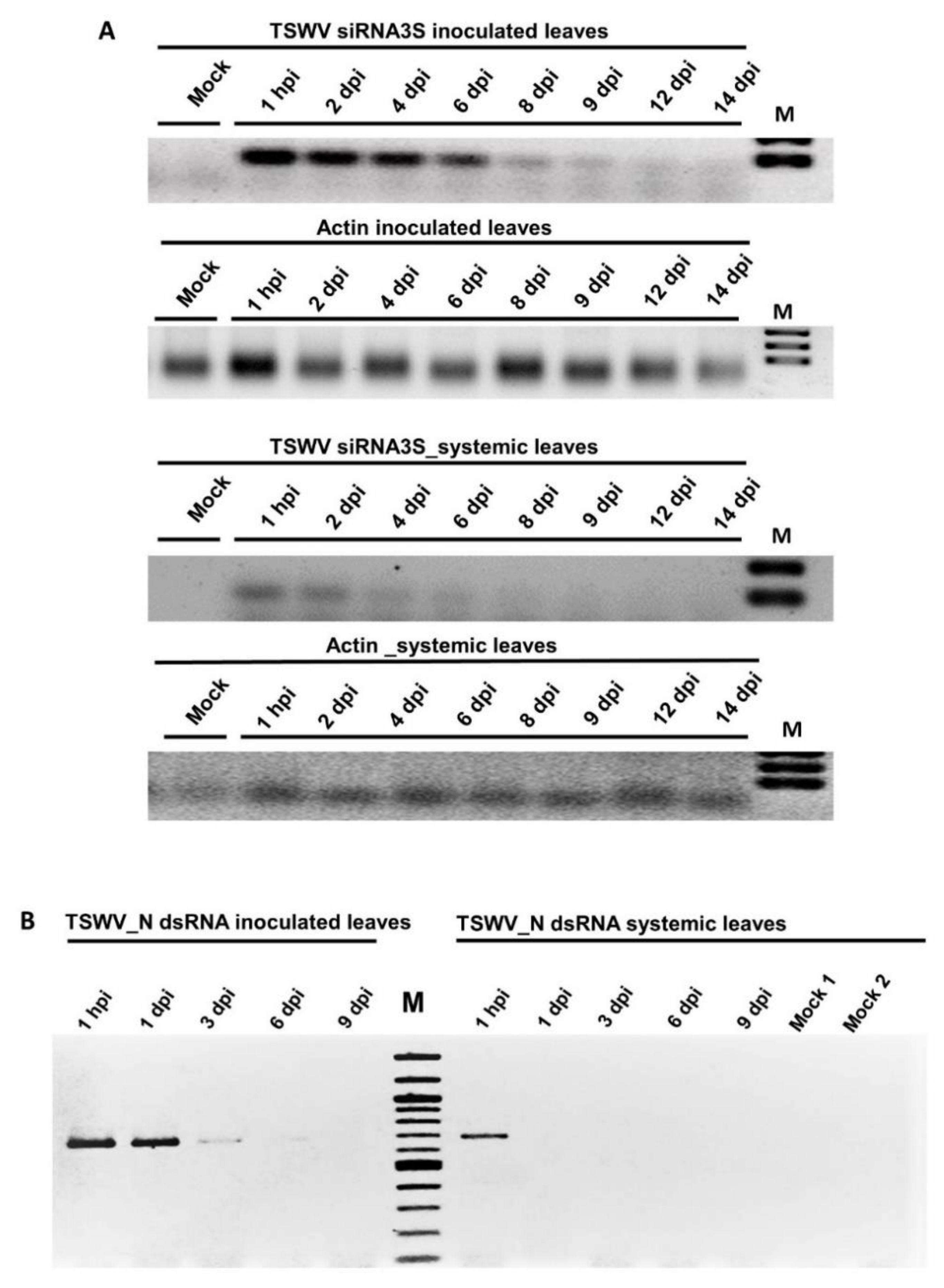
3.3. Systemic Movement of TSWV dsRNA_N

To determine if the dsRNA produced against the N gene could move systemically in tobacco, its presence over time was determined by semi-quantitative RT-PCR. RT-PCR results showed that the N gene dsRNA levels in the inoculated leaves continuously declined from six dpi, at nine dpi the signal was very low in agarose gel and was non-detectable at 12 dpi. Interestingly, dsRNA_N was detected in the non-inoculated leaves beginning from one hpi and no bands were detected afterward (Figure 4B). This experiment showed the movement of dsRNA from the inoculated to younger, non-inoculated leaves (Figure 4B).

3.4. Detection of a siRNA of N Gene by Stem-Loop RT-PCR

The presence of siRNA3S was monitored in the dsRNA-inoculated and non-inoculated leaves by stem-loop RT-PCR [45]. The putative siRNA, resolved as a specific band of 60 bp, was found beginning from one-day post dsRNA application and lasted up to nine dpi in inoculated leaves and six dpi in non-inoculated leaves. No amplification was seen in the inoculated leaves of plants treated with water (Mock, Figure 4A).

4. Discussion

TSWV continues to be a production constraint to several agronomic and horticultural crops in the USA and other parts of the world [15]. Resistant varieties offer the most cost-effective and environmentally friendly option for reducing the impact of this virus. RNAi was shown to be the mechanism behind host plant resistance to virus infection [14]. Transgenic technology has been used to induce RNAi in plants to attain pathogen-derived resistance against TSWV by targeting the N and NSm genes [58,59,60,61,62,63]. Due to the public concerns about the biosafety issues surrounding genetically modified crops, search for alternate, non-transgenic approaches for inducing RNAi for virus resistance has been the subject of research for the last several years. Here we showed that exogenously applied dsRNA targeted to the N and NSs genes of TSWV triggered RNAi in tobacco plants and the treated plants showed resistance to the virus. The strategy has several advantages compared to the use of transgenic plants. Firstly, this approach is faster since plant transformation, regeneration, and subsequent characterization of transgenic events are not needed. Secondly, dsRNAs for various genes could be prepared in a relatively short period of time, and their efficacy could be tested in a ‘shotgun approach’. Thirdly, mixed viral infections could be managed by introducing dsRNAs for multiple viruses at once. Furthermore, this strategy could target both vectors (such as thrips) and plant viruses simultaneously [64]. It has been shown that RNA-based vaccination was effective against a bipartite geminivirus and where a single dsRNA molecule could protect against two tomato-infecting viruses, tomato yellow leaf curl virus and cucumber mosaic virus [65].
The dsRNA molecules produced in vitro for N and NSs genes of TSWV, when topically applied on tobacco leaves, proved to be effective against TSWV infection. Interestingly, a recent study by Tabein et al. (2020) [66] used a similar strategy against TSWV in N. benthamina and emphasized that the choice of the target viral sequence in designing RNAi-based vaccines is crucial for its success. The observed differences in antiviral efficiency of the N- and NSs-targeting dsRNAs could be due to the biological role of the N and NSs gene products. In our study, dsRNA_N provided a higher level of resistance against TSWV when compared to that of the dsRNA_NSs. NSs sequesters the siRNAs resulting in the inhibition of the formation of replication initiation and silencing complex (RISC) [67]. It is possible that the exogenously applied dsRNA targeted to NSs inactivated the silencing suppressor, and thereby facilitating the spread of viral siRNAs. Successful establishment of TSWV infection requires the replication and transcription of the viral genes to produce infectious ribonucleocapsid proteins (RNPs), the minimal infectious unit containing the three genomic RNAs that are tightly packed by the N protein and few copies of the viral RdRp. In this context, the degradation of the N-coding small RNA by RNAi could impact viral accumulation due to the central role of the N protein, which was manifested in a higher level of resistance induced by a dsRNA targeted to the N gene. There are similar findings of the greater efficacy of dsRNAs targeting the capsid coding genes and silencing suppressor genes in the case of several other viruses [68].
There is a decrease in lesion size on tobacco leaves that were inoculated with either dsRNA_N or dsRNA_NSs compared to TSWV-inoculated plants (Figure 2B). This may indicate that the vsiRNAs produced from the exogenously applied dsRNA by endogenous RNAi machinery of tobacco are reducing or interfering with virus infection in inoculated leaves.
The systemic transport and stability of dsRNA molecules produced against TSWV genes in tobacco were studied to explore other possible means of dsRNA application (i.e., drenching) or clay nanosheets/Bio Clay [29,69]. dsRNA_N, upon application to the leaves, was quickly transported to the systemic leaves at one hpi. The movement of dsRNA might be similar to that of viroids. Viroids do not encode any proteins and are not encapsidated but utilize the host cellular factors for their spread throughout the plant [70]. The existing hypothesis about long-distance RNA transport is that once an RNA molecule enters the phloem sieve tube, it moves from the source to sink organs by simple diffusion [71]. The dsRNA used in this study may contain some motifs which could interact with cellular proteins facilitating their transport. Several previous reports described the systemic movement of dsRNA in plants [44,72,73,74,75]; however, topically applied dsRNAs remained mostly in treated leaves in watermelon and squash plants [45], or in tomato [73]. The differences in these studies could be explained by the existence of an active dsRNA long-distance movement process mediated by proteins binding to specific RNA motifs [75]. In contrast, Northern blot studies by Tenllado and Díaz-Ruíz (2001) [76] did not reveal the presence of dsRNA in the non-inoculated leaves when dsRNA was exogenously applied against pepper mild mottle virus in N. tabacum. A possible explanation for this difference could be the lower sensitivity of Northern blot assays relative to semi-quantitative and real-time RT-PCR used in the present study.
Finally, the accumulation of vsiRNAs in plants treated with dsRNA_N suggests that the RNAi processed the externally applied N dsRNA. SiRNA3S was detected in the treated leaf (where dsRNA was applied) and in the non-treated leaf at one hpi. The latter indicates either that siRNAs produced in the dsRNA-treated leaf are transported rapidly to non-inoculated leaves or that they are produced in the non-inoculated leaves from the rapidly transported dsRNA. Further studies on the successful application/delivery of dsRNA vaccine at field conditions as well as preventive and curative experiments to demonstrate the effectiveness of TSWV dsRNA on tobacco remains to be carried out.
In conclusion, we demonstrated that topical application of dsRNAs derived from the N and NSs genes conferred resistance to TSWV in tobacco. It remains to be seen if the resistance will be equally effective in other TSWV-affected crops such as peanut, pepper, and tomato.

Author Contributions

Conceived and designed the experiments: N.C.K. and S.B.; Performed the experiments: N.C.K. and A.S.D.; Analysed the data: N.C.K.; Project administration, planned the experiments, funding acquisition: S.B.; Writing, and editing of the manuscript: N.C.K., S.B., A.K.C. and H.R.P. All authors have read and agreed to the published version of the manuscript.

Funding

This work is funded in part by the Georgia Agricultural Commodity Commission for Tobacco (CPES 2019-2020 TOBB 2004), Georgia Agricultural Commodity Commission for Peanut (UGAT-30-19/21), and USDA-HATCH project fund to SB #1020319. The funders had no role in study design, data collection, and analysis, decision to publish, or manuscript preparation.

Institutional Review Board Statement

Not Applicable.

Informed Consent Statement

Not Applicable.

Data Availability Statement

Not Applicable.

Acknowledgments

The authors thank J. Michael Moore and Steven S. LaHue for their continued support of the tobacco research. SB acknowledges the support provided by UGA administrative staff for executing the project. The authors acknowledge the constructive and critical comments by the anonymous reviewers to improve the manuscript.

Conflicts of Interest

The authors declare no conflict of interest.

References

  1. Halliwel, R.S.; Philley, G. Spotted wilt of peanut in Texas. Plant Dis. Report. 1974, 58, 23–25. [Google Scholar]
  2. Black, M.C.; Lummus, P.F.; Smith, D.H.; Demski, J.W. An epidemic of spotted wilt disease in south Texas peanuts in 1985. Proc. Am. Peanut Res. Ed. Soc. 1986, 18, 66. [Google Scholar]
  3. Rotenberg, D.; Jacobson, A.L.; Schneweis, D.J.; Whitfield, A.E. Thrips transmission of tospoviruses. Curr. Opin. Virol. 2015, 15, 80–89. [Google Scholar] [CrossRef] [PubMed]
  4. Riley, D.G.; Joseph, S.V.; Srinivasan, R.; Diffie, S. Thrips vectors of tospoviruses. J. Integr. Pest Manag. 2011, 2, 1–10. [Google Scholar]
  5. Culbreath, A.K.; Csinos, A.S.; Bertrand, P.F.; Demski., J.W. Tomato Spotted Wilt Virus Epidemic in Flue-Cured Tobacco in Georgia. Plant Dis. 1991, 75, 483–485. [Google Scholar] [CrossRef]
  6. Gitaitis, R.D.; Dowler, C.C.; Chalfant, R.B. Epidemiology of tomato spotted wilt in pepper and tomato in southern Georgia. Plant Dis. 1998, 82, 752–756. [Google Scholar] [CrossRef] [Green Version]
  7. Martinez, A. Georgia Plant Disease Loss Estimates; Univ. Georgia Coop. Ext. Special Bulletin: Athens, GA, USA, 2005. [Google Scholar]
  8. Olatinwo, R.O.; Paz, J.O.; Brown, S.L.; Kemerait, R.C., Jr.; Culbreath, A.K.; Beasley, J.P., Jr.; Hoogenboom, G. A predictive model for spotted wilt epidemics in peanut based on local weather conditions and the tomato spotted wilt virus risk. Phytopathology 2008, 98, 1066–1074. [Google Scholar] [CrossRef] [PubMed] [Green Version]
  9. Csinos, A.S.; Pappu, H.R.; McPherson, R.M.; Stephenson, M.G. Management of tomato spotted wilt tospovirus in flue-cured tobacco with acibenzolar-S-methyl and imidacloprid. Plant Dis. 2001, 85, 292–296. [Google Scholar] [CrossRef] [Green Version]
  10. Pappu, H.R.; Csinos, A.S.; McPherson, R.M.; Jones, D.C.; Stephenson, M.G. Effect of acibenzolar-S-methyl and imidacloprid on suppression of tomato spotted wilt tospovirus in flue-cured tobacco. Crop Prot. 2000, 19, 349–354. [Google Scholar] [CrossRef]
  11. McPherson, R.M.; Pappu, H.R.; Csinos, A.S.; Bertrand, P.F. Influence of Dichloropene, volunteer peanuts, and thrips control practices on the abundance of insect pests and incidence of tomato spotted wilt in flue-cured tobacco. Tob. Sci. 1997, 41, 94–95. [Google Scholar]
  12. McPherson, R.M.; Pappu, H.R.; Jones, D.C. Occurrence of five thrips species on flue-cured tobacco and impact on spotted wilt disease incidence in Georgia. Plant Dis. 1999, 83, 765–767. [Google Scholar] [CrossRef] [Green Version]
  13. McPherson, R.M.; Jones, D.C.; Pappu, H.R.; Moore, J.M. Reducing the risks of spotted wilt virus in tobacco with selected thrips control practices. J. Agric. Urban Entomol. 2003, 11-23.–23. [Google Scholar]
  14. Nilon, A.; Robinson, K.; Pappu, H.R.; Mitter, N. Current status and potential of RNA interference for the management of tomato spotted wilt virus and thrips vectors. Pathogens 2021, 10, 320. [Google Scholar] [CrossRef]
  15. Srinivasan, R.; Abney, M.R.; Culbreath, A.K.; Kemerait, R.C.; Tubbs, R.S.; Monfort, W.S.; Pappu, H.R. Three decades of managing tomato spotted wilt virus in peanut in southeastern United States. Virus Res. 2017, 241, 203–212. [Google Scholar] [CrossRef]
  16. Black, L.L.; Hobbs, H.A.; Kammerlohr, D.S. Resistance of Capsicum chinenselines to tomato spotted wilt virus from Louisiana, USA, and inheritance of resistance. Acta Hortic. 1996, 431, 393–401. [Google Scholar] [CrossRef]
  17. Boiteux, L.S. Allelic relationships between genes for resistance to tomato spotted wilt tospovirus in Capsicum chinense. Theor. Appl. Genet. 1995, 90, 146–149. [Google Scholar] [CrossRef] [PubMed]
  18. Boiteux, L.S.; de Avila, A.C. Inheritance of a resistance specific to tomato spotted wilt tospovirus in Capsicum chinense. Euphytica 1994, 75, 139–142. [Google Scholar] [CrossRef]
  19. Moury, B.; Palloix, A.; Selassie, K.G.; Marchoux, G. Hypersensitive resistance to tomato spotted wilt virus in three Capsicum chinense accessions is controlled by a single gene and is overcome by virulent strains. Euphytica 1997, 94, 45–52. [Google Scholar] [CrossRef]
  20. Batuman, O.; Turini, T.A.; Oliveira, P.V.; Rojas, M.R.; Macedo, M.; Mellinger, H.C.; Adkins, S.; Gilbertson, R.L. First report of a resistance-breaking strain of tomato spotted wilt virus infecting tomatoes with the Sw-5 tospovirus-resistance gene in California. Plant Dis. 2017, 101, 637. [Google Scholar] [CrossRef]
  21. Macedo, M.A.; Rojas, M.R.; Gilbertson, R.L. First report of a resistance-breaking strain of tomato spotted wilt orthotospovirus infecting sweet pepper with the Tsw resistance gene in California, USA. Plant Dis. 2019, 103, 1048. [Google Scholar] [CrossRef]
  22. Patil, B.L.; Ogwok, E.; Wagaba, H.; Mohammed, I.U.; Yadav, J.S.; Bagewadi, B.; Taylor, N.J.; Kreuze, J.F.; Maruthi, M.N.N.; Alicai, T.; et al. RNAi-mediated resistance to diverse isolates belonging to two virus species involved in cassava brown streak disease. Mol. Plant. Pathol. 2011, 12, 31–41. [Google Scholar] [CrossRef]
  23. Jia, R.; Zhao, H.; Huang, J.; Kong, H.; Zhang, Y.; Guo, J.; Huang, Q.; Guo, Y.; Wei, Q.; Zuo, J.; et al. Use of RNAi technology to develop a PRSV-resistant transgenic papaya. Sci. Rep. 2017, 7, 1–9. [Google Scholar] [CrossRef] [PubMed] [Green Version]
  24. Fermin, G.A.; Castro, L.T.; Tennant, P.F. CP-transgenic and non-transgenic approaches for the control of papaya ringspot: Current situation and challenges. Transgenic Plant J. 2010, 4, 1–15. [Google Scholar]
  25. Lin, H.T.; Yen, G.C.; Huang, T.T.; Chan, L.F.; Cheng, Y.H.; Wu, J.H.; Yeh, S.D.; Wang, S.Y.; Liao, J.W. Toxicity assessment of transgenic papaya ringspot virus of 823-2210 line papaya fruits. J. Agric. Food Chem. 2013, 61, 1585–1596. [Google Scholar] [CrossRef]
  26. Hamim, I.; Borth, W.B.; Marquez, J.; Green, J.C.; Melzer, M.J.; Hu, J.S. Transgene-mediated resistance to papaya ringspot virus: Challenges and solutions. Phytoparasitica 2018, 46, 1–18. [Google Scholar] [CrossRef]
  27. Liu, H.M.; Zhu, C.X.; Zhu, X.P.; Guo, X.Q.; Song, Y.Z.; Wen, F.J. A link between PVYN CP gene-mediated virus resistance and transgene arrangement. J. Phytopathol. 2017, 155, 676–682. [Google Scholar] [CrossRef]
  28. Zhu, C.X.; Song, Y.Z.; Yin, G.H.; Wen, F.J. Induction of RNA-mediated Multiple Virus Resistance to Potato virus Y, tobacco mosaic virus and cucumber mosaic virus. J. Phytopathol. 2009, 157, 101–107. [Google Scholar] [CrossRef]
  29. Worrall, E.A.; Bravo-Cazar, A.; Nilon, A.T.; Fletcher, S.J.; Robinson, K.E.; Carr, J.P.; Mitter, N. Exogenous application of RNAi-inducing double-stranded RNA inhibits aphid-mediated transmission of a plant virus. Front. Plant Sci. 2019, 10, 265. [Google Scholar] [CrossRef] [Green Version]
  30. Dykxhoorn, D.M.; Novina, C.D.; Sharp, P.A. Killing the messenger: Short RNAs that silence gene expression. Nat. Rev. Mol. Cell Biol. 2003, 4, 457–467. [Google Scholar] [CrossRef] [PubMed]
  31. Meister, G.; Tuschl, T. Mechanisms of gene silencing by double-stranded RNA. Nature 2004, 431, 343–349. [Google Scholar] [CrossRef] [PubMed]
  32. Padmanabhan, C.; Zhang, X.; Jin, H. Host small RNAs are big contributors to plant innate immunity. Curr. Opin. Plant Biol. 2009, 4, 465–472. [Google Scholar] [CrossRef] [PubMed]
  33. Wang, M.B.; Masuta, C.; Smith, N.A.; Shimura, H. RNA silencing and plant viral diseases. Mol. Plant Microbe Interact. 2012, 25, 1275–1285. [Google Scholar] [CrossRef] [Green Version]
  34. Brodersen, P.; Voinnet, O. The diversity of RNA silencing pathways in plants. Trends Genet. 2006, 22, 268–280. [Google Scholar] [CrossRef] [PubMed]
  35. Blevins, T.; Rajeswaran, R.; Shivaprasad, P.V.; Beknazariants, D.; Si-Ammour, A.; Park, H.S.; Vazquez, F.; Robertson, D.; Meins, F., Jr.; Hohn, T.; et al. Four plant dicers mediate viral small RNA biogenesis and DNA virus induced silencing. Nucleic Acids Res. 2006, 34, 6233–6246. [Google Scholar] [CrossRef] [Green Version]
  36. Deleris, A.; Gallego-Bartolome, J.; Bao, J.; Kasschau, K.D.; Carrington, J.C.; Voinnet, O. Hierarchical action and inhibition of plant Dicer-like proteins in antiviral defense. Science 2006, 313, 68–71. [Google Scholar] [CrossRef]
  37. Tabara, H.; Sarkissian, M.; Kelly, W.G.; Fleenor, J.; Grishok, A.; Timmons, L.; Fire, A.; Mello, C.C. The rde-1 gene, RNA interference, and transposon silencing in C. elegans. Cell 1999, 99, 123–132. [Google Scholar] [CrossRef] [Green Version]
  38. Hammond, S.M.; Boettcher, S.; Caudy, A.A.; Kobayashi, R.; Hannon, G.J. Argonaute2, a link between genetic and biochemical analyses of RNAi. Science 2001, 293, 1146–1150. [Google Scholar] [CrossRef] [Green Version]
  39. Liu, J.; Carmell, M.A.; Rivas, F.V.; Marsden, C.G.; Thomson, J.M.; Song, J.; Hammond, S.M.; Joshua-Tor, L.; Hannon, G.J. Argonaute2 is the catalytic engine of mammalian RNAi. Science 2004, 305, 1437–1441. [Google Scholar] [CrossRef] [PubMed] [Green Version]
  40. Vadlamudi, T.; Patil, B.L.; Kaldis, A.; Gopal, D.V.R.S.; Mishra, R.; Berbati, M.; Voloudakis, A. DsRNA-mediated protection against two isolates of papaya ringspot virus through topical application of dsRNA in papaya. J. Virol. Methods. 2020, 275, 113750. [Google Scholar] [CrossRef]
  41. Patil, B.L.; Raghu, R.; Dangwal, M.; Byregowda, M.; Voloudakis, A. Exogenous dsRNA-mediated field protection against pigeonpea sterility mosaic emaravirus. J. Plant Biochem. Biotechnol. 2021, 1–6. [Google Scholar]
  42. Konakalla, N.C.; Kaldis, A.; Masarapu, H.; Voloudakis, A.E. Topical application of double stranded RNA molecules deriving from sesbania mosaic virus (SeMV) CP and MP genes protects sesbania plants against SeMV. Eur. J. Plant Pathol. 2019, 155, 1345–1352. [Google Scholar] [CrossRef]
  43. Yin, G.; Sun, Z.; Liu, N.; Zhang, L.; Song, Y.; Zhu, C.; Wen, F. Production of double-stranded RNA for interference with TMV infection utilizing a bacterial prokaryotic expression system. Appl. Microbiol. Biotechnol. 2009, 84, 323–333. [Google Scholar] [CrossRef] [PubMed]
  44. Konakalla, N.C.; Kaldis, A.; Berbati, M.; Masarapu, H.; Voloudakis, A.E. Exogenous application of double-stranded RNA molecules from TMV p126 and CP genes confers resistance against TMV in tobacco. Planta 2016, 244, 961–969. [Google Scholar] [CrossRef] [PubMed]
  45. Kaldis, A.; Berbati, M.; Melita, O.; Reppa, C.; Holeva, M.; Otten, P.; Voloudakis, A. Exogenously applied dsRNA molecules deriving from the zucchini yellow mosaic virus (ZYMV) genome move systemically and protect cucurbits against ZYMV. Mol. Plant. Pathol. 2018, 19, 883–895. [Google Scholar] [CrossRef] [Green Version]
  46. Aalto, A.P.; Sarin, L.P.; Van Dijk, A.; Saarma, M.; Poranen, M.M.; Arumäe, U.; Bamford, D.H. Large-scale production of dsRNA and siRNA pools for RNA interference utilizing bacteriophage ϕ6 RNA-dependent RNA polymerase. RNA 2007, 13, 422–429. [Google Scholar] [CrossRef] [PubMed] [Green Version]
  47. Voloudakis, A.E.; Holeva, M.C.; Sarin, L.P.; Bamford, D.H.; Vargas, M.; Poranen, M.M.; Tenllado, F. Efficient double-stranded RNA production methods for utilization in plant virus control. In Plant Virology Protocols; Methods in Molecular Biology (Methods and Protocols); Uyeda, I., Masuta, C., Eds.; Humana Press: New York, NY, USA, 2015; Volume 1236, pp. 255–274. [Google Scholar]
  48. Sun, Y.; Qiao, X.; Mindich, L. Construction of carrier state viruses with partial genomes of the segmented dsRNA bacteriophages. Virology 2004, 319, 274–279. [Google Scholar] [CrossRef] [Green Version]
  49. Makeyev, E.V.; Bamford, D.H. The polymerase subunit of a dsRNA virus plays a central role in the regulation of viral RNA metabolism. EMBO J. 2000, 19, 6275–6284. [Google Scholar] [CrossRef] [PubMed] [Green Version]
  50. Goto, K.; Kobori, T.; Kosaka, Y.; Natsuaki, T.; Masuta, C. Characterization of silencing suppressor 2b of cucumber mosaic virus based on examination of its small RNA-binding abilities. Plant Cell Physiol. 2007, 48, 1050–1060. [Google Scholar] [CrossRef] [Green Version]
  51. Soellick, T.R.; Uhrig, J.F.; Bucher, G.L.; Kellmann, J.W.; Schreier, P.H. The movement protein NSm of tomato spotted wilt tospovirus (TSWV): RNA binding, interaction with the TSWV N protein, and identification of interacting plant proteins. Proc. Natl. Acad. Sci. USA 2000, 97, 2373–2378. [Google Scholar] [CrossRef] [Green Version]
  52. Takeda, A.; Sugiyama, K.; Nagano, H.; Mori, M.; Kaido, M.; Mise, K.; Tsuda, S.; Okuno, T. Identification of a novel RNA silencing suppressor, NSs protein of tomato spotted wilt virus. FEBS Lett. 2002, 532, 75–79. [Google Scholar] [CrossRef] [Green Version]
  53. Zhai, Y.; Gnanasekaran, P.; Pappu, H.R. Identification and characterization of plant-interacting targets of tomato spotted wilt virus silencing suppressor. Pathogens 2021, 10, 27. [Google Scholar] [CrossRef]
  54. Varkonyi-Gasic, E.; Wu, R.; Wood, M.; Walton, E.F.; Hellens, R.P. Protocol: A highly sensitive RT-PCR method for detection and quantification of microRNAs. Plant Methods 2007, 3, 1–12. [Google Scholar] [CrossRef] [PubMed] [Green Version]
  55. Corpet, F. Multiple sequence alignment with hierarchical clustering. Nucl. Acids Res. 1988, 16, 10881–10890. [Google Scholar] [CrossRef] [PubMed]
  56. Koressaar, T.; Lepamets, M.; Kaplinski, L.; Raime, K.; Andreson, R.; Remm, M. Primer3_masker: Integrating masking of template sequence with primer design software. Bioinformatics 2018, 34, 1937–1938. [Google Scholar] [CrossRef] [PubMed]
  57. Sundaraj, S.; Srinivasan, R.; Culbreath, A.K.; Riley, D.G.; Pappu, H.R. Host plant resistance against tomato spotted wilt virus in peanut (Arachis hypogaea) and its impact on susceptibility to the virus, virus population genetics, and vector feeding behavior and survival. Phytopathology 2014, 104, 202–210. [Google Scholar] [CrossRef] [Green Version]
  58. Carbonell, A.; López, C.; Daròs, J.A. Fast-forward identification of highly effective artificial small RNAs against different tomato spotted wilt virus isolates. Mol. Plant Microbe Interact. 2019, 32, 142–156. [Google Scholar] [CrossRef] [Green Version]
  59. MacKenzie, D.J.; Ellis, P.J. Resistance to tomato spotted wilt virus infection in transgenic tobacco expressing the viral nucleocapsid gene. Mol. Plant Microbe Interact. 1992, 5, 34–40. [Google Scholar] [CrossRef]
  60. Vaira, A.M.; Semeria, L.; Crespi, S.; Lisa, V.; Allavena, A.; Accotto, G.P. Resistance to tospoviruses in Nicotiana benthamiana transformed with the N gene of tomato spotted wilt virus: Correlation between transgene expression and protection in primary transformants. Mol. Plant Microbe Interact. 1995, 8, 66–73. [Google Scholar] [CrossRef]
  61. Prins, M.; Resende, R.D.; Anker, C.; van Schepen, A.; de Haan, P.; Goldbach, R. Engineered RNA-mediated resistance to tomato spotted wilt virus is sequence specific. Mol. Plant Microbe Interact. 1996, 9, 416–418. [Google Scholar] [CrossRef]
  62. Herrero, S.; Culbreath, A.K.; Csinos, A.S.; Pappu, H.R.; Rufty, R.C.; Daub, M.E. Nucleocapsid gene-mediated transgenic resistance provides protection against tomato spotted wilt virus epidemics in the field. Phytopathology 2000, 90, 139–147. [Google Scholar] [CrossRef] [Green Version]
  63. Jan, F.J.; Fagoaga, C.; Pang, S.Z.; Gonsalves, D. A minimum length of N gene sequence in transgenic plants is required for RNA-mediated tospovirus resistance. J. Gen. Virol. 2000, 81, 235–242. [Google Scholar] [CrossRef]
  64. Turner, C.T.; Davy, M.W.; MacDiarmid, R.M.; Plummer, K.M.; Birch, N.P.; Newcomb, R.D. RNA interference in the light brown apple moth, Epiphyas postvittana (Walker) induced by double-stranded RNA feeding. Insect Mol. Biol. 2006, 15, 383–391. [Google Scholar] [CrossRef] [PubMed]
  65. Namgial, T.; Kaldis, A.; Chakraborty, S.; Voloudakis, A. Topical application of double-stranded RNA molecules containing sequences of tomato leaf curl virus and cucumber mosaic virus confers protection against the cognate viruses. Physiol. Mol. Plant Pathol. 2019, 108, 101432. [Google Scholar] [CrossRef]
  66. Tabein, S.; Jansen, M.; Noris, E.; Vaira, A.M.; Marian, D.; Behjatnia, S.A.A.; Accotto, G.P.; Miozzi, L. The induction of an effective dsRNA-mediated resistance against tomato spotted wilt virus by exogenous application of double-stranded RNA largely depends on the selection of the viral RNA target region. Front. Plant Sci. 2020, 11, 1896. [Google Scholar] [CrossRef] [PubMed]
  67. Jin, Y.; Zhao, J.H.; Guo, H.S. Recent advances in understanding plant antiviral RNAi and viral suppressors of RNAi. Curr. Opin. Virol. 2021, 46, 65–72. [Google Scholar]
  68. Taliansky, M.; Samarskaya, V.; Zavriev, S.K.; Fesenko, I.; Kalinina, N.O.; Love, A.J. RNA-Based Technologies for Engineering Plant Virus Resistance. Plants 2021, 10, 82. [Google Scholar] [CrossRef] [PubMed]
  69. Mitter, N.; Worrall, E.; Robinson, K. Clay nanosheets for topical delivery of RNAi for sustained protection against plant viruses. Nat. Plants. 2017, 3, 16207. [Google Scholar] [CrossRef]
  70. Itaya, A.; Zhong, X.; Bundschuh, R.; Qi, Y.; Wang, Y.; Takeda, R.; Harris, A.R.; Molina, C.; Nelson, R.S.; Ding, B. A structured viroid RNA serves as a substrate for dicer-like cleavage to produce biologically active small RNAs but is resistant to RNA-induced silencing complex-mediated degradation. J. Virol. 2007, 81, 2980–2994. [Google Scholar] [CrossRef] [Green Version]
  71. Sjolund, R.D. The phloem sieve element: A river runs through it. Plant Cell 1997, 9, 1137. [Google Scholar] [CrossRef] [PubMed] [Green Version]
  72. Hunter, W.B.; Glick, E.; Paldi, N.; Bextine, B.R. Advances in RNA interference: dsRNA treatment in trees and grapevines for insect pest suppression. Southwest. Entomol. 2012, 37, 85–87. [Google Scholar] [CrossRef]
  73. San Miguel, K.; Scott, J.G. The next generation of insecticides: dsRNA is stable as a foliar-applied insecticide. Pest Manag. Sci. 2016, 72, 801–809. [Google Scholar] [CrossRef] [PubMed]
  74. Gogoi, A.; Sarmah, N.; Kaldis, A.; Perdikis, D.; Voloudakis, A. Plant insects and mites uptake double-stranded RNA upon its exogenous application on tomato leaves. Planta 2017, 246, 1233–1241. [Google Scholar] [CrossRef] [PubMed]
  75. Kehr, J.; Kragler, F. Long distance RNA movement. New Phytol. 2018, 218, 29–40. [Google Scholar] [CrossRef] [PubMed] [Green Version]
  76. Tenllado, F.; Dıaz-Ruız, J.R. Double-stranded RNA-mediated interference with plant virus infection. J. Virol. 2001, 75, 12288–12297. [Google Scholar] [CrossRef] [PubMed] [Green Version]
Figure 1. Production of dsRNA molecules specific to the tomato spotted wilt virus nucleocapsid (N) gene and non-structural (NSs) genes by two-step PCR method and in vitro transcription. M: 100 bp DNA ladder.
Viruses 13 00662 g001
Figure 2. Effect of double-stranded RNA on tomato spotted wilt virus (TSWV) infection in tobacco. (A) TSWV + tobacco; dsRNA_N + TSWV; dsRNA_Nss + TSWV and Control (Buffer). Disease incidence was calculated based on the number of plants exhibiting systemic disease symptoms out of 10 plants used in each treatment. Results were expressed as mean values of three independent experiments. Error bars represent standard error. Different letters indicate significant differences between groups (p ≤ 0.05) (B) Tobacco plants that received dsRNAs + TSWV exhibited no disease phenotype, (C) Effect of dsRNAs on lesion formation on tobacco leaves. Leaves of infected plans in dsRNA treatments had fewer lesions compared to TSWV treatment alone.
Viruses 13 00662 g002
Figure 3. Accumulation of tomato spotted wilt virus (TSWV) in inoculated and younger, non-inoculated leaves of tobacco at different time points. Logarithmic graph of the viral copy numbers of four different treatments (dsRNA_N + TSWV, dsRNA_NSs + TSWV, TSWV+ tobacco, and control. (A) TSWV copy number in co-inoculated leaves and (B) TSWV copy number in younger, non-inoculated leaves. Different letters indicate statistically significant differences (p < 0.05) between means. Error bars represent standard error.
Viruses 13 00662 g003
Figure 4. Movement of tomato spotted wilt virus (TSWV) dsRNA_N and siRNA3S in tobacco (A) Accumulation of siRNA3S from 1 hpi to 14 dpi. M: 100 bpt DNA ladder. (B) Detection of the TSWV_N dsRNA in inoculated and non-inoculated leaves at different time points. Mock 1 is mock-inoculated leaves, Mock 2 is younger, non-inoculated leaves from the same Mock 1 plants.
Viruses 13 00662 g004
Table 1. Primers for RT-PCR amplification of TSWV N and NSs genes, qRT-PCR and stem-loop RT-PCR.
PrimerOligonucleotide Sequence (5′-3′)Target GeneAmplicon Size (bp)
TSWV N-FGTCTAAGGTTAAGCTCACTAANucleocapsid (N)717
TSWV N-RAAGAGTTTCACTGTAATGTTC
TSWV NSs-FAGTCTGGGGATCAACTGCATCNonstructural protein (NSs)646
TSWV NSs-RGATGTTGTTTTCTGCTGACAT
T7-Linker-FGAGAATTCTAATACGACTCACTATAGGGGATCCN/Nss
qTSWV_N_SYBR_FGCTTCCCACCCTTTGATTCN139
qTSWV_N_SYBR_RATAGCCAAGACAACACTGATC
siRNA3S_FGCGGCGTGTGAGTGAGCTTAACN60
siRNA3S_RTGTCGTATCCAGTGCAGGGTCCGAGGTATTCG-
CACTGGATACGACTCTAAG
Universal-RGTGCAGGGTCCGAGGT
Publisher’s Note: MDPI stays neutral with regard to jurisdictional claims in published maps and institutional affiliations.

