Article

Diversity and Reassortment Rate of Influenza A Viruses in Wild Ducks and Gulls

by
Yulia Postnikova
1,
Anastasia Treshchalina
1,
Elizaveta Boravleva
1,
Alexandra Gambaryan
1,*,
Aydar Ishmukhametov
1,
Mikhail Matrosovich
2,
Ron A. M. Fouchier
3,
Galina Sadykova
4,
Alexey Prilipov
4 and
Natalia Lomakina
4
1
Chumakov Federal Scientific Center for the Research and Development of Immune-and-Biological Products, Village of Institute of Poliomyelitis, Settlement “Moskovskiy”, 108819 Moscow, Russia
2
Institute of Virology, Philipps University, Hans-Meerwein-Str. 2, D-35043 Marburg, Germany
3
Department of Virology, Erasmus Medical Centre, Dr. Molewaterplein 50, 3015GE Rotterdam, The Netherlands
4
The Gamaleya National Center of Epidemiology and Microbiology of the Russian Ministry of Health, 123098 Moscow, Russia
*
Author to whom correspondence should be addressed.
Submission received: 29 April 2021 / Revised: 22 May 2021 / Accepted: 24 May 2021 / Published: 27 May 2021
(This article belongs to the Special Issue State-of-the-Art Respiratory Viruses Research in Russia)

Abstract

:
Influenza A viruses (IAVs) evolve via point mutations and reassortment of viral gene segments. The patterns of reassortment in different host species differ considerably. We investigated the genetic diversity of IAVs in wild ducks and compared it with the viral diversity in gulls. The complete genomes of 38 IAVs of H1N1, H1N2, H3N1, H3N2, H3N6, H3N8, H4N6, H5N3, H6N2, H11N6, and H11N9 subtypes isolated from wild mallard ducks and gulls resting in a city pond in Moscow, Russia were sequenced. The analysis of phylogenetic trees showed that stable viral genotypes do not persist from year to year in ducks owing to frequent gene reassortment. For comparison, similar analyses were carried out using sequences of IAVs isolated in the same period from ducks and gulls in The Netherlands. Our results revealed a significant difference in diversity and rates of reassortment of IAVs in ducks and gulls.

1. Introduction

Wild aquatic birds of the orders Anseriformes (ducks, geese, and swans) and Charadriiformes (gulls, terns, waders, and plovers) are natural hosts of IAVs [1]. In these birds, 16 antigenic subtypes of hemagglutinin (HA) and 9 antigenic subtypes of neuraminidase (NA) in various combinations were found [2]. IAVs of H1–H12, H14, and H15 HA subtypes mainly circulate in ducks; these IAVs replicate in the intestine, cause asymptomatic infections, and transmit by the fecal-oral route through the water [1]. IAVs of H13 and H16 HA subtypes mainly circulate in gulls, causing epidemics among chicks in densely populated breeding colonies. These gull IAVs replicate in the intestine and the respiratory tract, and can transmit both by the fecal-oral route and through the air [3,4,5]. Thus, the patterns of replication and transmission differ between IAVs of ducks and gulls.
IAVs in domestic poultry evolve from IAVs of wild aquatic birds. IAVs of poultry can occasionally transmit and initiate host-specific evolutionary lineages in pigs, humans, and other mammals [1,2]. Long-term adaptation in ducks enables efficient replication and release of the virus into the environment without significant pathogenic effects to the host [2]. In gulls, the virus also replicates asymptomatically, while in poultry it may evolve towards increased pathogenicity [3]. In mammals, the influenza virus usually causes non-fatal disease [1].
The evolution of IAVs is caused by mutations and reassortment of viral genes. Reassortment occurs when a single host cell is infected with different IAVs [6]. Reassortment plays a central role in the microevolution of influenza viruses. In wild ducks, there is a continuous mixing of all eight genome segments due to reassortment. This process may be less intense in secondary hosts.
Reassortment between IAVs of different hosts, including human viruses, contributes to initiation of influenza pandemics [7]. However, once emerged, human IAVs of a novel lineage evolve as a whole, without reassortment. Only intra-subtype reassortants between co-circulating clades are viable and persist in the population on a small spatial-temporal scale [8,9,10,11,12]. Rarely, reassortants between H1N1 and H3N2 were found. For example, prior to the 2009 pandemic, clinical IAV isolates of the H1N2 subtype were described [13,14,15,16]. However, all these cases are rather the exception to the rule. Such reassortants are not sufficiently viable [17]. Studies of H1N2 reassortants in the human population showed that they are under negative selection [18,19]. Reassortment of IAVs in pigs is an example of the formation of new variants. Pigs were called the “mixing vessel” of IAVs because they can be infected with both avian and human viruses and enable their reassortment [20]. The IAVs that had been formed in pigs initiated the 2009 pandemic and, possibly, other pandemics. In recent decades, numerous new lineages of reassortant viruses were discovered in pigs [21,22,23,24]. However, IAVs of the same evolutionary lineage can circulate in pigs for many years. The “classical” lineage of swine IAVs, originating from the precursor of the 1918 “Spanish” influenza, remained genetically stable until the 1980s [25,26]. In horses, two lineages of influenza viruses (H7N7 and H3N8) evolved for decades [27].
Several lineages of highly pathogenic and low pathogenic IAVs circulate in chickens. Asian H5N1 and H7N9 IAVs are reassortants. They all carry a cassette of internal genes derived from chicken H9N2 viruses [28,29]. However, after initial formation, such variants circulate largely without reassortment. Chicken viruses of the H5 and H9 subtypes circulate in Egypt together, and co-infection with these viruses was often observed. Nevertheless, reassortment of these subtypes has not been reported yet [30]. Introduction of the H5N1 IAVs into ducks and their reassortment with duck viruses of other subtypes led to the emergence of novel H5N3, H5N6, and H5N8 IAV lineages [31,32,33].
The reassortment of IAVs in their natural animal hosts has a different pattern than in all secondary host species. It is impossible to detect persistent evolutionary lineages of duck IAVs containing stable constellations of all eight gene segments, as duck IAVs constantly exchange segments during mixed infections with neither significant fitness costs nor selective advantages of newly emerging combinations of segments [34].
Gulls are usually considered natural hosts of influenza viruses along with ducks. IAVs of almost all subtypes were found in gulls. However, the H1-H12 and H14 subtypes were only rarely isolated from gulls, and, as a rule, the gull viruses were evolutionarily close to the contemporary duck IAVs that circulated in the same region. These cases can be classified as occasional spillovers of duck H1-H12 and H14 IAVs to gulls. By contrast, the IAVs of H13 and H16 subtypes were almost exclusively detected in gulls and terns [35,36,37].
Although gene reassortment has been found among gull viruses, a relatively stable gene cassette containing PB2, PA, M, NS, NP, and PB1 gene segments has been formed in gulls and could persist for at least four years [38,39]. All gene segments of gull H13 and H16 IAVs are phylogenetically distant from segments of duck IAVs [35,36,40].
Recent studies have shown that reassortment can occur in any living system (cell culture, chicken embryos, laboratory animals) if the dose of infection is large enough to provide simultaneous access of different viral particles to the host cell [41,42,43]. However, the emergence of new variants of IAVs via reassortment in some hosts, such as humans, occurs once in about a decade, whereas in other hosts, such as ducks, the reassortment occurs continuously. This notion raises the key question of whether or not the rate of reassortment of IAVs differs significantly between viral host species [6].
The aim of our work was to compare the IAV reassortment rate in wild ducks and gulls. From 2006 to 2019, we isolated IAVs from wild birds resting in a city pond in Moscow, Russia during their autumn migration. Thirty-seven duck IAVs and one duck-origin spillover H6N2 gull isolate were fully sequenced. We did not find stable IAV genotypes perpetuated from year to year. For comparison, a similar study on genome constellations was carried using sequences of duck and gull IAVs isolated in the same years in The Netherlands.

2. Materials and Methods

2.1. Viruses

Fresh feces of mallard ducks and gulls were collected in 2006–2019 on the shore of a city pond in Moscow. Feces were suspended in 1 mL of phosphate-buffered saline (PBS) supplemented with 0.4 mg/mL gentamicin, 0.1 mg/mL kanamycin, 0.01 mg/mL nystatin, and 2% MycoKill AB solution (PAA Laboratories GmbH, Pasching, Austria). The suspension was centrifuged for 10 min at 4000 rpm, and 0.2 mL of the supernatant were inoculated into 10-day-old chicken embryos. Allantoic fluid was collected after 48 h and tested by hemagglutination assay with chicken red blood cells. Positive samples were taken for further passaging. All isolated IAV strains are stored in the virus repository of the Chumakov Federal Scientific Center (Moscow, Russia). Full names, designations of the viruses, and GenBank accession numbers are given in Table S1 of Supplementary Materials.

2.2. Sequencing

Viral RNA was isolated from the allantoic fluid using QIAamp Viral RNA mini kit (Qiagen, # 52904) according to the instructions of the manufacturer. Reverse transcription was carried out at 42 °C for 1 h in a 25 μL reaction mixture containing 8 μL RNA, 1 μL uni12 primer with a concentration of 50 ng/μL (13.5 pmol/μL), 10 μL water, 1 μL 10 mM dNTP, 5 µL 5× buffer and 100 units of MMLV (Alpha-Ferment Ltd., Moscow, Russia). The resulting cDNA was used in PCR with specific terminal primers to synthesize full-length genome segments. The amplified fragments were separated by electrophoresis in 1–1.3% agarose gel in the presence of ethidium bromide and were eluted from the gel with a Diatom DNA Elution kit (Isogen Laboratory Ltd., Russia, Moscow # D1031). Sequencing reactions were performed with terminal or internal primers [44] using the BrightDye™ Terminator Cycle Sequencing Kit v3.1 (Nimagen, The Netherlands), followed by analysis on an ABI PRISM 3100-Avant Genetic analyzer (Applied Biosystems 3100-Avant Genetic Analyzer, Foster City, CA, USA,). For assembly and analysis of nucleotide sequences, the Lasergene software package (DNASTAR, Inc. Madison, WI, USA) was used.

2.3. Downloading of Sequences and Evolutionary Tree Construction

The complete nucleotide sequences of IAV internal genes (PA, PB1, PB2, NP, MP, and NS) and external genes (H1, H3, H4, H5, N1, H6, H11, N1, N2, N3, N6, and N8) were downloaded from the Influenza Research Database (https://www.fludb.org, accessed on 25 April 2021). The selected sequences were aligned by the MUSCLE method using the software package MEGA X (https://www.megasoftware.net/, accessed on 25 April 2021) [45]. Time-scaled trees were generated for each internal segment with known isolation dates using BEAST under GTR model with 1000 bootstrap replicates [46]. A strict molecular clock model was chosen for all segments.

2.4. Classification of Gene Variants and Detection of Gene Reassortants

All lineages and subordinate lineages were classified according to the topology of the phylogenetic trees using the approach described in [47], but with more detail. The major clades on each gene tree were defined by strong bootstrap support (>95%) and numbered. The minor clades within the major clade were designated by corresponding number and a letter.

3. Results

During the autumn periods of 2006–2019, feces of wild waterfowl were collected on the bank of a pond in Moscow city, and IAVs were isolated. Over 14 years, about 4000 samples were collected, and 38 strains of influenza A viruses of subtypes H1N1, H1N2, H3N1, H3N2, H3N6, H3N8, H4N6, H5N3, H6N2, H11N6, and H11N9 were isolated and completely sequenced (Table 1 and Table S1 of Supplementary Materials). All isolates replicated in chick embryos to high titers, were infectious and immunogenic in mice, although these animals were generally not killed by the viruses. Five viruses tested were non-pathogenic in chickens [48,49].
The subtypes of isolates differed in different years. Until 2013, IAVs with hemagglutinins H3 and H4 dominated. These subtypes were no longer isolated after 2014 and were substituted by IAVs of the subtypes H1N2, H1N1, and H11N6. This change in the virus isolation pattern correlated with data from databases on the isolation of IAVs from ducks in Europe.

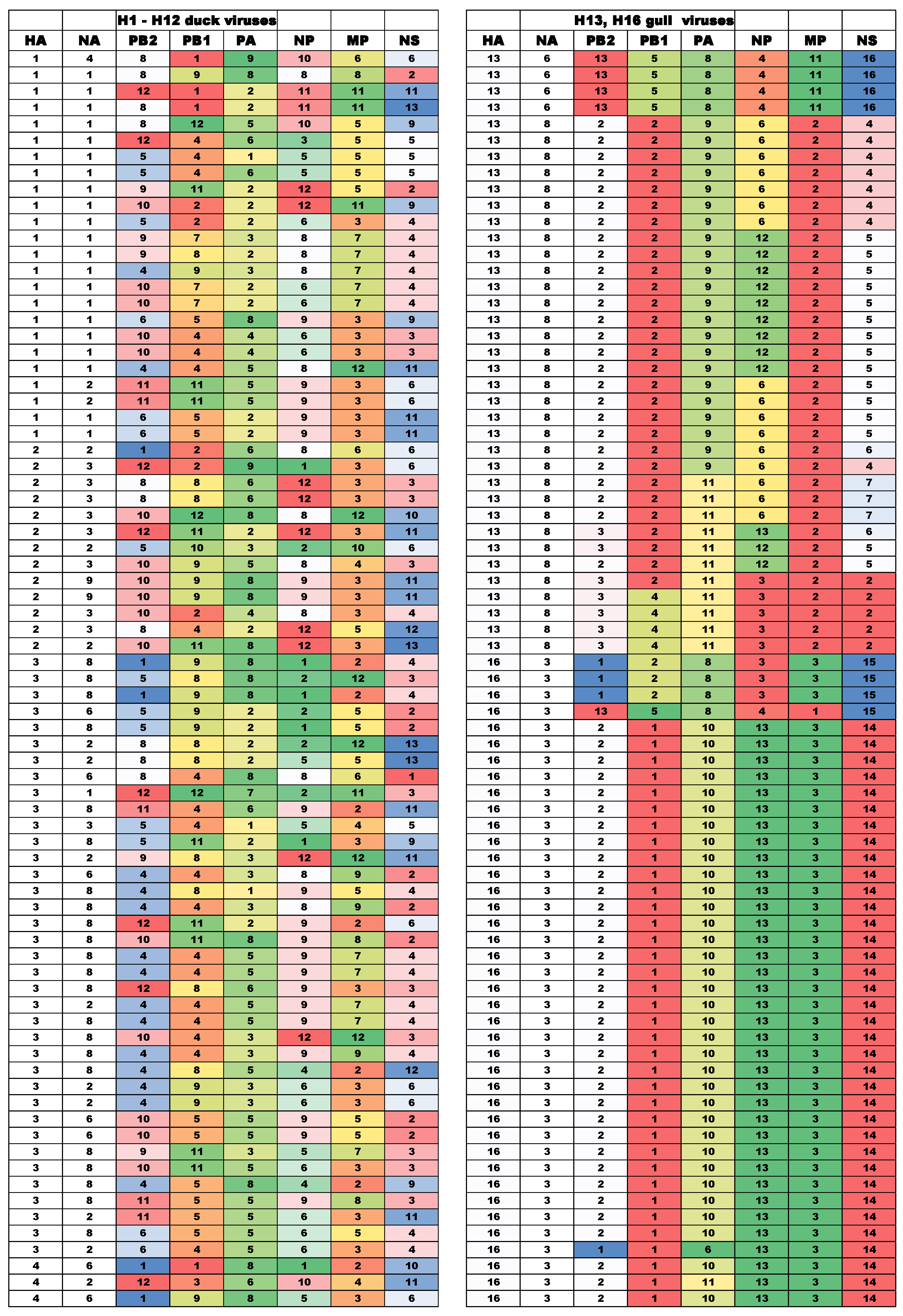
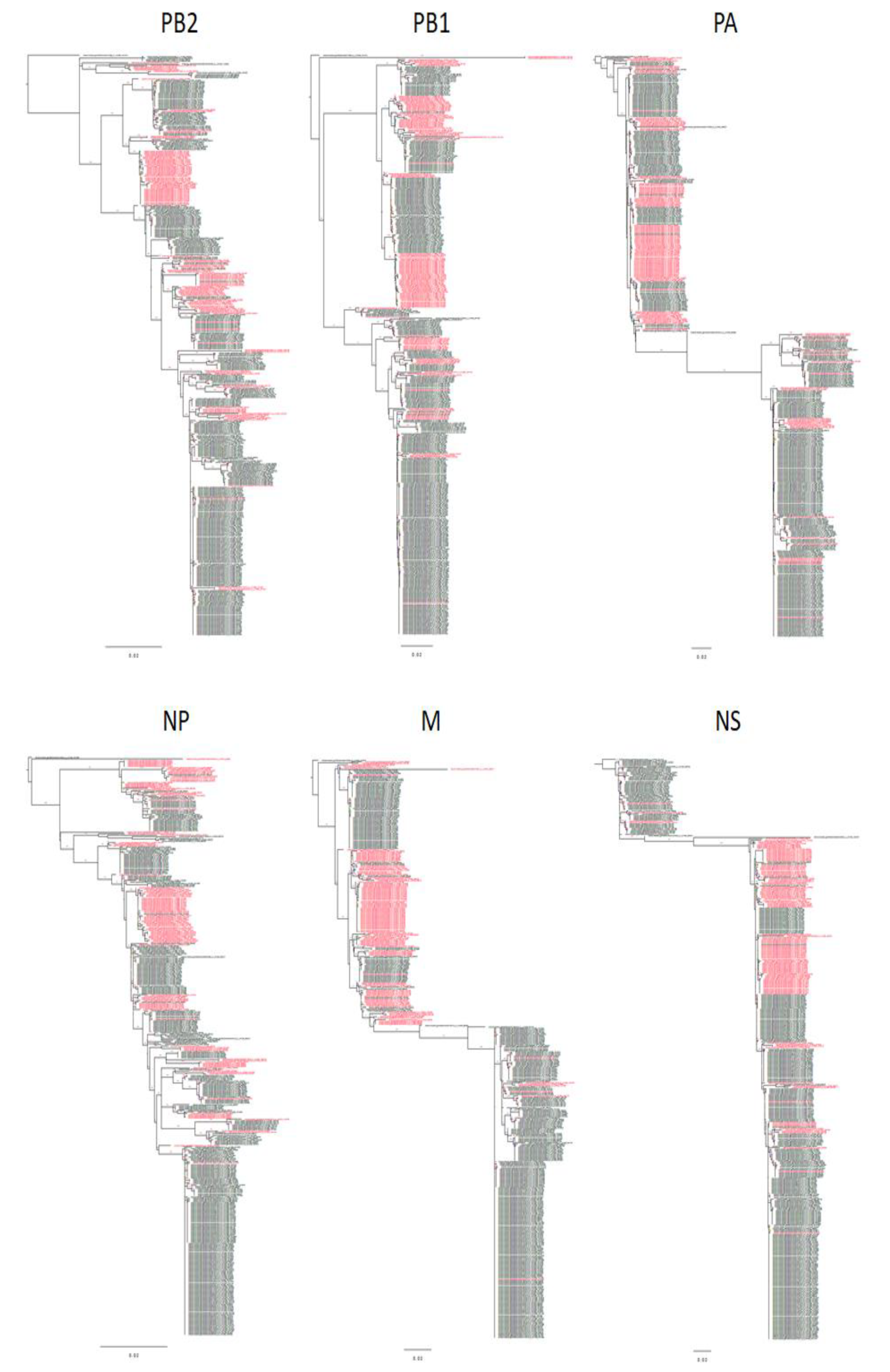
3.1. Evolutionary Relationships of Gene Segments

To study evolutionary relationships of gene segments, we expanded the set of IAVs isolated in Moscow by including the 191 mallard IAVs isolated in the Netherlands in the same period of time (2006–2019) [39]. We built phylogenetic trees for internal gene segments; clades/subclades on each tree were identified and numbered (Figures S1–S6 of Supplementary Materials).
The A/gull/Moscow/3100/2006 (H6N2) was not fundamentally separated from mallard IAVs. Thus, for all gene segments of the gull isolate, closely related variants can be found among the duck viruses (Table 1 and Table S2 of Supplementary Materials). A majority of IAVs isolated from mallards in Moscow had different gene constellations. The gene segments of the same clade/subclade were perpetuated in duck IAVs over several years. For example, PB2 of clade 11 was detected in 2009, 2010, 2013, 2014, 2018, and 2019 (Table 1).
Importantly, although segments of the same clade/subclade were found in duck IAVs isolated in different years, these viruses never preserved their full genome constellations and always contained a unique mixture of segments that belonged to different clades/subclades. Although four pairs of IAVs with identical constellations were detected among the Moscow viruses (Table 1), the viruses of each pair were isolated almost simultaneously and in the same place. These results support previous conclusion of Dugan and colleagues [34] that IAVs circulate in wild birds as a pool of gene segments, which reassort extensively and appear in a new combination each year.

3.2. Diversity of Gene Segment Constellations of IAVs in Ducks and Gulls

To compare the rate of natural reassortment of IAVs in ducks and gulls, we analyzed viruses isolated from ducks and viruses isolated from gulls in the Netherlands in the same period of time (2006–2019) [39]. The gull viruses were almost exclusively isolated from black headed gulls and were represented by 252 viruses of the H13 subtype, 94 viruses of the H16 subtype, and 11 mixed isolates.
The evolutionary trees for the gene segments were generated, and the clades and subclades were numbered (Figures S7–S12 of Supplementary Materials). The data on genome constellations of 228 duck IAVs viruses and 357 gull IAVs are presented in Tables S2–S5 of the Supplementary Materials; selected data are shown in Figure 1 and Table 3. Among 228 duck IAVs, we identified 187 distinctive genotypes. Pairs of isolates that matched in all gene segments, as a rule, were isolated on the same day.
In only three cases, pairs of IAVs listed below with matching genomes were isolated in different years. They were: d/N/20/2012|H1N1 and d/N/5/2013|H1N1, d/N/48/2011|H7N1 and d/N/31/2012|H7N1, d/N/71/2008|H10N7 and d/N/1/2009|H10N7.
Table 2 shows the diversity of IAVs of different subtypes, calculated as the ratio of the number of mismatched variants of genomes to the number of IAV strains in the group. The diversity is very high among the H1, H2, H3, H4, H5, H6, and H11 subtype IAVs, with 83 to 100% of the virus genomes representing unique genome constellations (Table S2 of Supplementary Materials). In contrast, gull IAVs were represented by 84 genome constellations per 252 H13 isolates (33% of genomes unique) and 38 constellations per 94 H16N3 isolates (40% of genome unique) (Tables S3 and S4 of Supplementary Materials).
Evolutionary histories of the internal gene segments of H13 and H16 IAVs are shown in Figure 2 with H16 IAVs colored in red. Non-persistent reassortment events are clearly visible on these trees as single strains of one color surrounded by strains with different color. Transition of gene segment from one subtype to another is also clearly visible as the alternation of groups of different colors on the same evolutionary branch. However, it is difficult to study the constellation of all gene segments on the trees, as this requires simultaneous comparison of patterns on different trees. To facilitate the analysis, we compiled tables in which we designated the clade numbers of the gene segments of all viruses in the same way as it was done for duck IAVs.
The gull virus isolates are naturally separated into large groups of closely related viruses, matching by subclades of all genes. Apparently, these groups represent variants of the same virus that caused the epidemic, e.g., in the nesting colonies. The genomic constellations of such variants are not very stable, they can vary from group to group and can change from year to year. Nevertheless, in some cases, clusters of the same variants of gene segment persist in the subsequent seasons. For example: HA, NA, PB2, PA, and NP of the H13N2/2016 isolates are direct descendants of the H13N2/2014 isolates; PB1, MP, and NS of both virus groups are descendants of the common close ancestors, HA, PB2, PB1, PA, and NP H13N2/2009 are descendants of H13N8/2008 (Figures S7–S12 of Supplementary Materials).
The internal gene segments of H16N3 viruses usually do not match the corresponding segments of H13 viruses; however, this rule is not very strict. Some of the exceptions are listed below.
H16N3/2008, H13N2/2009 and H13N2/2010 shared PA.
In 2007, H13N6 and H16N3 had matching PA.
H13N6/2007 and H16N3/2008 shared NP.
In 2011, H13N6 and H16N3 had matching NP (complete data are given in Tables S3–S4).
The trees are based on all published full-length sequences of H13 and H16 gull viruses isolated in The Netherlands. The route is placed on A/Black-headed gull/Netherlands/1/2000 (H13N8). Red color depicts sequences of H16 IAVs. Scale bars, 0.02. The trees were made using FigTree v1.4.4 (http://tree.bio.ed.ac.uk/software/figtree/, accessed on 25 April 2021).
In some seasons, large groups of gull IAVs had highly homologous gene segments, suggesting that all these IAVs represented descendants of the same precursor circulated during this season. The presence of such groups in our sets of gull IAVs allowed reliable identification of reassortant viruses, which clearly differed from the other isolates in the group by the origin of either one or a few gene segments (see Table S3 of Supplementary Materials; some of data are given in Table 3).
For example, one isolate from the group of H13N6/2014 IAVs, bhg/16/2014, contained PB2 from H16N3/2014. Bhg/31/2009/H13N2 is a reassortant, in which all internal genes coincide with the H13N6 viruses of the same year, and only NA belongs to another clade (Table S3).
Of particular interest are two H13N3 IAVs, bhg/17/2009 and bhg/8/2011, which contained heterologous N3 neuraminidase from H16N3. The former virus shared PB2, PA, NP, and NA with H16N3/2009, the remaining 4 segments were shared with other H13N6/2009 IAVs. Seven gene segments of bhg/8/2011 matched corresponding segments of H16N3/2011, only the HA being shared with H13N8/2011(Table 3 and Table S4).
One can conclude that two H13N3 isolates originated from H16N3 IAVs, in which HA and a number of other genes were replaced by gene segments of H13 IAVs via reassortment. Apparently, such reassortants are not viable enough; they were found in only two isolates despite the fact that there were seven mixed isolates in the same set (Table S4).
Some of the H16N3 IAVs represent reassortants containing internal gene segments of the H13 viruses. Thus, among 94 H16N3 viruses analyzed, we recognized 17 non-persistent reassortants, with 13 of them carrying gene segments of H13 viruses (Table S4 of Supplementary Materials). Among 252 H13 viruses studied, we found 14 non-persistent reassortants, nine of which acquired at least one gene segment from H16 viruses. About 10% of the gull viruses isolated within one season were reassortants, this number being of the same order as the number of mixed isolates (3%) (see Table S3 of Supplementary Materials).
Thus, although partial reassortment of gene segments can be observed over years among H13 and H16 IAVs, constellations of several segments remained stable for prolonged periods of time. The observed pattern is very different from what we observed in duck viruses of H1-H6 and H11 subtypes. No stable gene constellations could be detected in the sets of duck IAVs studied.

4. Discussion

Comparison of duck and gull viruses showed a large difference in the detection rate of reassortment in the main duck subtypes (H1, H2, H3, H4, H5, H6, and H11) and the gull subtypes H13 and H16. In gull viruses, reassortment was a fairly frequent, well-recorded phenomenon, while in duck viruses, gene mixing was so intense that it was almost impossible to find viruses with the same genome constellation. There may be several explanations for this difference.
A key difference between gulls and ducks is that gulls breed in dense colonies with much mixing of chicks, whereas ducks are dispersed during breeding. As virus spreads among the gull chicks in nesting colonies, a single newly introduced variant can infect many birds. Large numbers of nearly identical strains can be isolated at this time. In the separated gull colonies, an outbreak of another variant of the virus may occur. Thus, three clusters of closely related isolates from 2008 (one H16N3 cluster and two H13N8 clusters) represent independent epidemics in three different colonies [5]. Sometimes, the virus is carried from one colony to another, leading to emergence of mix-isolates and reassortants. However, these events are rather exceptions than the rule. There are even lower chances of mixed infections in gulls during their seasonal migration, as gulls do not form large flocks during migration.
Because ducks do not breed in colonies, transmission among young ducks is limited during breeding. On the other hand, during the moult and the fall flight, ducks from multiple breeding areas may mix, creating ideal conditions for mixed infections with distinctive influenza viruses. On the pond where the Moscow duck viruses were isolated, hundreds of mallards accumulate in the fall gathering along the edges of the pond, where children throw pieces of bread. Ducks arrive from the north of Europe [50,51]. Mallards spend about 2 months on this pond. The first birds arrive in mid-September, followed by constantly increasing numbers of birds. As shown by Wille and colleagues, ducks can be infected sequentially by several variants of IAVs, and excrete virus intensively in their feces. Therefore, each introduced virus multiplies and infects other ducks, thus promoting multiple infections and reassortments [52].
The second factor affecting the rate of reassortment is the pressure of natural selection, which sweeps unsuccessful combinations. In the secondary hosts, such as chickens and humans, this factor is probably the main reason for the persistence of certain optimal gene constellations over the years. For example, during the epidemics in humans, when two subtypes co-circulate, co-infection and even reassortment is quite possible [13,14,15,16], but reassortants are usually less viable than the parental variants and do not become fixed in the population [18,19].
Probably, the same reason explains the stability of the genomes of highly pathogenic influenza viruses. After moving from wild birds to poultry, the viruses adapt to a new host and a new route of virus transmission. The adaptation of IAVs to chickens is associated with an increase of the virus pathogenicity [53]. This effect can be explained, at least in part, by the IAV evolution towards efficient transmission in infected poultry owing to cannibalism (that is, pecking and eating of sick individuals). A characteristic feature of chicken influenza is the selection of IAVs with polybasic cleavage site in the HA, which enables the virus to infect endothelial cells and to cause generalized infection [54,55]. Acquisition of new properties requires the coordinated evolution of all genes. In each of the evolutionary branches of IAVs that adapt to chickens, unique concerted changes in the genes could take place, so that exchange of some segments by reassortment can destroy an interplay between the genes and/or their products and make the virus less viable. Naturally, such reassortants will be swept by natural selection, leading to the persistence of specific constellations of the gene segments.
In duck viruses, stable genome constellations are largely absent [34]. Gull viruses, by contrast, have semi-stable genome constellations, among them, stable combinations of HA and NA. The H16 HA is tightly associated with the N3 NA. The NAs of IAVs with H13 HA (N2, N6, and N8 NAs) separated in the course of evolution from the ancestral viruses of ducks and adapted to the viruses of gulls. The variants with H13 and N3 did not form stable evolutionary lineages, probably, they are not viable enough. The functional balance of HA and NA is an essential element of the viability of IAVs [56]. The receptor specificity of gull IAVs differs from that of duck IAVs. In contrast to duck IAVs, gull IAVs efficiently bind to fucosylated receptors. Unlike all other IAVs of aquatic birds, H16 IAVs recognize both 2–3 and 2–6 sialyl-galactose receptors, being in this respect more similar to swine viruses than to duck viruses [57]. Likely, the neuraminidase of H16N3 viruses is specifically adapted to H16 HA.
The internal gene segment of the H13 and H16N3 viruses, as well as the HA and NA segments, represent separate evolutionary branches adapted to gulls. The internal genes of the H13 and H16 viruses are still interchangeable, but tend to form relatively stable constellations in each subtype.

5. Conclusions

Duck AIVs represent a unique variant of the symbiosis of the virus with the host, where the virus does not persist as a specific genome, but as a pool of genes, from which new genomic combinations are constantly formed [34]. However, in non-duck species, including gulls, a different evolution scenario is common, when the virus evolves in the host in the form of a whole genome.

Supplementary Materials

The following materials are available online at https://0-www-mdpi-com.brum.beds.ac.uk/article/10.3390/v13061010/s1, Figure S1: Evolutionary tree of the PB2 gene of duck IAV isolated in Moscow and the Netherlands in 2006–2019. Branches are numbered as described in the Materials and Methods section. Figure S2: Evolutionary tree of the PB1 gene of duck IAV isolated in Moscow and Netherlands in 2006–2019. Branches are numbered as described in the Materials and Methods section, Figure S3: Evolutionary tree of the PA gene of duck IAV isolated in Moscow and Netherlands in 2006–2019. Branches are numbered as described in the Materials and Methods section. Figure S4: Evolutionary tree of the NP gene of duck IAV isolated in Moscow and the Netherlands in 2006–2019. Branches are numbered as described in the Materials and Methods section. Figure S5: Evolutionary tree of the MP gene of duck IAV isolated in Moscow and the Netherlands in 2006–2019. Branches are numbered as described in the Materials and Methods section, Figure S6: Evolutionary tree of the NS gene of duck IAV isolated in Moscow and the Netherlands in 2006–2019. Branches are numbered as described in the Materials and Methods section. Figure S7: Evolutionary tree of the PB2 gene of gull IAV isolated in the Netherlands in 2006–2019. Branches are numbered as described in the Materials and Methods section, Figure S8: Evolutionary tree of the PB1 gene of gull IAV isolated in Netherlands in 2006–2019. Branches are numbered as described in the Materials and Methods section. Figure S9: Evolutionary tree of the PA gene of gull IAV isolated in the Netherlands in 2006–2019. Branches are numbered as described in the Materials and Methods section. Figure S10: Evolutionary tree of the NP gene of gull IAV isolated in the Netherlands in 2006–2019. Branches are numbered as described in the Materials and Methods section, Figure S11: Evolutionary tree of the MP gene of gull IAV isolated in the Netherlands in 2006–2019. Branches are numbered as described in the Materials and Methods section. Figure S12: Evolutionary tree of the NS gene of gull IAV isolated in the Netherlands in 2006–2019. Branches are numbered as described in the Materials and Methods section, Table S1: Full names, designations and accession number of genes of IAV isolated in Moscow in 2006–2019., Table S2: Duck IAV isolated in Moscow and the Netherlands in 2006–2019. Viruses are grouped by hemagglutinin subtype. The cells contain clade and subclade numbers for the corresponding genes. The strain names of IAV isolates containing all gene segments of the same subclade highlighted in orange. If the date of collection coincides, the names of strains is written in black. If the date of collection differed within the same year, the names of strains are written in red. If the strains were collected on different years, the name is written in green. Table S3: Gull IAV of H13 subtype isolated in the Netherlands in 2007–2016. The subheadings contain the group names of the viruses. The cells contain clade and subclade numbers for the corresponding genes. The strain names of IAV isolates containing all gene segments of the same subclade highlighted in orange. The cells colored yellow correspond to the gene variants presented in both H13 and H16 viruses. Light green cells indicate non-persistent reassortment events. Table S4: Gull IV of H16 subtype isolated in the Netherlands in 2007–2017. The subheadings contain the group names of the viruses. The cells contain clade and subclade numbers for the corresponding genes. The strain names of IAV isolates containing all gene segments of the same subclade highlighted in orange. The cells colored yellow correspond to the gene variants presented in both H13 and H16 viruses. Light green cells indicate a non-persistent reassortment events. Table S5: Genome constellations of IAVs isolated from ducks and gulls in the Netherlands in 2007–2017. Colors in each column depict clades of the indicated gene segment on the phylogenetic tree.

Author Contributions

Y.P., A.T., A.G., conceived and designed the experiments; Y.P., A.T., E.B., G.S., A.P., A.G., and N.L. performed the experiments; A.G., A.T., M.M., A.I. and Y.P. analyzed the data; Y.P., A.G., M.M. and R.A.M.F. wrote the paper. All authors have read and agreed to the published version of the manuscript.

Funding

This research was supported by the research grant 17-04-00148 from the Russian Foundation for Basic Research and by the Deutsche Forschungsgemeinschaft (DFG, German Research Foundation)—Project number 197785619—SFB 1021. Publication was supported by Chumakov Federal Scientific Center for the Research and Development of Immune-and-Biological Products.

Acknowledgments

We thank Oanh Vuong, Rachel Scheuer, Josanne Verhagen and Marjolein Poen of Erasmus MC, who generated the Dutch sequence data.

Conflicts of Interest

The authors declare no conflict of interest.

References

  1. Webster, R.G.; Bean, W.J.; Gorman, O.T.; Chambers, T.M.; Kawaoka, Y. Evolution and ecology of influenza A viruses. Microbiol. Rev. 1992, 56, 152–179. [Google Scholar] [CrossRef] [PubMed]
  2. Olsen, B.; Munster, V.J.; Wallensten, A.; Waldenström, J.; Osterhaus, A.D.; Fouchier, R.A. Global patterns of influenza a virus in wild birds. Science 2006, 312, 384–388. [Google Scholar] [CrossRef] [PubMed] [Green Version]
  3. Munster, V.J.; Fouchier, R.A. Avian influenza virus: Of virus and bird ecology. Vaccine 2009, 45, 6340–6344. [Google Scholar] [CrossRef]
  4. Arnal, A.I.; Vittecoq, M.; Pearce-Duvet, J.; Gauthier-Clerc, M.; Boulinier, T.; Jourdain, E. Laridae: A neglected reservoir that could play a major role in avian influenza virus epidemiological dynamics. Crit. Rev. Microbiol. 2014, 41, 508–519. [Google Scholar] [CrossRef] [Green Version]
  5. Verhagen, J.H.; Majoor, F.; Lexmond, P.; Vuong, O.; Kasemir, G.; Lutterop, D.; Osterhaus, A.D.; Fouchier, R.A.; Kuiken, T. Epidemiology of influenza A virus among black-headed gulls, the Netherlands, 2006–2010. Emerging Infect. Dis. 2014, 1, 138–141. [Google Scholar] [CrossRef]
  6. Steel, J.; Lowen, A.C. Influenza A virus reassortment. Curr. Top Microbiol. Immunol. 2014, 385, 377–401. [Google Scholar] [CrossRef]
  7. Reid, A.H.; Taubenberger, J.K. The origin of the 1918 pandemic influenza virus: A continuing enigma. J. Gen. Virol. 2003, 84 Pt 9, 2285–2292. [Google Scholar] [CrossRef]
  8. Holmes, E.C.; Ghedin, E.; Miller, N.; Taylor, J.; Bao, Y.; St George, K.; Grenfell, B.T.; Salzberg, S.L.; Fraser, C.M.; Lipman, D.J.; et al. Whole-genome analysis of human influenza A virus reveals multiple persistent lineages and reassortment among recent H3N2 viruses. PLoS Biol. 2005, 9, e300. [Google Scholar] [CrossRef] [PubMed]
  9. Nelson, M.I.; Edelman, L.; Spiro, D.J.; Boyne, A.R.; Bera, J.; Halpin, R.; Ghedin, E.; Miller, M.A.; Simonsen, L.; Viboud, C.; et al. Molecular epidemiology of A/H3N2 and A/H1N1 influenza virus during a single epidemic season in the United States. PLoS Pathog. 2008, 4, e1000133. [Google Scholar] [CrossRef]
  10. Nelson, M.I.; Viboud, C.; Simonsen, L.; Bennett, R.T.; Griesemer, S.B.; George, K.S.; Taylor, J.; Spiro, D.J.; Sengamalay, N.A.; Ghedin, E.; et al. Multiple reassortment events in the evolutionary history of H1N1 influenza A virus since 1918. PLoS Pathog. 2008, 4, e1000012. [Google Scholar] [CrossRef] [PubMed] [Green Version]
  11. Rambaut, A.; Pybus, O.G.; Nelson, M.I.; Viboud, C.; Taubenberger, J.K.; Holmes, E.C. The genomic and epidemiological dynamics of human influenza A virus. Nature 2008, 453, 615–619. [Google Scholar] [CrossRef] [Green Version]
  12. Westgeest, K.B.; Russell, C.A.; Lin, X.; Spronken, M.I.; Bestebroer, T.M.; Bahl, J.; van Beek, R.; Skepner, E.; Halpin, R.A.; de Jong, J.C.; et al. Genomewide analysis of reassortment and evolution of human influenza A(H3N2) viruses circulating between 1968 and 2011. J. Virol. 2014, 88, 2844–2857. [Google Scholar] [CrossRef] [Green Version]
  13. Nishikawa, F.; Sugiyama, T. Direct isolation of H1N2 recombinant virus from a throat swab of a patient simultaneously infected with H1N1 and H3N2 influenza A viruses. J. Clin. Microbiol. 1983, 18, 425–427. [Google Scholar] [CrossRef] [Green Version]
  14. Mukherjee, T.R.; Agrawal, A.S.; Chakrabarti, S.; Chawla-Sarkar, M. Full genomic analysis of an influenza A (H1N2) virus identified during 2009 pandemic in Eastern India: Evidence of reassortment event between co-circulating A(H1N1)pdm09 and A/Brisbane/10/2007-like H3N2 strains. Virol. J. 2012, 9, 233. [Google Scholar] [CrossRef] [Green Version]
  15. Chen, M.J.; La, T.; Zhao, P.; Tam, J.S.; Rappaport, R.; Cheng, S.M. Genetic and phylogenetic analysis of multi-continent human influenza A(H1N2) reassortant viruses isolated in 2001 through 2003. Virus Res. 2006, 122, 200–205. [Google Scholar] [CrossRef] [PubMed]
  16. Al Faress, S.; Ferraris, O.; Moules, V.; Valette, M.; Hay, A.; Lina, B. Identification and characterization of a late AH1N2 human reassortant in France during the 2002–2003 influenza season. Virus Res. 2008, 132, 33–41. [Google Scholar] [CrossRef] [PubMed]
  17. Neverov, A.D.; Lezhnina, K.V.; Kondrashov, A.S.; Bazykin, G.A. Intrasubtype reassortments cause adaptive amino acid replacements in H3N2 influenza genes. PLoS Genet. 2014, 10, e1004037. [Google Scholar] [CrossRef] [PubMed]
  18. Villa, M.; Lässig, M. Fitness cost of reassortment in human influenza. PLoS Pathog. 2017, 13, e1006685. [Google Scholar] [CrossRef] [Green Version]
  19. Phipps, K.L.; Marshall, N.; Tao, H.; Danzy, S.; Onuoha, N.; Steel, J.; Lowen, A.C. Seasonal H3N2 and 2009 Pandemic H1N1 Influenza A Viruses Reassort Efficiently but Produce Attenuated Progeny. J. Virol. 2017, 91, e00830-17. [Google Scholar] [CrossRef] [PubMed] [Green Version]
  20. Scholtissek, C. Molecular evolution of influenza viruses. Virus Genes 1995, 11, 209. [Google Scholar] [CrossRef]
  21. Karasin, A.I.; Schutten, M.M.; Cooper, L.A.; Smith, C.B.; Subbarao, K.; Anderson, G.A.; Carman, S.; Olsen, C.W. Genetic characterization of H3N2 influenza viruses isolated from pigs in North America; 1977–1999: Evidence for wholly human and reassortant virus genotypes. Virus Res. 2000, 68, 71–85. [Google Scholar] [CrossRef] [Green Version]
  22. Karasin, A.I.; Carman, S.; Olsen, C.W. Identification of human H1N2 and human-swine reassortant H1N2 and H1N1 influenza A viruses among pigs in Ontario; Canada (2003 to 2005). J. Clin. Microbiol. 2006, 44, 1123–1126. [Google Scholar] [CrossRef] [PubMed] [Green Version]
  23. Sobolev, I.; Kurskaya, O.; Leonov, S.; Kabilov, M.; Alikina, T.; Alekseev, A.; Yushkov, Y.; Saito, T.; Uchida, Y.; Mine, J.; et al. Novel reassortant of H1N1 swine influenza virus detected in pig population in Russia. Emerg. Microbes Infect. 2019, 8, 1456–1464. [Google Scholar] [CrossRef] [PubMed] [Green Version]
  24. Yu, H.; Zhang, P.C.; Zhou, Y.J.; Li, G.X.; Pan, J.; Yan, L.P.; Shi, X.X.; Liu, H.L.; Tong, G.Z. Isolation and genetic characterization of avian-like H1N1 and novel ressortant H1N2 influenza viruses from pigs in China. Biochem. Biophys Res. Commun. 2009, 386, 278–283. [Google Scholar] [CrossRef]
  25. Nardelli, L.; Pascucci, S.; Gualandi, G.L.; Loda, P. Outbreaks of classical swine influenza in Italy in 1976. Zent. Vet. B 1978, 25, 853–857. [Google Scholar] [CrossRef] [PubMed]
  26. Chambers, T.M.; Hinshaw, V.S.; Kawaoka, Y.; Easterday, B.C.; Webster, R.G. Influenza viral infection of swine in the United States 1988–1989. Arch. Virol. 1991, 116, 261–265. [Google Scholar] [CrossRef]
  27. Worobey, M.; Han, G.Z.; Rambaut, A. A synchronized global sweep of the internal genes of modernavian influenza virus. Nature 2014, 508, 254–257. [Google Scholar] [CrossRef]
  28. Lin, Y.P.; Shaw, M.; Gregory, V.; Cameron, K.; Lim, W.; Klimov, A.; Subbarao, K.; Guan, Y.; Krauss, S.; Shortridge, K.; et al. Avian-to-human transmission of H9N2 subtype influenza A viruses: Relationship between H9N2 and H5N1 human isolates. Proc. Natl. Acad. Sci. USA 2000, 97, 9654–9658. [Google Scholar] [CrossRef] [Green Version]
  29. White, M.C.; Lowen, A.C. Implications of segment mismatch for influenza A virus evolution. J. Gen. Virol. 2018, 99, 3–16. [Google Scholar] [CrossRef] [PubMed] [Green Version]
  30. Mostafa, A.; Mahmoud, S.H.; Shehata, M.; Müller, C.; Kandeil, A.; El-Shesheny, R.; Nooh, H.Z.; Kayali, G.; Ali, M.A.; Pleschka, S. PA from a Recent H9N2 (G1-Like) Avian Influenza a Virus (AIV) Strain Carrying Lysine 367 Confers Altered Replication Efficiency and Pathogenicity to Contemporaneous H5N1 in Mammalian Systems. Viruses 2020, 12, 1046. [Google Scholar] [CrossRef]
  31. Feare, C.J. The role of wild birds in the spread of HPAI H5N1. Avian Dis. 2007, 51, 440–447. [Google Scholar] [CrossRef]
  32. Fan, S.; Zhou, L.; Wu, D.; Gao, X.; Pei, E.; Wang, T.; Gao, Y.; Xia, X. A novel highly pathogenic H5N8 avian influenza virus isolated from a wild duck in China. Influenza Other Respi. Viruses 2014, 8, 646–653. [Google Scholar] [CrossRef]
  33. Krauss, S.; Stallknecht, D.E.; Slemons, R.D.; Bowman, A.S.; Poulson, R.L.; Nolting, J.M.; Knowles, J.P.; Webster, R.G. The enigma of the apparent disappearance of Eurasian highly pathogenic H5 clade 2.3.4.4 influenza A viruses in North American waterfowl. Proc. Natl. Acad. Sci. USA 2016, 113, 9033–9038. [Google Scholar] [CrossRef] [Green Version]
  34. Dugan, V.G.; Chen, R.; Spiro, D.J.; Sengamalay, N.; Zaborsky, J.; Ghedin, E.; Nolting, J.; Swayne, D.E.; Runstadler, J.A.; Happ, G.M.; et al. The evolutionary genetics and emergence of avian influenza viruses in wild birds. PLoS Pathog. 2008, 4, e1000076. [Google Scholar] [CrossRef] [Green Version]
  35. Chambers, T.M.; Yamnikova, S.; Kawaoka, Y.; Lvov, D.K.; Webster, R.G. Antigenic and molecular characterization of subtype H13 hemagglutinin of influenza virus. Virology 1989, 172, 180–188. [Google Scholar] [CrossRef]
  36. Fouchier, R.A.; Munster, V.; Wallensten, A.; Bestebroer, T.M.; Herfst, S.; Smith, D.; Rimmelzwaan, G.F.; Olsen, B.; Osterhaus, A.D. Characterization of a novel influenza A virus hemagglutinin subtype (H16) obtained from black-headed gulls. J. Virol. 2005, 79, 2814–2822. [Google Scholar] [CrossRef] [PubMed] [Green Version]
  37. Wille, M.; Holmes, E.C. The Ecology and Evolution of Influenza Viruses. Cold Spring Harb. Perspect. Med. 2020, 10, a038489. [Google Scholar] [CrossRef] [Green Version]
  38. Hall, J.S.; Hallgrimsson, G.T.; Suwannanarn, K.; Sreevatsen, S.; Ip, H.S.; Magnusdottir, E.; TeSlaa, J.L.; Nashold, S.W.; Dusek, R.J. Avian influenza virus ecology in Iceland shorebirds: Intercontinental reassortment and movement. Infect. Genet. Evol. 2014, 28, 130–136. [Google Scholar] [CrossRef]
  39. Hall, J.S.; Teslaa, J.L.; Nashold, S.W.; Halpin, R.A.; Stockwell, T.; Wentworth, D.E.; Dugan, V.; Ip, H.S. Evolution of a reassortant North American gull influenza virus lineage: Drift; shift and stability. Virol. J. 2013, 10, 179. [Google Scholar] [CrossRef] [PubMed] [Green Version]
  40. Verhagen, J.H.; Poen, M.; Stallknecht, D.E.; van der Vliet, S.; Lexmond, P.; Sreevatsan, S.; Poulson, R.L.; Fouchier, R.A.M.; Lebarbenchon, C. Phylogeography and Antigenic Diversity of Low-Pathogenic Avian Influenza H13 and H16 Viruses. J. Virol. 2020, 94, e00537-20. [Google Scholar] [CrossRef]
  41. Marshall, N.; Priyamvada, L.; Ende, Z.; Steel, J.; Lowen, A.C. Influenza virus reassortment occurs with high frequency in the absence of segment mismatch. PLoS Pathog. 2013, 9, e1003421. [Google Scholar] [CrossRef] [Green Version]
  42. Fonville, J.M.; Marshall, N.; Tao, H.; Steel, J.; Lowen, A.C. Influenza Virus Reassortment Is Enhanced by Semi-infectious Particles but Can Be Suppressed by Defective Interfering Particles. PLoS Pathog. 2015, 11, e1005204. [Google Scholar] [CrossRef]
  43. Tao, H.; Li, L.; White, M.C.; Steel, J.; Lowen, A.C. Influenza A virus coinfection through transmission can support high levels of reassortment. J. Virol. 2015, 89, 8453–8461. [Google Scholar] [CrossRef] [Green Version]
  44. Li, O.T.; Barr, I.; Leung, C.Y.; Chen, H.; Guan, Y.; Peiris, J.S.; Poon, L.L. Reliable universal RT-PCR assays for studying influenza polymerase subunit gene sequences from all 16 haemagglutinin subtypes. J. Virol. Methods 2007, 142, 218–222. [Google Scholar] [CrossRef]
  45. Kumar, S.; Stecher, G.; Li, M.; Knyaz, C.; Tamura, K. MEGA X: Molecular Evolutionary Genetics Analysis across Computing Platforms. Mol. Biol. Evol. 2018, 35, 1547–1549. [Google Scholar] [CrossRef] [PubMed]
  46. Drummond, A.J.; Rambaut, A. BEAST: Bayesian evolutionary analysis by sampling trees. BMC Evol. Biol. 2007, 7, 214. [Google Scholar] [CrossRef] [PubMed] [Green Version]
  47. Zhuang, Q.; Wang, S.; Liu, S.; Hou, G.; Li, J.; Jiang, W.; Wang, K.; Peng, C.; Liu, D.; Guo, A.; et al. Diversity and distribution of type A influenza viruses: An updated panorama analysis based on protein sequences. Virol. J. 2019, 16, 85. [Google Scholar] [CrossRef] [PubMed] [Green Version]
  48. Boravleva, E.Y.; Lomakina, N.F.; Gambaryan, A.S. Isolation of influenza A viruses from birds on ponds of Moscow. Kazarka 2012, 15, 13–30. (In Russian) [Google Scholar]
  49. Heydarov, R.N.; Lomakina, N.F.; Boravleva, E.Y.; Kholodilov, I.S.; Gambaryan, A.S.; Mikhailovich, V.M.; Fesenko, E.E. The use of microarrays for the identification of the origin of genes of avian influenza viruses in wild birds. MIR J. 2017, 4, 21–30. [Google Scholar] [CrossRef] [Green Version]
  50. Verhagen, J.H.; van der Jeugd, H.P.; Nolet, B.A.; Slaterus, R.; Kharitonov, S.P.; de Vries, P.P.; Vuong, O.; Majoor, F.; Kuiken, T.; Fouchier, R.A. Wild bird surveillance around outbreaks of highly pathogenic avian influenza A(H5N8) virus in the Netherlands; 2014; within the context of global flyways. Eurosurveillance 2015, 20, 21069. [Google Scholar] [CrossRef] [PubMed]
  51. Venkatesh, D.; Poen, M.J.; Bestebroer, T.M.; Scheuer, R.D.; Vuong, O.; Chkhaidze, M.; Machablishvili, A.; Mamuchadze, J.; Ninua, L.; Fedorova, N.B.; et al. Avian Influenza Viruses in Wild Birds: Virus Evolution in a Multihost Ecosystem. J. Virol. 2018, 92, e00433-18. [Google Scholar] [CrossRef] [PubMed] [Green Version]
  52. Wille, M.; Tolf, C.; Avril, A.; Latorre-Margalef, N.; Wallerström, S.; Olsen, B.; Waldenström, J. Frequency and patterns of reassortment in natural influenza A virus infection in a reservoir host. J. Virol. 2013, 443, 150–160. [Google Scholar] [CrossRef] [PubMed]
  53. Swayne, D.E. Understanding the complex pathobiology of high pathogenicity avian influenza viruses in birds. Avian Dis. 2007, 51 (Suppl. 1), 242–249. [Google Scholar] [CrossRef]
  54. Munster, V.J.; Schrauwen, E.J.; de Wit, E.; van den Brand, J.M.; Bestebroer, T.M.; Herfst, S.; Rimmelzwaan, G.F.; Osterhaus, A.D.; Fouchier, R.A. Insertion of a multibasic cleavage motif into the hemagglutinin of a low-pathogenic avian influenza H6N1 virus induces a highly pathogenic phenotype. J. Virol. 2010, 84, 7953–7960. [Google Scholar] [CrossRef] [Green Version]
  55. Suguitan, A.L., Jr.; Matsuoka, Y.; Lau, Y.F.; Santos, C.P.; Vogel, L.; Cheng, L.I.; Orandle, M.; Subbarao, K. The multibasic cleavage site of the hemagglutinin of highly pathogenic A/Vietnam/1203/2004 (H5N1) avian influenza virus acts as a virulence factor in a host-specific manner in mammals. J. Virol. 2012, 8, 2706–2714. [Google Scholar] [CrossRef] [Green Version]
  56. Kaverin, N.V.; Gambaryan, A.S.; Bovin, N.V.; Rudneva, I.A.; Shilov, A.A.; Khodova, O.M.; Varich, N.L.; Sinitsin, B.V.; Makarova, N.V.; Kropotkina, E.A. Postreassortment changes in influenza A virus hemagglutinin restoring HA-NA functional match. Virology 1998, 244, 315–321. [Google Scholar] [CrossRef] [PubMed] [Green Version]
  57. Gambaryan, A.S.; Matrosovich, T.Y.; Boravleva, E.Y.; Lomakina, N.F.; Yamnikova, S.S.; Tuzikov, A.B.; Pazynina, G.V.; Bovin, N.V.; Fouchier, R.A.M.; Klenk, H.D.; et al. Receptor-binding properties of influenza viruses isolated from gulls. Virology 2018, 522, 37–45. [Google Scholar] [CrossRef]
Figure 1. Genome constellations of IAVs isolated from ducks and gulls in The Netherlands in 2007–2009 (colors in each column depict clades of the indicated gene segment on the phylogenetic tree (see Tables S2–S4 of Supplementary Materials). The names of the strains are not shown in the figure for clarity, they are shown in the Tables S2–S5 of Supplementary Materials).
Viruses 13 01010 g001
Figure 2. Evolutionary history of six internal gene segments of H13 and H16 AIVs.
Viruses 13 01010 g002
Table 1. Clades and subclades of the gene segments of IAVs isolated in Moscow.
Isolation Date Strain Name aSubtypeClade/Subclade b
PB2PB1PANPMNS
15 October 2019M/5743/2019H1N1652a10b4EB11
15 October 2019M/5744/2019H1N1652a10b4EB11
1 October 2013M/4970/2013H1N1658a10c4DB9d
17 October 2018M/5586/2018H1N21112A5a10b4E6a
1 November 2018M/5662/2018H1N21112A5a10b4E6a
13 October 2008M/4203/2010H3N844C3a9B102b
26 November 2010M/4238/2010H3N644C3a9B102b
13 October 2010M/4298/2010H3N849C110e5e4d
27 September 2011M/4494/2011H3N844A5b10d74c
10 October 2011M/4681/2011H3N844A5b10d74c
1 October 2011M/4524/2011mixH3N244C5b10d74c
1 October 2011M/4524/2011mixH3N844C5b10d74c
1 October 2012M/4780/2012H3N849C5a43B12
1 October 2012M/4788/2012H3N844C3a10d104c
4 September 2009M/3806/2009H3N8114C6b10d3B11
1 October 2014M/5037/2014H3N81155b10a93c
13 October 2008M/3556/2008H3N1121372B123d
13 November 2010M/4242/2010H3N81212A2b10d36a
10 November 2011M/4661/2011H3N8129C6b10d4E3e
19 September 2009M/3740/2009H4N629C110e5eB11
4 September 2009M/3799/2009H4N629C110e1B11
19 September 2009M/3735/2009H4N639C110d1B11
31 October 2012M/4781/2012H4N644C3a10d73e
17 October 2012M/4843/2012H4N644C3a374c
10 October 2012M/4771/2012H4N664A3a374c
11 October 2011M/4643/2011H4N610108b9B4E2c
21 October 2008M/3661/2008H4N6121371263d
4 October 2011M/4518/2011H4N6129B5a9B5e2c
4 October 2011M/4528/2011H4N6124C5a10d5e4c
19 October 2011M/4641/2011H4N6124C5a10d5e4c
16 November 2010M/4182/2010H5N3114A2a10d33d
19 November 2013M/4971/2013H5N344C6b10d102b
26 November 2013M/4952/2013H5N3114A5b10b62b
1 September 2009M/3720/2009H6N264A2b312B11
1 September 2010M/4031/2010H6N2124A2a13133c
11 October 2006gull/M/3100/2006H6N21c107166b
4 November 2008M/3641/2008H11N9129A3b5113d
21 October 2019M/5712/2019H11N61112A5a10b4E3e
a With the exception of one gull isolate (gull/M/3100/2006), all IAVs were isolated from ducks. The duck viruses are designated by isolation place (M), strain number, and isolation year, for example, M/5743/2019 stands for A/duck/Moscow/5743/2019. The viruses are ordered according to the HA subtype. b Clade numbers and subclade letters of the indicated segments. IAV isolates containing all gene segments of the same subclade are highlighted in yellow, with the strain names highlighted in orange.
Table 2. Diversity of influenza viruses of different subtypes a.
Duck Viruses Isolated in Moscow and The NetherlandsGull Viruses
H1H2H3H4H5H6H11H13H16N3
NI241337311934925294
NG20113429182998438
%8385929495851003340
a NI, number of isolates; NG, number of genotypes; %, percentage of NG relative to NI.
Table 3. Genome constellations of IAVs isolated in The Netherlands from black-headed gulls in the years 2007–2016.
Strain NumberClade/Subclade aStrain NumberClade/Subclade
PB2PB1PANPMPNSPB2PB1PANPMPNS
bhg/N/xx/2007|H13N6 b bhg/N/xx/2007|H16N3
21358141116a812b833f15
41358141116a312b833f15
61358141116a912b833f15
101358141116a713584115
bhg/N/xx/2008|H13N8 bhg/N/xx/2008|H16N3
152b2a962b6842a1a10b133a14a
552b2a962b4562a1a10b133a14a
513c2a11b132b63311a8133a14a
523c2a9132b6922a1a10b133b14a
bhg/N/xx/2009|H13N2, N6, N3 bhg/N/xx/2009|H16N3
27 (N2)11a3d10a3112c3511c3d10b73b2e
38 (N2)3a1b10b7516b2511c3d10b153b2e
31 (N2)126b6a21016b2111c3d10b153b2e
39 (N6)126b6a21016b2811c3d10b153b2e
17 (N3)11a6b11c111016b2311a3a11c113c2c
bhg/N/xx/2011|H13N8, N32211c3b10c153b2e
1911c3c10c157c11 bhg/N/xx/2011|H16N3
911c3c10c157c112811c3c10c93b2e
3911c3c10c153d8 bhg/N/xx/2011|H16N3
8 (N3)11b1010a153d2c3311b1010a153d2c
bhg/N/xx/2012|H13N63011b1010a153d2c
968a8e3b8d8e9a3411b1010a153d2c
978a8e3b8d8e9a2711b1010a153d2c
bhg/N/xx/2014|H13N6111b1010c153d2c
118c9b6b8c3c10d3111b10c10a153d2c
158c9b6b8c3c10d bhg/N/xx/2012|H16N3
288c9b6b8c3c10d107103c6d153c2c
38c9b6b8c3c10d114103c6d153c2c
328c9b6b8c3c10d bhg/N/xx/2013|H16N3
16109b6b8c3c10d57c8a3c8b3d9a
bhg/N/xx/2014|H13N227c8a3c8b3d9a
259a5a8c69a37c8a3c8b3d9a
659a5a8c69a47c8a3c8b3d12
3759a48a616b bhg/N/xx/2015|H16N3
2459a48a616b17c9b5c8c3d9a
bhg/N/xx/2016|H13N2 bhg/N/xx/2016|H16N3
1198a5b8c7b16b15123b8a3d9a
298a5b8c7b16b35123b8a3d9a
a Clade numbers and subclade letters of the indicated segments. IAV isolates containing all gene segments with matching subclades are depicted by orange color. Genome subclades highlighted in yellow are present in both H13 and H16 IAVs. Green color highlights non-persistent reassortment events. b Group name of the IAVs grouped in accord with isolation year and gene constellation. bhg, black headed gull; N, Netherlands; xx, strain number (specific strain numbers are listed in the first column).
Publisher’s Note: MDPI stays neutral with regard to jurisdictional claims in published maps and institutional affiliations.

