Article

Hyperendemic Dengue and Possible Zika Circulation in the Westernmost Region of the Indonesian Archipelago

1
Medical Research Unit, School of Medicine, Universitas Syiah Kuala, Banda Aceh 23111, Indonesia
2
Tropical Disease Centre, School of Medicine, Universitas Syiah Kuala, Banda Aceh 23111, Indonesia
3
Department of Microbiology, School of Medicine, Universitas Syiah Kuala, Banda Aceh 23111, Indonesia
4
School of Biomedical Sciences, University of Western Australia, Nedlands, WA 6009, Australia
5
Pathwest Laboratory Medicine, Nedlands, WA 6009, Australia
6
Department of Internal Medicine, School of Medicine, Universitas Syiah Kuala, Banda Aceh 23111, Indonesia
7
Department of Parasitology, School of Medicine, Universitas Syiah Kuala, Banda Aceh 23111, Indonesia
8
Eijkman Institute for Molecular Biology, Jakarta 10430, Indonesia
*
Author to whom correspondence should be addressed.
Submission received: 2 December 2021 / Revised: 16 January 2022 / Accepted: 20 January 2022 / Published: 24 January 2022
(This article belongs to the Topic Infectious Diseases)

Abstract

:
The transmission of dengue and other medically important mosquito-borne viruses in the westernmost region of Indonesia is not well described. We assessed dengue and Zika virus seroprevalence in Aceh province, the westernmost area of the Indonesian archipelago. Serum samples collected from 199 randomly sampled healthy residents of Aceh Jaya in 2017 were analyzed for neutralizing antibodies by plaque reduction neutralization test (PRNT). Almost all study participants (198/199; 99.5%) presented with multitypic profiles of neutralizing antibodies to two or more DENV serotypes, indicating transmission of multiple DENV in the region prior to 2017. All residents were exposed to one or more DENV serotypes by the age of 30 years. The highest geometric mean titers were measured for DENV-4, followed by DENV-1, DENV-2 and DENV-3. Among a subset of 116 sera, 27 neutralized ZIKV with a high stringency (20 with PRNT90 > 10 and 7 with PRNT90 > 40). This study showed that DENV is hyperendemic in the westernmost region of the Indonesian archipelago and suggested that ZIKV may have circulated prior to 2017.

1. Introduction

Dengue is endemic in Indonesia and was first reported in 1968 [1,2,3]. Dengue molecular epidemiology studies were conducted in major Indonesian cities and regions, including Bali [4], Jakarta [5], Jambi [6], Makassar [7], Semarang [8], Sukabumi [9], Surabaya [10], and Purwokerto [11]. These studies characterized circulating DENV serotypes, genotypes and lineages (reviewed in [3]). Understanding population seroprevalence data is also important, as an indicator of the magnitude of dengue infection and to inform dynamic transmission models. A recent assessment of sera collected during 2014 in Indonesia found that 98.6% of serum samples neutralized one or more DENV serotypes and 50.9% neutralized more than one serotype [12]. However few samples (23/776 sera; 3%) from the westernmost areas of Indonesia were included in the study, compared to other regions. We therefore undertook a seroprevalence assessment among randomly sampled residents of Ache Jaya to provide a comprehensive understanding of dengue transmission in this westernmost region of Indonesia.
Zika, caused by Zika virus (ZIKV), re-emerged as a public health threat in 2007, and multiple ZIKV outbreaks were subsequently reported [13,14,15,16,17]. Microcephaly associated with ZIKV infection was also reported in Asian countries [18,19,20]. The presence of ZIKV in Southeast Asia, prior to the 2007 re-emergence, was confirmed by detection of the virus in Aedes aegypti mosquitoes in Malaysia in 1966 [21]. Recent autochthonous transmission of ZIKV was confirmed with detection of cases in Thailand [22], the Philippines [23], Vietnam [24], Cambodia [25], Indonesia [26], Malaysia [27], as well as in travelers returning from a number of Southeast Asian countries [28]. However, only one large outbreak was reported in the region: in Singapore in 2016 [29]. It was postulated that the background herd immunity to ZIKV or cross-protection by antibodies against other flaviviruses may have prevented the development of large ZIKV outbreaks [30]. A recent study in Nicaragua revealed that recent DENV infection was significantly associated with a decreased risk of symptomatic ZIKV infection, although both prior and recent DENV infections did not affect the rate of total ZIKV infections [31].
Zika outbreaks were not reported in Indonesia, although there is evidence of ZIKV transmission [26,32,33,34,35,36]. Indonesia was ranked as the third country most at risk of ZIKV exposure due to its volume of airline travelers [37]. These studies indicate Zika is potentially a major health problem in Indonesia; however, relatively few studies assessed ZIKV seroprevalence in the country [34,38] and the number of samples included from Aceh province, the westernmost point of Indonesia, was very limited. Therefore, more data regarding ZIKV seroprevalence in Aceh are required to enhance our understanding of previous ZIKV transmission in this region. The aim of this study was to determine the prevalence of neutralizing antibodies (NAb) against DENV and ZIKV in residents of Aceh, Indonesia.

2. Materials and Methods

2.1. Serum Samples

A cross-sectional study was conducted in 2017 in two sub-districts within Aceh Jaya regency. Aceh Jaya is located 120 km south of the capital of Aceh province, Banda Aceh, and had a population of 89,618 residents in 2017 [39]. Dengue outbreaks were reported in Aceh regularly in each regency and Aceh Jaya has a similar pattern of dengue incidence to other regencies. Healthy residents, aged between 4 to 67 years old, who had resided for at least five years in the current regency were considered eligible for inclusion as participants of the study. Blood samples were collected and kept at −80 °C before use. Individuals with a chronic infection, autoimmune diseases or allergic diseases were excluded.

2.2. Viruses for PRNT

Two Indonesian DENV isolates were used as targets in the PRNT assay: DENV-1 (D1/IDN/Bali_005/2010, accession: KM216665) and DENV-2 (D2/IDN/Bali_070/2011). Both viruses were isolated from Western Australian (WA) travelers returning from Bali in 2010 [40] and in 2011, respectively. DENV-3 and DENV-4 WHO reference strains [41], DENV-3H-87 and DENV-4H-241, were selected for the assays. The prototype ZIKV Asian lineage, PRVABC59, a clinical isolate sampled in Puerto Rico in 2005, was used; this lineage was the first to be sampled in Indonesia [26].

2.3. DENV and ZIKV PRNT

The PRNT procedure, as described previously [42], was optimized for each virus with slight modifications. The serum samples were tested in a 24-well plate template using Vero cells (ATCC CCL-81). Cells were maintained in Dulbecco’s Modified Eagle Media (Thermo Fisher Scientific, Waltham, MA, USA), supplemented with 5% fetal bovine serum, 1% L-glutamine and 1% penicillin/streptomycin (Life Technologies, Carlsbad, CA, USA). Sera were heat-inactivated and two, serial, two-fold dilutions (1/10 and 1/40) were prepared using serum diluent (DMEM medium). Pre-titrated virus was mixed with respective plasma dilutions at an equal volume and incubated at 37 °C and 5% CO2. After incubation, 200 μL of plasma/virus mix was inoculated onto Vero monolayer and incubated for an hour. Following incubation, 1.2% methylcellulose was overlain on the assay and incubated for 4–14 days, until plaque formation was observed microscopically. Virus was fixed and plaques were visualized using methylene blue with 1% formalin. For each assay, a virus-only control and media-only controls were included for assay validation, and to enable the calculation of plaque reduction induced by the dilutions of sera, in comparison with the serum-free, virus control.
Plaques produced were manually counted, and the neutralization titer was expressed as the reciprocal value of the serum dilution. A nonlinear regression curve was fit and used to interpolate Nab titer at 50% and 90% reduction. A Tobit regression model with random effects was used [43]. As this was a largely explorative study, the serostatus of each sample was categorized using various criteria, a combination of two PRNT thresholds (PRNT50 and PRNT90) and two cut-offs of serum dilutions (10 and 40). The geometric mean titer (GMT), the mean PRNT titer, for each age group, gender and dengue serotype was calculated based on DENV PRNT results described previously [12].

2.4. PRNT Criteria

For dengue-specific PRNT profiles, sera were categorized as naïve, monotypic or multitypic based on the interpolated NAb titers at PRNT50 as follows: (a) naïve, if NAb titers were <10 for all four serotypes; (b) monotypic, if NAb titers were >10 for only one serotype or if titers were ≥10 for different serotypes, with a single serotype having a high titer (>80 and >5-fold higher than other serotype titers); and (c) multitypic, if the titers were ≥10 for different serotypes without a single predominant titer [12]. ZIKV infection was defined using criteria developed in the Indonesian context of endemic dengue [34], where ZIKV-specific PRNT90 was >4-fold higher than any anti-DENV NAb titer.

3. Results

3.1. Participants’ Characteristics

A total of 199 serum samples from the residents of Aceh were tested, of which 129 (64.8%) were female. The distribution of age groups and the gender of participants within each age group, is presented in Figure 1. The majority of the participants were aged between 11–50 years old. There were seven participants aged over 61 years old.

3.2. Prevalence of DENV Neutralizing Antibodies

PRNT seropositivity was defined with two thresholds, PRNT50 (50% reduction) and PRNT90 (90% reduction), and the above titers of 10 and 40. The proportion of anti-DENV NAb-positive individuals for each serotype is presented in Table 1. Using the standard cut-off point [12] of PRNT50 > 10, DENV NAb responses against at least one serotype were detected in all samples. Responses were highest against DENV-4 (100%), followed by DENV-1 (99.5%), while a slightly lower seropositivity was observed for DENV-3 (96.0%) and DENV-2 (99.0%). At the more stringent threshold of PRNT90 > 10, more than 75% of study volunteers had NAb responses against at least one serotype.

3.3. DENV Infection Status

No samples demonstrated a naïve response of dengue, with NAb titers that were <10 for all four serotypes. Multitypic NAb responses were observed in 198/199 (99.5%) samples, and one sample had a monotypic DENV-4 response at threshold, PRNT50 > 10 (Figure 2). At a higher threshold, PRNT90 > 10, monotypic profiles were observed in eight samples for DENV-1 and two samples for DENV-4, all from individuals aged younger than 30 years old.

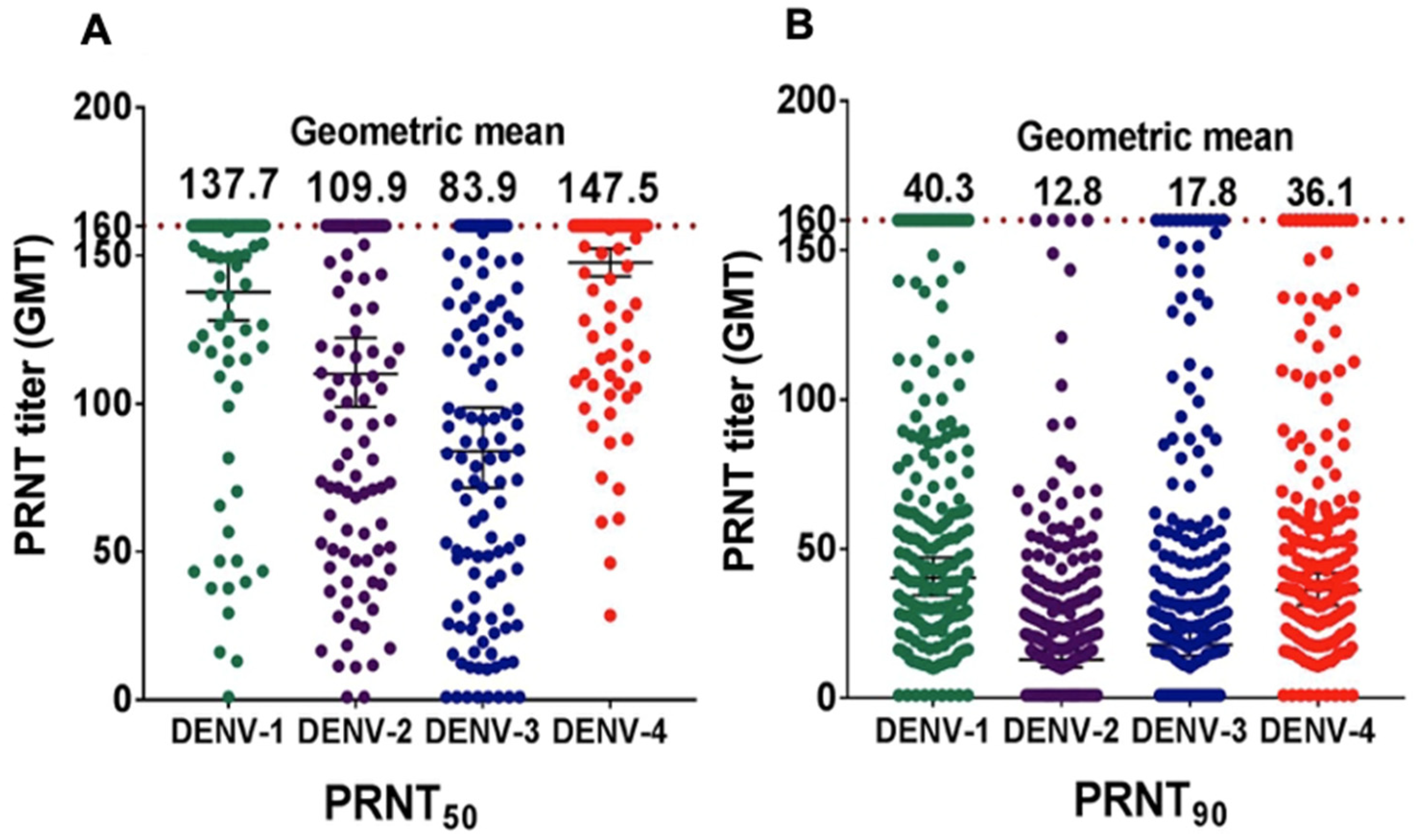
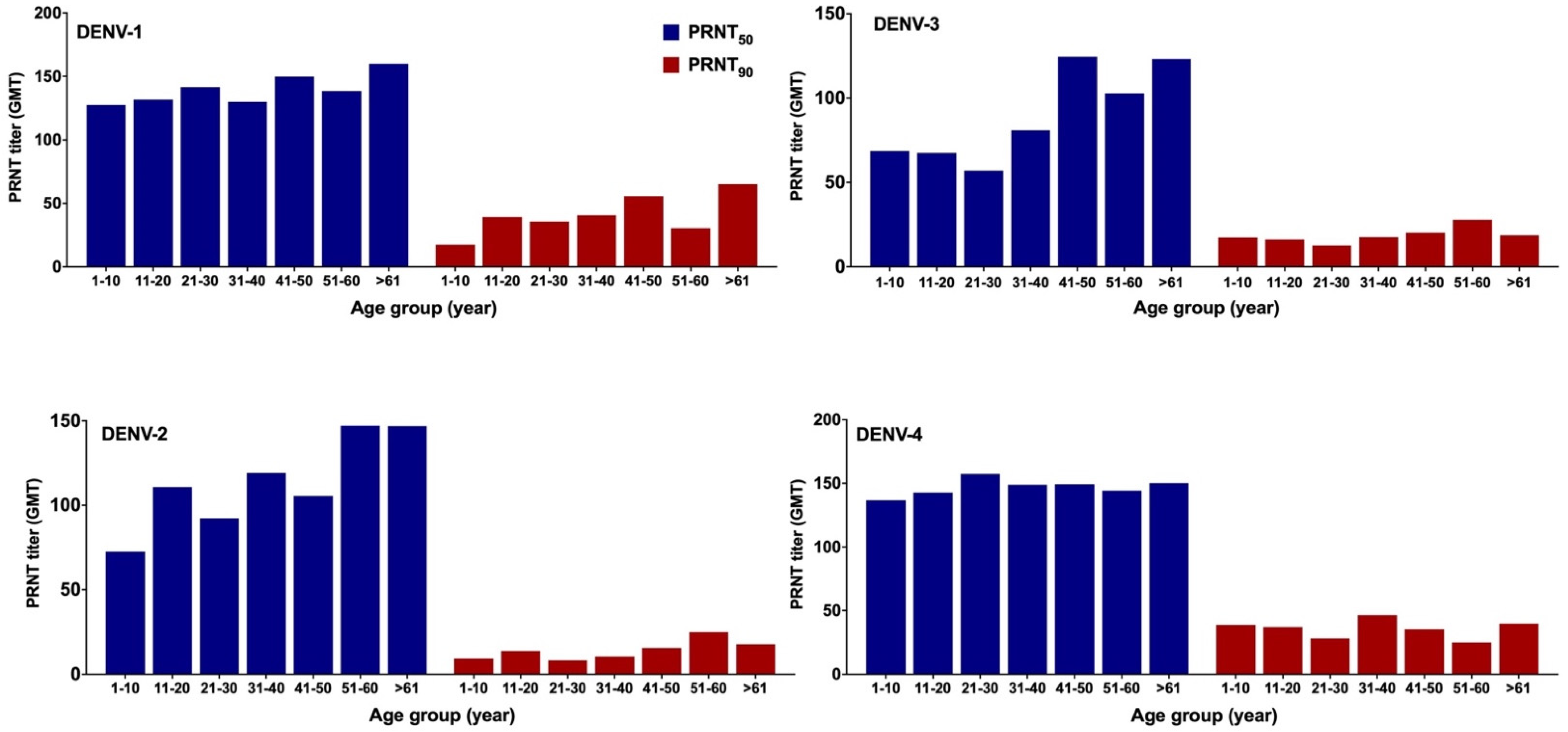
3.4. DENV NAb Geometric Mean Titer (GMT)

Interpolated NAb titers were determined using a non-linear regression curve at two different thresholds, PRNT50 and PRNT90. NAb GMTs were calculated for each DENV serotype. At a PRNT50 threshold, DENV-4 had the highest GMT (147.5) followed by DENV-1 (137.7), DENV-2 (109.9) and DENV-3 (83.9) (Figure 3A). At a higher PRNT90 threshold, DENV-1 GMT was of the greatest magnitude followed by DENV-4, DENV-3 and DENV-2 (Figure 3B).
GMTs of NAb for each DENV serotype were stratified based on age and gender, which increased with age for all serotypes for both thresholds (Figure 4). This pattern, typically seen in dengue-endemic areas, was clearer for DENV-2 and DENV-3. At PRNT50, the GMTs for DENV-1 and DENV-4 were greater than 120 for all age groups (Figure 4). The GMTs were not influenced by gender for all four DENV serotypes. Although GMTs were slightly lower among females, in particular for DENV-1 and DENV-4, t-test analyses revealed that differences were not statistically significant (not shown).

3.5. Prevalence of ZIKV NAb

Sufficient volumes were available for 116/199 serum samples volume to assess ZIKV NAb responses. NAb GMTs at two thresholds (PRNT50, and PRNT90) are depicted in Figure 5A. ZIKV NAb GMTs were 40.9 and 10.3 for PRNT50 and PRNT90, respectively. The proportion of NAb-positive sera for each cut-off are presented in Figure 5B. At threshold PRNT50 > 10, NAb were detected in all specimens, while a total of 73 (62.9%) serum samples were seropositive at PRNT90 > 10, respectively. At the highest cut-off, PRNT90 > 40, 7 (6.0%) specimens were seropositive for ZIKV (Figure 5B).

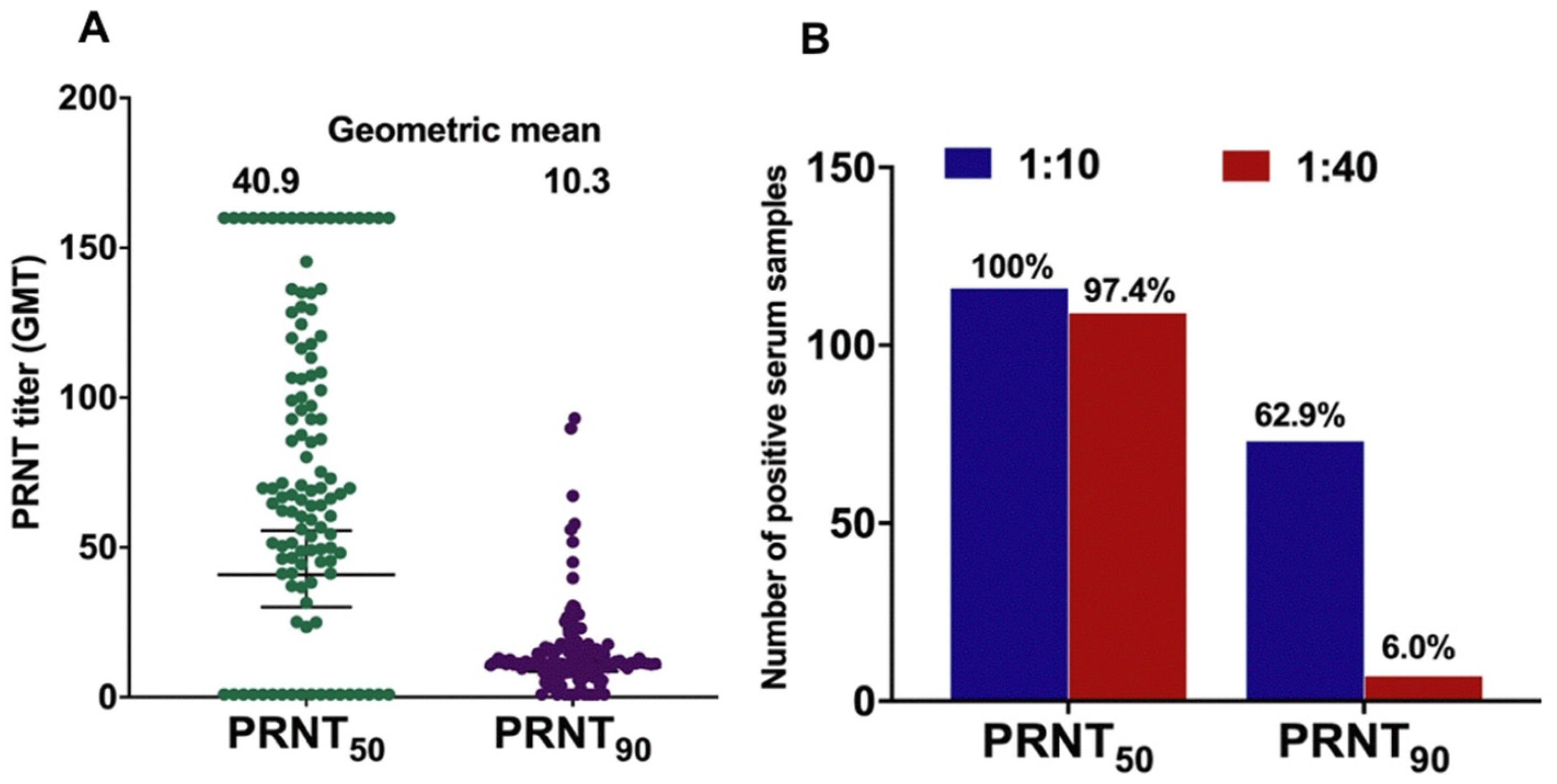
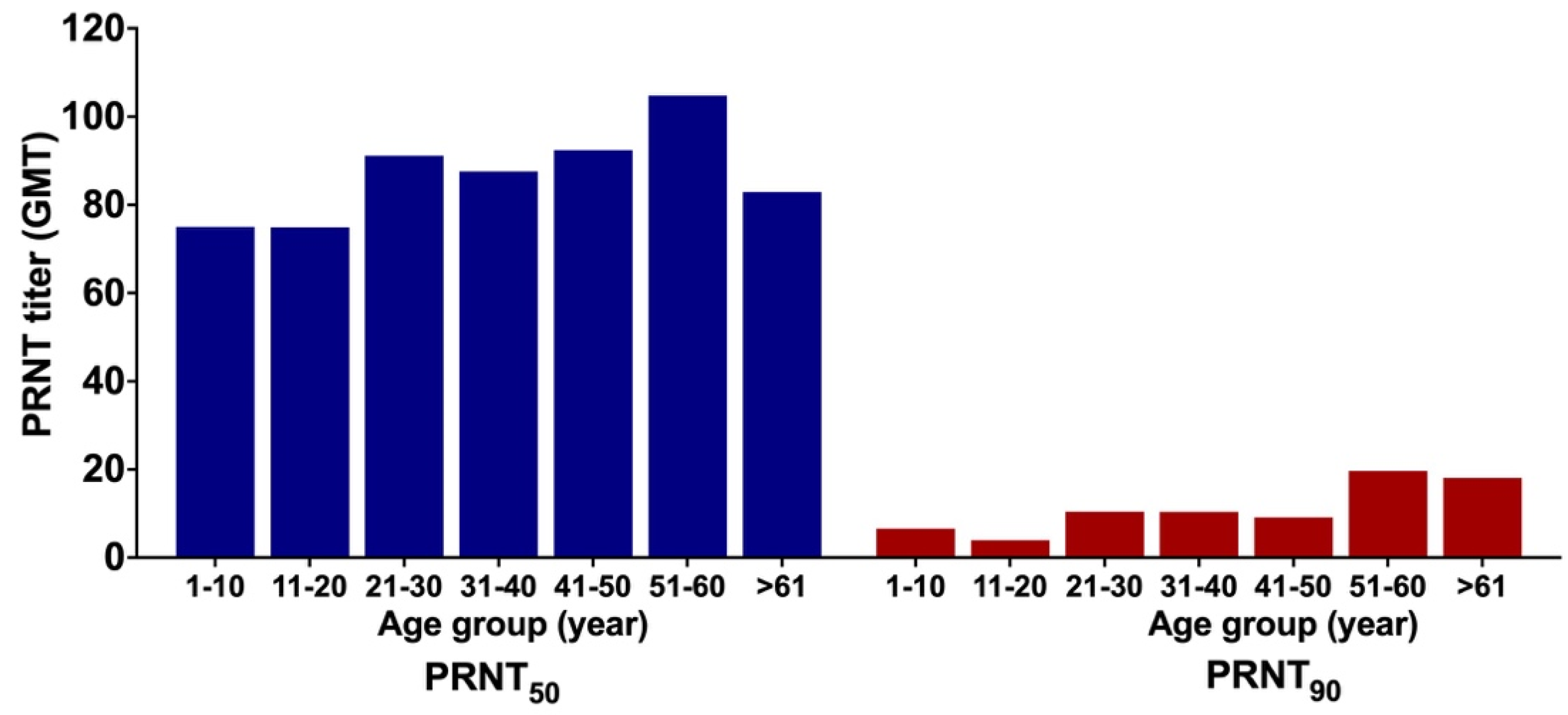
3.6. GMT of ZIKV NAb

GMTs of ZIKV NAb were stratified based on age group and gender. The GMTs increased with age (Figure 6). At PRNT50, GMT for individuals aged 1–10 years old was 75.07 and peaked to 104.78 among those aged between 51 and 60 years. At PRNT90, GMT for individuals aged 1–10 years old was 6.63, increasing to 10.39 for the middle age group (31–40 years old), and to 18.12 for the oldest group (>61 years old) (Figure 6). There was no significant difference of GMT in ZIKV NAb titers between males and females for each threshold with t-test p-values of 0.343 and 0.223 for PRNT50 and PRNT90, respectively (Figure 6).

3.7. Confirmation of ZIKV Infection

To confirm the validity of ZIKV infection among ZIKV-seropositive samples at the highest threshold (PRNT90), an additional confirmational step using stringent criteria was conducted. In Indonesia, due to the cross-reactivity between DENV and ZIKV, it was recommended that serum samples were classified: (a) ZIKV seropositive, if ZIKV PRNT90 titers > 4-fold higher than all DENV serotype PRNT90 titers; and (b) Flavivirus seropositive, if ZIKV NAb are present but at PRNT90 titers < 4-fold higher than any DENV PRNT90 titer [34]. The distribution of DENV NAb and ZIKV NAb titers at PRNT90 for all 116 serum samples and NAb GMTs for all four DENV serotypes and ZIKV are presented in Figure 7A. In this study, at the threshold PRNT90, 73 and 7 samples had ZIKV NAb titers > 10 and >40, respectively. Using these criteria, none of the samples were ZIKV-seropositive. However, 76 (65.5%) samples were classified as Flavivirus seropositive.
Detailed profiles of seven sera that had ZIKV NAb titers > 40 at PRNT90 were explored (Figure 7B and Table 2). When dengue infection background was assessed, all seven sera had multitypic anti-DENV NAb responses at PRNT50 > 10 (Table 2). However, at PRNT90 > 40, for both DENV and ZIKV, two of the samples, ID A-71 and A-119, were classified as naïve for DENV infection and seropositive for ZIKV, and thus suggested a ZIKV infection.

4. Discussion

4.1. Prevalence of DENV-Specific NAb

In this cross-sectional study, we assessed DENV seroprevalence among randomly sampled healthy inhabitants in Aceh Province, Indonesia using the gold standard plaque reduction neutralization test. Using a threshold PRNT50 > 10, all sera neutralized one or more DENV serotypes; 198 (99.4%) showed a multitypic profile and a single serum sample suggested prior monotypic infection with DENV-4. These data indicate dengue is hyperendemic in this westernmost region of Indonesia. The results from this present study are comparable with seroprevalence data among children (mean age of 9.6 years) from urban areas in Indonesia, in which 98.6% of 765 samples neutralized one or more DENV serotypes, with multitypic profiles in 50.9% of study participants [12]. In the present study, 99.4% of samples demonstrated multitypic DENV-neutralizing antibodies. This is unsurprising given the participants in the present study were older, with 75.2% aged 20 years or more, and were exposed to hyperendemic dengue transmission over their lifetimes.
In this present study, no monotypic DENV infections were observed in participants aged more than 30 years old. In addition, NAb GMTs increased with age indicating likely long-term dengue circulation in the area. This classic pattern was also observed in many studies in endemic and hyperendemic countries including India [44], Sri Lanka [45], Vietnam [46], and Mexico [47]. Our data confirm hyperendemic dengue circulation occurred for a significant period of time, likely many decades, in the westernmost region of Indonesia. A 1995 study in Yogyakarta, Central Java, found that 56.2% of 1837 children aged between 4–9 years old had PRNT70 NAb titers > 60. These data indicate that dengue was hyperendemic in different regions of Indonesia with intense transmission for decades, a trend that continued in recent years.
Data from this present study indicate that 100% of samples were positive for DENV-4 with fewer samples (191; 96%) seropositive for DENV-3. Across all four serotypes, DENV-4 demonstrated the highest NAb GMTs, and DENV-3 demonstrated the lowest, suggesting differential DENV serotype dominance within Aceh. However, serotype dominance could not be elucidated clearly as the proportion of monotypic infections was low. A previous study in 2014 found that, of 14 provinces assessed, DENV-4 predominance was observed in Aceh only, while in other provinces, DENV-1, DENV-2, and DENV-3 or a combination of these serotypes, were dominant, with DENV-2 dominance being more common [12]. Altogether, these findings indicate that the distribution of predominant serotypes varies by region in Indonesia. Given this diversity of dengue exposure history, it is unsurprising that the rates of symptomatic infection, and the pattern of dengue outbreaks, vary across Indonesian localities, although this is not entirely influenced by serotype distributions.

4.2. Prevalence of ZIKV-Specific NAb

Various thresholds were previously used to define ZIKV infection, for example PRNT50 ≥ 10 [48,49]. However, the high degree of structural and nucleotide sequence homology between ZIKV and other flaviviruses [50] means that distinguishing ZIKV infection in samples from dengue-hyperendemic regions, such as Aceh, is challenging particularly if samples are collected from adults. The WHO recommend the use of the PRNT90 threshold as the gold standard confirmatory assay for ZIKV serology [51], and previous studies used endpoints of PRNT90 ≥ 10 [52,53,54] or ≥20 [55,56] to define the ZIKV seropositivity. In our study, at threshold PRNT90 > 10 and >40, there were 75 (64.6%) and 7 (6.0%) samples classified as ZIKV-seropositive. However, as serum samples used in this study had continuous exposure to DENV, interpretation was more complex.
Using a previously defined criterion for assessing ZIKV seropositivity in Indonesia, ZIKV-specific PRNT90 were > 4-fold higher than any anti-DENV NAb titer [34]; none of the serum samples were positive for ZIKV infection. Nevertheless, more than 75% of the specimens were Flavivirus-seropositive. One reason for this high proportion of nonspecific Flavivirus seropositivity is the large proportion of older participants in this study, with 75.2% of participants being aged 20 years old or older and 100% of the participants exposed to at least one DENV serotype. Nevertheless, by using stringent criteria of PRNT90 > 40, the possibility of ZIKV infection could not be excluded for samples A-71 and A-119 (Table 2). If the same PRNT90 > 40 threshold was used to characterize ZIKV and DENV infections, these two samples would be classified as naïve for all four DENV serotypes and seropositive for ZIKV, suggesting a true ZIKV infection. Other assays to distinguish between ZIKV and DENV antibodies were not available for the present study; we aim to develop more specific protocols in the near future.
The possibility of DENV and ZIKV co-infection cannot be ruled out. In one study, when ZIKV NAb titers were measured 12–19 months after illness onset among PCR-confirmed Zika cases, only 70.1% of 62 samples could be characterized as ZIKV infections based on the described criteria [57]. Furthermore, five (8%) samples had DENV NAb titers > 4-fold higher than ZIKV NAb titers [57]. Due to this fact, diagnosing ZIKV based on the criteria of a ZIKV PRNT90 titer being > 4-fold higher than that of any DENV NAb titer potentially underreports Zika cases in dengue endemic areas.
The cross-reactivity between ZIKV and DENV is not only a diagnostic dilemma but may also have clinical consequences. DENV-induced antibodies were shown to enhance ZIKV infection in FcγR-bearing monocytic cell lines [58]. In addition, other studies reported that anti-DENV antibodies may enhance ZIKV infection through the process of antibody-dependent enhancement (ADE) of infection [59,60,61,62]. Although growing evidence also suggests that prior DENV has a protective role in ZIKV infection [31,63,64,65], studies in rhesus macaques [66,67] and humans [68] do not support ZIKV enhancement. Other studies suggested that protection or enhancement in ZIKV infection may depend upon the level of anti-DENV antibodies present [59].

4.3. Study Limitations

In this study, most serum samples were collected from participants aged five years old or older. This means that most samples had multitypic infection profiles; therefore, it was difficult to determine the current predominant serotypes in circulation. Therefore, the analysis of samples from a younger population is required for better discrimination of predominant DENV serotypes. In addition, limited molecular data of previous ZIKV infections means we are unable to describe ZIKV transmission in this region.

5. Conclusions

All four DENV serotypes circulated in Aceh prior to 2017. DENV-4 seropositivity was identified at the highest frequency in the present study and the highest mean GMT titers were measured for DENV-4 followed by DENV-1, DENV-2 and DENV-3; however, the small sample size limits any generalization to DENV-4 seroprevalence in Aceh. All respondents were exposed to one or more DENV serotypes by the age of 30 years. This indicates DENV is hyperendemic in Aceh. Among 116 sera that had a sufficient volume for analysis, 20 were reactive with ZIKV at a threshold of PRNT90 > 10 and 7 at PRNT90 > 40. However, when ZIKV seropositivity was defined as ZIKV-specific PRNT90 > 4-fold higher than any anti-DENV NAb titer, none of the samples could be classified as ZIKV-seropositive; however, 76 (65.5%) of samples were classified as Flavivirus-seropositive.

Author Contributions

Conceptualization, H.H. and A.I.; methodology, H.H., A.I., K.P., A.M., T.E., S.M., M.M. (Muhsin Muhsin), S.S., T.M.Z. and M.M. (Mudatsir Mudatsir); software, A.I.; validation, H.H., K.P., A.M., T.E., S.M., M.M. (Muhsin Muhsin), S.S., T.M.Z., R.T.S. and A.I.; formal analysis, H.H. and K.P.; investigation, H.H. and K.P.; resources, M.M. (Mudatsir Mudatsir), S.S., T.M.Z. and A.I.; data curation, H.H.; writing—original draft preparation, H.H.; writing—review and editing, H.H., K.P., A.M., T.E., R.T.S. and A.I.; visualization, H.H.; supervision, R.T.S. and A.I.; project administration, H.H.; funding acquisition, A.I. All authors have read and agreed to the published version of the manuscript.

Funding

This research was funded by the Department of Foreign Affairs and Trade thought Australia Awards Scholarship (AAS) (Grant No. ST000DMX2), Scholarship for International Research Fees (SIRF) (Grant No. F51416) from The University of Western Australia and NHMRC Centre of Research Excellence in Emerging Infectious Diseases (CREID) Scholarship. The APC was funded by The University of Western Australia.

Institutional Review Board Statement

The study was conducted according to the guidelines of the Declaration of Helsinki, and approved by The University of Western Australia Human Research Ethics Committee (No.: RA/4/1/8550 and RA/4/1/5420) and Ethical Clearance Committee Universitas Syiah Kuala, Banda Aceh, Indonesia (No.: 269/KE/FK/2014).

Informed Consent Statement

Informed consent was obtained from all subjects involved in the study.

Data Availability Statement

The NAb titers of samples are available from the corresponding author upon reasonable request.

Acknowledgments

We would like to thank Benediktus Yohan and Dionisius Denis from Eijkman Institute for Molecular Biology, Jakarta for the assistance during sample storage and shipment.

Conflicts of Interest

The authors declare no conflict of interest. The funders had no role in the design of the study; in the collection, analyses, or interpretation of data; in the writing of the manuscript, or in the decision to publish the results.

References

  1. Setiati, T.E. Changing epidemiology of dengue haemorrhagic fever in Indonesia. Dengue Bull. 2006, 30, 1–14. [Google Scholar]
  2. Dhewantara, P.W.; Jamil, K.F.; Fajar, J.K.; Saktianggi, P.P.; Nusa, R.; Garjito, T.A.; Anwar, S.; Firzan, F.; Megawati, D.; Sasmono, R.T. Decline of notified dengue infections in Indonesia in 2017: Discussion of the possible determinants. Narra J. 2021, 1. [Google Scholar] [CrossRef]
  3. Harapan, H.; Michie, A.; Yohan, B.; Shu, P.; Mudatsir, M.; Sasmono, R.T.; Imrie, A. Dengue viruses circulating in Indonesia: A systematic review and phylogenetic analysis of data from five decades. Rev. Med Virol. 2019, 29, e2037. [Google Scholar] [CrossRef]
  4. Megawati, D.; Masyeni, S.; Yohan, B.; Lestarini, A.; Hayati, R.F.; Meutiawati, F.; Suryana, K.; Widarsa, T.; Budiyasa, D.G.; Budiyasa, N.; et al. Dengue in Bali: Clinical characteristics and genetic diversity of circulating dengue viruses. PLoS Negl. Trop. Dis. 2017, 11, e0005483. [Google Scholar] [CrossRef]
  5. Lestari, C.S.W.; Yohan, B.; Yunita, A.; Meutiawati, F.; Hayati, R.F.; Trimarsanto, H.; Sasmono, R.T. Phylogenetic and evolutionary analyses of dengue viruses isolated in Jakarta, Indonesia. Virus Genes 2017, 53, 778–788. [Google Scholar] [CrossRef]
  6. Haryanto, S.; Hayati, R.F.; Yohan, B.; Sijabat, L.; Sihite, I.F.; Fahri, S.; Meutiawati, F.; Halim, J.A.N.; Halim, S.N.; Soebandrio, A.; et al. The molecular and clinical features of dengue during outbreak in Jambi, Indonesia in 2015. Pathog. Glob. Health 2016, 110, 119–129. [Google Scholar] [CrossRef] [Green Version]
  7. Sasmono, R.T.; Wahid, I.; Trimarsanto, H.; Yohan, B.; Wahyuni, S.; Hertanto, M.; Yusuf, I.; Mubin, H.; Ganda, I.J.; Latief, R.; et al. Genomic analysis and growth characteristic of dengue viruses from Makassar, Indonesia. Infect. Genet. Evol. 2015, 32, 165–177. [Google Scholar] [CrossRef] [Green Version]
  8. Fahri, S.; Yohan, B.; Trimarsanto, H.; Sayono, S.; Hadisaputro, S.; Dharmana, E.; Syafruddin, D.; Sasmono, R.T. Molecular Surveillance of Dengue in Semarang, Indonesia Revealed the Circulation of an Old Genotype of Dengue Virus Serotype-1. PLoS Negl. Trop. Dis. 2013, 7, e2354. [Google Scholar] [CrossRef] [Green Version]
  9. Nusa, R.; Prasetyowati, H.; Meutiawati, F.; Yohan, B.; Trimarsanto, H.; Setianingsih, T.Y.; Sasmono, R.T. Molecular surveillance of Dengue in Sukabumi, West Java province, Indonesia. J. Infect. Dev. Ctries. 2014, 8, 733–741. [Google Scholar] [CrossRef] [Green Version]
  10. Wardhani, P.; Aryati, A.; Yohan, B.; Trimarsanto, H.; Setianingsih, T.Y.; Puspitasari, D.; Arfijanto, M.V.; Bramantono, B.; Suharto, S.; Sasmono, R.T. Clinical and virological characteristics of dengue in Surabaya, Indonesia. PLoS ONE 2017, 12, e0178443. [Google Scholar] [CrossRef] [Green Version]
  11. Kusmintarsih, E.S.; Hayati, R.F.; Turnip, O.N.; Yohan, B.; Suryaningsih, S.; Pratiknyo, H.; Denis, D.; Sasmono, R.T. Molecular characterization of dengue viruses isolated from patients in Central Java, Indonesia. J. Infect. Public Health 2018, 11, 617–625. [Google Scholar] [CrossRef]
  12. Sasmono, R.T.; Taurel, A.-F.; Prayitno, A.; Sitompul, H.; Yohan, B.; Hayati, R.F.; Bouckenooghe, A.; Hadinegoro, S.R.; Nealon, J. Dengue virus serotype distribution based on serological evidence in pediatric urban population in Indonesia. PLoS Negl. Trop. Dis. 2018, 12, e0006616. [Google Scholar] [CrossRef] [Green Version]
  13. Cauchemez, S.; Besnard, M.; Bompard, P.; Dub, T.; Guillemette-Artur, P.; Eyrolle-Guignot, D.; Salje, H.; Van Kerkhove, M.D.; Abadie, V.; Gael, C.; et al. Association between Zika virus and microcephaly in French Polynesia, 2013–15: A retrospective study. Lancet 2016, 387, 2125–2132. [Google Scholar] [CrossRef] [Green Version]
  14. de Araújo, T.V.B.; Rodrigues, L.C.; de Alencar Ximenes, R.A.; de Barros Miranda-Filho, D.; Montarroyos, U.R.; de Melo, A.P.L.; Valongueiro, S.; de Albuquerque, M.D.F.P.M.; Souza, W.V.; Braga, C.; et al. Association between Zika virus infection and microcephaly in Brazil, January to May 2016: Preliminary report of a case-control study. Lancet Infect. Dis. 2016, 16, 1356–1363. [Google Scholar] [CrossRef] [Green Version]
  15. de Araújo, T.V.B.; Ximenes, R.A.D.A.; Miranda-Filho, D.D.B.; Souza, W.; Montarroyos, U.R.; de Melo, A.P.L.; Valongueiro, S.; Albuquerque, M.D.F.P.M.D.; Braga, C.; Filho, S.B.; et al. Association between microcephaly, Zika virus infection, and other risk factors in Brazil: Final report of a case-control study. Lancet Infect. Dis. 2018, 18, 328–336. [Google Scholar] [CrossRef] [Green Version]
  16. Schuler-Faccini, L.; Ribeiro, E.M.; Feitosa, I.M.L.; Horovitz, D.D.G.; Cavalcanti, D.P.; Pessoa, A.; Doriqui, M.J.R.; Neri, J.I.; Neto, J.M.D.; Wanderley, H.Y.C.; et al. Possible association between Zika virus Infection and microcephaly—Brazil, 2015. Morbid. Mortal. Wkly. Rep. 2016, 65, 59–62. [Google Scholar] [CrossRef]
  17. de Oliveira, W.K.; de França, G.V.A.; Carmo, E.H.; Duncan, B.B.; Kuchenbecker, R.D.S.; Schmidt, M.I. Infection-related microcephaly after the 2015 and 2016 Zika virus outbreaks in Brazil: A surveillance-based analysis. Lancet 2017, 390, 861–870. [Google Scholar] [CrossRef] [Green Version]
  18. Moi, M.L.; Nguyen, T.T.T.; Nguyen, C.T.; Vu, T.B.H.; Tun, M.M.N.; Pham, T.D.; Pham, N.T.; Tran, T.; Morita, K.; Le, T.Q.M.; et al. Zika virus infection and microcephaly in Vietnam. Lancet Infect. Dis. 2017, 17, 805–806. [Google Scholar] [CrossRef] [Green Version]
  19. Wongsurawat, T.; Athipanyasilp, N.; Jenjaroenpun, P.; Jun, S.-R.; Kaewnapan, B.; Wassenaar, T.M.; Leelahakorn, N.; Angkasekwinai, N.; Kantakamalakul, W.; Ussery, D.W.; et al. Case of Microcephaly after Congenital Infection with Asian Lineage Zika Virus, Thailand. Emerg. Infect. Dis. 2018, 24, 1758–1761. [Google Scholar] [CrossRef]
  20. Lim, S.K.; Lim, J.K.; Yoon, I.K. An Update on Zika Virus in Asia. Infect. Chemother. 2017, 49, 91–100. [Google Scholar] [CrossRef] [Green Version]
  21. Marchette, N.J.; Garcia, R.; Rudnick, A. Isolation of Zika Virus from Aedes Aegypti Mosquitoes in Malaysia. Am. J. Trop. Med. Hyg. 1969, 18, 411–415. [Google Scholar] [CrossRef] [PubMed]
  22. Buathong, R.; Hermann, L.; Thaisomboonsuk, B.; Rutvisuttinunt, W.; Klungthong, C.; Chinnawirotpisan, P.; Manasatienkij, W.; Nisalak, A.; Fernandez, S.; Yoon, I.-K.; et al. Detection of Zika Virus Infection in Thailand, 2012–2014. Am. J. Trop. Med. Hyg. 2015, 93, 380–383. [Google Scholar] [CrossRef] [PubMed] [Green Version]
  23. Alera, M.T.; Hermann, L.; Tac-An, I.A.; Klungthong, C.; Rutvisuttinunt, W.; Manasatienkij, W.; Villa, D.; Thaisomboonsuk, B.; Velasco, J.M.; Chinnawirotpisan, P.; et al. Zika virus infection, Philippines, 2012. Emerg. Infect. Dis. 2015, 21, 722–724. [Google Scholar] [CrossRef] [PubMed]
  24. Chu, D.-T.; Ngoc, V.T.N.; Tao, Y. Zika virus infection in Vietnam: Current epidemic, strain origin, spreading risk, and perspective. Eur. J. Clin. Microbiol. Infect. Dis. 2017, 36, 2041–2042. [Google Scholar] [CrossRef] [PubMed]
  25. Heang, V.; Yasuda, C.Y.; Sovann, L.; Haddow, A.D.; Travassos da Rosa, A.P.; Tesh, R.B.; Kasper, M.R. Zika virus infection, Cambodia, 2010. Emerg. Infect. Dis. 2012, 18, 349–351. [Google Scholar] [CrossRef] [PubMed]
  26. Perkasa, A.; Yudhaputri, F.; Haryanto, S.; Hayati, R.F.; Ma’roef, C.N.; Antonjaya, U.; Yohan, B.; Myint, K.S.A.; Ledermann, J.P.; Rosenberg, R.; et al. Isolation of Zika Virus from Febrile Patient, Indonesia. Emerg. Infect. Dis. 2016, 22, 924–925. [Google Scholar] [CrossRef] [PubMed] [Green Version]
  27. Jeffree, S.; Dony, J.J.F.; Ahmed, K. The first outbreak of autochthonous Zika virus in Sabah, Malaysian Borneo. Int. J. Infect. Dis. 2018, 73, 213. [Google Scholar] [CrossRef]
  28. Duong, V.; Dussart, P.; Buchy, P. Zika virus in Asia. Int. J. Infect. Dis. 2017, 54, 121–128. [Google Scholar] [CrossRef] [Green Version]
  29. Singapore Zika Study Group. Outbreak of Zika virus infection in Singapore: An epidemiological, entomological, virological, and clinical analysis. Lancet Infect. Dis. 2017, 17, 813–821. [Google Scholar] [CrossRef] [Green Version]
  30. Musso, D.; Lanteri, M.C. Zika virus in Singapore: Unanswered questions. Lancet Infect. Dis. 2017, 17, 782–783. [Google Scholar] [CrossRef] [Green Version]
  31. Gordon, A.; Gresh, L.; Ojeda, S.; Katzelnick, L.C.; Sanchez, N.; Mercado, J.C.; Chowell, G.; Lopez, B.; Elizondo, D.; Coloma, J.; et al. Prior dengue virus infection and risk of Zika: A pediatric cohort in Nicaragua. PLoS Med. 2019, 16, e1002726. [Google Scholar] [CrossRef] [PubMed]
  32. Olson, J.G.; Ksiazek, T.G.; Gubler, D.; Lubis, S.I.; Simanjuntak, G.; Lee, V.H.; Nalim, S.; Juslis, K.; See, R. A survey for arboviral antibodies in sera of humans and animals in Lombok, Republic of Indonesia. Ann. Trop. Med. Parasitol. 1983, 77, 131–137. [Google Scholar] [CrossRef] [PubMed]
  33. Olson, J.G.; Ksiazek, T.G.; Suhandiman; Triwibowo. Zika virus, a cause of fever in Central Java, Indonesia. Trans. R. Soc. Trop. Med. Hyg. 1981, 75, 389–393. [Google Scholar] [CrossRef]
  34. Sasmono, R.T.; Dhenni, R.; Yohan, B.; Pronyk, P.; Hadinegoro, S.R.; Soepardi, E.J.; Ma’roef, C.N.; Satari, H.I.; Menzies, H.; Hawley, W.A.; et al. Zika Virus Seropositivity in 1–4-Year-Old Children, Indonesia, 2014. Emerg. Infect. Dis. 2018, 24, 1740. [Google Scholar] [CrossRef]
  35. Kwong, J.C.; Druce, J.D.; Leder, K. Case report: Zika virus infection acquired during brief travel to Indonesia. Am. J. Trop. Med. Hyg. 2013, 89, 516–517. [Google Scholar] [CrossRef] [PubMed] [Green Version]
  36. Leung, G.H.Y.; Baird, R.; Druce, J.; Anstey, N.M. Zika virus infection in australia following a monkey bite in indonesia. Southeast Asian J. Trop. Med. Public Health 2015, 46, 460. [Google Scholar] [PubMed]
  37. Bogoch, I.I.; Brady, O.J.; Kraemer, M.U.G.; German, M.; Creatore, I.M.; Brent, S.; Watts, A.G.; Hay, S.; A Kulkarni, M.; Brownstein, J.S.; et al. Potential for Zika virus introduction and transmission in resource-limited countries in Africa and the Asia-Pacific region: A modelling study. Lancet Infect. Dis. 2016, 16, 1237–1245. [Google Scholar] [CrossRef] [Green Version]
  38. Sasmono, R.T.; Johar, E.; Yohan, B.; Ma’roef, C.N.; Pronyk, P.; Hadinegoro, S.R.; Soepardi, E.J.; Bouckenooghe, A.; Hawley, W.A.; Rosenberg, R.; et al. Spatiotemporal Heterogeneity of Zika Virus Transmission in Indonesia: Serosurveillance Data from a Pediatric Population. Am. J. Trop. Med. Hyg. 2021, 104, 2220–2223. [Google Scholar] [CrossRef]
  39. Badan Pusat Statistik. Proyeksi Jumlah Penduduk Kabupaten Aceh Jaya (Jiwa), 2017–2019; Badan Pusat Statistik Kabupaten Aceh Jaya: Jakarta, Indonesia, 2019. [Google Scholar]
  40. Ernst, T.; McCarthy, S.; Chidlow, G.; Luang-Suarkia, D.; Holmes, E.C.; Smith, D.W.; Imrie, A. Emergence of a New Lineage of Dengue Virus Type 2 Identified in Travelers Entering Western Australia from Indonesia, 2010–2012. PLoS Negl. Trop. Dis. 2015, 9, e0003442. [Google Scholar] [CrossRef]
  41. WHO. Guidelines for Plaque Reduction Neutralization Testing of Human Antibodies to Dengue Viruses; WHO: Geneva, Switzerland, 2008; Available online: http://whqlibdoc.who.int/hq/2007/who_ivb_07.07_eng.pdf (accessed on 29 August 2018).
  42. Timiryasova, T.M.; Bonaparte, M.I.; Luo, P.; Zedar, R.; Hu, B.T.; Hildreth, S.W. Optimization and Validation of a Plaque Reduction Neutralization Test for the Detection of Neutralizing Antibodies to Four Serotypes of Dengue Virus Used in Support of Dengue Vaccine Development. Am. J. Trop. Med. Hyg. 2013, 88, 962–970. [Google Scholar] [CrossRef]
  43. Panta, K. Antibody Mediated Immune Response Againts Flavivirus Infection in Western Australia Travellers; The University of Western Australia: Perth, Australia, 2018. [Google Scholar]
  44. Garg, S.; Chakravarti, A.; Singh, R.; Masthi, N.R.; Goyal, R.C.; Jammy, G.R.; Ganguly, E.; Sharma, N.; Singh, M.M.; Ferreira, G.; et al. Dengue serotype-specific seroprevalence among 5- to 10-year-old children in India: A community-based cross-sectional study. Int. J. Infect. Dis. 2017, 54, 25–30. [Google Scholar] [CrossRef] [PubMed] [Green Version]
  45. Jeewandara, C.; Gomes, L.; Paranavitane, S.A.; Tantirimudalige, M.; Panapitiya, S.S.; Jayewardene, A.; Fernando, S.; Fernando, R.H.; Prathapan, S.; Ogg, G.S.; et al. Change in Dengue and Japanese Encephalitis Seroprevalence Rates in Sri Lanka. PLoS ONE 2015, 10, e0144799. [Google Scholar] [CrossRef] [Green Version]
  46. Thai, K.T.D.; Binh, T.Q.; Giao, P.T.; Phuöng, H.L.; Hung, L.Q.; Van Nam, N.; Nga, T.T.; Groen, J.; Nagelkerke, N.; Vries, P.J. Seroprevalence of dengue antibodies, annual incidence and risk factors among children in southern Vietnam. Trop. Med. Int. Health 2005, 10, 379–386. [Google Scholar] [CrossRef]
  47. Amaya-Larios, I.Y.; Ramos-Castañeda, J.; Sepúlveda-Salinas, K.J.; Mayer, S.V.; Falcón-Lezama, J.A.; Galeana-Hernández, M.; Vasilakis, N.; Comas-García, A.; Martínez-Vega, R.A. Seroprevalence of Neutralizing Antibodies Against Dengue Virus in Two Localities in the State of Morelos, Mexico. Am. J. Trop. Med. Hyg. 2014, 91, 1057–1065. [Google Scholar] [CrossRef] [PubMed] [Green Version]
  48. Fortuna, C.; Remoli, M.E.; Rizzo, C.; Benedetti, E.; Fiorentini, C.; Bella, A.; Argentini, C.; Farchi, F.; Castilletti, C.; Capobianchi, M.R.; et al. Imported arboviral infections in Italy, July 2014–October 2015: A National Reference Laboratory report. BMC Infect. Dis. 2017, 17, 216. [Google Scholar] [CrossRef] [Green Version]
  49. Kim, Y.H.; Lee, J.; Kim, Y.-E.; Chong, C.-K.; Pinchemel, Y.; Reisdörfer, F.; Coelho, J.B.; Dias, R.F.; Bae, P.K.; Gusmão, Z.P.M.; et al. Development of a Rapid Diagnostic Test Kit to Detect IgG/IgM Antibody against Zika Virus Using Monoclonal Antibodies to the Envelope and Non-structural Protein 1 of the Virus. Korean J. Parasitol. 2018, 56, 61–70. [Google Scholar] [CrossRef] [Green Version]
  50. Chua, A.; Prat, I.; Nuebling, C.M.; Wood, D.; Moussy, F. Update on Zika Diagnostic Tests and WHO’s Related Activities. PLoS Negl. Trop. Dis. 2017, 11, e0005269. [Google Scholar] [CrossRef]
  51. Roehrig, J.T.; Hombach, J.; Barrett, A.D.T. Guidelines for Plaque-Reduction Neutralization Testing of Human Antibodies to Dengue Viruses. Viral Immunol. 2008, 21, 123–132. [Google Scholar] [CrossRef]
  52. Shan, C.; Xie, X.; Ren, P.; Loeffelholz, M.J.; Yang, Y.; Furuya, A.; Dupuis, A.P., 2nd; Kramer, L.D.; Wong, S.J.; Shi, P.Y. A Rapid Zika Diagnostic Assay to Measure Neutralizing Antibodies in Patients. EBioMedicine 2017, 17, 157–162. [Google Scholar] [CrossRef] [Green Version]
  53. Lee, W.T.; Wong, S.J.; Kulas, K.E.; Dupuis, A.P., 2nd; Payne, A.F.; Kramer, L.D.; Dean, A.B.; St George, K.; White, J.L.; Sommer, J.N.; et al. Development of Zika Virus Serological Testing Strategies in New York State. J. Clin. Microbiol. 2018, 56, e01591-17. [Google Scholar] [CrossRef] [Green Version]
  54. Ribeiro, M.; Khouri, R.; Sousa, P.; Branco, M.; Batista, R.; Costa, E.; Alves, M.; Amaral, G.; Borges, M.; Takahasi, E.; et al. Plaque Reduction Neutralization Test (PRNT) in the Congenital Zika Syndrome: Positivity and Associations with Laboratory, Clinical, and Imaging Characteristics. Viruses 2020, 12, 1244. [Google Scholar] [CrossRef] [PubMed]
  55. Koishi, A.C.; Suzukawa, A.A.; Zanluca, C.; Camacho, D.E.; Comach, G.; Dos Santos, C.N.D. Development and evaluation of a novel high-throughput image-based fluorescent neutralization test for detection of Zika virus infection. PLoS Negl. Trop. Dis. 2018, 12, e0006342. [Google Scholar] [CrossRef] [PubMed]
  56. L’Huillier, A.G.; Hamid-Allie, A.; Kristjanson, E.; Papageorgiou, L.; Hung, S.; Wong, C.F.; Stein, D.R.; Olsha, R.; Goneau, L.W.; Dimitrova, K.; et al. Evaluation of Euroimmun Anti-Zika Virus IgM and IgG Enzyme-Linked Immunosorbent Assays for Zika Virus Serologic Testing. J. Clin. Microbiol. 2017, 55, 2462–2471. [Google Scholar] [CrossRef] [PubMed] [Green Version]
  57. Griffin, I.; Martin, S.W.; Fischer, M.; Chambers, T.V.; Kosoy, O.; Al, I.G.E.; Ponomareva, O.; Gillis, L.D.; Blackmore, C.; Jean, R. Zika Virus IgM Detection and Neutralizing Antibody Profiles 12–19 Months after Illness Onset. Emerg. Infect. Dis. 2019, 25, 299–303. [Google Scholar] [CrossRef] [PubMed]
  58. Priyamvada, L.; Quicke, K.M.; Hudson, W.; Onlamoon, N.; Sewatanon, J.; Edupuganti, S.; Pattanapanyasat, K.; Chokephaibulkit, K.; Mulligan, M.J.; Wilson, P.C.; et al. Human antibody responses after dengue virus infection are highly cross-reactive to Zika virus. Proc. Natl. Acad. Sci. USA 2016, 113, 7852–7857. [Google Scholar] [CrossRef] [PubMed] [Green Version]
  59. Bardina, S.V.; Bunduc, P.; Tripathi, S.; Duehr, J.; Frere, J.J.; Brown, J.A.; Nachbagauer, R.; Foster, G.A.; Krysztof, D.; Tortorella, D.; et al. Enhancement of Zika virus pathogenesis by preexisting antiflavivirus immunity. Science 2017, 356, 175–180. [Google Scholar] [CrossRef] [PubMed] [Green Version]
  60. Paul, L.M.; Carlin, E.R.; Jenkins, M.M.; Tan, A.L.; Barcellona, C.M.; Nicholson, C.O.; Michael, S.F.; Isern, S. Dengue virus antibodies enhance Zika virus infection. Clin. Transl. Immunol. 2016, 5, e117. [Google Scholar] [CrossRef] [PubMed]
  61. Castanha, P.; Nascimento, E.J.M.; Cynthia, B.; Cordeiro, M.T.; De Carvalho, O.V.; De Mendonça, L.R.; Azevedo, E.; França, R.F.; Rafael, D.; Marques, E.T. Dengue virus (DENV)-specific antibodies enhance Brazilian Zika virus (ZIKV) infection. J. Infect. Dis. 2016, 215, 781–785. [Google Scholar] [CrossRef] [Green Version]
  62. Dejnirattisai, W.; Supasa, P.; Wongwiwat, W.; Rouvinski, A.; Barba-Spaeth, G.; Duangchinda, T.; Sakuntabhai, A.; Cao-Lormeau, V.-M.; Malasit, P.; Rey, F.A.; et al. Dengue virus sero-cross-reactivity drives antibody-dependent enhancement of infection with zika virus. Nat. Immunol. 2016, 17, 1102–1108. [Google Scholar] [CrossRef]
  63. Swanstrom, J.A.; Plante, J.A.; Plante, K.S.; Young, E.F.; McGowan, E.; Gallichotte, E.N.; Widman, D.G.; Heise, M.T.; de Silva, A.M.; Baric, R.S. Dengue Virus Envelope Dimer Epitope Monoclonal Antibodies Isolated from Dengue Patients Are Protective against Zika Virus. mBio 2016, 7, e01123-16. [Google Scholar] [CrossRef] [Green Version]
  64. Ribeiro, G.S.; Kikuti, M.; Tauro, L.B.; Nascimento, L.C.J.; Cardoso, C.; Campos, G.S.; Ko, A.; Weaver, S.C.; Reis, M.G.; Kitron, U.; et al. Does immunity after Zika virus infection cross-protect against dengue? Lancet Glob. Health 2018, 6, e140–e141. [Google Scholar] [CrossRef] [Green Version]
  65. Wen, J.; Elong Ngono, A.; Regla-Nava, J.A.; Kim, K.; Gorman, M.J.; Diamond, M.S.; Shresta, S. Dengue virus-reactive CD8+ T cells mediate cross-protection against subsequent Zika virus challenge. Nat. Commun. 2017, 8, 1459. [Google Scholar] [CrossRef] [PubMed] [Green Version]
  66. Pantoja, P.; Pérez-Guzmán, E.X.; Rodríguez, I.V.; White, L.J.; González, O.; Serrano, C.; Giavedoni, L.; Hodara, V.; Cruz, L.; Arana, T.; et al. Zika virus pathogenesis in rhesus macaques is unaffected by pre-existing immunity to dengue virus. Nat. Commun. 2017, 8, 15674. [Google Scholar] [CrossRef] [PubMed]
  67. McCracken, M.; Gromowski, G.D.; Friberg, H.L.; Lin, X.; Abbink, P.; De La Barrera, R.; Eckles, K.H.; Garver, L.S.; Boyd, M.; Jetton, D.; et al. Impact of prior flavivirus immunity on Zika virus infection in rhesus macaques. PLoS Pathog. 2017, 13, e1006487. [Google Scholar] [CrossRef] [PubMed]
  68. Terzian, A.C.B.; Schanoski, A.S.; Mota, M.T.D.O.; Da Silva, R.A.; Estofolete, C.F.; Colombo, T.E.; Rahal, P.; Hanley, K.A.; Vasilakis, N.; Kalil, J.; et al. Viral Load and Cytokine Response Profile Does Not Support Antibody-Dependent Enhancement in Dengue-Primed Zika Virus–Infected Patients. Clin. Infect. Dis. 2017, 65, 1260–1265. [Google Scholar] [CrossRef] [Green Version]
Figure 1. Number of participants based on age and gender (n = 199).
Viruses 14 00219 g001
Figure 2. Status of DENV infection by age group (n = 199). Status of individuals with naïve, monotypic, or multitypic infections classified using two thresholds (PRNT50 and PRNT90 at >10). For each threshold, all monotypic infections were observed in individuals younger than 30 years old.
Viruses 14 00219 g002
Figure 3. Geometric mean titers (GMT) of DENV NAb at two different thresholds: PRNT50 (A) and PRNT90 (B) (n = 199). Neutralizing antibody (NAb) GMTs were interpolated using non-linear regression.
Viruses 14 00219 g003
Figure 4. Stratification of geometric mean titer (GMT) of DENV neutralizing antibody (NAb) titers by age (n = 199). DENV NAb GMTs for each age group for all serotypes were calculated based on two thresholds PRNT50 (blue), and PRNT90 (red).
Viruses 14 00219 g004
Figure 5. Geometric mean titer (GMT) of ZIKV neutralizing antibody (NAb) and the distribution of ZIKV NAb-positive sera at different thresholds (n = 116). (A) GMT of ZIKV NAb using two thresholds (PRNT50 and PRNT90). (B) ZIKV seropositive sera were classified using a combination of four cut-offs (two thresholds of plaque reductions percentage and two cut-offs for serum dilutions).
Viruses 14 00219 g005
Figure 6. Geometric mean titers (GMTs) of ZIKV neutralizing antibody (NAb) at two thresholds (PRNT50 and PRNT90).
Viruses 14 00219 g006
Figure 7. Neutralizing antibody (NAb) titers for ZIKV and DENV at PRNT90 (n = 116). (A) ZIKV NAb titers and corresponding DENV NAb titers were plotted to show dengue infection background. (B) For seven serum samples that had ZIKV NAb titers > 40, the DENV NAb titers were plotted individually and the GMTs were calculated. Using criteria developed in the Indonesian context [34] none of the samples can be classified as ZIKV-seropositive. One dot indicates an individual sample; grey dots indicate samples that have ZIKV NAb titers < 40 at PRNT90, while colored dots represent samples that had ZIKV NAb titers > 40 at PRNT90.
Viruses 14 00219 g007
Table 1. Proportion of DENV NAb-positive sera at different thresholds (n = 199).
Threshold and Titer Cut-OffDENV-1
n (%)
DENV-2
n (%)
DENV-3
n (%)
DENV-4
n (%)
PRNT50
10198 (99.5)197 (99.0)191 (96.0)199 (100.0)
40192 (96.5)181 (91.0)168 (84.4)198 (99.5)
PRNT90
10190 (95.5)151 (75.9)156 (78.4)190 (95.5)
40111 (55.8)45 (22.6)68 (34.2)97 (48.7)
Table 2. Comparison of DENV and ZIKV NAb-positive sera for (n = 7).
IDAge (Year)PRNT90 Titer for DENV Status of Dengue Infection ZIKV PRNT90 TiterInfection Status *
DENV-1DENV-2DENV-3DENV-4PRNT90 > 10PRNT90 > 40
A-15753100.05120.75>160>160Multitypic Multitypic93.119Flavivirus
A-526339.1911.5536.86100.23MultitypicDENV-489.746Flavivirus
A-9760104.8643.2757.94117.66MultitypicMultitypic67.247Flavivirus
A-715332.4316.6131.4036.67MultitypicNaïve57.835Flavivirus
A-1192227.4836.1129.5539.67MultitypicNaïve55.986Flavivirus
A-9450>16024.3128.4289.72MultitypicMultitypic51.872Flavivirus
A-135548.8324.4015.89160.00MultitypicDENV-445.076Flavivirus
* ZIKV seropositive: ZIKV PRNT90 titers > 4-fold higher than all DENV serotype. Flavivirus seropositive: ZIKV NAb are present but at titers < 4-fold higher than any DENV NAb titer [34].
Publisher’s Note: MDPI stays neutral with regard to jurisdictional claims in published maps and institutional affiliations.

