
Feline Injection-Site Sarcoma and Other Adverse Reactions to Vaccination in Cats

 , , , , ,
  , , ,
, , ,  , ,
, , 
Abstract
:1. Introduction
2. Non-Specific Systemic Reactions
3. Hypersensitivity Reactions
3.1. Type I Hypersensitivity Reactions
3.2. Type II Hypersensitivity Reactions
3.3. Type III Hypersensitivity Reactions
3.4. Type IV Hypersensitivity Reactions
4. Immunosuppression
5. Mild Local Reactions at the Injection Site
6. Feline Injection-Site Sarcoma (FISS)
6.1. Epidemiology
6.2. Pathogenesis
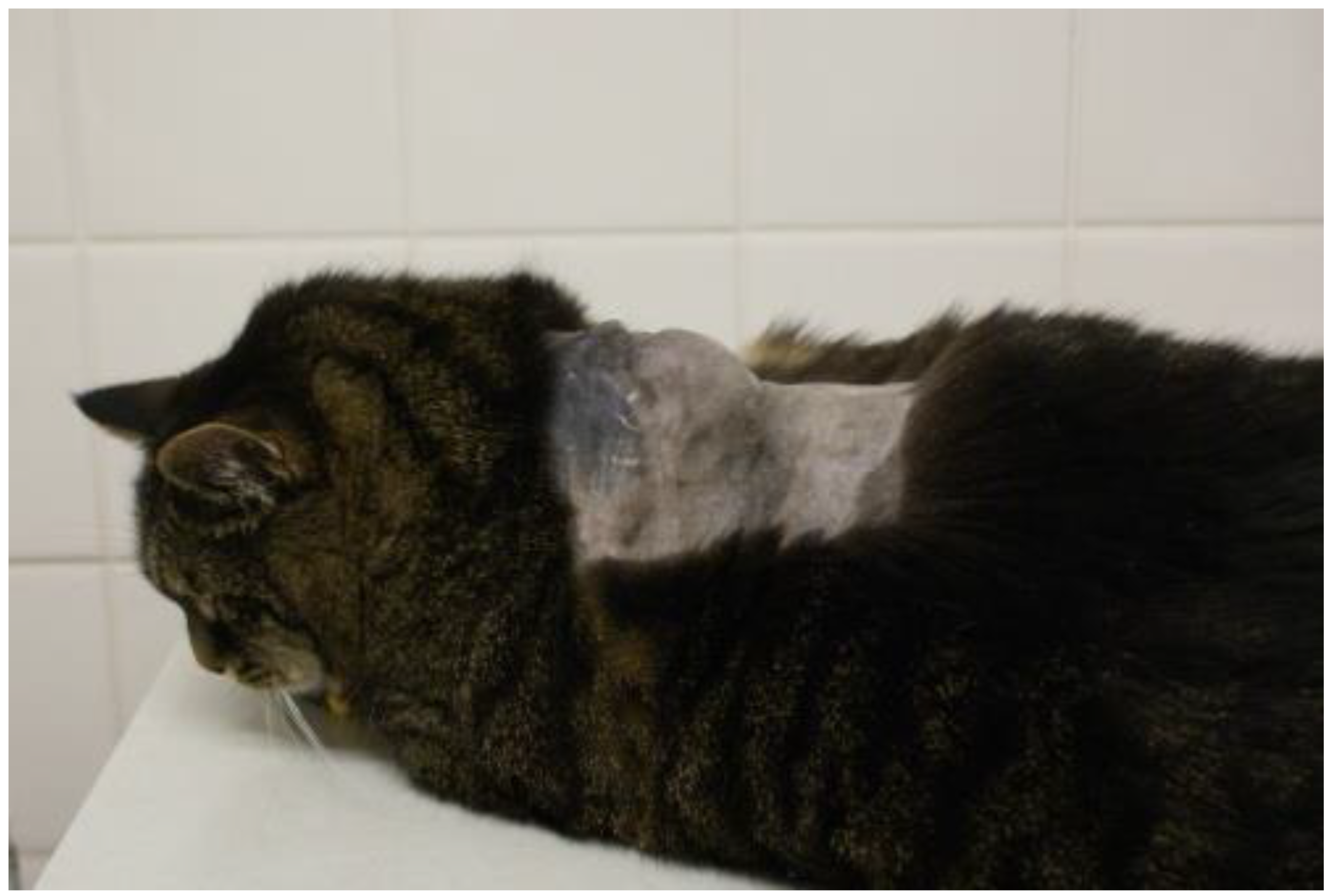
6.3. Clinical Signs
6.4. Prevention
- -
- -
- It is a fact that that vaccines containing adjuvant cause more inflammation than vaccines without adjuvants [41];
- -
- It has been reported that inflammation might contribute to FISS development [83];
- -
- -
- It is currently difficult to truly compare vaccine efficacy studies because of differences in study designs.
7. Disease Induced by the Vaccine Organism or Contamination
8. Lack of Efficacy
9. Conclusions
- Vaccination of cats provides essential protection and should not be stopped because of the risk of FISS. It is important to realize that vaccines are not the only injectable medical products associated with FISSs.
- An individually tailored vaccination schedule is important for each cat. Cats should be vaccinated only as often as necessary in accordance with current guidelines.
- Mucosal intranasal vaccines are preferred over injectable vaccines, if available.
- Among injectable vaccines, there is insufficient information to make definitive recommendations on the preferred vaccine type.
- Vaccines should be brought to room temperature prior to administration but should not be kept unrefrigerated for hours.
- Multi-dose vaccine vials should not be used in cats.
- Subcutaneous injection is preferred to intramuscular injection.
- Vaccines with a long duration of immunity are preferred over those with a short duration of immunity.
- Appropriate sites for injections should be selected. The interscapular region as well as the lateral thoracic wall should generally be avoided for any injection. Vaccines should be injected at sites where any subsequent mass could be easily surgically removed, preferably distally in a limb.
- Generally, any skin or subcutaneous mass in a cat requires further diagnostics. Specifically (the “3-2-1”-rule), thorough post-vaccination monitoring should be performed. Any lump at the site of injection that is still present three months after vaccination, or that is larger than two cm in diameter, or that is increasing in size one month after vaccination, should be surgically removed and investigated through histopathology.
Funding
Conflicts of Interest
References
- European Advisory Board on Cat Diseases. Guideline for Adverse Reactions to Vaccination. Available online: http://www.abcdcatsvets.org/adverse-reactions-to-vaccination/ (accessed on 13 January 2023).
- Hosie, M.J.; Addie, D.; Belák, S.; Boucraut-Baralon, C.; Egberink, H.; Frymus, T.; Gruffydd-Jones, T.; Hartmann, K.; Lloret, A.; Lutz, H.; et al. Matrix vaccination guidelines: ABCD recommendations for indoor/outdoor cats, rescue shelter cats and breeding catteries. J. Feline Med. Surg. 2013, 15, 540–544. [Google Scholar] [CrossRef]
- Hartmann, K.; Day, M.J.; Thiry, E.; Lloret, A.; Frymus, T.; Addie, D.; Boucraut-Baralon, C.; Egberink, H.; Gruffydd-Jones, T.; Horzinek, M.C.; et al. Feline injection-site sarcoma: ABCD guidelines on prevention and management. J. Feline Med. Surg. 2015, 17, 606–613. [Google Scholar] [CrossRef]
- Moore, G.E.; HogenEsch, H. Adverse vaccinal events in dogs and cats. Vet. Clin. N. Am. Small Anim. Pract. 2010, 40, 393–407. [Google Scholar] [CrossRef] [PubMed]
- Day, M.J.; Horzinek, M.C.; Schultz, R.D.; Squires, R.A. WSAVA Guidelines for the vaccination of dogs and cats. J. Small Anim. Pract. 2016, 57, e1–e45. [Google Scholar] [CrossRef] [Green Version]
- Stone, A.E.; Brummet, G.O.; Carozza, E.M.; Kass, P.H.; Petersen, E.P.; Sykes, J.; Westman, M.E. 2020 AAHA/AAFP Feline Vaccination Guidelines. J. Feline Med. Surg. 2020, 22, 813–830. [Google Scholar] [CrossRef]
- Zaugg, I.; Ottiger, H.P. Vaccinovigilance: Adverse reaction reports of animal vaccines in 2020. Schweiz. Arch. Tierheilkd. 2021, 163, 545–552. [Google Scholar] [CrossRef]
- Gaskell, R.M.; Gettinby, G.; Graham, S.J.; Skilton, D. Veterinary Products Committee working group report on feline and canine vaccination. Vet. Rec. 2002, 150, 126–134. [Google Scholar]
- Miller, E.R.; McNeil, M.M.; Moro, P.L.; Duffy, J.; Su, J.R. The reporting sensitivity of the Vaccine Adverse Event Reporting System (VAERS) for anaphylaxis and for Guillain-Barré syndrome. Vaccine 2020, 38, 7458–7463. [Google Scholar] [CrossRef]
- Davis, G.; Cooles, S.; Diesel, G.; Blenkinsop, J. Summary of suspected adverse events, 2014. Vet. Rec. 2016, 178, 187–189. [Google Scholar] [CrossRef] [PubMed]
- Davis, G.; Rooney, A.; Cooles, S.; Evans, G. Suspected adverse events, 2012. Vet. Rec. 2013, 173, 573–576. [Google Scholar] [CrossRef] [PubMed]
- Dyer, F.; Brown, E.; Cooles, S.; Tait, A. Suspected adverse reactions, 2008. Vet. Rec. 2009, 165, 162–164. [Google Scholar] [CrossRef]
- Dyer, F.; Diesel, G.; Cooles, S.; Tait, A. Suspected adverse reactions, 2009. Vet. Rec. 2010, 167, 118–121. [Google Scholar] [CrossRef]
- Dyer, F.; Diesel, G.; Cooles, S.; Tait, A. Suspected adverse reaction surveillance scheme. Suspected adverse events, 2010. Vet. Rec. 2011, 168, 610–613. [Google Scholar] [CrossRef]
- Dyer, F.; Diesel, G.; Cooles, S.; Tait, A. Suspected adverse events, 2011. Vet. Rec. 2012, 170, 640–643. [Google Scholar] [CrossRef] [PubMed]
- Dyer, F.; Spagnuolo-Weaver, M.; Coolees, S.; Tait, A. Suspected adverse reactions, 2007. Vet. Rec. 2008, 163, 69–72. [Google Scholar] [CrossRef]
- Dyer, F.; Spagnuolo-Weaver, M.; Cooles, S.; Tait, A. Suspected adverse reactions, 2006. Vet. Rec. 2007, 160, 748–750. [Google Scholar] [CrossRef]
- Davis, G.; Cooles, S.; Vasan, N. Suspected adverse events, 2013. Vet. Rec. 2015, 176, 11–14. [Google Scholar] [CrossRef] [PubMed]
- Day, M.J. Vaccine side effects: Fact and fiction. Vet. Microbiol. 2006, 117, 51–58. [Google Scholar] [CrossRef]
- Moore, G.E.; DeSantis-Kerr, A.C.; Guptill, L.F.; Glickman, N.W.; Lewis, H.B.; Glickman, L.T. Adverse events after vaccine administration in cats: 2560 cases (2002–2005). J. Am. Vet. Med. Assoc. 2007, 231, 94–100. [Google Scholar] [CrossRef] [PubMed] [Green Version]
- Moore, G.E.; Guptill, L.F.; Ward, M.P.; Glickman, N.W.; Faunt, K.K.; Lewis, H.B.; Glickman, L.T. Adverse events diagnosed within three days of vaccine administration in dogs. J. Am. Vet. Med. Assoc. 2005, 227, 1102–1108. [Google Scholar] [CrossRef] [Green Version]
- Bergmann, M.; Schwertler, S.; Reese, S.; Speck, S.; Truyen, U.; Hartmann, K. Antibody response to feline panleukopenia virus vaccination in healthy adult cats. J. Feline Med. Surg. 2018, 20, 1087–1093. [Google Scholar] [CrossRef]
- Starr, R.M. Reaction rate in cats vaccinated with a new controlled-titer feline panleukopenia-rhinotracheitis-calicivirus-Chlamydia psittaci vaccine. Cornell Vet. 1993, 83, 311–323. [Google Scholar]
- Day, M.J.; Schultz, R.D. Hypersensitivity mechanisms. In Veterinary Immunology—Principles and Practice, 2nd ed.; Day, M.J., Schultz, R.D., Eds.; CRC Press Taylor & Francis Group: Boca Raton, FL, USA, 2014; pp. 139–152. [Google Scholar]
- Tizard, I.R. The major histocompatibility complex. In Veterinary Immunology, 10th ed.; Tizard, I.R., Ed.; Elsevier Inc: St. Louis, MO, USA, 2018; pp. 100–107. [Google Scholar]
- Davis-Wurzler, G.M. Current vaccination strategies in puppies and kittens. Vet. Clin. N. Am. Small Anim. Pract. 2006, 36, 607–640. [Google Scholar] [CrossRef] [PubMed]
- Richards, J.R.; Elston, T.H.; Ford, R.B.; Gaskell, R.M.; Hartmann, K.; Hurley, K.F.; Lappin, M.R.; Levy, J.K.; Rodan, I.; Scherk, M.; et al. The 2006 American association of feline practitioners feline vaccine advisory panel report. J. Am. Vet. Med. Assoc. 2006, 229, 1405–1441. [Google Scholar] [CrossRef]
- Brooks, R. Adverse reactions to canine and feline vaccines. Aust. Vet. J. 1991, 68, 342–344. [Google Scholar] [PubMed]
- Meyer, E.K. Vaccine-associated adverse events. Vet. Clin. N. Am. Small Anim. Pract. 2001, 31, 493–514. [Google Scholar] [CrossRef]
- Gershwin, L.J. Adverse reactions to vaccination: From anaphylaxis to autoimmunity. Vet. Clin. N. Am. Small Anim. Pract. 2018, 48, 279–290. [Google Scholar] [CrossRef]
- Ohmori, K.; Masuda, K.; Maeda, S.; Kaburagi, Y.; Kurata, K.; Ohno, K.; Deboer, D.J.; Tsujimoto, H.; Sakaguchi, M. IgE reactivity to vaccine components in dogs that developed immediate-type allergic reactions after vaccination. Vet. Immunol. Immunopathol. 2005, 104, 249–256. [Google Scholar] [CrossRef] [PubMed]
- Ohmori, K.; Sakaguchi, M.; Kaburagi, Y.; Maeda, S.; Masuda, K.; Ohno, K.; Tsujimoto, H. Suspected allergic reactions after vaccination in 85 dogs in Japan. Vet. Rec. 2005, 156, 87–88. [Google Scholar] [CrossRef]
- Yoshida, M.; Mizukami, K.; Hisasue, M.; Imanishi, I.; Kurata, K.; Ochiai, M.; Itoh, M.; Nasukawa, T.; Uchiyama, J.; Tsujimoto, H.; et al. Anaphylaxis after vaccination for cats in Japan. J. Vet. Med. Sci. 2022, 84, 149–152. [Google Scholar] [CrossRef]
- Sykes, J. Immunization. In Greene’s Infectious Diseases of the Dog and Cat, 5th ed.; Sykes, J., Ed.; Elsevier: St. Louis, MO, USA, 2022; pp. 238–255. [Google Scholar]
- Duval, D.; Giger, U. Vaccine-associated immune-mediated hemolytic anemia in the dog. J. Vet. Intern. Med. 1996, 10, 290–295. [Google Scholar] [CrossRef]
- Lappin, M.R.; Jensen, W.A.; Jensen, T.D.; Basaraba, R.J.; Brown, C.A.; Radecki, S.V.; Hawley, J.R. Investigation of the induction of antibodies against Crandell-Rees feline kidney cell lysates and feline renal cell lysates after parenteral administration of vaccines against feline viral rhinotracheitis, calicivirus, and panleukopenia in cats. Am. J. Vet. Res. 2005, 66, 506–511. [Google Scholar] [CrossRef] [PubMed]
- Whittemore, J.C.; Hawley, J.R.; Jensen, W.A.; Lappin, M.R. Antibodies against Crandell Rees feline kidney (CRFK) cell line antigens, alpha-enolase, and annexin A2 in vaccinated and CRFK hyperinoculated cats. J. Vet. Intern. Med. 2010, 24, 306–313. [Google Scholar] [CrossRef]
- Finch, N.C.; Syme, H.M.; Elliott, J. Risk factors for development of chronic kidney disease in cats. J. Vet. Intern. Med. 2016, 30, 602–610. [Google Scholar] [CrossRef] [Green Version]
- Bennett, D.; Gaskell, R.M.; Mills, A.; Knowles, J.; Carter, S.; McArdle, F. Detection of feline calicivirus antigens in the joints of infected cats. Vet. Rec. 1989, 124, 329–332. [Google Scholar] [CrossRef] [PubMed]
- Dawson, S.; Bennett, D.; Carter, S.D.; Bennett, M.; Meanger, J.; Turner, P.C.; Carter, M.J.; Milton, I.; Gaskell, R.M. Acute arthritis of cats associated with feline calicivirus infection. Res. Vet. Sci. 1994, 56, 133–143. [Google Scholar] [CrossRef] [PubMed]
- Day, M.J.; Schoon, H.A.; Magnol, J.P.; Saik, J.; Devauchelle, P.; Truyen, U.; Gruffydd-Jones, T.J.; Cozette, V.; Jas, D.; Poulet, H.; et al. A kinetic study of histopathological changes in the subcutis of cats injected with non-adjuvanted and adjuvanted multi-component vaccines. Vaccine 2007, 25, 4073–4084. [Google Scholar] [CrossRef] [PubMed]
- Mak, T.W.; Saunders, M.E. Allergy and Hypersensitivity. In The Immune Response. Basic and Clinical Principles, 1st ed.; Mak, T.W., Saunders, M.E., Eds.; Academic Press: New York, NY, USA, 2006; pp. 923–962. [Google Scholar]
- Strasser, A.; May, B.; Teltscher, A.; Wistrela, E.; Niedermüller, H. Immune modulation following immunization with polyvalent vaccines in dogs. Vet. Immunol. Immunopathol. 2003, 94, 113–121. [Google Scholar] [CrossRef]
- Foley, J.E.; Orgad, U.; Hirsh, D.C.; Poland, A.; Pedersen, N.C. Outbreak of fatal salmonellosis in cats following use of a high-titer modified-live panleukopenia virus vaccine. J. Am. Vet. Med. Assoc. 1999, 214, 67–70, 43–44. [Google Scholar]
- Scherk, M.A.; Ford, R.B.; Gaskell, R.M.; Hartmann, K.; Hurley, K.F.; Lappin, M.R.; Levy, J.K.; Little, S.E.; Nordone, S.K.; Sparkes, A.H. 2013 AAFP Feline Vaccination Advisory Panel Report. J. Feline Med. Surg. 2013, 15, 785–808. [Google Scholar] [CrossRef] [Green Version]
- Jas, D.; Frances-Duvert, V.; Brunet, S.; Oberli, F.; Guigal, P.M.; Poulet, H. Evaluation of safety and immunogenicity of feline vaccines with reduced volume. Vaccine 2021, 39, 1051–1057. [Google Scholar] [CrossRef]
- Vaccine-Associated Feline Sarcoma Task Force Guidelines. Diagnosis and treatment of suspected sarcomas. J. Am. Vet. Med. Assoc. 1999, 214, 1745. [Google Scholar]
- Morrison, W.B.; Starr, R.M. Vaccine-associated feline sarcomas. J. Am. Vet. Med. Assoc. 2001, 218, 697–702. [Google Scholar] [CrossRef] [Green Version]
- Hendrick, M.J.; Dunagan, C.A. Focal necrotizing granulomatous panniculitis associated with subcutaneous injection of rabies vaccine in cats and dogs: 10 cases (1988–1989). J. Am. Vet. Med. Assoc. 1991, 198, 304–305. [Google Scholar] [PubMed]
- Hendrick, M.J.; Goldschmidt, M.H. Do injection site reactions induce fibrosarcomas in cats? J. Am. Vet. Med. Assoc. 1991, 199, 968. [Google Scholar] [PubMed]
- Kass, P.H.; Barnes, W.G., Jr.; Spangler, W.L.; Chomel, B.B.; Culbertson, M.R. Epidemiologic evidence for a causal relation between vaccination and fibrosarcoma tumorigenesis in cats. J. Am. Vet. Med. Assoc. 1993, 203, 396–405. [Google Scholar]
- Carroll, E.E.; Dubielzig, R.R.; Schultz, R.D. Cats differ from mink and ferrets in their response to commercial vaccines: A histologic comparison of early vaccine reactions. Vet. Pathol. 2002, 39, 216–227. [Google Scholar] [CrossRef] [PubMed]
- Munday, J.S.; Stedman, N.L.; Richey, L.J. Histology and immunohistochemistry of seven ferret vaccination-site fibrosarcomas. Vet. Pathol. 2003, 40, 288–293. [Google Scholar] [CrossRef]
- Vascellari, M.; Melchiotti, E.; Bozza, M.A.; Mutinelli, F. Fibrosarcomas at presumed sites of injection in dogs: Characteristics and comparison with non-vaccination site fibrosarcomas and feline post-vaccinal fibrosarcomas. J. Vet. Med. A Physiol. Pathol. Clin. Med. 2003, 50, 286–291. [Google Scholar] [CrossRef]
- Wei, Q.; Ramsey, S.A.; Larson, M.K.; Berlow, N.E.; Ochola, D.; Shiprack, C.; Kashyap, A.; Séguin, B.; Keller, C.; Löhr, C.V. Elucidating the transcriptional program of feline injection-site sarcoma using a cross-species mRNA-sequencing approach. BMC Cancer 2019, 19, 311. [Google Scholar] [CrossRef] [PubMed]
- Dubielzig, R.R.; Hawkins, K.L.; Miller, P.E. Myofibroblastic sarcoma originating at the site of rabies vaccination in a cat. J. Vet. Diagn. Investig. 1993, 5, 637–638. [Google Scholar] [CrossRef] [PubMed] [Green Version]
- Graf, R.; Guscetti, F.; Welle, M.; Meier, D.; Pospischil, A. Feline injection site sarcomas: Data from Switzerland 2009–2014. J. Comp. Pathol. 2018, 163, 1–5. [Google Scholar] [CrossRef]
- Hendrick, M.J.; Brooks, J.J. Postvaccinal sarcomas in the cat: Histology and immunohistochemistry. Vet. Pathol. 1994, 31, 126–129. [Google Scholar] [CrossRef] [PubMed]
- Srivastav, A.; Kass, P.H.; McGill, L.D.; Farver, T.B.; Kent, M.S. Comparative vaccine-specific and other injectable-specific risks of injection-site sarcomas in cats. J. Am. Vet. Med. Assoc. 2012, 241, 595–602. [Google Scholar] [CrossRef] [Green Version]
- Dean, R.; Adams, V.; Whitbread, T.; Scase, T.; Dunham, S.; Mellor, D.; Philbey, A.; McCandlish, I.; Pfeiffer, D.; Smith, K. Study of feline injection site sarcomas. Vet. Rec. 2006, 159, 641–642. [Google Scholar] [CrossRef]
- Hendrick, M.J.; Goldschmidt, M.H.; Shofer, F.S.; Wang, Y.Y.; Somlyo, A.P. Postvaccinal sarcomas in the cat: Epidemiology and electron probe microanalytical identification of aluminum. Cancer Res. 1992, 52, 5391–5394. [Google Scholar]
- Kass, P.H.; Spangler, W.L.; Hendrick, M.J.; McGill, L.D.; Esplin, D.G.; Lester, S.; Slater, M.; Meyer, E.K.; Boucher, F.; Peters, E.M.; et al. Multicenter case-control study of risk factors associated with development of vaccine-associated sarcomas in cats. J. Am. Vet. Med. Assoc. 2003, 223, 1283–1292. [Google Scholar] [CrossRef] [PubMed] [Green Version]
- Macy, D.W. The potential role and mechanisms of FeLV vaccine-induced neoplasms. Semin. Vet. Med. Surg. Small Anim. 1995, 10, 234–237. [Google Scholar]
- Coyne, M.J.; Reeves, N.C.; Rosen, D.K. Estimated prevalence of injection-site sarcomas in cats during 1992. J. Am. Vet. Med. Assoc. 1997, 210, 249–251. [Google Scholar] [PubMed]
- Gobar, G.M.; Kass, P.H. World Wide Web-based survey of vaccination practices, postvaccinal reactions, and vaccine site-associated sarcomas in cats. J. Am. Vet. Med. Assoc. 2002, 220, 1477–1482. [Google Scholar] [CrossRef] [Green Version]
- Ladlow, J. Injection site-associated sarcoma in the cat: Treatment recommendations and results to date. J. Feline Med. Surg. 2013, 15, 409–418. [Google Scholar] [CrossRef]
- Doddy, F.D.; Glickman, L.T.; Glickman, N.W.; Janovitz, E.B. Feline fibrosarcomas at vaccination sites and non-vaccination sites. J. Comp. Pathol. 1996, 114, 165–174. [Google Scholar] [CrossRef]
- Kliczkowska, K.; Jankowska, U.; Jagielski, D.; Czopowicz, M.; Sapierzyński, R. Epidemiological and morphological analysis of feline injection site sarcomas. Pol. J. Vet. Sci. 2015, 18, 313–322. [Google Scholar] [CrossRef]
- Dean, R.S.; Pfeiffer, D.U.; Adams, V.J. The incidence of feline injection site sarcomas in the United Kingdom. BMC Vet. Res. 2013, 9, 17. [Google Scholar] [CrossRef]
- Wilcock, B.; Wilcock, A.; Bottoms, K. Feline postvaccinal sarcoma: 20 years later. Can. Vet. J. 2012, 53, 430–434. [Google Scholar] [PubMed]
- Hendrick, M.J. Feline vaccine-associated sarcomas: Current studies on pathogenesis. J. Am. Vet. Med. Assoc. 1998, 213, 1425–1426. [Google Scholar]
- Hendrick, M.J. Feline vaccine-associated sarcomas. Cancer Investig. 1999, 17, 273–277. [Google Scholar] [CrossRef] [PubMed]
- Nieto, A.; Sánchez, M.A.; Martínez, E.; Rollán, E. Immunohistochemical expression of p53, fibroblast growth factor-b, and transforming growth factor-alpha in feline vaccine-associated sarcomas. Vet. Pathol. 2003, 40, 651–658. [Google Scholar] [CrossRef] [PubMed]
- O’Byrne, K.J.; Dalgleish, A.G. Chronic immune activation and inflammation as the cause of malignancy. Br. J. Cancer 2001, 85, 473–483. [Google Scholar] [CrossRef]
- Santelices Iglesias, O.A.; Wright, C.; Duchene, A.G.; Risso, M.A.; Risso, P.; Zanuzzi, C.N.; Nishida, F.; Lavid, A.; Confente, F.; Díaz, M.; et al. Association between degree of anaplasia and degree of inflammation with the expression of COX-2 in feline injection site sarcomas. J. Comp. Pathol. 2018, 165, 45–51. [Google Scholar] [CrossRef]
- Williams, C.S.; Shattuck-Brandt, R.L.; DuBois, R.N. The role of COX-2 in intestinal cancer. Expert. Opin. Investig. Drugs 1999, 8, 1–12. [Google Scholar] [CrossRef]
- Hsueh, C.S.; Wu, C.H.; Shih, C.H.; Yeh, J.L.; Jeng, C.R.; Pang, V.F.; Chiou, H.Y.; Chang, H.W. Role of nuclear factor-kappa B in feline injection site sarcoma. BMC Vet. Res. 2019, 15, 365. [Google Scholar] [CrossRef] [Green Version]
- Shih, C.H.; Chang, Y.C.; Lai, Y.C.; Chiou, H.Y. Investigating the role of signal transducer and activator of transcription 3 in feline injection site sarcoma. BMC Vet. Res. 2022, 18, 276. [Google Scholar] [CrossRef]
- Zanuncio, V.V.; Conceição, L.G.; Loures, F.H.; Cassali, G.D.; Rocha, K.; Lima, B.M. Hormone receptor expression, clinical and histopathological analysis in feline injection site sarcomas. Vet. Comp. Oncol. 2021, 19, 473–481. [Google Scholar] [CrossRef] [PubMed]
- Deim, Z.; Palmai, N.; Cserni, G. Feline vaccine-associated fibrosarcoma induced by aluminium compound in two cats: Short communication. Acta Vet. Hung. 2008, 56, 111–116. [Google Scholar] [CrossRef] [Green Version]
- Madewell, B.R.; Griffey, S.M.; McEntee, M.C.; Leppert, V.J.; Munn, R.J. Feline vaccine-associated fibrosarcoma: An ultrastructural study of 20 tumors (1996–1999). Vet. Pathol. 2001, 38, 196–202. [Google Scholar] [CrossRef] [Green Version]
- Coussens, L.M.; Werb, Z. Inflammation and cancer. Nature 2002, 420, 860–867. [Google Scholar] [CrossRef]
- Mikiewicz, M.; Paździor-Czapula, K.; Fiedorowicz, J.; Gesek, M.; Otrocka-Domagała, I. Metallothionein expression in feline injection site fibrosarcomas. BMC Vet. Res. 2023, 19, 42. [Google Scholar] [CrossRef] [PubMed]
- Burton, G.; Mason, K.V. Do postvaccinal sarcomas occur in Australian cats? Aust. Vet. J. 1997, 75, 102–106. [Google Scholar] [CrossRef]
- De Man, M.M.; Ducatelle, R.V. Bilateral subcutaneous fibrosarcomas in a cat following feline parvo-, herpes- and calicivirus vaccination. J. Feline Med. Surg. 2007, 9, 432–434. [Google Scholar] [CrossRef] [PubMed]
- Lester, S.; Clemett, T.; Burt, A. Vaccine site-associated sarcomas in cats: Clinical experience and a laboratory review (1982–1993). J. Am. Anim. Hosp. Assoc. 1996, 32, 91–95. [Google Scholar] [CrossRef] [PubMed]
- Esplin, D.G.; Bigelow, M.; McGill, L.D.; Wilson, S.R. Fibrosarcoma at the site of a lufenuron injection in a cat. Vet. Cancer Soc. Newsletter 1999, 23, 8–9. [Google Scholar]
- Gagnon, A.C. Drug injection-associated fibrosarcoma in a cat. Feline Pract. 2000, 28, 18–21. [Google Scholar]
- Martano, M.; Morello, E.; Iussich, S.; Buracco, P. A case of feline injection-site sarcoma at the site of cisplatin injections. J. Feline Med. Surg. 2012, 14, 751–754. [Google Scholar] [CrossRef]
- Munday, J.S.; Banyay, K.; Aberdein, D.; French, A.F. Development of an injection site sarcoma shortly after meloxicam injection in an unvaccinated cat. J. Feline Med. Surg. 2011, 13, 988–991. [Google Scholar] [CrossRef]
- Buracco, P.; Martano, M.; Morello, E.; Ratto, A. Vaccine-associated-like fibrosarcoma at the site of a deep nonabsorbable suture in a cat. Vet. J. 2002, 163, 105–107. [Google Scholar] [CrossRef] [Green Version]
- Haddad, J.L.; Goldschmidt, M.H.; Patel, R.T. Fibrosarcoma arising at the site of a retained surgical sponge in a cat. Vet. Clin. Pathol. 2010, 39, 241–246. [Google Scholar] [CrossRef] [PubMed]
- Carminato, A.; Vascellari, M.; Marchioro, W.; Melchiotti, E.; Mutinelli, F. Microchip-associated fibrosarcoma in a cat. Vet. Dermatol. 2011, 22, 565–569. [Google Scholar] [CrossRef]
- Daly, M.K.; Saba, C.F.; Crochik, S.S.; Howerth, E.W.; Kosarek, C.E.; Cornell, K.K.; Roberts, R.E.; Northrup, N.C. Fibrosarcoma adjacent to the site of microchip implantation in a cat. J. Feline Med. Surg. 2008, 10, 202–205. [Google Scholar] [CrossRef]
- McLeland, S.M.; Imhoff, D.J.; Thomas, M.; Powers, B.E.; Quimby, J.M. Subcutaneous fluid port-associated soft tissue sarcoma in a cat. J. Feline Med. Surg. 2013, 15, 917–920. [Google Scholar] [CrossRef]
- Graf, R.; Grüntzig, K.; Boo, G.; Hässig, M.; Axhausen, K.W.; Fabrikant, S.; Welle, M.; Meier, D.; Guscetti, F.; Folkers, G.; et al. Swiss feline cancer registry 1965–2008: The influence of sex, breed and age on tumour types and tumour locations. J. Comp. Pathol. 2016, 154, 195–210. [Google Scholar] [CrossRef] [Green Version]
- Graf, R.; Grüntzig, K.; Hässig, M.; Axhausen, K.W.; Fabrikant, S.; Welle, M.; Meier, D.; Guscetti, F.; Folkers, G.; Otto, V.; et al. Swiss feline cancer registry: A retrospective study of the occurrence of tumours in sats in Switzerland from 1965 to 2008. J. Comp. Pathol. 2015, 153, 266–277. [Google Scholar] [CrossRef] [PubMed]
- Ellis, J.A.; Jackson, M.L.; Bartsch, R.C.; McGill, L.G.; Martin, K.M.; Trask, B.R.; Haines, D.M. Use of immunohistochemistry and polymerase chain reaction for detection of oncornaviruses in formalin-fixed, paraffin-embedded fibrosarcomas from cats. J. Am. Vet. Med. Assoc. 1996, 209, 767–771. [Google Scholar] [PubMed]
- Carneiro, C.S.; de Queiroz, G.F.; Pinto, A.C.; Dagli, M.L.; Matera, J.M. Feline injection site sarcoma: Immunohistochemical characteristics. J. Feline Med. Surg. 2019, 21, 314–321. [Google Scholar] [CrossRef]
- Kidney, B.A.; Ellis, J.A.; Haines, D.M.; Jackson, M.L. Evaluation of formalin-fixed paraffin-embedded tissues obtained from vaccine site-associated sarcomas of cats for DNA of feline immunodeficiency virus. Am. J. Vet. Res. 2000, 61, 1037–1041. [Google Scholar] [CrossRef] [PubMed]
- Kidney, B.A.; Haines, D.M.; Ellis, J.A.; Burnham, M.; Jackson, M.L. Evaluation of formalin-fixed paraffin-embedded tissues from vaccine site-associated sarcomas of cats for polyomavirus DNA and antigen. Am. J. Vet. Res. 2001, 62, 828–832. [Google Scholar] [CrossRef]
- Kidney, B.A.; Haines, D.M.; Ellis, J.A.; Burnham, M.L.; Jackson, M.L. Evaluation of formalin-fixed paraffin-embedded tissues from feline vaccine site-associated sarcomas for feline foamy virus DNA. Am. J. Vet. Res. 2002, 63, 60–63. [Google Scholar] [CrossRef]
- Kidney, B.A.; Haines, D.M.; Ellis, J.A.; Burnham, M.L.; Teifke, J.P.; Czerwinski, G.; Jackson, M.L. Evaluation of formalin-fixed paraffin-embedded tissues from vaccine site-associated sarcomas of cats for papillomavirus DNA and antigen. Am. J. Vet. Res. 2001, 62, 833–839. [Google Scholar] [CrossRef] [PubMed]
- Kalat, M.; Mayr, B.; Schleger, W.; Wagner, B.; Reifinger, M. Chromosomal hyperdiploidy in a feline sarcoma. Res. Vet. Sci. 1991, 51, 227–228. [Google Scholar] [CrossRef]
- Mayr, B.; Bockstahler, B.; Loupal, G.; Reifinger, M.; Schleger, W. Cytogenetic variation between four cases of feline fibrosarcoma. Res. Vet. Sci. 1996, 61, 268–270. [Google Scholar] [CrossRef]
- Mayr, B.; Eschborn, U.; Kalat, M. Near triploidy in a feline fibrosarcoma. Zentralbl. Vet. A 1991, 38, 617–620. [Google Scholar] [CrossRef] [PubMed]
- Banerji, N.; Kanjilal, S. Somatic alterations of the p53 tumor suppressor gene in vaccine-associated feline sarcoma. Am. J. Vet. Res. 2006, 67, 1766–1772. [Google Scholar] [CrossRef]
- Mayr, B.; Reifinger, M.; Alton, K.; Schaffner, G. Novel p53 tumour suppressor mutations in cases of spindle cell sarcoma, pleomorphic sarcoma and fibrosarcoma in cats. Vet. Res. Commun. 1998, 22, 249–255. [Google Scholar] [CrossRef] [PubMed]
- Mayr, B.; Schaffner, G.; Kurzbauer, R.; Schneider, A.; Reifinger, M.; Loupal, G. Mutations in tumour suppressor gene p53 in two feline fibrosarcomas. Br. Vet. J. 1995, 151, 707–713. [Google Scholar] [CrossRef] [PubMed]
- Nambiar, P.R.; Haines, D.M.; Ellis, J.A.; Kidney, B.A.; Jackson, M.L. Mutational analysis of tumor suppressor gene p53 in feline vaccine site-associated sarcomas. Am. J. Vet. Res. 2000, 61, 1277–1281. [Google Scholar] [CrossRef] [PubMed]
- Nambiar, P.R.; Jackson, M.L.; Ellis, J.A.; Chelack, B.J.; Kidney, B.A.; Haines, D.M. Immunohistochemical detection of tumor suppressor gene p53 protein in feline injection site-associated sarcomas. Vet. Pathol. 2001, 38, 236–238. [Google Scholar] [CrossRef] [PubMed] [Green Version]
- Banerji, N.; Kapur, V.; Kanjilal, S. Association of germ-line polymorphisms in the feline p53 gene with genetic predisposition to vaccine-associated feline sarcoma. J. Hered. 2007, 98, 421–427. [Google Scholar] [CrossRef] [Green Version]
- Mucha, D.; Laberke, S.; Meyer, S.; Hirschberger, J. Lack of association between p53 SNP and FISS in a cat population from Germany. Vet. Comp. Oncol. 2014, 12, 130–137. [Google Scholar] [CrossRef]
- Hirschberger, J.; Kessler, M. Das feline Fibrosarkom. Tierarztl. Prax. 2001, 29, 66–71. [Google Scholar]
- Esplin, D.G.; McGill, L.D.; Meininger, A.C.; Wilson, S.R. Postvaccination sarcomas in cats. J. Am. Vet. Med. Assoc. 1993, 202, 1245–1247. [Google Scholar]
- Couto, C.G.; Macy, D.W. Review of treatment options for vaccine-associated feline sarcoma. J. Am. Vet. Med. Assoc. 1998, 213, 1426–1427. [Google Scholar] [PubMed]
- Hershey, A.E.; Sorenmo, K.U.; Hendrick, M.J.; Shofer, F.S.; Vail, D.M. Prognosis for presumed feline vaccine-associated sarcoma after excision: 61 cases (1986–1996). J. Am. Vet. Med. Assoc. 2000, 216, 58–61. [Google Scholar] [CrossRef] [PubMed]
- Kobayashi, T.; Hauck, M.L.; Dodge, R.; Page, R.L.; Price, G.S.; Williams, L.E.; Hardie, E.M.; Mathews, K.G.; Thrall, D.E. Preoperative radiotherapy for vaccine associated sarcoma in 92 cats. Vet. Radiol. Ultrasound 2002, 43, 473–479. [Google Scholar] [CrossRef] [PubMed]
- Sandler, I.; Teeger, M.; Best, S. Metastatic vaccine associated fibrosarcoma in a 10-year-old cat. Can. Vet. J. 1997, 38, 374. [Google Scholar] [PubMed]
- Travetti, O.; di Giancamillo, M.; Stefanello, D.; Ferrari, R.; Giudice, C.; Grieco, V.; Saunders, J.H. Computed tomography characteristics of fibrosarcoma—A histological subtype of feline injection-site sarcoma. J. Feline Med. Surg. 2013, 15, 488–493. [Google Scholar] [CrossRef] [PubMed]
- Porcellato, I.; Menchetti, L.; Brachelente, C.; Sforna, M.; Reginato, A.; Lepri, E.; Mechelli, L. Feline Injection-Site Sarcoma. Vet. Pathol. 2017, 54, 204–211. [Google Scholar] [CrossRef] [Green Version]
- Cronin, K.; Page, R.L.; Spodnick, G.; Dodge, R.; Hardie, E.N.; Price, G.S.; Ruslander, D.; Thrall, D.E. Radiation therapy and surgery for fibrosarcoma in 33 cats. Vet. Radiol. Ultrasound 1998, 39, 51–56. [Google Scholar] [CrossRef]
- Phelps, H.A.; Kuntz, C.A.; Milner, R.J.; Powers, B.E.; Bacon, N.J. Radical excision with five-centimeter margins for treatment of feline injection-site sarcomas: 91 cases (1998–2002). J. Am. Vet. Med. Assoc. 2011, 239, 97–106. [Google Scholar] [CrossRef] [Green Version]
- Rousset, N.; Holmes, M.A.; Caine, A.; Dobson, J.; Herrtage, M.E. Clinical and low-field MRI characteristics of injection site sarcoma in 19 cats. Vet. Radiol. Ultrasound 2013, 54, 623–629. [Google Scholar] [CrossRef] [Green Version]
- Eckstein, C.; Guscetti, F.; Roos, M.; Martín de las Mulas, J.; Kaser-Hotz, B.; Rohrer Bley, C. A retrospective analysis of radiation therapy for the treatment of feline vaccine-associated sarcoma. Vet. Comp. Oncol. 2009, 7, 54–68. [Google Scholar] [CrossRef] [Green Version]
- Mayer, M.N.; Treuil, P.L.; LaRue, S.M. Radiotherapy and surgery for feline soft tissue sarcoma. Vet. Radiol. Ultrasound 2009, 50, 669–672. [Google Scholar] [CrossRef] [PubMed]
- Steger-Lieb, A.; Kostorz, A.; Hauser, B.; Sumova, A.; Kaser-Hotz, B. Einsatz der Strahlentherapie beim vakzineassozierten Sarkom der Katze, Erfahrungen aus 18 Fällen. Tierarztl. Prax. 2002, 30, 35–40. [Google Scholar]
- Hüttinger, C.; Hirschberger, J.; Jahnke, A.; Köstlin, R.; Brill, T.; Plank, C.; Küchenhoff, H.; Krieger, S.; Schillinger, U. Neoadjuvant gene delivery of feline granulocyte-macrophage colony-stimulating factor using magnetofection for the treatment of feline fibrosarcomas: A phase I trial. J. Gene Med. 2008, 10, 655–667. [Google Scholar] [CrossRef] [PubMed]
- Jahnke, A.; Hirschberger, J.; Fischer, C.; Brill, T.; Köstlin, R.; Plank, C.; Küchenhoff, H.; Krieger, S.; Kamenica, K.; Schillinger, U. Intra-tumoral gene delivery of feIL-2, feIFN-gamma and feGM-CSF using magnetofection as a neoadjuvant treatment option for feline fibrosarcomas: A phase-I study. J. Vet. Med. A Physiol. Pathol. Clin. Med. 2007, 54, 599–606. [Google Scholar] [CrossRef]
- Jas, D.; Soyer, C.; De Fornel-Thibaud, P.; Oberli, F.; Vernes, D.; Guigal, P.M.; Poulet, H.; Devauchelle, P. Adjuvant immunotherapy of feline injection-site sarcomas with the recombinant canarypox virus expressing feline interleukine-2 evaluated in a controlled monocentric clinical trial when used in association with surgery and brachytherapy. Trials Vaccinol. 2015, 4, 1–8. [Google Scholar] [CrossRef] [Green Version]
- Chiti, L.E.; Martano, M.; Ferrari, R.; Boracchi, P.; Giordano, A.; Grieco, V.; Buracco, P.; Iussich, S.; Giudice, C.; Miniscalco, B.; et al. Evaluation of leukocyte counts and neutrophil-to-lymphocyte ratio as predictors of local recurrence of feline injection site sarcoma after curative intent surgery. Vet. Comp. Oncol. 2020, 18, 105–116. [Google Scholar] [CrossRef]
- Shaw, S.C.; Kent, M.S.; Gordon, I.K.; Collins, C.J.; Greasby, T.A.; Beckett, L.A.; Hammond, G.M.; Skorupski, K.A. Temporal changes in characteristics of injection-site sarcomas in cats: 392 cases (1990–2006). J. Am. Vet. Med. Assoc. 2009, 234, 376–380. [Google Scholar] [CrossRef] [Green Version]
- Haas, J. Klinik, Labordiagnostik und Verwendete Impfstoffe bei Katzen Mit Einem Fibrosarkom; LMU: Munich, Germany, 2009. [Google Scholar]
- Cecco, B.S.; Henker, L.C.; De Lorenzo, C.; Schwertz, C.I.; Bianchi, R.M.; da Costa, F.V.A.; Driemeier, D.; Pavarini, S.P.; Sonne, L. Epidemiological and pathological characterization of feline injection site sarcomas in southern Brazil. J. Comp. Pathol. 2019, 172, 31–36. [Google Scholar] [CrossRef]
- Carwardine, D.; Friend, E.; Toscano, M.; Bowlt, K. UK owner preferences for treatment of feline injection site sarcomas. J. Small Anim. Pract. 2014, 55, 84–88. [Google Scholar] [CrossRef]
- Hendricks, C.G.; Levy, J.K.; Tucker, S.J.; Olmstead, S.M.; Crawford, P.C.; Dubovi, E.J.; Hanlon, C.A. Tail vaccination in cats: A pilot study. J. Feline Med. Surg. 2014, 16, 275–280. [Google Scholar] [CrossRef]
- Kass, P.H. Prevention of Feline Injection-Site Sarcomas: Is There a Scientific Foundation for Vaccine Recommendations at This Time? Vet. Clin. N. Am. Small Anim. Pract. 2018, 48, 301–306. [Google Scholar] [CrossRef] [PubMed]
- Hosie, M.J.; Addie, D.D.; Boucraut-Baralon, C.; Egberink, H.; Frymus, T.; Gruffydd-Jones, T.; Hartmann, K.; Horzinek, M.C.; Lloret, A.; Lutz, H.; et al. Matrix vaccination guidelines: 2015 ABCD recommendations for indoor/outdoor cats, rescue shelter cats and breeding catteries. J. Feline Med. Surg. 2015, 17, 583–587. [Google Scholar] [CrossRef]
- Egberink, H.; Frymus, T.; Hartmann, K.; Möstl, K.; Addie, D.D.; Belák, S.; Boucraut-Baralon, C.; Hofmann-Lehmann, R.; Lloret, A.; Marsilio, F.; et al. Vaccination and Antibody Testing in Cats. Viruses 2022, 14, 1602. [Google Scholar] [CrossRef]
- Truyen, U.; Addie, D.; Belák, S.; Boucraut-Baralon, C.; Egberink, H.; Frymus, T.; Gruffydd-Jones, T.; Hartmann, K.; Hosie, M.J.; Lloret, A.; et al. Feline panleukopenia. ABCD guidelines on prevention and management. J. Feline Med. Surg. 2009, 11, 538–546. [Google Scholar] [CrossRef]
- Ohe, K.; Sakai, S.; Takahasi, T.; Sunaga, F.; Murakami, M.; Kiuchi, A.; Fukuyama, M.; Furuhata, K.; Hara, M.; Ishikawa, Y.; et al. Genogrouping of vaccine breakdown strains (VBS) of feline calicivirus in Japan. Vet. Res. Commun. 2007, 31, 497–507. [Google Scholar] [CrossRef] [PubMed]
- Radford, A.D.; Dawson, S.; Wharmby, C.; Ryvar, R.; Gaskell, R.M. Comparison of serological and sequence-based methods for typing feline calcivirus isolates from vaccine failures. Vet. Rec. 2000, 146, 117–123. [Google Scholar] [CrossRef]
- Radford, A.D.; Sommerville, L.; Ryvar, R.; Cox, M.B.; Johnson, D.R.; Dawson, S.; Gaskell, R.M. Endemic infection of a cat colony with a feline calicivirus closely related to an isolate used in live attenuated vaccines. Vaccine 2001, 19, 4358–4362. [Google Scholar] [CrossRef] [PubMed]
- Radford, A.D.; Bennett, M.; McArdle, F.; Dawson, S.; Turner, P.C.; Glenn, M.A.; Gaskell, R.M. The use of sequence analysis of a feline calicivirus (FCV) hypervariable region in the epidemiological investigation of FCV related disease and vaccine failures. Vaccine 1997, 15, 1451–1458. [Google Scholar] [CrossRef] [PubMed]
- Povey, R.C.; Carman, P.S. Risks of vaccination. In Veterinary Vaccinology, 2nd ed.; Pastoret, P.P., Blancou, J., Vannier, P., Verschueren, C., Eds.; Elsevier Science B.V.: Amsterdam, NY, USA, 1997; pp. 546–551. [Google Scholar]
- Erickson, G.A.; Bolin, S.R.; Landgraf, J.G. Viral contamination of fetal bovine serum used for tissue culture: Risks and concerns. Dev. Biol. Stand. 1991, 75, 173–175. [Google Scholar]
- Bolin, S.R.; Black, J.W.; Frey, M.L.; Katz, J.B.; Ridpath, J.F.; Roblin, R.O. Detection of a cell line contaminated with hog cholera virus. J. Am. Vet. Med. Assoc. 1994, 205, 742–745. [Google Scholar]
- Bolin, S.R.; Ridpath, J.F.; Black, J.; Macy, M.; Roblin, R. Survey of cell lines in the American Type Culture Collection for bovine viral diarrhea virus. J. Virol. Methods 1994, 48, 211–221. [Google Scholar] [CrossRef] [PubMed]
- Greene, C.E.; Levy, J. Immunoprophylaxis. In Infectious Diseases of the Dog and Cat, 4th ed.; Greene, C.E., Ed.; Saunders Elsevier: St. Louis, MO, USA, 2012; pp. 1163–1205. [Google Scholar]
- Wellemans, G.; Van Opdenbosch, E. Mise en évidence du virus BVD (bovine viral diarrhoea virus) dans plusieurs lignées cellulaires. Ann. Rech. Vet. 1987, 18, 99–102. [Google Scholar] [PubMed]
- Van Oirschot, J.T. Classical attenuated vaccines. In Veterinary Vaccinology, 2nd ed.; Pastoret, P.P., Blancou, J., Vannier, P., Verschueren, C., Eds.; Elsevier Science B.V.: Amsterdam, NY, USA, 1997; pp. 260–262. [Google Scholar]
- Luff, P.; Soulebot, J.-P. Good manufacturing practices. In Veterinary Vaccinology, 2nd ed.; Pastoret, P.P., Blancou, J., Vannier, P., Verschueren, C., Eds.; Elsevier Science B.V.: Amsterdam, NY, USA, 1997; pp. 201–202. [Google Scholar]
- Luff, P.; Soulebot, J.-P. Production and biosafety. In Veterinary Vaccinology, 2nd ed.; Pastoret, P.P., Blancou, J., Vannier, P., Verschueren, C., Eds.; Elsevier Science B.V.: Amsterdam, NY, USA, 1997; pp. 202–204. [Google Scholar]
- European Advisory Board on Cat Diseases. Guidelines for Good Vaccination Practice. Available online: https://www.abcdcatsvets.org/guideline-for-good-vaccination-practices/ (accessed on 13 January 2023).


Disclaimer/Publisher’s Note: The statements, opinions and data contained in all publications are solely those of the individual author(s) and contributor(s) and not of MDPI and/or the editor(s). MDPI and/or the editor(s) disclaim responsibility for any injury to people or property resulting from any ideas, methods, instructions or products referred to in the content. |
© 2023 by the authors. Licensee MDPI, Basel, Switzerland. This article is an open access article distributed under the terms and conditions of the Creative Commons Attribution (CC BY) license (https://creativecommons.org/licenses/by/4.0/).
Share and Cite
Hartmann, K.; Egberink, H.; Möstl, K.; Addie, D.D.; Belák, S.; Boucraut-Baralon, C.; Frymus, T.; Lloret, A.; Hofmann-Lehmann, R.; Marsilio, F.; et al. Feline Injection-Site Sarcoma and Other Adverse Reactions to Vaccination in Cats. Viruses 2023, 15, 1708. https://0-doi-org.brum.beds.ac.uk/10.3390/v15081708
Hartmann K, Egberink H, Möstl K, Addie DD, Belák S, Boucraut-Baralon C, Frymus T, Lloret A, Hofmann-Lehmann R, Marsilio F, et al. Feline Injection-Site Sarcoma and Other Adverse Reactions to Vaccination in Cats. Viruses. 2023; 15(8):1708. https://0-doi-org.brum.beds.ac.uk/10.3390/v15081708
Chicago/Turabian StyleHartmann, Katrin, Herman Egberink, Karin Möstl, Diane D. Addie, Sándor Belák, Corine Boucraut-Baralon, Tadeusz Frymus, Albert Lloret, Regina Hofmann-Lehmann, Fulvio Marsilio, and et al. 2023. "Feline Injection-Site Sarcoma and Other Adverse Reactions to Vaccination in Cats" Viruses 15, no. 8: 1708. https://0-doi-org.brum.beds.ac.uk/10.3390/v15081708