Freezing Weakens the Barrier Function of Reconstructed Human Epidermis as Evidenced by Raman Spectroscopy and Percutaneous Permeation

 , ,
, ,
Abstract
:
1. Introduction
2. Materials and Methods
2.1. Chemicals
2.2. EpiSkin™ RHE
2.3. EpiSkin™ RHE Storage
2.4. Non-Exposed EpiSkin™ RHE Preparation
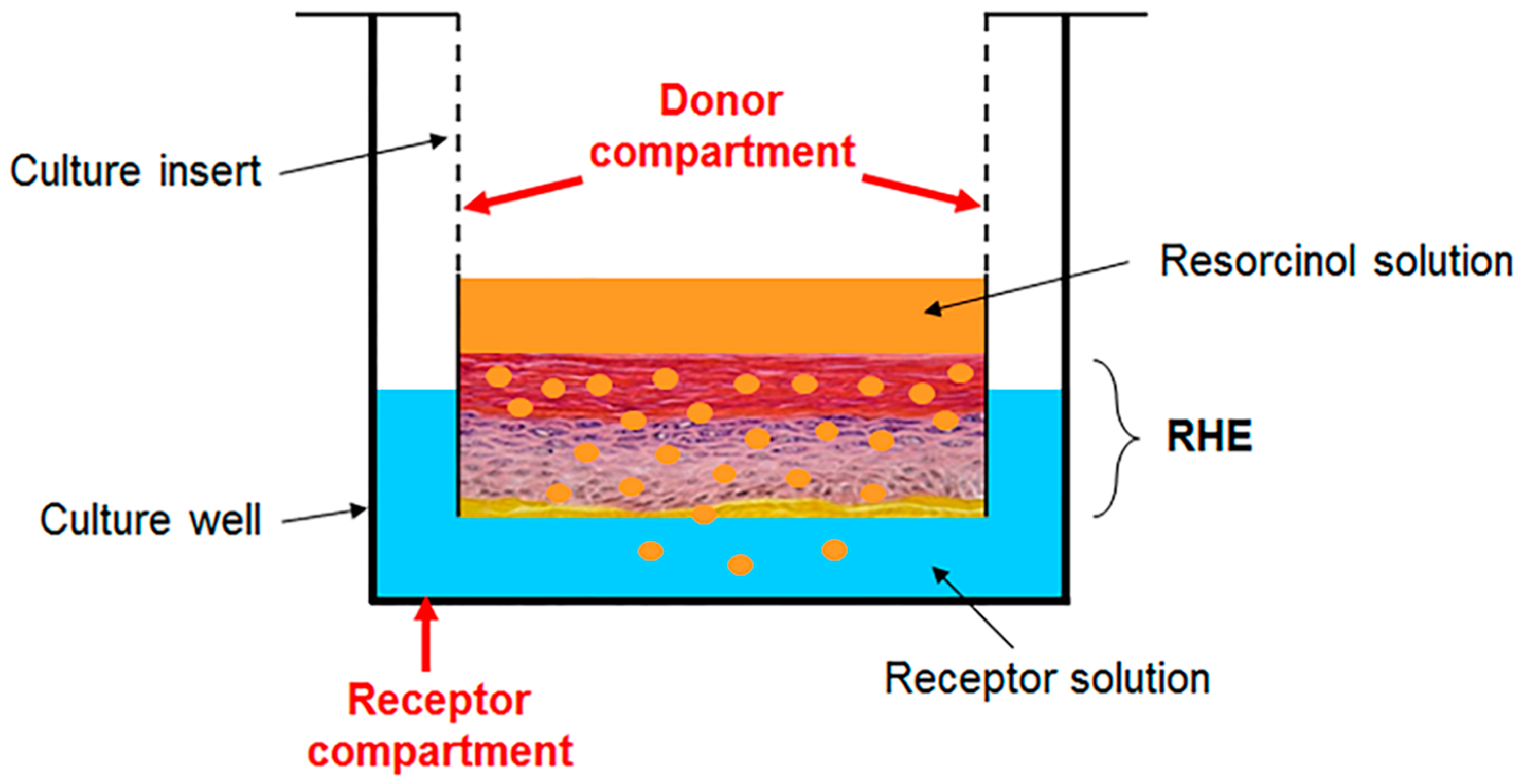
2.5. Pemeation of Resorcinol
2.5.1. RHE Samples for Raman Spectroscopy
2.5.2. RHE Samples for Time-Dependent Permeation
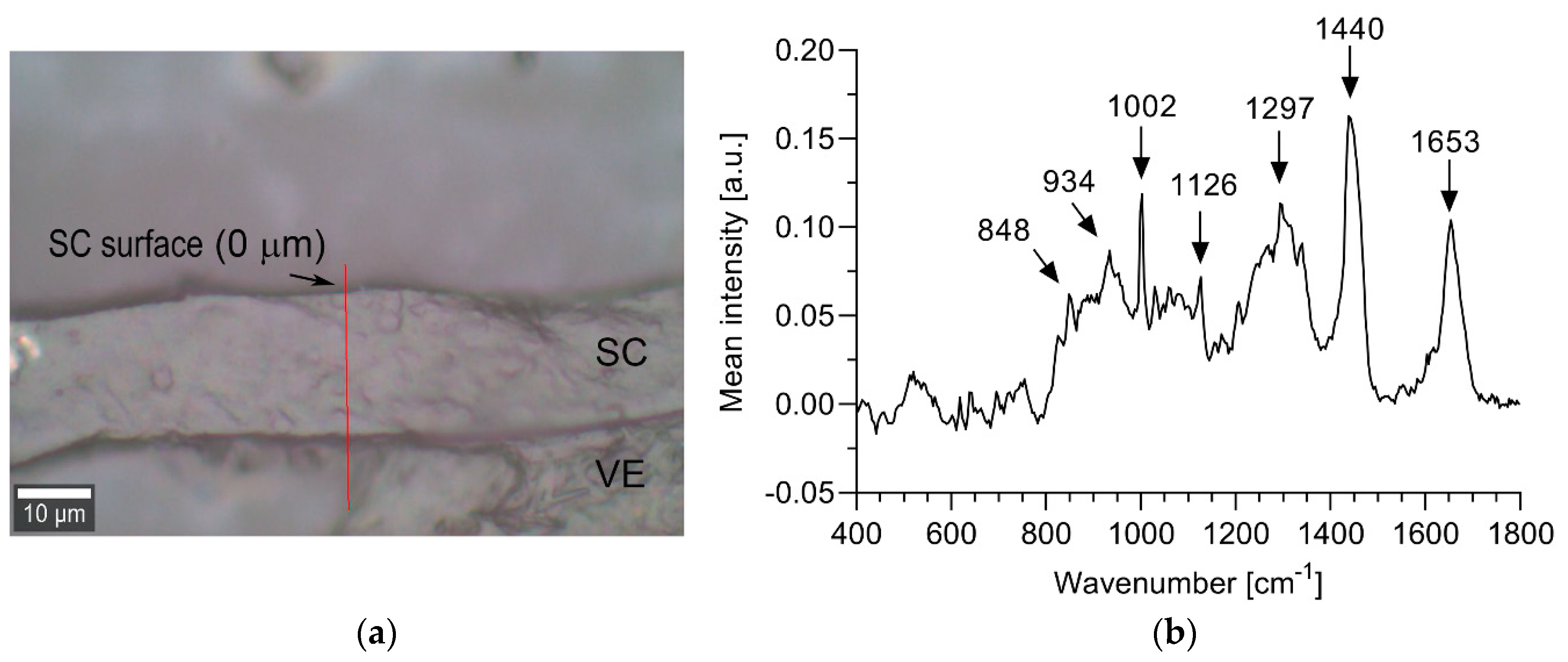
2.6. Confocal Raman Spectroscopy
2.7. Analysis of Raman Spectra
2.8. HPLC Analysis
2.9. Analysis of Time-Dependent Permeation Data
3. Results
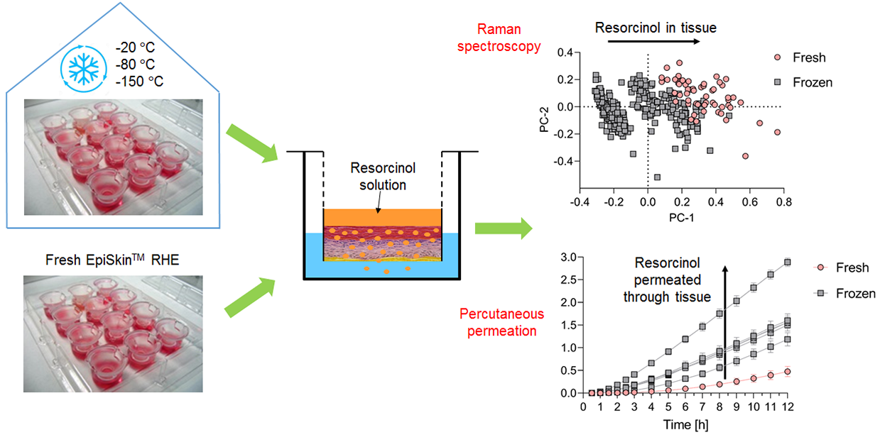
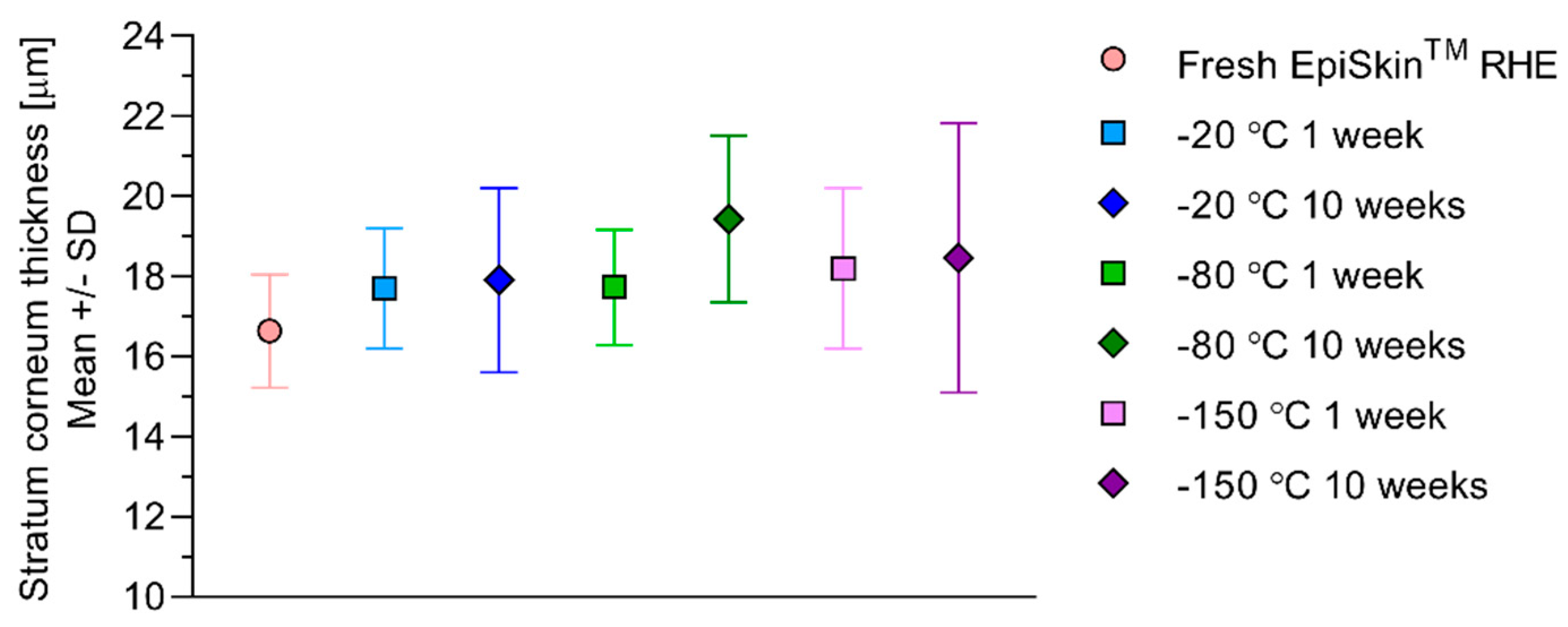
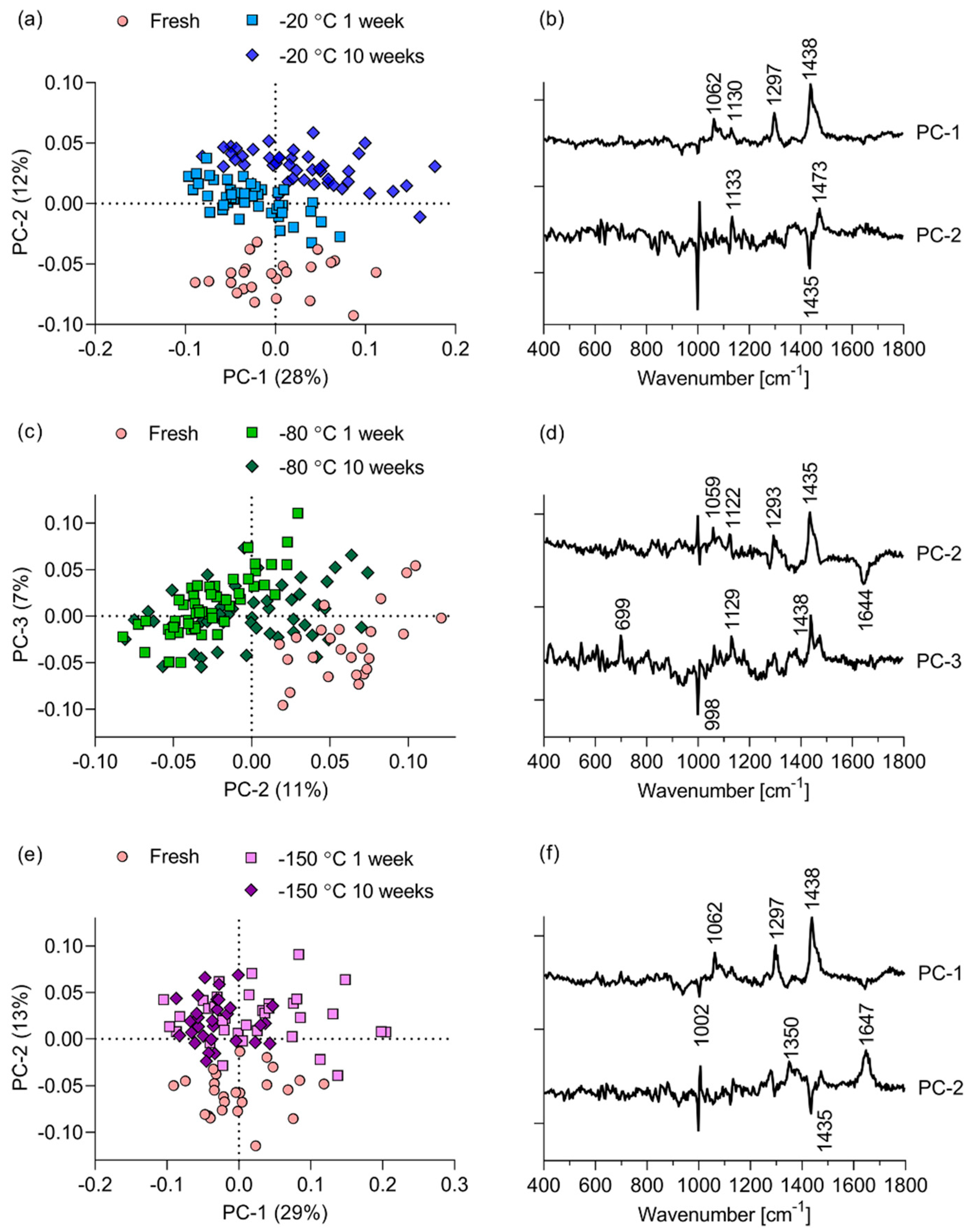
3.1. Intrinsic Effects of Freezing on EpiSkin™ RHE
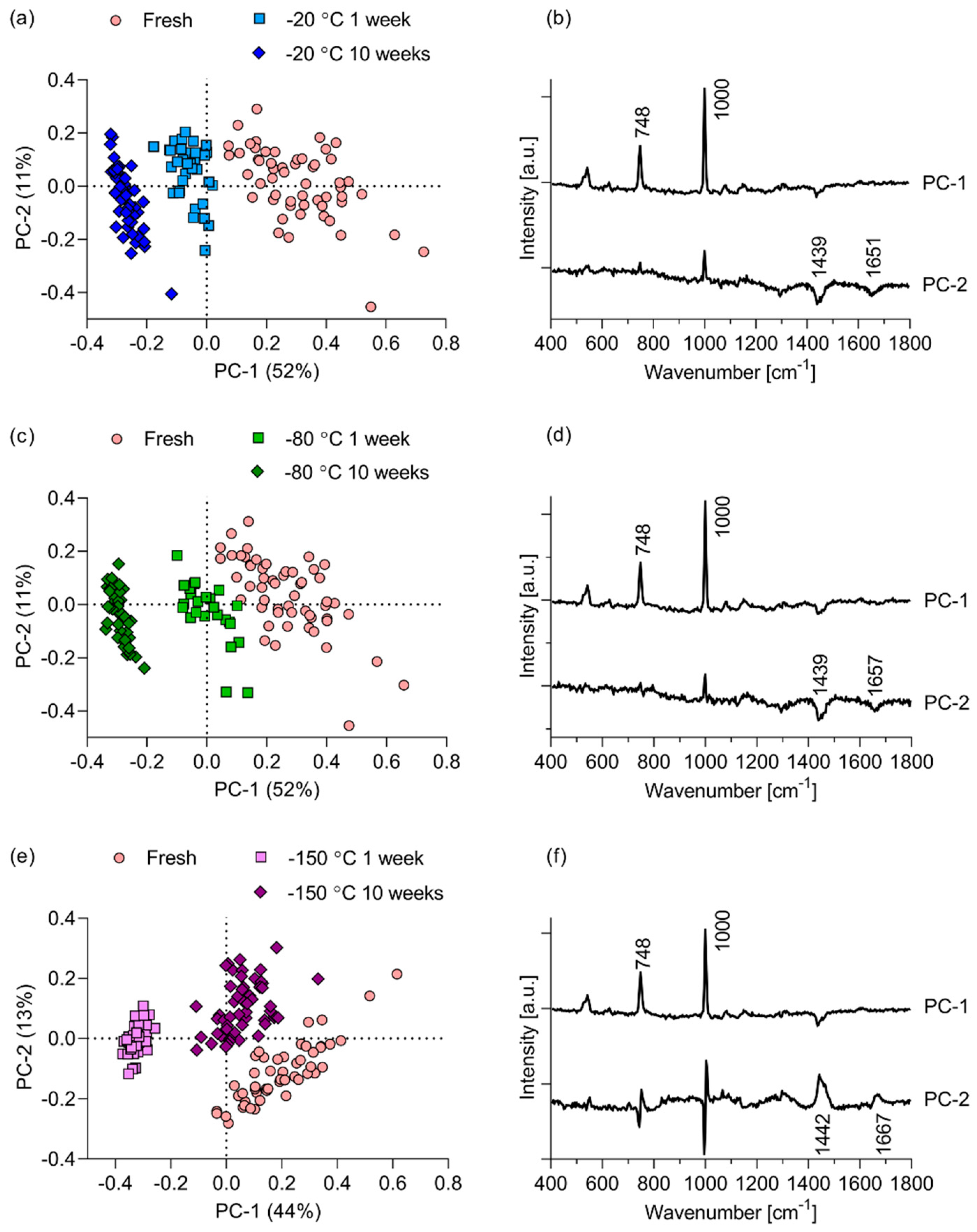
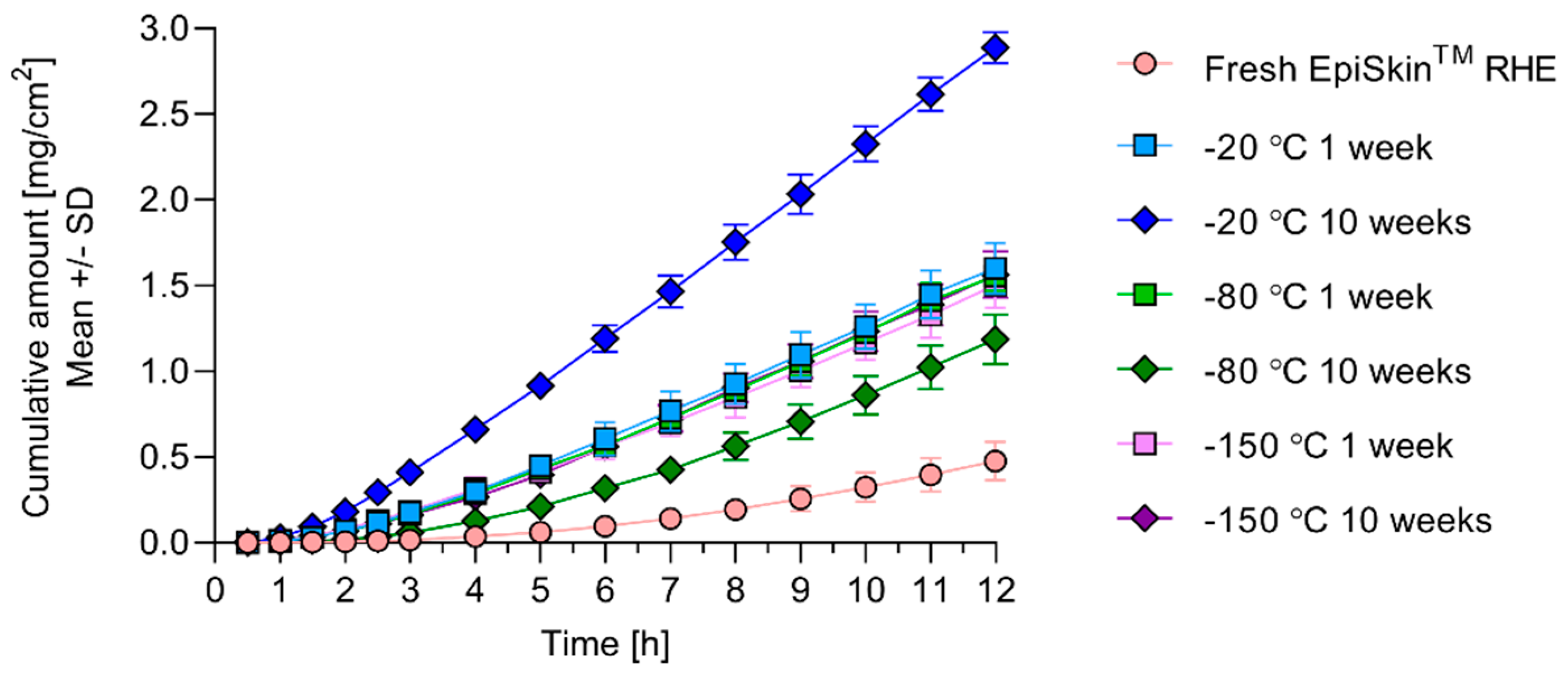
3.2. Effects of Freezing on Resorcinol Permeation through EpiSkin™ RHE
4. Discussion
5. Conclusions
Author Contributions
Funding
Acknowledgments
Conflicts of Interest
References
- Gordon, S.; Daneshian, M.; Bouwstra, J.; Caloni, F.; Constant, S.; Davies, D.E.; Dandekar, G.; Guzman, C.A.; Fabian, E.; Haltner, E.; et al. Non-animal models of epithelial barriers (skin, intestine and lung) in research, industrial applications and regulatory toxicology. ALTEX 2015, 32, 327–378. [Google Scholar] [CrossRef] [PubMed]
- Planz, V.; Lehr, C.M.; Windbergs, M. In vitro models for evaluating safety and efficacy of novel technologies for skin drug delivery. J. Control. Release 2016, 242, 89–104. [Google Scholar] [CrossRef] [PubMed]
- Huet, F.; Severino-Freire, M.; Chéret, J.; Gouin, O.; Praneuf, J.; Pierre, O.; Misery, L.; Le Gall-Ianotto, C. Reconstructed human epidermis for in vitro studies on atopic dermatitis: A review. J. Dermatol. Sci. 2018, 89, 213–218. [Google Scholar] [CrossRef] [PubMed] [Green Version]
- OECD. OECD Guidelines for the Testing of Chemicals, Section 4. Test No. 431: In Vitro Skin Corrosion: Reconstructed Human Epidermis (RHE) Test Method; OECD Publishing: Paris, France, 2019. [Google Scholar]
- OECD. OECD Guidelines for the Testing of Chemicals, Section 4. Test No. 439: In Vitro Skin Irritation: Reconstructed Human Epidermis Test Method; OECD Publishing: Paris, France, 2019. [Google Scholar]
- Schreiber, S.; Mahmoud, A.; Vuia, A.; Rübbelke, M.K.; Schmidt, E.; Schaller, M.; Kandárová, H.; Haberland, A.; Schäfer, U.F.; Bock, U.; et al. Reconstructed epidermis versus human and animal skin in skin absorption studies. Toxicol. Vitr. 2005, 19, 813–822. [Google Scholar] [CrossRef]
- Netzlaff, F.; Kaca, M.; Bock, U.; Haltner-Ukomadu, E.; Meiers, P.; Lehr, C.M.; Schaefer, U.F. Permeability of the reconstructed human epidermis model Episkin® in comparison to various human skin preparations. Eur. J. Pharm. Biopharm. 2007, 66, 127–134. [Google Scholar] [CrossRef]
- Küchler, S.; Strüver, K.; Friess, W. Reconstructed skin models as emerging tools for drug absorption studies. Expert Opin. Drug Metab. Toxicol. 2013, 9, 1255–1263. [Google Scholar] [CrossRef]
- Schäfer-Korting, M.; Bock, U.; Diembeck, W.; Düsing, H.J.; Gamer, A.; Haltner-Ukomadu, E.; Hoffmann, C.; Kaca, M.; Kamp, H.; Kersen, S.; et al. The use of reconstructed human epidermis for skin absorption testing: Results of the validation study. ATLA 2008, 36, 161–187. [Google Scholar] [CrossRef]
- Schäfer-Korting, M.; Mahmoud, A.; Borgia, S.L.; Brüggener, B.; Kleuser, B.; Schreiber, S.; Mehnert, W. Reconstructed epidermis and full-thickness skin for absorption testing: Influence of the vehicles used on steroid permeation. ATLA 2008, 36, 441–452. [Google Scholar] [CrossRef]
- Hoffmann, C.; Müller-Goymann, C.C. Use of artificial skin constructs in permeation studies of clindamycin phosphate. Pharmazie 2005, 60, 350–353. [Google Scholar]
- Pouliot, R. Effects of Freezing on Functionality and Physicochemical Properties of A 3D-Human Skin Model. J. Dermatol. Cosmetol. 2017, 1, 24–31. [Google Scholar] [CrossRef] [Green Version]
- Vyumvuhore, R.; Tfayli, A.; Biniek, K.; Duplan, H.; Delalleau, A.; Manfait, M.; Dauskardt, R.; Baillet-Guffroy, A. The relationship between water loss, mechanical stress, and molecular structure of human stratum corneum ex vivo. J. Biophotonics 2015, 8, 217–225. [Google Scholar] [CrossRef] [PubMed]
- Choe, C.; Lademann, J.; Darvin, M.E. A depth-dependent profile of the lipid conformation and lateral packing order of the stratum corneum in vivo measured using Raman microscopy. Analyst 2016, 141, 1981–1987. [Google Scholar] [CrossRef] [PubMed]
- Caspers, P.J.; Lucassen, G.W.; Puppels, G.J. Combined in vivo confocal Raman spectroscopy and confocal microscopy of human skin. Biophys. J. 2003, 85, 572–580. [Google Scholar] [CrossRef] [Green Version]
- Dąbrowska, A.K.; Adlhart, C.; Spano, F.; Rotaru, G.-M.; Derler, S.; Zhai, L.; Spencer, N.D.; Rossi, R.M. In vivo confirmation of hydration-induced changes in human-skin thickness, roughness and interaction with the environment. Biointerphases 2016, 11, 031015. [Google Scholar] [CrossRef] [Green Version]
- Choe, C.; Lademann, J.; Darvin, M.E. Depth profiles of hydrogen bound water molecule types and their relation to lipid and protein interaction in the human stratum corneum: In vivo. Analyst 2016, 141, 6329–6337. [Google Scholar] [CrossRef]
- Sdobnov, A.Y.; Darvin, M.E.; Schleusener, J.; Lademann, J.; Tuchin, V.V. Hydrogen bound water profiles in the skin influenced by optical clearing molecular agents—Quantitative analysis using confocal Raman microscopy. J. Biophotonics 2019, 12, 1–11. [Google Scholar] [CrossRef] [Green Version]
- Choe, C.S.; Schleusener, J.; Lademann, J.; Darvin, M.E. Age related depth profiles of human Stratum Corneum barrier-related molecular parameters by confocal Raman microscopy in vivo. Mech. Ageing Dev. 2018, 172, 6–12. [Google Scholar] [CrossRef]
- Verzeaux, L.; Vyumvuhore, R.; Boudier, D.; Le Guillou, M.; Bordes, S.; Essendoubi, M.; Manfait, M.; Closs, B. Atopic skin: In vivo Raman identification of global molecular signature, a comparative study with healthy skin. Exp. Dermatol. 2018, 27, 403–408. [Google Scholar] [CrossRef]
- Mahrhauser, D.S.; Nagelreiter, C.; Gehrig, S.; Geyer, A.; Ogris, M.; Kwizda, K.; Valenta, C. Assessment of Raman spectroscopy as a fast and non-invasive method for total stratum corneum thickness determination of pig skin. Int. J. Pharm. 2015, 495, 482–484. [Google Scholar] [CrossRef]
- Tfayli, A.; Piot, O.; Draux, F.; Pitre, F.; Manfait, M. Molecular characterization of reconstructed skin model by Raman microspectroscopy: Comparison with excised human skin. Biopolymers 2007, 87, 261–274. [Google Scholar] [CrossRef]
- Ali, S.M.; Bonnier, F.; Ptasinski, K.; Lambkin, H.; Flynn, K.; Lyng, F.M.; Byrne, H.J. Raman spectroscopic mapping for the analysis of solar radiation induced skin damage. Analyst 2013, 138, 3946–3956. [Google Scholar] [CrossRef] [Green Version]
- Tfayli, A.; Bonnier, F.; Farhane, Z.; Libong, D.; Byrne, H.J.; Baillet-Guffroy, A. Comparison of structure and organization of cutaneous lipids in a reconstructed skin model and human skin: Spectroscopic imaging and chromatographic profiling. Exp. Dermatol. 2014, 23, 441–443. [Google Scholar] [CrossRef] [Green Version]
- Miloudi, L.; Bonnier, F.; Bertrand, D.; Byrne, H.J.; Perse, X.; Chourpa, I.; Munnier, E. Quantitative analysis of curcumin-loaded alginate nanocarriers in hydrogels using Raman and attenuated total reflection infrared spectroscopy. Anal. Bioanal. Chem. 2017, 409, 4593–4605. [Google Scholar] [CrossRef] [PubMed]
- Sriram, G.; Alberti, M.; Dancik, Y.; Wu, B.; Wu, R.; Feng, Z.; Ramasamy, S.; Bigliardi, P.L.; Bigliardi-Qi, M.; Wang, Z. Full-thickness human skin-on-chip with enhanced epidermal morphogenesis and barrier function. Mater. Today 2018, 21, 326–340. [Google Scholar] [CrossRef]
- Dancik, Y.; Sriram, G.; Rout, B.; Zou, Y.; Bigliardi-Qi, M.; Bigliardi, P.L. Physical and compositional analysis of differently cultured 3D human skin equivalents by confocal Raman spectroscopy. Analyst 2018, 143, 1065–1076. [Google Scholar] [CrossRef] [PubMed]
- Roguet, R. The use of reconstructed human epidermis EPISKINTM in the assessment of local tolerance of cosmetics and chemicals. ATLA Altern. Lab. Anim. 2004, 32, 83–91. [Google Scholar] [CrossRef] [PubMed]
- Schäfer-Korting, M.; Bock, U.; Gamer, A.; Haberland, A.; Haltner-Ukomadu, E.; Kaca, M.; Kamp, H.; Kietzmann, M.; Korting, H.C.; Krächter, H.U.; et al. Reconstructed human epidermis for skin absorption testing: Results of the German prevalidation study. ATLA Altern. Lab. Anim. 2006, 34, 283–294. [Google Scholar] [CrossRef]
- Alberti, M.; Dancik, Y.; Sriram, G.; Wu, B.; Teo, Y.L.; Feng, Z.; Bigliardi-Qi, M.; Wu, R.G.; Wang, Z.P.; Bigliardi, P.L. Multi-chamber microfluidic platform for high-precision skin permeation testing. Lab Chip 2017, 17, 1625–1634. [Google Scholar] [CrossRef] [Green Version]
- Butler, H.J.; Ashton, L.; Bird, B.; Cinque, G.; Curtis, K.; Dorney, J.; Esmonde-White, K.; Fullwood, N.J.; Gardner, B.; Martin-Hirsch, P.L.; et al. Using Raman spectroscopy to characterize biological materials. Nat. Protoc. 2016, 11, 664–687. [Google Scholar] [CrossRef] [Green Version]
- Byrne, H.J.; Knief, P.; Keating, M.E.; Bonnier, F. Spectral pre and post processing for infrared and Raman spectroscopy of biological tissues and cells. Chem. Soc. Rev. 2016, 45, 1865–1878. [Google Scholar] [CrossRef] [Green Version]
- Gautam, R.; Vanga, S.; Ariese, F.; Umapathy, S. Review of multidimensional data processing approaches for Raman and infrared spectroscopy. EPJ Tech. Instrum. 2015, 2. [Google Scholar] [CrossRef] [Green Version]
- Jepps, O.G.; Dancik, Y.; Anissimov, Y.G.; Roberts, M.S. Modeling the human skin barrier—Towards a better understanding of dermal absorption. Adv. Drug Deliv. Rev. 2013, 65, 152–168. [Google Scholar] [CrossRef] [PubMed]
- Czekalla, C.; Schönborn, K.H.; Lademann, J.; Meinke, M.C. Noninvasive Determination of Epidermal and Stratum Corneum Thickness in vivo Using Two-Photon Microscopy and Optical Coherence Tomography: Impact of Body Area, Age, and Gender. Skin Pharmacol. Physiol. 2019, 32, 142–150. [Google Scholar] [CrossRef] [PubMed]
- Khiao In, M.; Richardson, K.C.; Loewa, A.; Hedtrich, S.; Kaessmeyer, S.; Plendl, J. Histological and functional comparisons of four anatomical regions of porcine skin with human abdominal skin. J. Vet. Med. Ser. C Anat. Histol. Embryol. 2019, 48, 207–217. [Google Scholar] [CrossRef]
- Albèr, C.; Brandner, B.D.; Björklund, S.; Billsten, P.; Corkery, R.W.; Engblom, J. Effects of water gradients and use of urea on skin ultrastructure evaluated by confocal Raman microspectroscopy. Biochim. Biophys. Acta Biomembr. 2013, 1828, 2470–2478. [Google Scholar] [CrossRef] [PubMed] [Green Version]
- Egawa, M.; Sato, Y. In vivo evaluation of two forms of urea in the skin by Raman spectroscopy after application of urea-containing cream. Ski. Res. Technol. 2015, 21, 259–264. [Google Scholar] [CrossRef] [PubMed]
- OECD. OECD Guideline for Testing of Chemicals. Test Guideline 428: Skin Absorption: In Vitro Method; OECD Publishing: Paris, France, 2004. [Google Scholar]
- Abd, E.; Yousef, S.A.; Pastore, M.N.; Telaprolu, K.; Mohammed, Y.H.; Namjoshi, S.; Grice, J.E.; Roberts, M.S. Skin models for the testing of transdermal drugs. Clin. Pharmacol. Adv. Appl. 2016, 8, 163–176. [Google Scholar] [CrossRef] [Green Version]
- Garcia, M.T.J.; de Vasconcellos, F.L.L.; Raffier, C.P.; Roberts, M.S.; Grice, J.E.; Benson, H.A.E.; Leite-Silva, V.R. Alternative Methods to Animal Studies for the Evaluation of Topical/Transdermal Drug Delivery Systems. Curr. Top. Med. Chem. 2018, 18, 287–299. [Google Scholar] [CrossRef]
- Suhail, S.; Sardashti, N.; Jaiswal, D.; Rudraiah, S.; Misra, M.; Kumbar, S.G. Engineered Skin Tissue Equivalents for Product Evaluation and Therapeutic Applications. Biotechnol. J. 2019, 14. [Google Scholar] [CrossRef]
- Flaten, G.E.; Palac, Z.; Engesland, A.; Filipović-Grčić, J.; Vanić, Ž.; Škalko-Basnet, N. In vitro skin models as a tool in optimization of drug formulation. Eur. J. Pharm. Sci. 2015, 75, 10–24. [Google Scholar] [CrossRef] [Green Version]
- Venkatesan, G.; Dancik, Y.; Sinha, A.; Bigliardi, M.; Srinivas, R.; Dawson, T.; Valiyaveettil, S.; Bigliardi, P.; Pastorin, G. Facile synthesis of oligo anilines as permanent hair dyes: How chemical modifications impart colour and avoid toxicity. New J. Chem. 2019, 43, 16188–16199. [Google Scholar] [CrossRef]
- Venkatesan, G.; Dancik, Y.; Sinha, A.; Kyaw, H.M.; Srinivas, R.; Dawson, T.L.; Bigliardi, M.; Bigliardi, P.; Pastorin, G. Development of novel alternative hair dyes to hazardous para-phenylenediamine. J. Hazard. Mater. 2020, 123712. [Google Scholar] [CrossRef]
- CIR (Cosmetic Ingredients Review) Final report on the safety assessment of 2-methylresorcinol and resorcinol. J. Am. Coll. Toxicol. 1986, 5, 167–203. [CrossRef]
- Porras, S.P.; Hartonen, M.; Ylinen, K.; Tornaeus, J.; Tuomi, T.; Santonen, T. Environmental and occupational exposure to resorcinol in Finland. Toxicol. Lett. 2018, 298, 125–133. [Google Scholar] [CrossRef]
- Dancik, Y.; Bigliardi, P.L.; Bigliardi-Qi, M. What happens in the skin? Integrating skin permeation kinetics into studies of developmental and reproductive toxicity following topical exposure. Reprod. Toxicol. 2015, 58, 252–281. [Google Scholar] [CrossRef] [PubMed]
- Barbero, A.M.; Frasch, H.F. Effect of Frozen Human Epidermis Storage Duration and Cryoprotectant on Barrier Function Using Two Model Compounds. Skin Pharmacol. Physiol. 2016, 29, 31–40. [Google Scholar] [CrossRef] [Green Version]
- Franzen, L.; Vidlářová, L.; Kostka, K.H.; Schaefer, U.F.; Windbergs, M. Freeze-drying as a preserving preparation technique for in vitro testing of human skin. Exp. Dermatol. 2013, 22, 54–56. [Google Scholar] [CrossRef]
- Franzen, L.; Anderski, J.; Planz, V.; Kostka, K.H.; Windbergs, M. Combining confocal Raman microscopy and freeze-drying for quantification of substance penetration into human skin. Exp. Dermatol. 2014, 23, 942–944. [Google Scholar] [CrossRef]
- Jacques-Jamin, C.; Duplan, H.; Rothe, H.; Vaillant, O.; Eilstein, J.; Grégoire, S.; Cubberley, R.; Lange, D.; Ellison, C.; Klaric, M.; et al. Comparison of the Skin Penetration of 3 Metabolically Stable Chemicals Using Fresh and Frozen Human Skin. Skin Pharmacol. Physiol. 2017, 30, 234–245. [Google Scholar] [CrossRef]
- Swarbrick, J.; Lee, G.; Brom, J. Drug permeation through human skin: I. Effect of storage conditions of skin. J. Investig. Dermatol. 1982, 78, 63–66. [Google Scholar] [CrossRef]
- Kemppainen, B.W.; Riley, R.T.; Russell, R.B.; Pace, J.G.; Detrick, F. Effects of skin storage conditions and concentration of applied dose on [3H]T-2 toxin penetration through excised human and monkey skin. Food Chem. Toxicol. 1986, 24, 221–227. [Google Scholar] [CrossRef]
- Nielsen, J.B.; Plasencia, I.; Sørensen, J.A.; Bagatolli, L.A. Storage conditions of skin affect tissue structure and subsequent in vitro percutaneous penetration. Skin Pharmacol. Physiol. 2011, 24, 93–102. [Google Scholar] [CrossRef] [PubMed]
- Payne, O.J.; Graham, S.J.; Dalton, C.H.; Spencer, P.M.; Mansson, R.; Jenner, J.; Azeke, J.; Braue, E. The effects of sulfur mustard exposure and freezing on transdermal penetration of tritiated water through ex vivo pig skin. Toxicol. Vitr. 2013, 27, 79–83. [Google Scholar] [CrossRef]
- Abdayem, R.; Roussel, L.; Zaman, N.; Pirot, F.; Gilbert, E.; Haftek, M. Deleterious effects of skin freezing contribute to variable outcomes of the predictive drug permeation studies using hydrophilic molecules. Exp. Dermatol. 2015, 24, 972–974. [Google Scholar] [CrossRef] [PubMed] [Green Version]
- Sintov, A.C. Cumulative evidence of the low reliability of frozen/thawed pig skin as a model for in vitro percutaneous permeation testing. Eur. J. Pharm. Sci. 2017, 102, 261–263. [Google Scholar] [CrossRef] [PubMed]
- Sintov, A.C.; Botner, S. Transdermal drug delivery using microemulsion and aqueous systems: Influence of skin storage conditions on the in vitro permeability of diclofenac from aqueous vehicle systems. Int. J. Pharm. 2006, 311, 55–62. [Google Scholar] [CrossRef]
- Sintov, A.C.; Greenberg, I. Comparative percutaneous permeation study using caffeine-loaded microemulsion showing low reliability of the frozen/thawed skin models. Int. J. Pharm. 2014, 471, 516–524. [Google Scholar] [CrossRef]
- Bajza, Á.; Kocsis, D.; Berezvai, O.; Laki, A.J.; Lukács, B.; Imre, T.; Iván, K.; Szabó, P.; Erdő, F. Verification of p-glycoprotein function at the dermal barrier in diffusion cells and dynamic “skin-on-a-chip” microfluidic device. Pharmaceutics 2020, 12, 804. [Google Scholar] [CrossRef]
- Ponec, M.; Boelsma, E.; Gibbs, S.; Mommaas, M. Characterization of reconstructed skin models. Skin Pharmacol. Appl. Skin Physiol. 2002, 15, 4–17. [Google Scholar] [CrossRef]
- Bäsler, K.; Bergmann, S.; Heisig, M.; Naegel, A.; Zorn-Kruppa, M.; Brandner, J.M. The role of tight junctions in skin barrier function and dermal absorption. J. Control. Release 2016, 242, 105–118. [Google Scholar] [CrossRef]
- Niehues, H.; Bouwstra, J.A.; El Ghalbzouri, A.; Brandner, J.M.; Zeeuwen, P.L.J.M.; van den Bogaard, E.H. 3D skin models for 3R research: The potential of 3D reconstructed skin models to study skin barrier function. Exp. Dermatol. 2018, 27, 501–511. [Google Scholar] [CrossRef] [Green Version]


| Condition | Temperature [°C] | Duration [Weeks] |
|---|---|---|
| 1 | −20 | 1 |
| 2 | 10 | |
| 3 | −80 | 1 |
| 4 | 10 | |
| 5 | −150 | 1 |
| 6 | 10 |
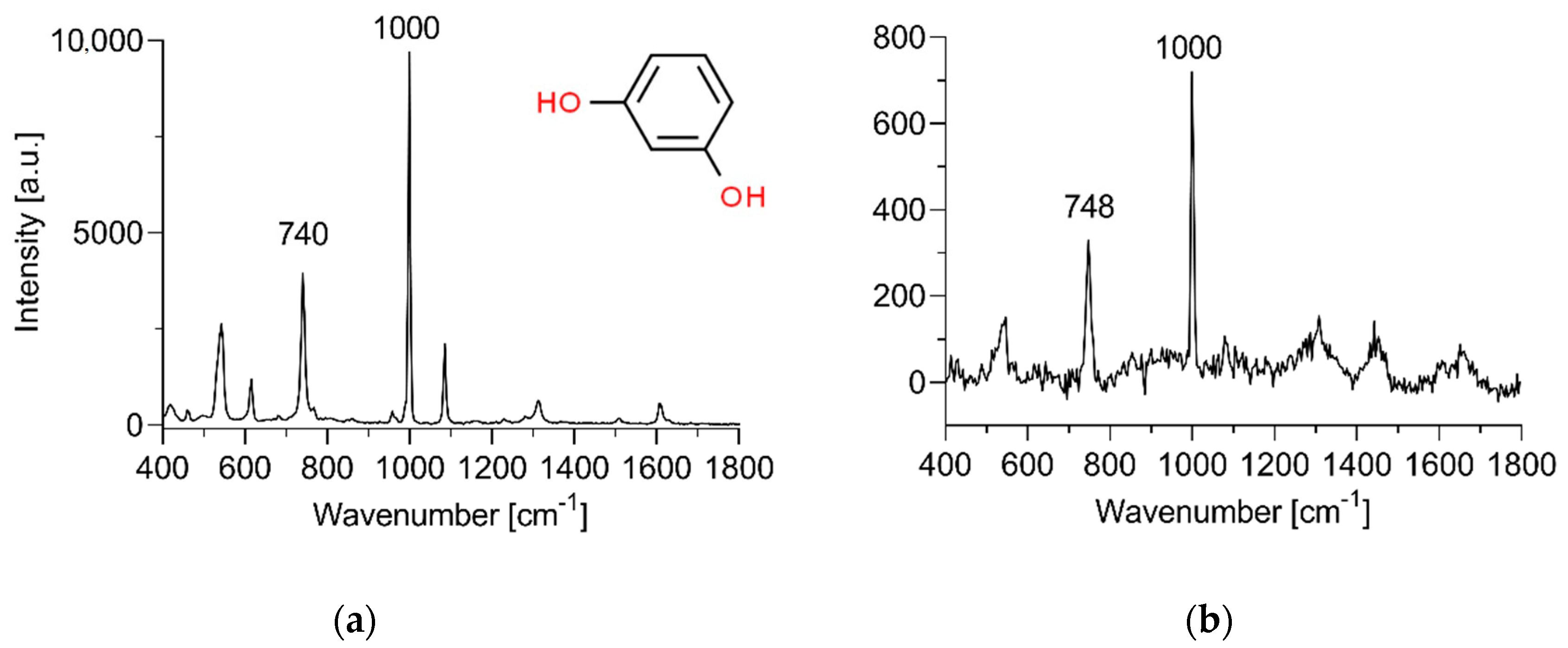
| Wavenumber [cm−1] | Vibrations and Assigned Components |
|---|---|
| 849 | Tyrosine Fermi doublet (ring) |
| 933 | ρ(CH3) terminal; ν(C–C): protein α helix (secondary structure); Phospholipids |
| 1002 | Phenylalanine symmetric ring breathing * |
| 1126 | Lipids hydrocarbon chains, trans conformation, ceramides |
| 1297 | Amide III; CH2 phospholipids |
| 1440 | δ(C–H): proteins and lipids |
| 1653 | Amide I |
| Fresh | −20 °C | −80 °C | −150 °C | ||||
|---|---|---|---|---|---|---|---|
| 1 wk. | 10 wks. | 1 wk. | 10 wks. | 1 wk. | 10 wks. | ||
| JSS [mg/(cm2 h)] | 0.073 ± 0.014 | 0.17 ± 0.0061 | 0.29 ± 0.0088 | 0.17 ± 0.0040 | 0.16 ± 0.015 | 0.17 ± 0.020 | 0.17 ± 0.015 |
| KP × 103 [cm/h] | 1.5 ± 0.022 | 3.4 ± 0.12 | 5.7 ± 0.18 | 3.4 ± 0.081 | 3.2 ± 0.31 | 3.3 ± 0.40 | 3.3 ± 0.31 |
| tlag [h] | 5.6 ± 0.32 | 2.6 ± 0.56 | 1.8 ± 0.62 | 2.7 ± 0.43 | 4.6 ± 0.20 | 3.0 ± 0.54 | 2.6 ± 0.48 |
| Compartment | Fresh | −20 °C | −80 °C | −150 °C | |||
|---|---|---|---|---|---|---|---|
| 1 wk. | 10 wks. | 1 wk. | 10 wks. | 1 wk. | 10 wks. | ||
| Donor solution | 83 ± 3.3 | 71 ± 4.6 | 63 ± 3.2 | 73 ± 5.8 | 80 ± 3.2 | 75 ± 6.3 | 69 ± 2.1 |
| Skin swabs | 0.89 ± 0.61 | 0.14 ± 0.12 | 2.3 ± 1.4 | 0.094 ± 0.016 | 0.42 ± 0.73 | 0.12 ± 0.20 | 0.0 ± 0.0 |
| Skin and support membrane | 9.7 ± 1.0 | 7.0 ± 1.3 | 2.8 ± 0.17 | 5.3 ± 0.11 | 3.4 ± 0.17 | 6.5 ± 0.45 | 4.1 ± 0.50 |
| Receptor solution (permeated) | 5.1 ± 1.2 | 17 ± 1.6 | 31 ± 0.97 | 17 ± 0.92 | 13 ± 1.6 | 16 ± 1.5 | 17 ± 1.5 |
| Total recovery | 98 ± 1.2 | 95 ± 2.0 | 99 ± 4.6 | 96 ± 5.0 | 96 ± 2.0 | 97 ± 4.4 | 90 ± 0.40 |
Publisher’s Note: MDPI stays neutral with regard to jurisdictional claims in published maps and institutional affiliations. |
© 2020 by the authors. Licensee MDPI, Basel, Switzerland. This article is an open access article distributed under the terms and conditions of the Creative Commons Attribution (CC BY) license (http://creativecommons.org/licenses/by/4.0/).
Share and Cite
Dancik, Y.; Kichou, H.; Eklouh-Molinier, C.; Soucé, M.; Munnier, E.; Chourpa, I.; Bonnier, F. Freezing Weakens the Barrier Function of Reconstructed Human Epidermis as Evidenced by Raman Spectroscopy and Percutaneous Permeation. Pharmaceutics 2020, 12, 1041. https://0-doi-org.brum.beds.ac.uk/10.3390/pharmaceutics12111041
Dancik Y, Kichou H, Eklouh-Molinier C, Soucé M, Munnier E, Chourpa I, Bonnier F. Freezing Weakens the Barrier Function of Reconstructed Human Epidermis as Evidenced by Raman Spectroscopy and Percutaneous Permeation. Pharmaceutics. 2020; 12(11):1041. https://0-doi-org.brum.beds.ac.uk/10.3390/pharmaceutics12111041
Chicago/Turabian StyleDancik, Yuri, Hichem Kichou, Christophe Eklouh-Molinier, Martin Soucé, Emilie Munnier, Igor Chourpa, and Franck Bonnier. 2020. "Freezing Weakens the Barrier Function of Reconstructed Human Epidermis as Evidenced by Raman Spectroscopy and Percutaneous Permeation" Pharmaceutics 12, no. 11: 1041. https://0-doi-org.brum.beds.ac.uk/10.3390/pharmaceutics12111041