
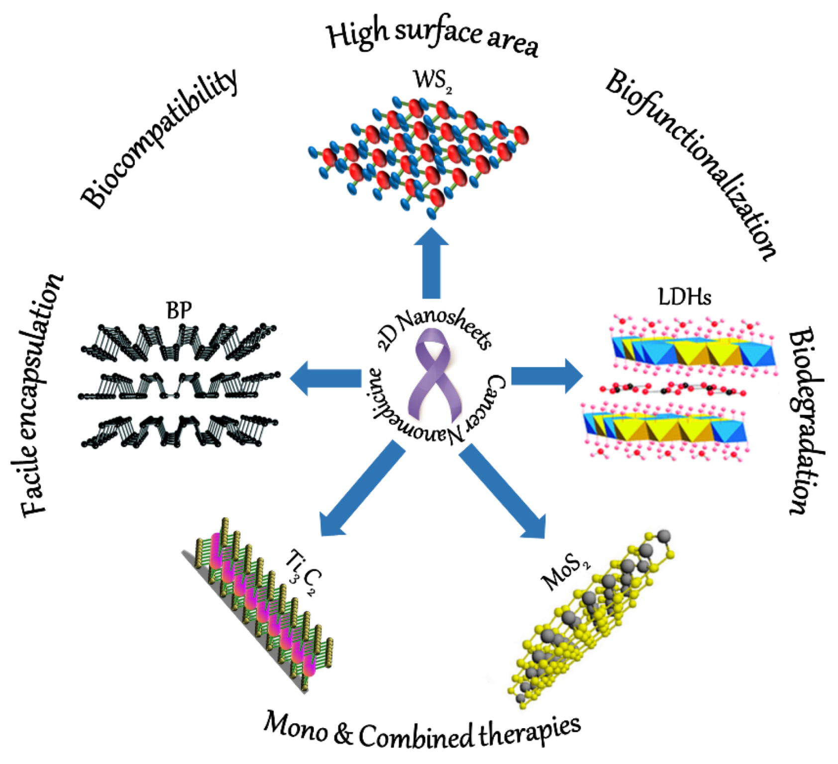
2D Nanosheets—A New Class of Therapeutic Formulations against Cancer

 ,
,  , , and
Abstract
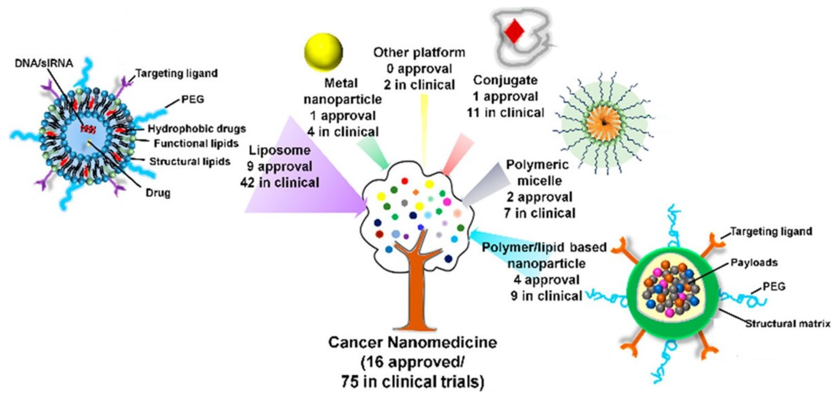
:1. Introduction
2. 2D Nanoformulations for Drug Delivery
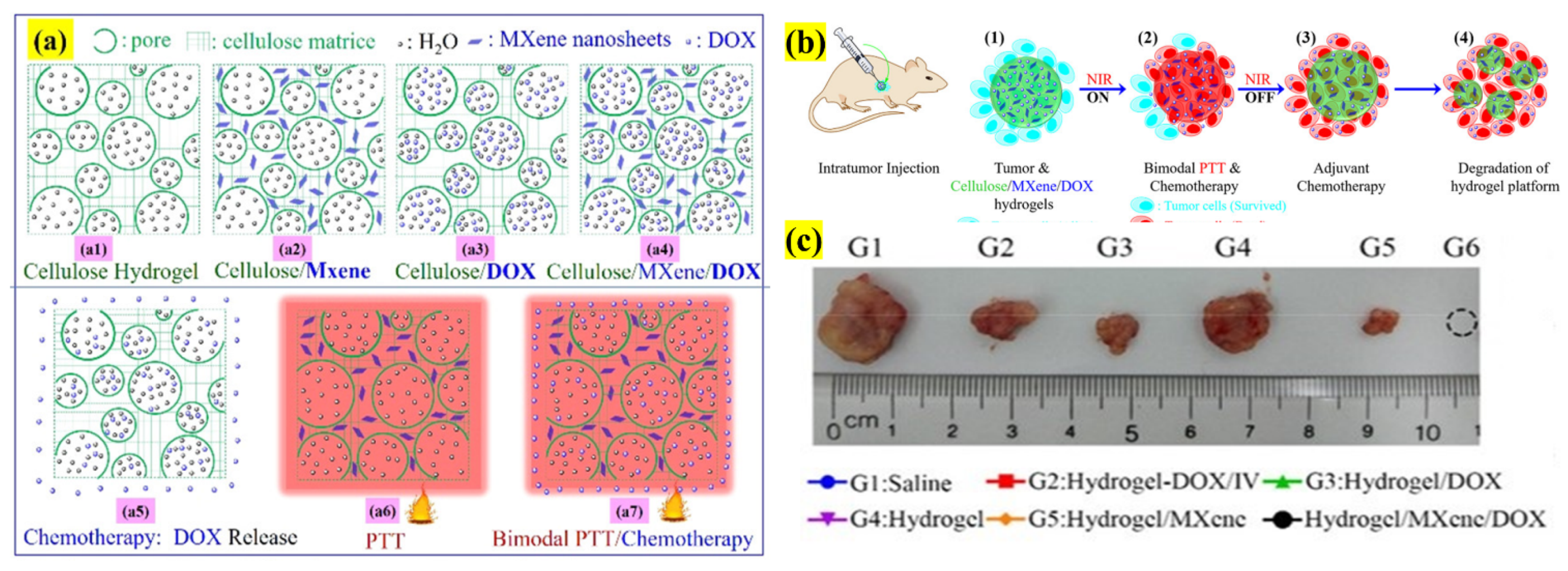
2.1. MXene Nanosheets
Titanium Carbide (Ti3C2) Nanosheets
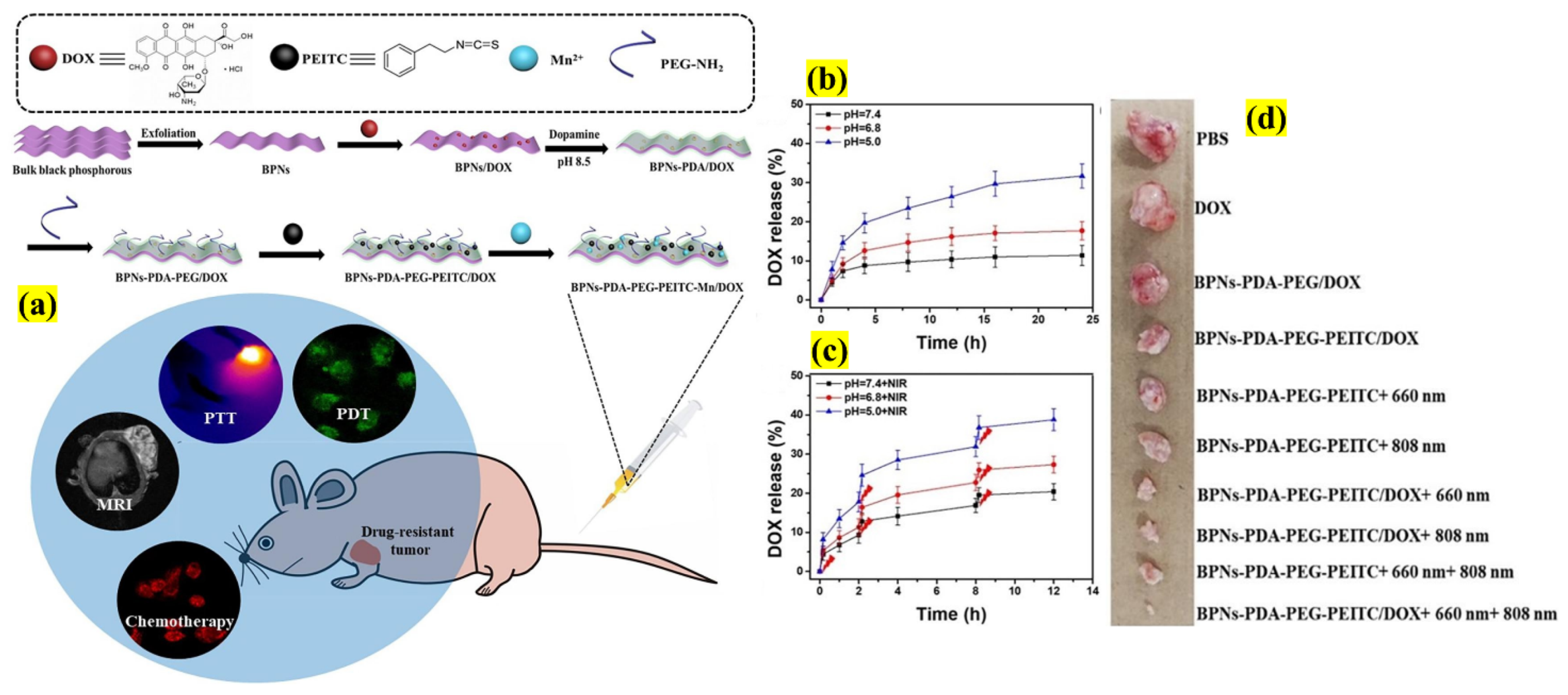
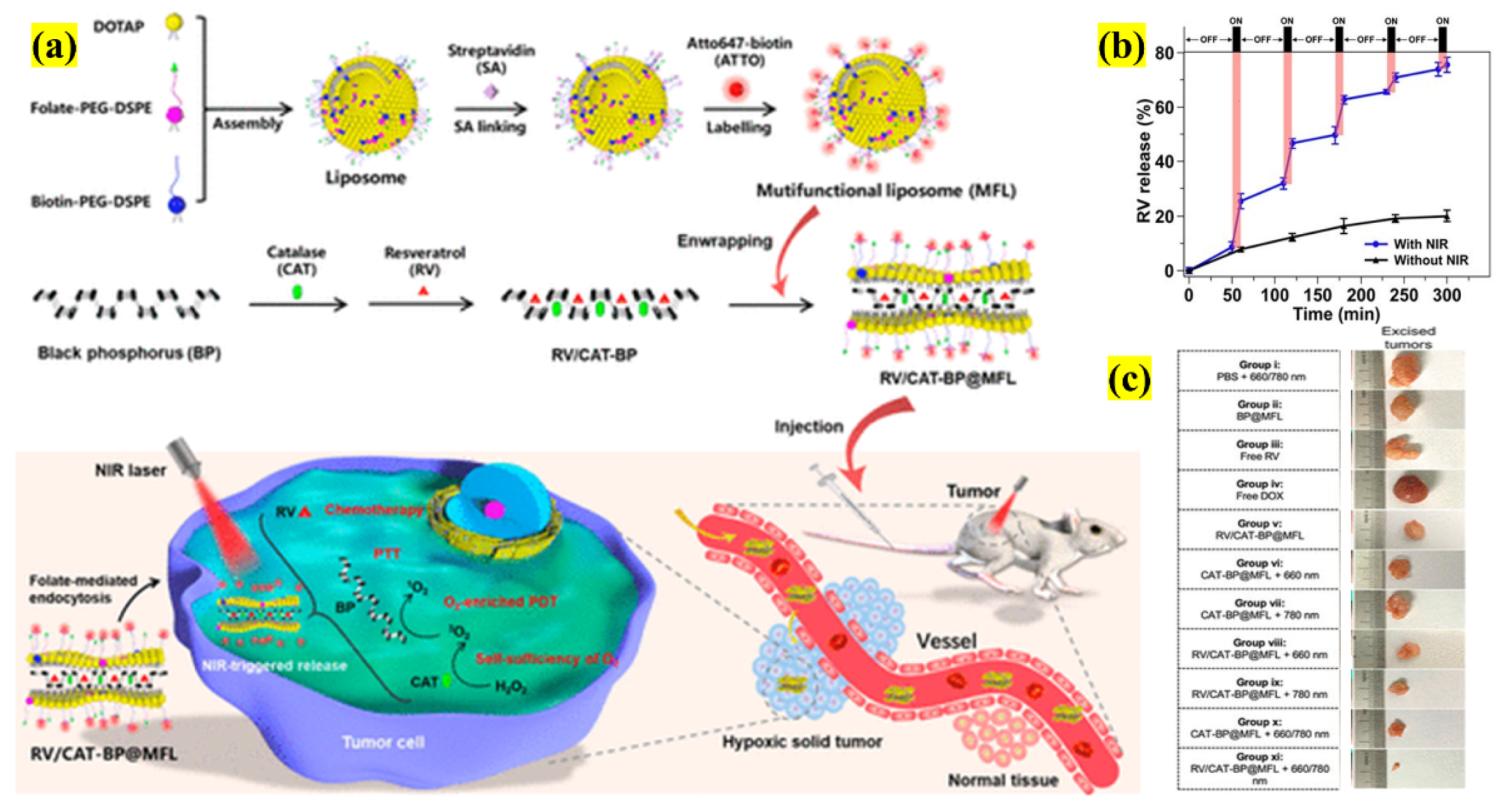
2.2. Black Phosphorous (BP) Nanosheets
2.3. Transition-Metal Dichalcogenides (TMDCs)
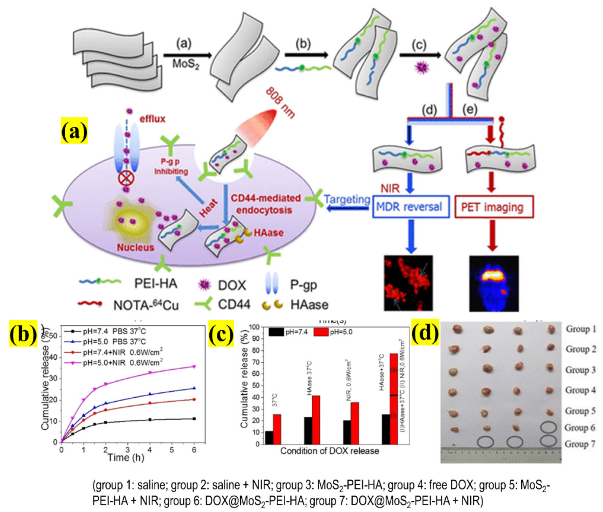
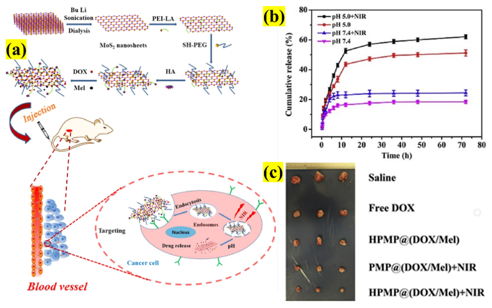
2.3.1. Molybdenum Disulfide (MoS2) Nanosheets
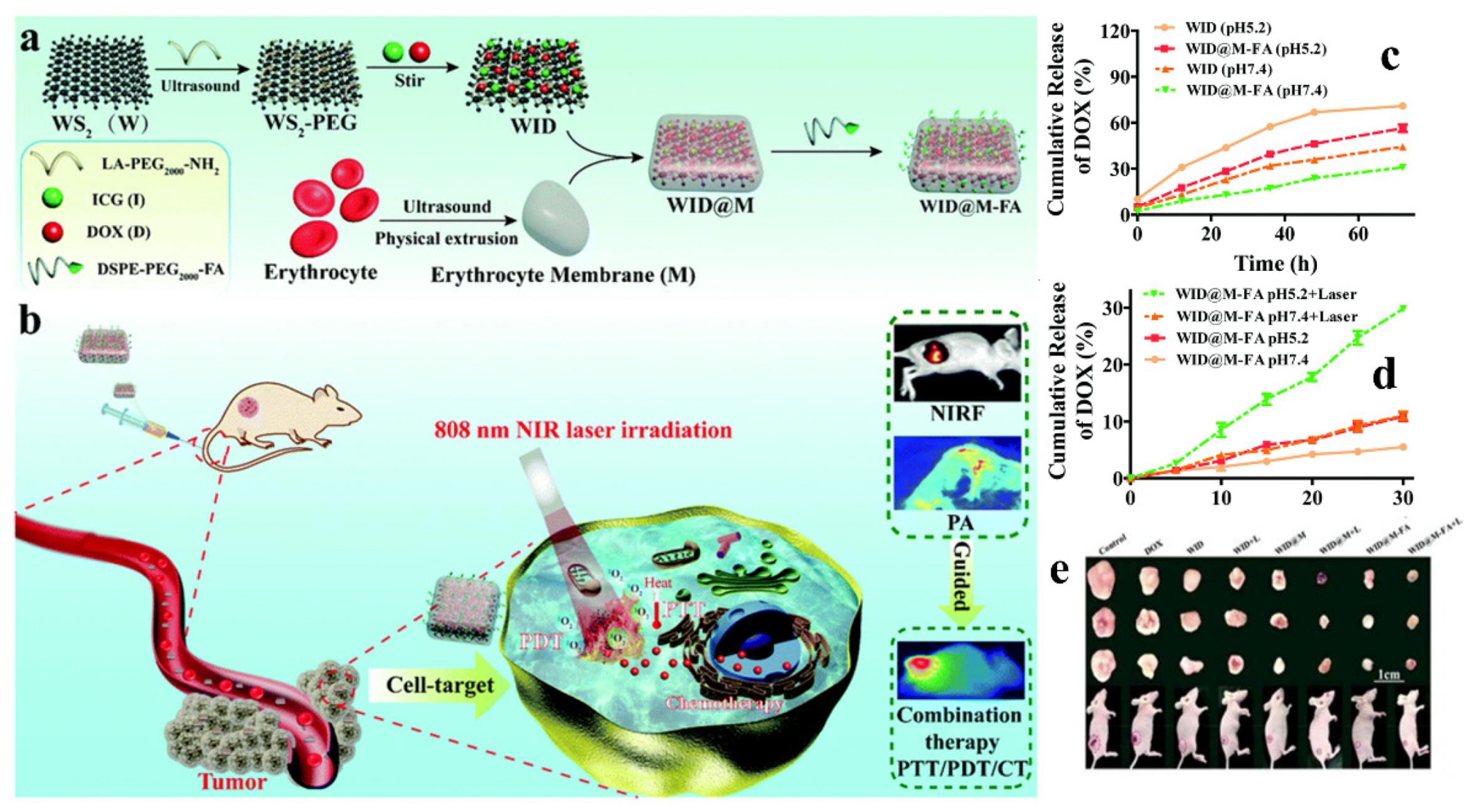
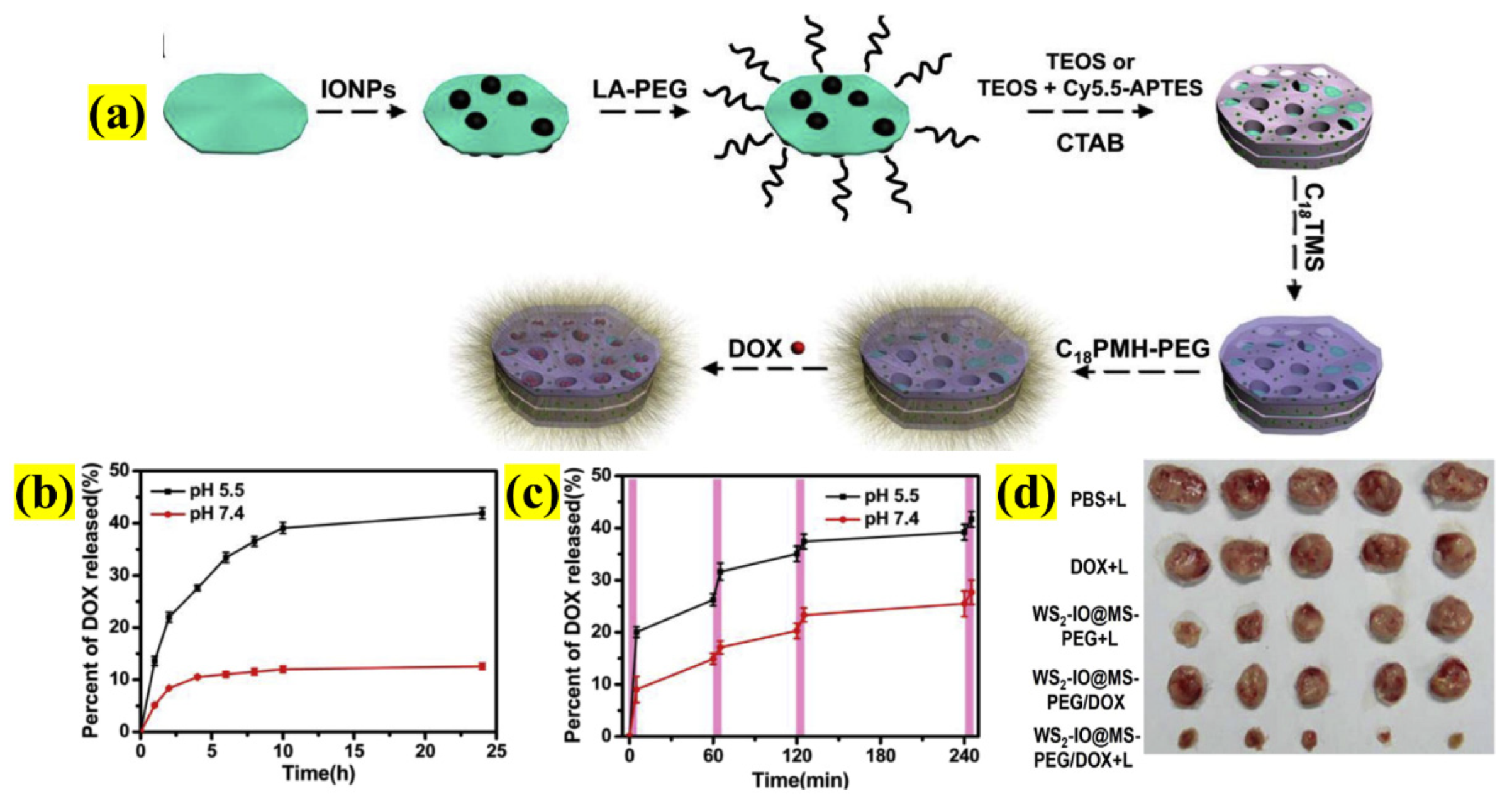
2.3.2. Tungsten Disulfide (WS2) Nanosheets
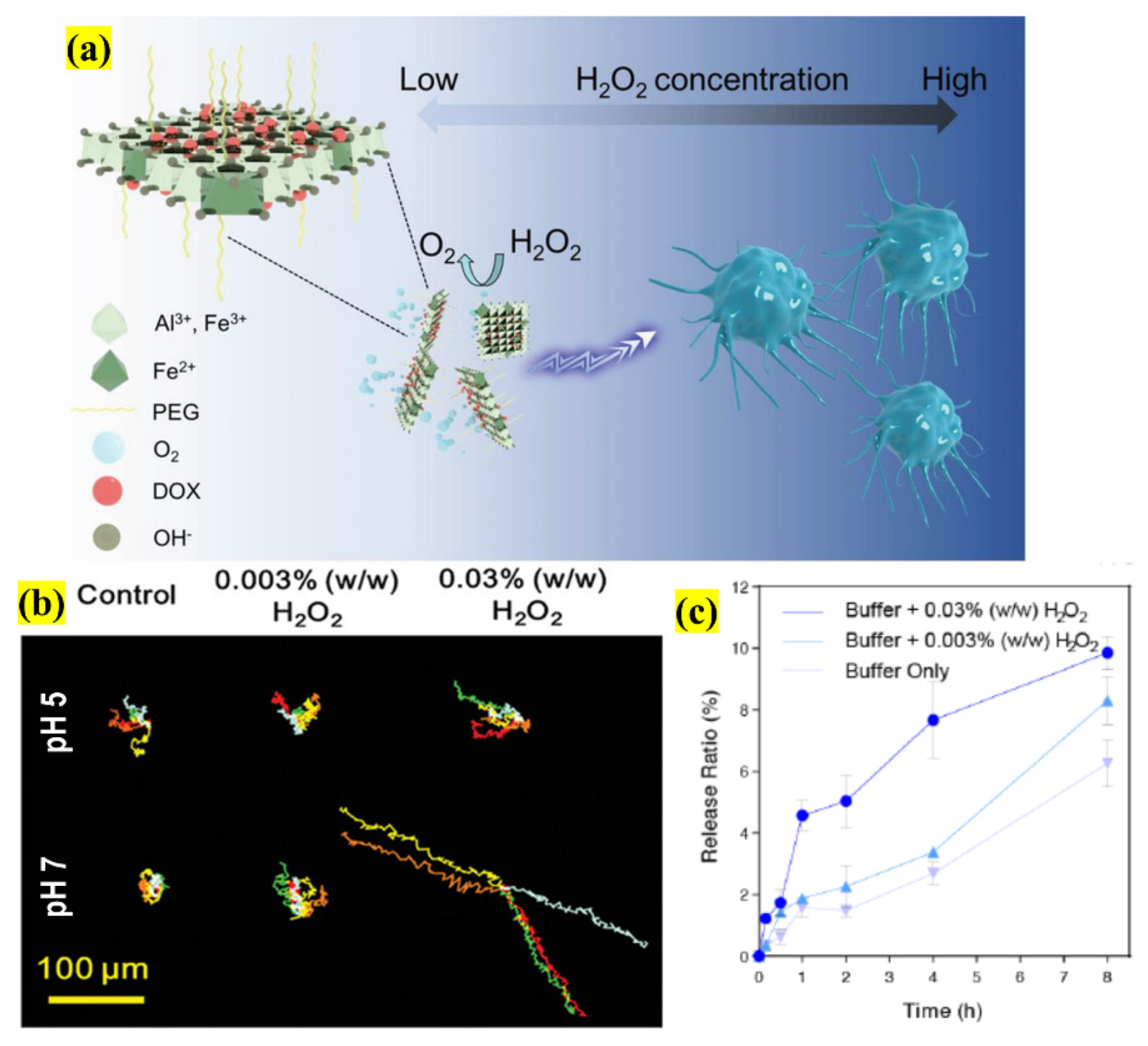
2.4. Layered Double Hydroxides (LDHs)
3. 2D Formulations with Biological Barriers (BBs)
- (i)
- Opsonization: To overcome this process, most of the NSs-based investigations have focused on polymeric or biomimetic cell membrane coating technology to encapsulate the functionalized NSs.
- (ii)
- Hemorheological limitations: The size, surface charge, and geometry play a vital role in blood fluid dynamics and circulation. So, researchers have designed NSs-based formulations with size ranging from 100–250 nm (maximum 300 nm) with multifunctionality. Considering surface charge, most NSs have a negative charge (around −20 mV), extending the circulation time. Lastly, a non-spherical structure that relies on margination and adhesion maximizes after administration seems favorable compared to spherical-shaped nanostructures.
- (iii)
- Cellular internalization is enhanced by decorating cationic moieties on the NSs surface with polypeptides such poly-l-lysine (PLL), PEI, and PAMAM for internalizing, and for endosomal or lysosomes compartmentalization.
4. Conclusions and Perspectives
- (1)
- Synthesis method: For all the above-discussed NSs, a high-yield, cost-effective, large-scale fabrication method is necessary to produce a tightly controlled size, thickness, and stability.
- (2)
- Toxicity: All four different NSs are biocompatible with either low or negligible cytotoxicity even at higher concentrations, and are biodegradable in biological environments.
Author Contributions
Funding
Institutional Review Board Statement
Informed Consent Statement
Data Availability Statement
Conflicts of Interest
Abbreviations
| α-TOS | α-tocopherol succinate |
| 2D | Two-Dimensional |
| 3D | Three-dimensional |
| 5-FU | 5-fluorouracil |
| ABX | Abraxane |
| ADR | Adverse drug reaction |
| ATP | Adenosine triphosphate |
| AuNPs | Gold nanoparticles |
| BSA | Bovine serum albumin |
| CAT | Catalase |
| CaP | Calcium phosphate |
| CD-siRNA | Cell death-siRNA |
| CDT | Chemodynamic therapy |
| CIS | Cisplatin |
| CS | Chitosan |
| CT | Computed tomography |
| CTX | Chemotherapy |
| Cur | Curcumin |
| CuS | Copper sulfide |
| CMC | Carboxymethyl-cellulose |
| DACH | Oxaliplatin |
| DDSs | Drug delivery systems |
| DNA | Deoxyribonucleic Acid |
| DOX | Doxorubicin |
| EE | Encapsulation efficiency |
| EPR | Enhanced permeability and retention |
| F-CDs | Folate-carbon dots |
| FA | Folic acid |
| FDA | Food and Drug Administration |
| Fe | Ferrous |
| G | Graphene |
| Gal | Galactose |
| GNRs | Gold nanorods |
| H2O2 | Hydrogen peroxide |
| HA | Hyaluronic acid |
| ICG | Indocyanine green |
| IM | Imatinib mesylate |
| LA | α-lipoic acid |
| LC | Loading capacity |
| LDHs | Layered double hydroxides |
| M | Million |
| MDR | Multiple drug resistance |
| Mel | Melanin |
| MET | Metformin |
| MRI | Magnetic resonance imaging |
| MTX | Methotrexate |
| NIR | Near-infrared |
| NSs | Nanosheets |
| O2 | Oxygen |
| PAMAM | Poly(amidoamine) |
| PC | Phosphatidylcholine |
| PC | Prostate cancer |
| PDA | Polydopamine |
| PDT | Photodynamic therapy |
| PEG | Polyethylene glycol |
| PEI | Polyethylenimine |
| PEITC | Phenethyl isothiocyanate |
| Pgp | P-glycoprotein |
| PDA | Polydopamine |
| PLGA | Poly(lactic-co-glycolic acid) |
| PLL | Poly-L-lysine |
| PTT | Photothermal therapy |
| QSAR | Quantitative-structural activity relationship |
| R&D | Research and development |
| RBC | Red blood cells |
| ROS | Reactive oxygen species |
| RV | Resveratrol |
| SLOT | Survive locate operate and terminate |
| SPIONs | Superparamagnetic iron oxide nanoparticles |
| TCS | Thiolated chitosan |
| TGI | Tumor growth inhibition |
| TIR | Tumor inhibition rate |
| TMDCs | Transition metal dichalcogenides |
| TME | Tumor Microenvironment |
| US | United States |
| UV | Ultraviolet |
| wt | Weight |
References
- Sung, H.; Ferlay, J.; Siegel, R.L.; Laversanne, M.; Soerjomataram, I.; Jemal, A.; Bray, F. Global Cancer Statistics 2020: GLOBOCAN Estimates of Incidence and Mortality Worldwide for 36 Cancers in 185 Countries. CA Cancer J. Clin. 2021, 71, 209–249. [Google Scholar] [CrossRef] [PubMed]
- Fan, W.; Yung, B.; Huang, P.; Chen, X. Nanotechnology for Multimodal Synergistic Cancer Therapy. Chem. Rev. 2017, 117, 13566–13638. [Google Scholar] [CrossRef]
- Ravichandran, M.; Oza, G.; Velumani, S.; Ramirez, J.T.; Garcia-Sierra, F.; Andrade, N.B.; Vera, A.; Leija, L.; Garza-Navarro, M.A. Plasmonic/magnetic multifunctional nanoplatform for cancer theranostics. Sci. Rep. 2016, 6, 34874. [Google Scholar] [CrossRef] [PubMed] [Green Version]
- Sharpless, N.E.; Singer, D.S. Progress and potential: The Cancer Moonshot. Cancer Cell 2021, 39, 889–894. [Google Scholar] [CrossRef]
- Mu, W.; Chu, Q.; Liu, Y.; Zhang, N. A Review on Nano-Based Drug Delivery System for Cancer Chemoimmunotherapy. Nano-Micro Lett. 2020, 12, 142. [Google Scholar] [CrossRef]
- Dang, Y.; Guan, J. Nanoparticle-based drug delivery systems for cancer therapy. Smart Mater. Med. 2020, 1, 10–19. [Google Scholar] [CrossRef] [PubMed]
- He, H.; Liu, L.; Morin, E.E.; Liu, M.; Schwendeman, A. Survey of Clinical Translation of Cancer Nanomedicines—Lessons Learned from Successes and Failures. Acc. Chem. Res. 2019, 52, 2673–2683. [Google Scholar] [CrossRef]
- Shi, J.; Kantoff, P.W.; Wooster, R.; Farokhzad, O.C. Cancer nanomedicine: Progress, challenges and opportunities. Nat. Rev. Cancer 2017, 17, 20–37. [Google Scholar] [CrossRef]
- Dasari Shareena, T.P.; McShan, D.; Dasmahapatra, A.K.; Tchounwou, P.B. A Review on Graphene-Based Nanomaterials in Biomedical Applications and Risks in Environment and Health. Nano-Micro Lett. 2018, 10, 53. [Google Scholar] [CrossRef]
- Ghosal, K.; Sarkar, K. Biomedical Applications of Graphene Nanomaterials and beyond. ACS Biomater. Sci. Eng. 2018, 4, 2653–2703. [Google Scholar] [CrossRef]
- Hu, T.; Mei, X.; Wang, Y.; Weng, X.; Liang, R.; Wei, M. Two-dimensional nanomaterials: Fascinating materials in biomedical field. Sci. Bull. 2019, 64, 1707–1727. [Google Scholar] [CrossRef] [Green Version]
- Pandey, A.; Nikam, A.N.; Padya, B.S.; Kulkarni, S.; Fernandes, G.; Shreya, A.B.; García, M.C.; Caro, C.; Páez-Muñoz, J.M.; Dhas, N.; et al. Surface architectured black phosphorous nanoconstructs based smart and versatile platform for cancer theranostics. Coord. Chem. Rev. 2021, 435, 213826. [Google Scholar] [CrossRef]
- Wang, Y.; Qiu, M.; Won, M.; Jung, E.; Fan, T.; Xie, N.; Chi, S.G.; Zhang, H.; Kim, J.S. Emerging 2D material-based nanocarrier for cancer therapy beyond graphene. Coord. Chem. Rev. 2019, 400, 213041. [Google Scholar] [CrossRef]
- Zhang, H.; Cao, Z.; Zhang, Q.; Xu, J.; Yun, S.L.J.; Liang, K.; Gu, Z. Chemotaxis-Driven 2D Nanosheet for Directional Drug Delivery toward the Tumor Microenvironment. Small 2020, 16, 2002732. [Google Scholar] [CrossRef] [PubMed]
- Li, B.; Hao, G.; Sun, B.; Gu, Z.; Xu, Z.P. Engineering a Therapy-Induced “Immunogenic Cancer Cell Death” Amplifier to Boost Systemic Tumor Elimination. Adv. Funct. Mater. 2020, 30, 1909745. [Google Scholar] [CrossRef]
- Cheng, L.; Wang, X.; Gong, F.; Liu, T.; Liu, Z. 2D Nanomaterials for Cancer Theranostic Applications. Adv. Mater. 2020, 32, 1902333. [Google Scholar] [CrossRef] [PubMed]
- Qian, X.; Gu, Z.; Chen, Y. Two-dimensional black phosphorus nanosheets for theranostic nanomedicine. Mater. Horiz. 2017, 4, 800–816. [Google Scholar] [CrossRef]
- Kumar, E.A.; Kokulnathan, T.; Wang, T.J.; Anthuvan, A.J.; Chang, Y.H. Two-dimensional titanium carbide (MXene) nanosheets as an efficient electrocatalyst for 4-nitroquinoline N-oxide detection. J. Mol. Liq. 2020, 312, 113354. [Google Scholar] [CrossRef]
- Mishra, R.; Nirala, N.R.; Pandey, R.K.; Ojha, R.P.; Prakash, R. Homogenous Dispersion of MoS2 Nanosheets in Polyindole Matrix at Air-Water Interface Assisted by Langmuir Technique. Langmuir 2017, 33, 13572–13580. [Google Scholar] [CrossRef]
- Lee, J.H.; Wu, C.; Sung, S.; An, H.; Kim, T.W. Highly flexible and stable resistive switching devices based on WS2 nanosheets:poly(methylmethacrylate) nanocomposites. Sci. Rep. 2019, 9, 1–8. [Google Scholar] [CrossRef]
- Yang, M.; McDermott, O.; Buffet, J.C.; O’Hare, D. Synthesis and characterisation of layered double hydroxide dispersions in organic solvents. RSC Adv. 2014, 4, 51676–51682. [Google Scholar] [CrossRef]
- Mohammadpour, Z.; Majidzadeh-A, K. Applications of Two-Dimensional Nanomaterials in Breast Cancer Theranostics. ACS Biomater. Sci. Eng. 2020, 6, 1852–1873. [Google Scholar] [CrossRef] [PubMed]
- Iravani, S.; Varma, R.S. MXenes for Cancer Therapy and Diagnosis: Recent Advances and Current Challenges. ACS Biomater. Sci. Eng. 2021, 7, 1900–1913. [Google Scholar] [CrossRef]
- Xing, C.; Chen, S.; Liang, X.; Liu, Q.; Qu, M.; Zou, Q.; Li, J.; Tan, H.; Liu, L.; Fan, D.; et al. Two-Dimensional MXene (Ti3C2)-Integrated Cellulose Hydrogels: Toward Smart Three-Dimensional Network Nanoplatforms Exhibiting Light-Induced Swelling and Bimodal Photothermal/Chemotherapy Anticancer Activity. ACS Appl. Mater. Interfaces 2018, 10, 27631–27643. [Google Scholar] [CrossRef]
- Han, X.; Huang, J.; Lin, H.; Wang, Z.; Li, P.; Chen, Y. 2D Ultrathin MXene-Based Drug-Delivery Nanoplatform for Synergistic Photothermal Ablation and Chemotherapy of Cancer. Adv. Healthc. Mater. 2018, 7, 1–13. [Google Scholar] [CrossRef] [PubMed]
- Guo, Y.; Wang, H.; Feng, X.; Zhao, Y.; Liang, C.; Yang, L.; Li, M.; Zhang, Y.; Gao, W. 3D MXene microspheres with honeycomb architecture for tumor photothermal/ photodynamic/chemo combination therapy. Nanotechnology 2021, 32, 195701. [Google Scholar] [CrossRef]
- Fernández-García, C.; Coggins, A.J.; Powner, M.W. A chemist’s perspective on the role of phosphorus at the origins of life. Life 2017, 7, 31. [Google Scholar] [CrossRef] [Green Version]
- Wei, Q.; Peng, X. Superior mechanical flexibility of phosphorene and few-layer black phosphorus. Appl. Phys. Lett. 2014, 104, 251915. [Google Scholar] [CrossRef]
- Choi, J.R.; Yong, K.W.; Choi, J.Y.; Nilghaz, A.; Lin, Y.; Xu, J.; Lu, X. Black phosphorus and its biomedical applications. Theranostics 2018, 8, 1005–1026. [Google Scholar] [CrossRef] [PubMed]
- Liu, W.; Dong, A.; Wang, B.; Zhang, H. Current Advances in Black Phosphorus-Based Drug Delivery Systems for Cancer Therapy. Adv. Sci. 2021, 8, 2003033. [Google Scholar] [CrossRef]
- Poudel, B.K.; Hwang, J.; Ku, S.K.; Kim, J.O.; Byeon, J.H. A batch-by-batch free route for the continuous production of black phosphorus nanosheets for targeted combination cancer therapy. NPG Asia Mater. 2018, 10, 727–739. [Google Scholar] [CrossRef]
- Wang, X.; Di Pasqua, A.J.; Govind, S.; McCracken, E.; Hong, C.; Mi, L.; Mao, Y.; Wu, J.Y.C.; Tomita, Y.; Woodrick, J.C.; et al. Selective depletion of mutant p53 by cancer chemopreventive isothiocyanates and their structure-activity relationships. J. Med. Chem. 2011, 54, 809–816. [Google Scholar] [CrossRef] [PubMed] [Green Version]
- Wu, F.; Zhang, M.; Chu, X.; Zhang, Q.; Su, Y.; Sun, B.; Lu, T.; Zhou, N.; Zhang, J.; Wang, J.; et al. Black phosphorus nanosheets-based nanocarriers for enhancing chemotherapy drug sensitiveness via depleting mutant p53 and resistant cancer multimodal therapy. Chem. Eng. J. 2019, 370, 387–399. [Google Scholar] [CrossRef]
- Liu, G.; Tsai, H.I.; Zeng, X.; Qi, J.; Luo, M.; Wang, X.; Mei, L.; Deng, W. Black phosphorus nanosheets-based stable drug delivery system via drug-self-stabilization for combined photothermal and chemo cancer therapy. Chem. Eng. J. 2019, 375, 121917. [Google Scholar] [CrossRef]
- Saraf, S.; Jain, A.; Tiwari, A.; Verma, A.; Panda, P.K.; Jain, S.K. Advances in liposomal drug delivery to cancer: An overview. J. Drug Deliv. Sci. Technol. 2020, 56, 101549. [Google Scholar] [CrossRef]
- Nisini, R.; Poerio, N.; Mariotti, S.; De Santis, F.; Fraziano, M. The multirole of liposomes in therapy and prevention of infectious diseases. Front. Immunol. 2018, 9, 1. [Google Scholar] [CrossRef]
- Hai, L.; Zhang, A.; Wu, X.; Cheng, H.; He, D.; Wang, T.; He, X.; Wang, K. Liposome-Stabilized Black Phosphorus for Photothermal Drug Delivery and Oxygen Self-Enriched Photodynamic Therapy. ACS Appl. Nano Mater. 2020, 3, 563–575. [Google Scholar] [CrossRef]
- Rawla, P. Epidemiology of Prostate Cancer. World J. Oncol. 2019, 10, 63–89. [Google Scholar] [CrossRef] [Green Version]
- Gao, L.; Teng, R.; Zhang, S.; Zhou, Y.; Luo, M.; Fang, Y.; Lei, L.; Ge, B. Zinc Ion-Stabilized Aptamer-Targeted Black Phosphorus Nanosheets for Enhanced Photothermal/Chemotherapy Against Prostate Cancer. Front. Bioeng. Biotechnol. 2020, 8, 1–13. [Google Scholar] [CrossRef]
- Sun, Z.; Song, C.; Wang, C.; Hu, Y.; Wu, J. Hydrogel-Based Controlled Drug Delivery for Cancer Treatment: A Review. Mol. Pharm. 2020, 17, 373–391. [Google Scholar] [CrossRef]
- Qiu, M.; Wang, D.; Liang, W.; Liu, L.; Zhang, Y.; Chen, X.; Sang, D.K.; Xing, C.; Li, Z.; Dong, B.; et al. Novel concept of the smart NIR-light-controlled drug release of black phosphorus nanostructure for cancer therapy. Proc. Natl. Acad. Sci. USA 2018, 115, 501–506. [Google Scholar] [CrossRef] [PubMed] [Green Version]
- Manzeli, S.; Ovchinnikov, D.; Pasquier, D.; Yazyev, O.V.; Kis, A. 2D transition metal dichalcogenides. Nat. Rev. Mater. 2017, 2, 17033. [Google Scholar] [CrossRef]
- Novotny, J.A. Molybdenum Nutriture in Humans. J. Evid. Based Complement. Altern. Med. 2011, 16, 164–168. [Google Scholar] [CrossRef]
- Liu, M.; Zhu, H.; Wang, Y.; Sevencan, C.; Li, B.L. Functionalized MoS2-Based Nanomaterials for Cancer Phototherapy and Other Biomedical Applications. ACS Mater. Lett. 2021, 462–496. [Google Scholar] [CrossRef]
- Lu, J.; Chen, M.; Dong, L.; Cai, L.; Zhao, M.; Wang, Q.; Li, J. Molybdenum disulfide nanosheets: From exfoliation preparation to biosensing and cancer therapy applications. Colloids Surf. B Biointerfaces 2020, 194, 111162. [Google Scholar] [CrossRef]
- Wang, J.; Sui, L.; Huang, J.; Miao, L.; Nie, Y.; Wang, K.; Yang, Z.; Huang, Q.; Gong, X.; Nan, Y.; et al. MoS2-based nanocomposites for cancer diagnosis and therapy. Bioact. Mater. 2021, 6, 4209–4242. [Google Scholar] [CrossRef]
- Chou, S.S.; Kaehr, B.; Kim, J.; Foley, B.M.; De, M.; Hopkins, P.E.; Huang, J.; Brinker, C.J.; Dravid, V.P. Chemically exfoliated MoS2 as near-infrared photothermal agents. Angew. Chem. Int. Ed. 2013, 52, 4160–4164. [Google Scholar] [CrossRef] [Green Version]
- Wang, S.; Chen, Y.; Li, X.; Gao, W.; Zhang, L.; Liu, J.; Zheng, Y.; Chen, H.; Shi, J. Injectable 2D MoS2-Integrated Drug Delivering Implant for Highly Efficient NIR-Triggered Synergistic Tumor Hyperthermia. Adv. Mater. 2015, 27, 7117–7122. [Google Scholar] [CrossRef]
- Majidinia, M.; Mirza-Aghazadeh-Attari, M.; Rahimi, M.; Mihanfar, A.; Karimian, A.; Safa, A.; Yousefi, B. Overcoming multidrug resistance in cancer: Recent progress in nanotechnology and new horizons. IUBMB Life 2020, 72, 855–871. [Google Scholar] [CrossRef]
- Bukowski, K.; Kciuk, M.; Kontek, R. Mechanisms of multidrug resistance in cancer chemotherapy. Int. J. Mol. Sci. 2020, 21, 3233. [Google Scholar] [CrossRef] [PubMed]
- Robinson, K.; Tiriveedhi, V. Perplexing Role of P-Glycoprotein in Tumor Microenvironment. Front. Oncol. 2020, 10, 265. [Google Scholar] [CrossRef]
- Dong, X.; Yin, W.; Zhang, X.; Zhu, S.; He, X.; Yu, J.; Xie, J.; Guo, Z.; Yan, L.; Liu, X.; et al. Intelligent MoS2 Nanotheranostic for Targeted and Enzyme-/pH-/NIR-Responsive Drug Delivery to Overcome Cancer Chemotherapy Resistance Guided by PET Imaging. ACS Appl. Mater. Interfaces 2018, 10, 4271–4284. [Google Scholar] [CrossRef] [PubMed]
- Yang, Y.; Wu, J.; Bremner, D.H.; Niu, S.; Li, Y.; Zhang, X.; Xie, X.; Zhu, L.M. A multifunctional nanoplatform based on MoS2-nanosheets for targeted drug delivery and chemo-photothermal therapy. Colloids Surf. B Biointerfaces 2020, 185, 110585. [Google Scholar] [CrossRef] [PubMed]
- Tenne, R.; Margulis, L.; Genut, M.; Hodes, G. Polyhedral and cylindrical structures of tungsten disulphide. Nature 1992, 360, 444–446. [Google Scholar] [CrossRef]
- Tan, C.; Zhang, H. Two-dimensional transition metal dichalcogenide nanosheet-based composites. Chem. Soc. Rev. 2015, 44, 2713–2731. [Google Scholar] [CrossRef]
- Chen, Y.; Tan, C.; Zhang, H.; Wang, L. Two-dimensional graphene analogues for biomedical applications. Chem. Soc. Rev. 2015, 44, 2681–2701. [Google Scholar] [CrossRef] [PubMed]
- Li, A.; Zhao, J.; Fu, J.; Cai, J.; Zhang, P. Recent advances of biomimetic nano-systems in the diagnosis and treatment of tumor. Asian J. Pharm. Sci. 2019, 16, 161–174. [Google Scholar] [CrossRef]
- Guido, C.; Maiorano, G.; Cortese, B.; D’amone, S.; Palamà, I.E. Biomimetic nanocarriers for cancer target therapy. Bioengineering 2020, 7, 111. [Google Scholar] [CrossRef]
- Liu, Y.; Castro Bravo, K.M.; Liu, J. Targeted liposomal drug delivery: A nanoscience and biophysical perspective. Nanoscale Horiz. 2021, 6, 78–94. [Google Scholar] [CrossRef]
- Liu, Y.; Liu, J. Hybrid nanomaterials of WS2 or MoS2 nanosheets with liposomes: Biointerfaces and multiplexed drug delivery. Nanoscale 2017, 9, 13187–13194. [Google Scholar] [CrossRef] [Green Version]
- Shen, H.; Xie, M.; Yang, M.; Sun, X.; Yang, N.; Deng, T.; Li, Y. WS2 nanosheets functionalized by biomimetic lipids with enhanced dispersibility for photothermal and chemo combination therapy. J. Mater. Chem. B 2020, 8, 2331–2342. [Google Scholar] [CrossRef]
- Rao, C.; Mishra, P.M.; Yadav, A.; Nandi, C.K. Cancer Cell Membrane Technology for Cancer Therapy. ChemNanoMat 2020, 6, 1712–1729. [Google Scholar] [CrossRef]
- Long, Y.; Wu, X.; Li, Z.; Fan, J.; Hu, X.; Liu, B. PEGylated WS2nanodrug system with erythrocyte membrane coating for chemo/photothermal therapy of cervical cancer. Biomater. Sci. 2020, 8, 5088–5105. [Google Scholar] [CrossRef] [PubMed]
- Yang, G.; Gong, H.; Liu, T.; Sun, X.; Cheng, L.; Liu, Z. Two-dimensional magnetic WS2@Fe3O4 nanocomposite with mesoporous silica coating for drug delivery and imaging-guided therapy of cancer. Biomaterials 2015, 60, 62–71. [Google Scholar] [CrossRef]
- Sharma, R.; Bhawna, K.; Sanjeev, S.; Poonam, G.; Akanksha, K. Layered Double Hydroxide Nanomaterials: Biomedical Applications, Current Status and Challenges. Nano Life 2021, 11, 2130008. [Google Scholar] [CrossRef]
- Yan, L.; Gonca, S.; Zhu, G.; Zhang, W.; Chen, X. Layered double hydroxide nanostructures and nanocomposites for biomedical applications. J. Mater. Chem. B 2019, 7, 5583–5601. [Google Scholar] [CrossRef] [Green Version]
- Andrade, K.N.; Pérez, A.M.P.; Arízaga, G.G.C. Passive and active targeting strategies in hybrid layered double hydroxides nanoparticles for tumor bioimaging and therapy. Appl. Clay Sci. 2019, 181, 105214. [Google Scholar] [CrossRef]
- Cao, Z.; Li, B.; Sun, L.; Li, L.; Xu, Z.P.; Gu, Z. 2D Layered Double Hydroxide Nanoparticles: Recent Progress toward Preclinical/Clinical Nanomedicine. Small Methods 2020, 4, 1–20. [Google Scholar] [CrossRef]
- Wen, J.; Yang, K.; Huang, J.; Sun, S. Recent advances in LDH-based nanosystems for cancer therapy. Mater. Des. 2021, 198, 109298. [Google Scholar] [CrossRef]
- Choi, G.; Choy, J.H. Recent progress in layered double hydroxides as a cancer theranostic nanoplatform. Wiley Interdiscip. Rev. Nanomed. Nanobiotechnol. 2021, 13, 1–19. [Google Scholar] [CrossRef]
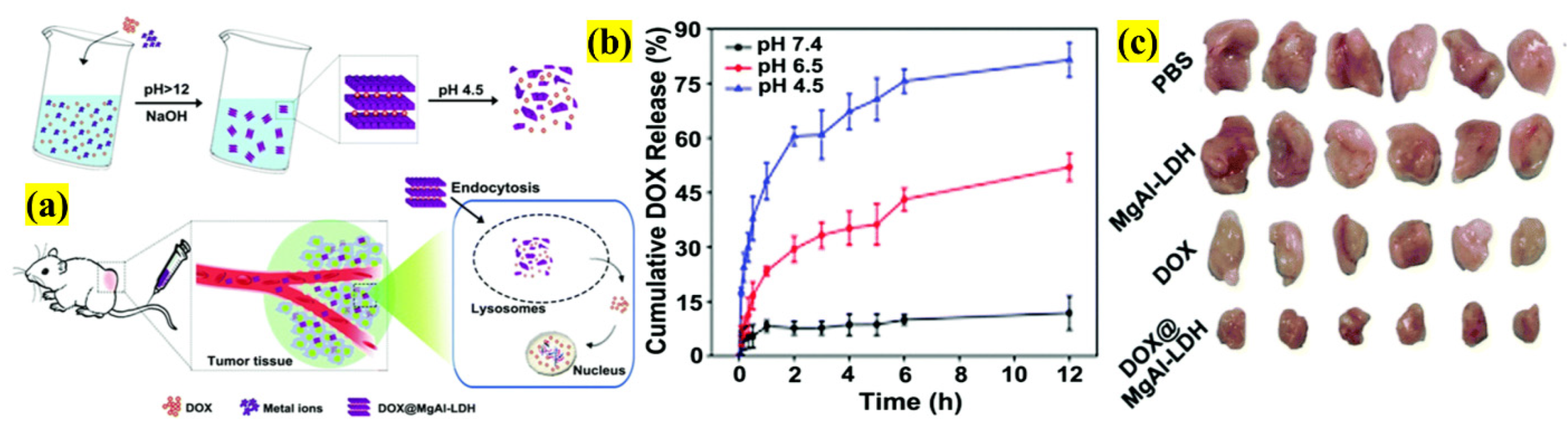
- Hakeem, A.; Zhan, G.; Xu, Q.; Yong, T.; Yang, X.; Gan, L. Facile synthesis of pH-responsive doxorubicin-loaded layered double hydroxide for efficient cancer therapy. J. Mater. Chem. B 2018, 6, 5768–5774. [Google Scholar] [CrossRef]
- Ameena Shirin, V.K.; Sankar, R.; Johnson, A.P.; Gangadharappa, H.V.; Pramod, K. Advanced drug delivery applications of layered double hydroxide. J. Control. Release 2021, 330, 398–426. [Google Scholar] [CrossRef]
- Kura, A.U.; Hussein, M.Z.; Fakurazi, S.; Arulselvan, P. Layered double hydroxide nanocomposite for drug delivery systems; bio-distribution, toxicity and drug activity enhancement. Chem. Cent. J. 2014, 8. [Google Scholar] [CrossRef] [PubMed] [Green Version]
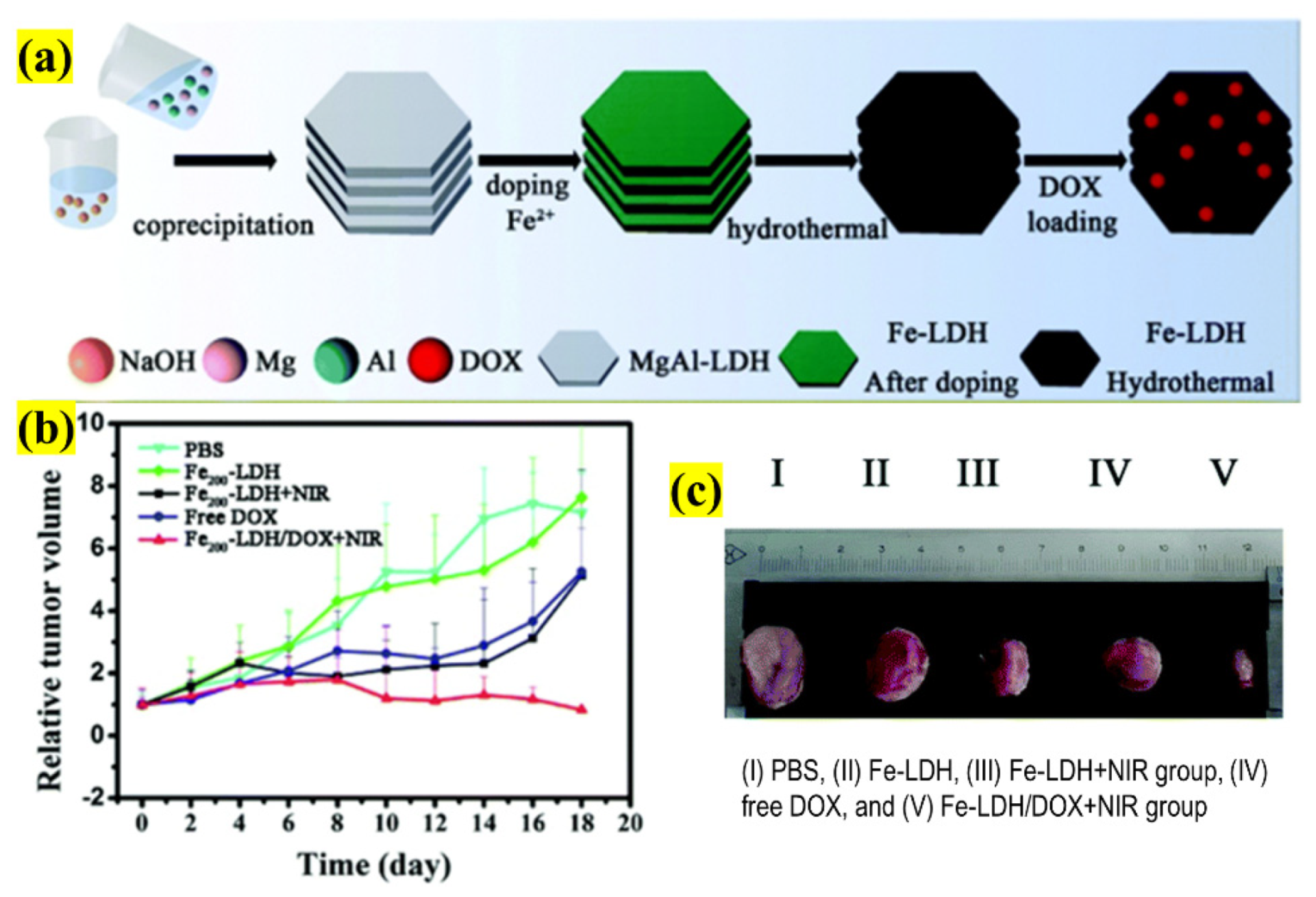
- Guo, Z.; Xie, W.; Lu, J.; Guo, X.; Chi, Y.; Wang, D.; Takuya, N.; Xu, W.; Ye, J.; Liu, X.; et al. Ferrous ions doped layered double hydroxide: Smart 2D nanotheranostic platform with imaging-guided synergistic chemo/photothermal therapy for breast cancer. Biomater. Sci. 2021, 9, 5928–5938. [Google Scholar] [CrossRef]
- Tang, L.; Li, J.; Zhao, Q.; Pan, T.; Zhong, H.; Wang, W. Advanced and Innovative Nano-Systems for Anticancer Targeted Drug Delivery. Pharmaceutics 2021, 13, 1151. [Google Scholar] [CrossRef] [PubMed]
- Neubi, G.M.N.; Opoku-Damoah, Y.; Gu, X.; Han, Y.; Zhou, J.; Ding, Y. Bio-inspired drug delivery systems: An emerging platform for targeted cancer therapy. Biomater. Sci. 2018, 6, 958–973. [Google Scholar] [CrossRef]
- Yadav, P.; Jain, J.; Sherje, A.P. Recent advances in nanocarriers-based drug delivery for cancer therapeutics: A review. React. Funct. Polym. 2021, 165, 104970. [Google Scholar] [CrossRef]
- Edis, Z.; Wang, J.; Waqas, M.K.; Ijaz, M.; Ijaz, M. Nanocarriers-Mediated Drug Delivery Systems for Anticancer Agents: An Overview and Perspectives. Int. J. Nanomed. 2021, 16, 1313–1330. [Google Scholar] [CrossRef] [PubMed]
- Aberoumandi, S.M.; Mohammadhosseini, M.; Abasi, E.; Saghati, S.; Nikzamir, N.; Akbarzadeh, A.; Panahi, Y.; Davaran, S. An update on applications of nanostructured drug delivery systems in cancer therapy: A review. Artif. Cells Nanomed. Biotechnol. 2017, 45, 1058–1068. [Google Scholar] [CrossRef]
- Balaji, A.; Vellayappan, M.V.; John, A.A.; Subramanian, A.P.; Jaganathan, S.K.; Supriyanto, E.; Razak, S.I.A. An insight on electrospun-nanofibers-inspired modern drug delivery system in the treatment of deadly cancers. RSC Adv. 2015, 5, 57984–58004. [Google Scholar] [CrossRef]
- Khodadadi, M.; Alijani, S.; Montazeri, M.; Esmaeilizadeh, N.; Sadeghi-Soureh, S.; Pilehvar-Soltanahmadi, Y. Recent advances in electrospun nanofiber-mediated drug delivery strategies for localized cancer chemotherapy. J. Biomed. Mater. Res.—Part A 2020, 108, 1444–1458. [Google Scholar] [CrossRef]
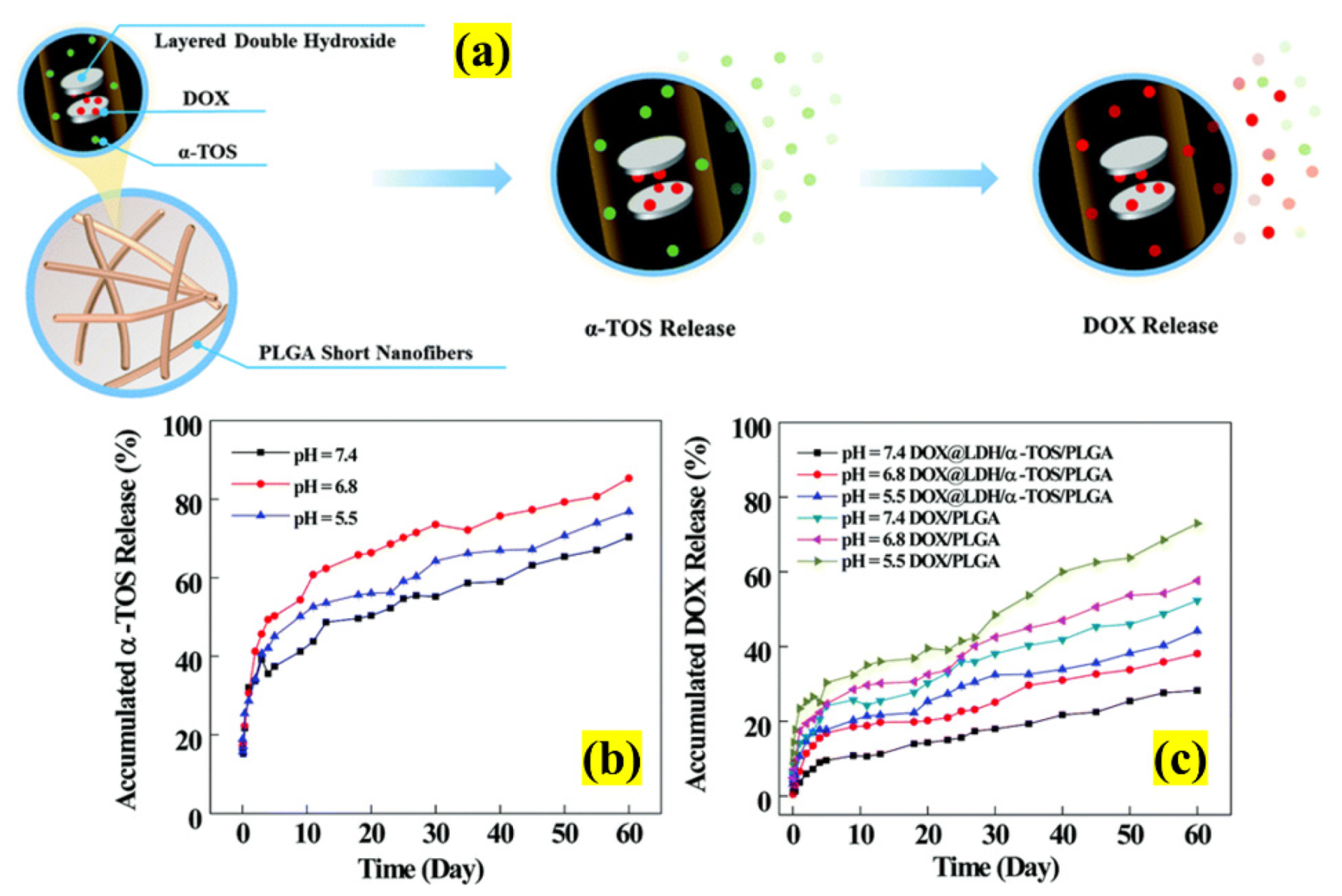
- Ma, Y.; Li, D.; Xiao, Y.; Ouyang, Z.; Shen, M.; Shi, X. LDH-doped electrospun short fibers enable dual drug loading and multistage release for chemotherapy of drug-resistant cancer cells. New J. Chem. 2021, 45, 13421–13428. [Google Scholar] [CrossRef]
- Wang, J.; Dong, R.; Wu, H.; Cai, Y.; Ren, B. A Review on Artificial Micro/Nanomotors for Cancer-Targeted Delivery, Diagnosis, and Therapy. Nano-Micro Lett. 2020, 12, 11. [Google Scholar] [CrossRef] [Green Version]
- Wang, W.; Zhou, C. A Journey of Nanomotors for Targeted Cancer Therapy: Principles, Challenges, and a Critical Review of the State-of-the-Art. Adv. Healthc. Mater. 2021, 10, 2001236. [Google Scholar] [CrossRef] [PubMed]
- Rastmanesh, A.; Yaraki, M.T.; Wu, J.; Wang, Z.; Ghoderao, P.; Gao, Y.; Tan, Y.N. Bioinspired micro/nanomotors towards a self-propelled noninvasive diagnosis and treatment of cancer. Mol. Syst. Des. Eng. 2021, 6, 566–593. [Google Scholar] [CrossRef]
- Ross, K.A.; Brenza, T.M.; Binnebose, A.M.; Phanse, Y.; Kanthasamy, A.G.; Gendelman, H.E.; Salem, A.K.; Bartholomay, L.C.; Bellaire, B.H.; Narasimhan, B. Nano-enabled delivery of diverse payloads across complex biological barriers. J. Control. Release 2015, 219, 548–559. [Google Scholar] [CrossRef] [Green Version]
- Nagamune, T. Biomolecular engineering for nanobio/bionanotechnology. Nano Converg. 2017, 4, 9. [Google Scholar] [CrossRef] [PubMed] [Green Version]
- Meng, H.; Leong, W.; Leong, K.W.; Chen, C.; Zhao, Y. Walking the line: The fate of nanomaterials at biological barriers. Biomaterials 2018, 174, 41–53. [Google Scholar] [CrossRef] [PubMed]
- Tang, Z.; Xiao, Y.; Kong, N.; Liu, C.; Chen, W.; Huang, X.; Xu, D.; Ouyang, J.; Feng, C.; Wang, C.; et al. Nano-bio interfaces effect of two-dimensional nanomaterials and their applications in cancer immunotherapy. Acta Pharm. Sin. B 2021. [Google Scholar] [CrossRef]
- Blanco, E.; Shen, H.; Ferrari, M. Principles of nanoparticle design for overcoming biological barriers to drug delivery. Nat. Biotechnol. 2015, 33, 941–951. [Google Scholar] [CrossRef]
- Chen, W.; Ouyang, J.; Liu, H.; Chen, M.; Zeng, K.; Sheng, J.; Liu, Z.; Han, Y.; Wang, L.; Li, J.; et al. Black Phosphorus Nanosheet-Based Drug Delivery System for Synergistic Photodynamic/Photothermal/Chemotherapy of Cancer. Adv. Mater. 2017, 29, 1603864. [Google Scholar] [CrossRef]
- Peng, F.; Zhao, F.; Shan, L.; Li, R.; Jiang, S.; Zhang, P. Black phosphorus nanosheets-based platform for targeted chemo-photothermal synergistic cancer therapy. Colloids Surf. B Biointerfaces 2021, 198, 111467. [Google Scholar] [CrossRef] [PubMed]
- Pan, T.; Fu, W.; Xin, H.; Geng, S.; Li, Z.; Cui, H.; Zhang, Y.; Chu, P.K.; Zhou, W.; Yu, X.F. Calcium Phosphate Mineralized Black Phosphorous with Enhanced Functionality and Anticancer Bioactivity. Adv. Funct. Mater. 2020, 30, 1–11. [Google Scholar] [CrossRef]
- Zhang, F.; Peng, F.; Qin, L.; Yang, D.; Li, R.; Jiang, S.; He, H.; Zhang, P. pH/near infrared dual-triggered drug delivery system based black phosphorus nanosheets for targeted cancer chemo-photothermal therapy. Colloids Surf. B Biointerfaces 2019, 180, 353–361. [Google Scholar] [CrossRef]
- Shi, J.; Zhang, H.; Chen, Z.; Xu, L.; Zhang, Z. A multi-functional nanoplatform for efficacy tumor theranostic applications. Asian J. Pharm. Sci. 2017, 12, 235–249. [Google Scholar] [CrossRef] [PubMed]
- Zhang, X.; Wu, J.; Williams, G.R.; Yang, Y.; Niu, S.; Qian, Q.; Zhu, L.M. Dual-responsive molybdenum disulfide/copper sulfide-based delivery systems for enhanced chemo-photothermal therapy. J. Colloid Interface Sci. 2019, 539, 433–441. [Google Scholar] [CrossRef] [PubMed] [Green Version]
- Zhang, X.; Wu, J.; Williams, G.R.; Niu, S.; Qian, Q.; Zhu, L.M. Functionalized MoS 2 -nanosheets for targeted drug delivery and chemo-photothermal therapy. Colloids Surf. B Biointerfaces 2019, 173, 101–108. [Google Scholar] [CrossRef] [Green Version]
- Xie, M.; Yang, N.; Cheng, J.; Yang, M.; Deng, T.; Li, Y.; Feng, C. Layered MoS2 nanosheets modified by biomimetic phospholipids: Enhanced stability and its synergistic treatment of cancer with chemo-photothermal therapy. Colloids Surf. B Biointerfaces 2020, 187, 110631. [Google Scholar] [CrossRef]
- Xie, M.; Li, J.; Deng, T.; Yang, N.; Yang, M. Modification of magnetic molybdenum disulfide by chitosan/carboxymethylcellulose with enhanced dispersibility for targeted photothermal-/chemotherapy of cancer. J. Mater. Chem. B 2021, 9, 1833–1845. [Google Scholar] [CrossRef] [PubMed]
- Liu, G.; Zou, J.; Tang, Q.; Yang, X.; Zhang, Y.; Zhang, Q.; Huang, W.; Chen, P.; Shao, J.; Dong, X. Surface Modified Ti3C2 MXene Nanosheets for Tumor Targeting Photothermal/Photodynamic/Chemo Synergistic Therapy. ACS Appl. Mater. Interfaces 2017, 9, 40077–40086. [Google Scholar] [CrossRef] [PubMed]
- Bai, L.; Yi, W.; Sun, T.; Tian, Y.; Zhang, P.; Si, J.; Hou, X.; Hou, J. Surface modification engineering of two-dimensional titanium carbide for efficient synergistic multitherapy of breast cancer. J. Mater. Chem. B 2020, 8, 6402–6417. [Google Scholar] [CrossRef]
- Zhu, B.; Shi, J.; Liu, C.; Li, J.; Cao, S. In-situ self-assembly of sandwich-like Ti3C2 MXene/gold nanorods nanosheets for synergistically enhanced near-infrared responsive drug delivery. Ceram. Int. 2021, 47, 24252–24261. [Google Scholar] [CrossRef]
- Anirudhan, T.S.; Chithra Sekhar, V. Fabrication of functionalized layered double hydroxide/chitosan nanocomposite with dual responsive drug release for the targeted therapy of breast cancer. Eur. Polym. J. 2020, 139, 109993. [Google Scholar] [CrossRef]
- Li, L.; Qian, Y.; Sun, L.; Han, F.Y.; Zhang, R.; Wang, P.Y.; Xu, Z.P. Albumin-stabilized layered double hydroxide nanoparticles synergized combination chemotherapy for colorectal cancer treatment. Nanomed. Nanotechnol. Biol. Med. 2021, 34, 102369. [Google Scholar] [CrossRef]
- Sun, L.; Wang, J.; Liu, J.; Li, L.; Xu, Z.P. Creating Structural Defects of Drug-Free Copper-Containing Layered Double Hydroxide Nanoparticles to Synergize Photothermal/Photodynamic/Chemodynamic Cancer Therapy. Small Struct. 2021, 2, 2000112. [Google Scholar] [CrossRef]
- Li, L.; Gu, W.; Chen, J.; Chen, W.; Xu, Z.P. Co-delivery of siRNAs and anticancer drugs using layered double hydroxide nanoparticles. Biomaterials 2014, 35, 3331–3339. [Google Scholar] [CrossRef] [PubMed] [Green Version]
- Mokhtari, S.; Solati-Hashjin, M.; Khosrowpour, Z.; Gholipourmalekabadi, M. Layered double hydroxide-galactose as an excellent nanocarrier for targeted delivery of curcumin to hepatocellular carcinoma cells. Appl. Clay Sci. 2021, 200, 105891. [Google Scholar] [CrossRef]
- Li, B.; Tang, J.; Chen, W.; Hao, G.; Kurniawan, N.; Gu, Z.; Xu, Z.P. Novel theranostic nanoplatform for complete mice tumor elimination via MR imaging-guided acid-enhanced photothermo-/chemo-therapy. Biomaterials 2018, 177, 40–51. [Google Scholar] [CrossRef]
- Choi, G.; Piao, H.; Aalothman, Z.; Vinu, A.; Yun, C.O.; Choy, J.H. Anionic clay as the drug delivery vehicle: Tumor targeting function of layered double hydroxide-methotrexate nanohybrid in C33A orthotopic cervical cancer model. Int. J. Nanomed. 2016, 11, 337–348. [Google Scholar] [CrossRef] [Green Version]
- Mei, X.; Xu, S.; Hu, T.; Peng, L.; Gao, R.; Liang, R.; Wei, M.; Evans, D.G.; Duan, X. Layered double hydroxide monolayers for controlled loading and targeted delivery of anticancer drugs. Nano Res. 2018, 11, 195–205. [Google Scholar] [CrossRef]
- Busa, P.; Koutavarapu, R.; Lee, D.-Y.; Shim, J.; Kuthati, Y. Hierarchical Two-Dimensional Layered Double Hydroxide Coated Polydopamine Nanocarriers for Combined Chemodynamic and Photothermal Tumor Therapy. Coatings 2021, 11, 1008. [Google Scholar] [CrossRef]
- Nava Andrade, K.; Knauth, P.; López, Z.; Hirata, G.A.; Guevara Martinez, S.J.; Carbajal Arízaga, G.G. Assembly of folate-carbon dots in GdDy-doped layered double hydroxides for targeted delivery of doxorubicin. Appl. Clay Sci. 2020, 192, 105661. [Google Scholar] [CrossRef]


| 2D NSs | Surface Functionalization | Targeting Ligand | Drug | LC (mg or μg)/EE (%) | Size (nm) | ζ Potential (mV) | Synergistic Effect | Cancer Type | Treatment Days | TGI Rates (%)/Observation after the Treatment | Reference | |
|---|---|---|---|---|---|---|---|---|---|---|---|---|
| BP | DOX | 950% (in wt) | 281 ± 9.5 | 1.5 | CTX/PTT/PDT | Breast cancer (4T1 cells) | 14 | 95.5% | [91] | |||
| PAMAM | HA | 95% | 291 ± 4.17 | −5.83 ± 2.31 | CTX/PTT | Reduction in tumor size | [92] | |||||
| CaP | 53.6% | 181.5 | −15.5 | CTX | Breast cancer (MCF-7 cells) | 18 | 85% | [93] | ||||
| HA | MTX | 2.6% | 276 | −18 | CTX/PTT | Breast cancer (4T1 cells) | 10 | Inhibit tumor growth | [94] | |||
| MoS2 | PEG | DOX | ~69% | 150 | −7.17 ± 1.8 | Sarcoma (S180 cells) | 14 | Decrease in tumor volume | [95] | |||
| CuS | 162.3 mg | 114.5 | −5.3 ± 1.5 | - | [96] | |||||||
| PEI, PEG, α-LA | FA, BSA | 185 mg | 196 | 6.8 | Breast cancer (MCF-7 cells) | 20 | Inhibit tumor growth | [97] | ||||
| Liposome | 104.4% | 250 | −38.26 | Breast cancer (4T1 cells) | 2 | [98] | ||||||
| CS, CMC, SPIONs | 95.69% | 429.07 ± 3.538 | −59.01 ± 3.629 | 14 | [99] | |||||||
| Ti3C2 | HA | 84.2% | 178 ± 32.4 | −20.71 ± 1.5 | CTX/PTT/PDT | Colorectal cancer (HCT-116 cells) | 16 | Tumor disappearance and no metastasis/recurrence | [100] | |||
| CP | MET | 96.2% | 310 | −20 | Breast cancer (MDA-MB-231 cells) | [101] | ||||||
| PDA, PEG-GNRs | DOX | 95.88% | 250 | −22.1 ± 1.2 | CTX/PTT | - | [102] | |||||
| LDHs | AuNPs | FA-TCS | 94.6% | 100–180 | −52.4 | Breast cancer (MCF7 cells) | - | Increased % of cell death at the G0/G1 phase | [103] | |||
| BSA | 5-FU | 56.4% ± 7.4% | 172.4 ± 10.6 | −14.0 | CTX | Colorectal cancer (HCT-116 cells) | 18 | Significant and effective inhibition of tumor growth | [104] | |||
| ABX | 93.2% ± 5.2% | |||||||||||
| ICG | - | 99.6% ± 0.1% | 38.8 ± 1.8 | 33.9 ± 0.9 | CTX/PTT/PDT | Breast cancer (4T1 cells) | 12 | [105] | ||||
| 5-FU and CD-siRNA | 22.60% (in wt) | 89 | 38.9 | CTX | Breast cancer (MCF7 cells) | - | Suppresses cell growth | [106] | ||||
| Gal | Cur | 31% | 116.1 ± 35.9 | 10.0 ± 0.7 | Hepatocellular cancer (HepG2 cells) | - | [107] | |||||
| BSA | 5-FU | - | 41.2 ± 5.4 | - | CTX/PTT | Colon cancer (HCT-116 cells) | 24 | Induces cancer cell death with no sign of recurrence | [108] | |||
| MTX | 42% ± 0.25% | 104 | - | CTX | Cervical cancer (C33A cells) | 4 | Tumor growth suppression | [109] | ||||
| FA | DOX | 3.6 mg/mg (w/w) | 60–100 | −3.2 | Oral epidermal cancer (KB cells) | - | Predominant apoptosis of cancer cells | [110] | ||||
| PDA | - | 125 | −14.61 | CDT/PTT | Lung cancer (A549 cells) | - | [111] | |||||
| F-CDs | 212.6 μg/g | 137 | 39.7 | CTX | HeLa cells | - | Enhanced cytotoxicity | [112] | ||||
Publisher’s Note: MDPI stays neutral with regard to jurisdictional claims in published maps and institutional affiliations. |
© 2021 by the authors. Licensee MDPI, Basel, Switzerland. This article is an open access article distributed under the terms and conditions of the Creative Commons Attribution (CC BY) license (https://creativecommons.org/licenses/by/4.0/).
Share and Cite
Manisekaran, R.; García-Contreras, R.; Rasu Chettiar, A.-D.; Serrano-Díaz, P.; Lopez-Ayuso, C.A.; Arenas-Arrocena, M.C.; Hernández-Padrón, G.; López-Marín, L.M.; Acosta-Torres, L.S. 2D Nanosheets—A New Class of Therapeutic Formulations against Cancer. Pharmaceutics 2021, 13, 1803. https://0-doi-org.brum.beds.ac.uk/10.3390/pharmaceutics13111803
Manisekaran R, García-Contreras R, Rasu Chettiar A-D, Serrano-Díaz P, Lopez-Ayuso CA, Arenas-Arrocena MC, Hernández-Padrón G, López-Marín LM, Acosta-Torres LS. 2D Nanosheets—A New Class of Therapeutic Formulations against Cancer. Pharmaceutics. 2021; 13(11):1803. https://0-doi-org.brum.beds.ac.uk/10.3390/pharmaceutics13111803
Chicago/Turabian StyleManisekaran, Ravichandran, René García-Contreras, Aruna-Devi Rasu Chettiar, Paloma Serrano-Díaz, Christian Andrea Lopez-Ayuso, Ma Concepción Arenas-Arrocena, Genoveva Hernández-Padrón, Luz M. López-Marín, and Laura Susana Acosta-Torres. 2021. "2D Nanosheets—A New Class of Therapeutic Formulations against Cancer" Pharmaceutics 13, no. 11: 1803. https://0-doi-org.brum.beds.ac.uk/10.3390/pharmaceutics13111803