
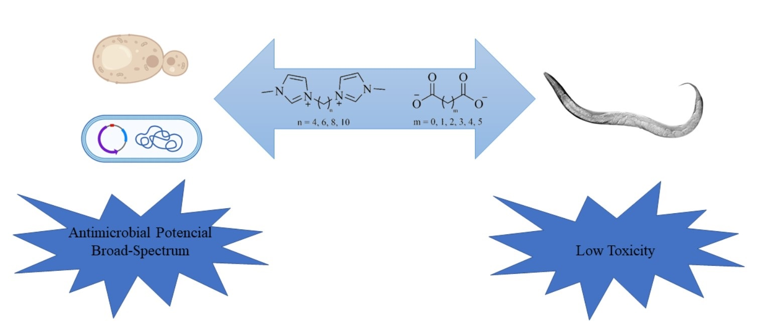
Antimicrobial and Toxicity Evaluation of Imidazolium-Based Dicationic Ionic Liquids with Dicarboxylate Anions

 ,
,
Abstract
:
1. Introduction
2. Materials and Methods
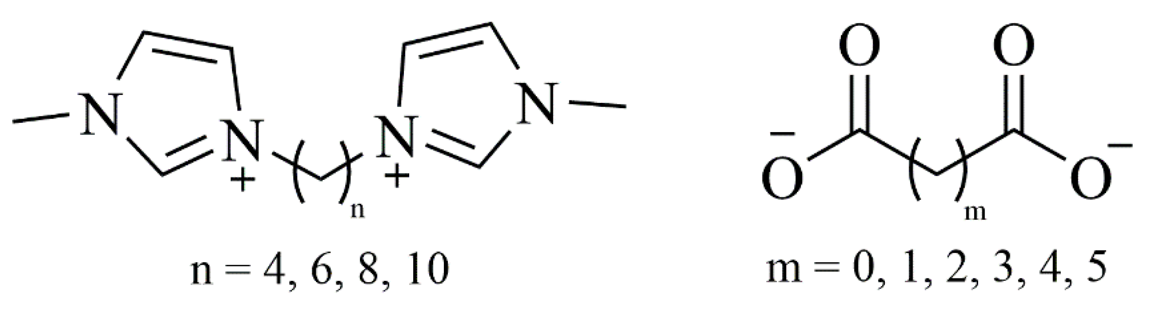
2.1. Synthesis of DILs
2.2. Antibacterial Activity
2.3. Antifungal Activity
2.3.1. Susceptibility Test
2.3.2. Cell Culture
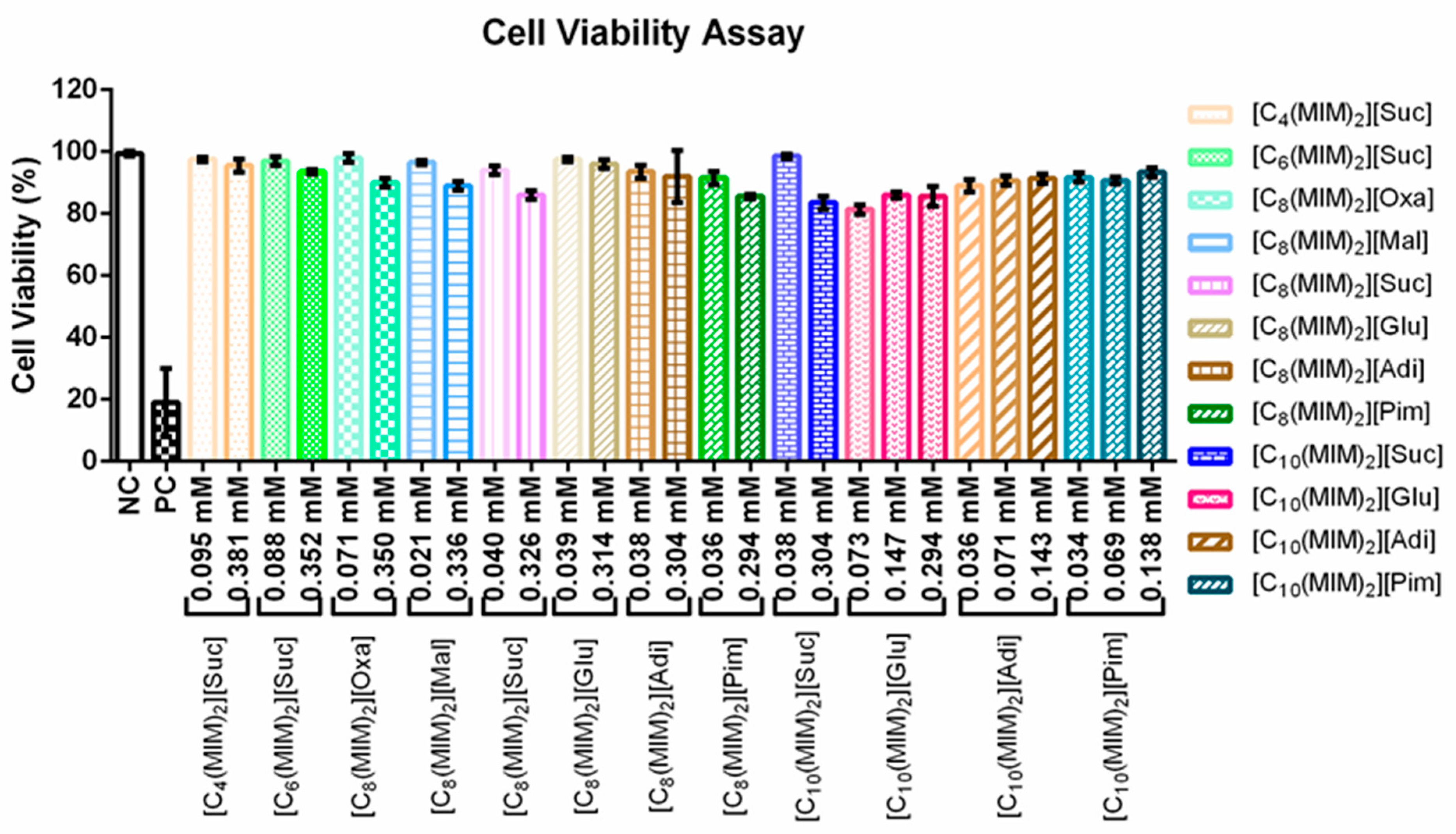
2.3.3. Cell Viability
2.4. C. elegans Strains: Maintenance and Treatment
Survival Assay
3. Results
3.1. Antifungal Activity and Cell Viability
3.2. Antibacterial Activity
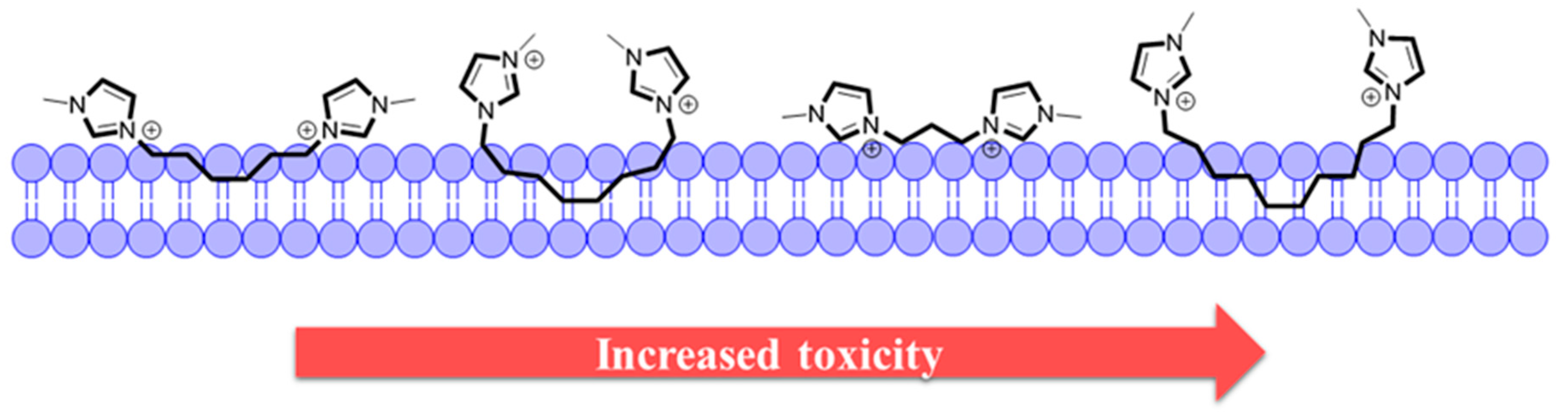
3.3. Toxicity Tests
4. Conclusions
Author Contributions
Funding
Institutional Review Board Statement
Informed Consent Statement
Data Availability Statement
Conflicts of Interest
References
- Pendleton, J.N.; Gorman, S.P.; Gilmore, B.F. Clinical relevance of the ESKAPE pathogens. Expert Rev. Anti. Infect. Ther. 2013, 11, 297–308. [Google Scholar] [CrossRef] [PubMed]
- Qin, J.; Guo, J.; Xu, Q.; Zheng, Z.; Mao, H.; Yan, F. Synthesis of Pyrrolidinium-Type Poly(ionic liquid) Membranes for Antibacterial Applications. ACS Appl. Mater. Interfaces 2017, 9, 10504–10511. [Google Scholar] [CrossRef] [PubMed]
- Florio, W.; Becherini, S.; D’Andrea, F.; Lupetti, A.; Chiappe, C.; Guazzelli, L. Comparative evaluation of antimicrobial activity of different types of ionic liquids. Mater. Sci. Eng. C 2019, 104, 109907. [Google Scholar] [CrossRef] [PubMed]
- Pendleton, J.N.; Gilmore, B.F. The antimicrobial potential of ionic liquids: A source of chemical diversity for infection and biofilm control. Int. J. Antimicrob. Agents 2015, 46, 131–139. [Google Scholar] [CrossRef]
- Docherty, K.M.; Kulpa, C.F. Toxicity and antimicrobial activity of imidazolium and pyridinium ionic liquids. Green Chem. 2005, 7, 185–189. [Google Scholar] [CrossRef]
- Garcia, M.T.; Ribosa, I.; Perez, L.; Manresa, A.; Comelles, F.; Garcia, M.T.; Ribosa, I.; Perez, L.; Manresa, A.; Comelles, F. Aggregation behavior and antimicrobial activity of ester-functionalized imidazolium- and pyridinium-based ionic liquids in aqueous solution. Langmuir 2013, 29, 2536–2545. [Google Scholar] [CrossRef] [PubMed]
- Montalbán, M.G.; Víllora, G.; Licence, P. Ecotoxicity assessment of dicationic versus monocationic ionic liquids as a more environmentally friendly alternative. Ecotoxicol. Environ. Saf. 2018, 150, 129–135. [Google Scholar] [CrossRef]
- Coleman, D.; Gathergood, N. Biodegradation studies of ionic liquids. Chem. Soc. Rev. 2010, 39, 600–637. [Google Scholar] [CrossRef]
- Jordan, A.; Gathergood, N. Biodegradation of ionic liquids-a critical review. Chem. Soc. Rev. 2015, 44, 8200–8237. [Google Scholar] [CrossRef]
- Gindri, I.M.; Siddiqui, D.A.; Bhardwaj, P.; Rodriguez, L.C.; Palmer, K.L.; Frizzo, C.P.; Martins, M.A.P.; Rodrigues, D.C. Dicationic imidazolium-based ionic liquids: A new strategy for non-toxic and antimicrobial materials. RSC Adv. 2014, 4, 62594–62602. [Google Scholar] [CrossRef]
- Pernak, J.; Goc, I.; Mirska, I. Anti-microbial activities of protic ionic liquids with lactate anion. Green Chem. 2004, 6, 323–329. [Google Scholar] [CrossRef]
- Dalla Lana, D.F.; Giuliani, L.M.; Reolon, J.B.; Lopes, W.; Vainstein, M.H.; Danielli, L.J.; Bergamo, V.Z.; Pippi, B.; Apel, M.A.; Teixeira, M.L.; et al. Nanoemulsion Improves the Antifungal Activity of Allylic Thiocyanates against Yeasts and Filamentous Pathogenic Fungi. ChemistrySelect 2018, 3, 11663. [Google Scholar] [CrossRef]
- Pappas, P.G.; Lionakis, M.S.; Arendrup, M.C.; Ostrosky-Zeichner, L.; Kullberg, B.J. Invasive candidiasis. Nat. Rev. Dis. Prim. 2018, 4, 1–20. [Google Scholar] [CrossRef]
- Steudte, S.; Bemowsky, S.; Mahrova, M.; Bottin-Weber, U.; Tojo-Suarez, E.; Stepnowski, P.; Stolte, S. Toxicity and biodegradability of dicationic ionic liquids. RSC Adv. 2014, 4, 5198–5205. [Google Scholar] [CrossRef]
- Egorova, K.S.; Ananikov, V.P. Toxicity of ionic liquids: Eco(cyto)activity as complicated, but unavoidable parameter for task-specific optimization. ChemSusChem 2014, 7, 336–360. [Google Scholar] [CrossRef] [PubMed]
- Zhao, D.; Liao, Y.; Zhang, Z.D. Toxicity of ionic liquids. Clean Soil Air Water 2007, 35, 42–48. [Google Scholar] [CrossRef]
- Höss, S.; Weltje, L. Endocrine disruption in nematodes: Effects and mechanisms. Ecotoxicology 2007, 16, 15–28. [Google Scholar] [CrossRef] [PubMed]
- Leung, M.C.K.; Williams, P.L.; Benedetto, A.; Au, C.; Helmcke, K.J.; Aschner, M.; Meyer, J.N. Caenorhabditis elegans: An emerging model in biomedical and environmental toxicology. Toxicol. Sci. 2008, 106, 5–28. [Google Scholar] [CrossRef]
- Frizzo, C.P.; Bender, C.R.; Salbego, P.R.S.; Farias, C.A.A.; da Silva, T.C.; Stefanello, S.T.; da Silveira, T.L.; Soares, F.A.A.; Villetti, M.A.; Martins, M.A.P. Impact of Anions on the Partition Constant, Self-Diffusion, Thermal Stability, and Toxicity of Dicationic Ionic Liquids. ACS Omega 2018, 3, 734–743. [Google Scholar] [CrossRef]
- Kuhn, B.L.; Osmari, B.F.; Heinen, T.M.; Bonacorso, H.G.; Zanatta, N.; Nielsen, S.O.; Ranathunga, D.T.S.; Villetti, M.A.; Frizzo, C.P. Dicationic imidazolium-based dicarboxylate ionic liquids: Thermophysical properties and solubility. J. Mol. Liq. 2020, 308, 112983. [Google Scholar] [CrossRef]
- Gindri, I.M.; Siddiqui, D.A.; Frizzo, C.P.; Martins, M.A.P.P.; Rodrigues, D.C. Improvement of tribological and anti-corrosive performance of titanium surfaces coated with dicationic imidazolium-based ionic liquids. RSC Adv. 2016, 6, 78795–78802. [Google Scholar] [CrossRef]
- Cockerill, F.R.; Wiker, M.A.; Alder, J.; Dudley, M.N.; Eliopoulos, G.M.; Ferraro, M.J.; Hardy, D.J.; Hecht, D.W.; Hindler, J.A.; Patel, J.B.; et al. Methods for Dilution Antimicrobial Susceptibility Tests for Bacteria That Grow Aerobically; Approved Standard, 9th ed.; Clinical and Laboratory Standards Institute: Wayne, PA, USA, 2012; Volume 32, ISBN 1562387839. [Google Scholar]
- CLSI. Reference Method for Broth Dilution. M27-A3 Ref. Method Broth Dilution Antifungal Susceptibility Test. Yeasts; Approved Standard, 3rd ed.; CLSI: Wayne, PA, USA, 2008; p. 28. [Google Scholar]
- dos Santos Montagner, G.F.F.; Sagrillo, M.; Machado, M.M.; Almeida, R.C.; Mostardeiro, C.P.; Duarte, M.M.M.F.; da Cruz, I.B.M. Toxicological effects of ultraviolet radiation on lymphocyte cells with different manganese superoxide dismutase Ala16Val polymorphism genotypes. Toxicol. In Vitro 2010, 24, 1410–1416. [Google Scholar] [CrossRef] [PubMed]
- Burow, M.E.; Weldon, C.B.; Tang, Y.; Navar, G.L.; Krajewski, S.; Reed, J.C.; Hammond, T.G.; Clejan, S.; Beckman, B.S. Differences in susceptibility to tumor necrosis factor α-induced apoptosis among MCF-7 breast cancer cell variants. Cancer Res. 1998, 58, 4940–4946. [Google Scholar]
- Brenner, S. The Genetics of Caenorhabditis elegans. Genetics 1974, 77, 71–94. [Google Scholar] [CrossRef] [PubMed]
- Wu, X.; Tong, Z.H.; Li, L.L.; Yu, H.Q. Toxic effects of imidazolium-based ionic liquids on Caenorhabditis elegans: The role of reactive oxygen species. Chemosphere 2013, 93, 2399–2404. [Google Scholar] [CrossRef] [PubMed]
- Stolte, S.; Arning, J.; Bottin-Weber, U.; Matzke, M.; Stock, F.; Thiele, K.; Uerdingen, M.; Welz-Biermann, U.; Jastorff, B.; Ranke, J. Anion effects on the cytotoxicity of ionic liquids. Green Chem. 2006, 8, 621–629. [Google Scholar] [CrossRef]
- Gal, N.; Malferarri, D.; Kolusheva, S.; Galletti, P.; Tagliavini, E.; Jelinek, R. Membrane interactions of ionic liquids: Possible determinants for biological activity and toxicity. Biochim. Biophys. Acta Biomembr. 2012, 1818, 2967–2974. [Google Scholar] [CrossRef] [PubMed] [Green Version]
- Lim, G.S.; Zidar, J.; Cheong, D.W.; Jaenicke, S.; Klahn, M. Impact of ionic liquids in aqueous solution on bacterial plasma membranes studied with molecular dynamics simulations. J. Phys. Chem. B 2014, 118, 10444–10459. [Google Scholar] [CrossRef] [PubMed]
- Frizzo, C.P.; Gindri, I.M.; Bender, C.R.; Tier, A.Z.; Villetti, M.A.; Rodrigues, D.C.; Machado, G.; Martins, M.A.P. Effect on aggregation behavior of long-chain spacers of dicationic imidazolium-based ionic liquids in aqueous solution. Colloids Surf. A Physicochem. Eng. Asp. 2015, 468, 285–294. [Google Scholar] [CrossRef]
- Piotrowska, A.; Syguda, A.; Wyrwas, B.; Chrzanowski, Ł.; Heipieper, H.J. Toxicity evaluation of selected ammonium-based ionic liquid forms with MCPP and dicamba moieties on Pseudomonas putida. Chemosphere 2017, 167, 114–119. [Google Scholar] [CrossRef] [PubMed]
- Cornellas, A.; Perez, L.; Comelles, F.; Ribosa, I.; Manresa, A.; Garcia, T.T. Self-aggregation and antimicrobial activity of imidazolium and pyridinium based ionic liquids in aqueous solution. J. Colloid Interface Sci. 2011, 355, 164–171. [Google Scholar] [CrossRef]
- Muñoz-Bonilla, A.; Fernández-García, M. Poly(ionic liquid)s as antimicrobial materials. Eur. Polym. J. 2018, 105, 135–149. [Google Scholar] [CrossRef]
- Sternberg, P.W. Working in the post-genomic C. elegans world. Cell 2001, 105, 173–176. [Google Scholar] [CrossRef] [Green Version]
- Swatloski, R.P.; Holbrey, J.D.; Memon, S.B.; Caldwell, G.A.; Caldwell, K.A.; Rogers, R.D. Using Caenorhabditis elegans to probe toxicity of 1-alkyl-3-methylimidazolium chloride based ionic liquids. Chem. Commun. 2004, 668–669. [Google Scholar] [CrossRef] [PubMed]
- Frizzo, C.P.; Bender, C.R.; Salbego, P.R.S.; Farias, C.A.A.; Villetti, M.A.; Martins, M.A.P. Heteroassembly Ability of Dicationic Ionic Liquids and Neutral Active Pharmaceutical Ingredients. ACS Omega 2018, 3. [Google Scholar] [CrossRef] [PubMed]
- Zhu, C.J.; Peng, Y.; Tong, Z.H.; Lu, L.Y.; Cui, Y.H.; Yu, H.Q. Hormetic effect and mechanism of imidazolium-based ionic liquids on the nematode Caenorhabditis elegans. Chemosphere 2016, 157, 65–70. [Google Scholar] [CrossRef] [PubMed]
- Peng, Y.; Tong, Z.H.; Chong, H.J.; Shao, X.Y. Toxic effects of prolonged exposure to [C14mim]Br on Caenorhabditis elegans. Chemosphere 2018, 208, 226–232. [Google Scholar] [CrossRef] [PubMed]


| IL | C. albicans CA02 | C. krusei CK03 | C. parapsilosis CP RL38 | C. tropicalis CT 08 |
|---|---|---|---|---|
| [C8(MIM)2][Oxa] | >0.312 | 0.039 | - | >0.312 |
| [C8(MIM)2][Mal] | >0.312 | >0.312 | >0.312 | 0.039 |
| [C8(MIM)2][[Suc] | >0.312 | 0.039 | >0.312 | 0.078 |
| [C8(MIM)2][Glu] | 0.156 | 0.039 | 0.156 | 0.078 |
| [C8(MIM)2][Adi] | >0.312 | 0.078 | >0.312 | 0.039 |
| [C8(MIM)2][[Pim] | >0.312 | 0.078 | >0.312 | 0.039 |
| Fluconazole | 0.013 | 0.013 | 0.026 | 0.013 |
| IL | C. albicans CA02 | C. krusei CK03 | C. parapsilosis CP RL38 | C. tropicalis CT 08 | ||||
|---|---|---|---|---|---|---|---|---|
| 24 h | 48 h | 24 h | 48 h | 24 h | 48 h | 24 h | 48 h | |
| [C4(MIM)2][Suc] | 0.156 | >0.312 | 0.078 | >0.312 | 0.156 | >0.312 | >0.312 | >0.312 |
| [C6(MIM)2][Suc] | 0.039 | 0.078 | 0.078 | 0.156 | 0.078 | >0.312 | 0.039 | 0.078 |
| [C8(MIM)2][Suc] | >0.312 | >0.312 | 0.039 | 0.078 | >0.312 | >0.312 | 0.078 | >0.312 |
| [C10(MIM)2][Suc] | 0.078 | 0.078 | 0.039 | 0.039 | 0.039 | >0.312 | 0.039 | 0.156 |
| IL | log P | ||
|---|---|---|---|
| ChemDraw a | Molinspiration (miLogP) b | ||
| [C4(MIM)2][Oxa] | dianion: −0.85 | dication: −5.32 | dianion: −3.76 |
| [C4(MIM)2][Mal] | dianion: −0.94 | dication: −5.32 | dianion: −3.49 |
| [C4(MIM)2][Suc] | dianion: −0.48 | dication: −5.32 | dianion: −3.22 |
| [C4(MIM)2][Glu] | dianion: −0.03 | dication: −5.32 | dianion: −2.71 |
| [C4(MIM)2][Adi] | dianion: 0.43 | dication: −5.32 | dianion: −2.21 |
| [C4(MIM)2][Pim] | dianion: 0.89 | dication: −5.32 | dianion: −1.70 |
| [C6(MIM)2][Oxa] | dianion: −0.85 | dication: −5.00 | dianion: −3.76 |
| [C6(MIM)2][Mal] | dianion: −0.94 | dication: −5.00 | dianion: −3.49 |
| [C6(MIM)2][Suc] | dianion: −0.48 | dication: −5.00 | dianion: −3.22 |
| [C6(MIM)2][Glu] | dianion: −0.03 | dication: −5.00 | dianion: −2.71 |
| [C6(MIM)2][Adi] | dianion: 0.43 | dication: −5.00 | dianion: −2.21 |
| [C6(MIM)2][Pim] | dianion: 0.89 | dication: −5.00 | dianion: −1.70 |
| [C8(MIM)2][Oxa] | dianion: −0.85 | dication: −4.54 | dianion: −3.76 |
| [C8(MIM)2][Mal] | dianion: −0.94 | dication: −4.54 | dianion: −3.49 |
| [C8(MIM)2][Suc] | dianion: −0.48 | dication: −4.54 | dianion: −3.22 |
| [C8(MIM)2][Glu] | dianion: −0.03 | dication: −4.54 | dianion: −2.71 |
| [C8(MIM)2][Adi] | dianion: 0.43 | dication: −4.54 | dianion: −2.21 |
| [C8(MIM)2][Pim] | dianion: 0.89 | dication: −4.54 | dianion: −1.70 |
| [C10(MIM)2][Oxa] | dianion: −0.85 | dication: −3.70 | dianion: −3.76 |
| [C10(MIM)2][Mal] | dianion: −0.94 | dication: −3.70 | dianion: −3.49 |
| [C10(MIM)2][Suc] | dianion: −0.48 | dication: −3.70 | dianion: −3.22 |
| [C10(MIM)2][Glu] | dianion: −0.03 | dication: −3.70 | dianion: −2.71 |
| [C10(MIM)2][Adi] | dianion: 0.43 | dication: −3.70 | dianion: −2.21 |
| [C10(MIM)2][Pim] | dianion: 0.89 | dication: −3.70 | dianion: −1.70 |
| IL | S. aureusa | S. aureusb | E. faecalisc | E. faecalisd |
|---|---|---|---|---|
| [C4(MIM)2][[Oxa] | >2.5 | >2.5 | >2.5 | >2.5 |
| [C4(MIM)2][Mal] | >2.5 | >2.5 | >2.5 | >2.5 |
| [C4(MIM)2][Suc] | 1.25 | 2.5 | >2.5 | >2.5 |
| [C4(MIM)2][Glu] | 2.5 | 1.25 | >2.5 | >2.5 |
| [C4(MIM)2][Adi] | 1.25 | 1.25 | 0.625 | 0.625 |
| [C4(MIM)2][Pim] | 0.625 | 0.078 | >2.5 | >2.5 |
| [C6(MIM)2][Oxa] | >2.5 | 2.5 | >2.5 | >2.5 |
| [C6(MIM)2][Mal] | 0.312 | 1.25 | >2.5 | >2.5 |
| [C6(MIM)2][Suc] | 2.5 | 1.25 | >2.5 | >2.5 |
| [C6(MIM)2][Glu] | 2.5 | >2.5 | >2.5 | >2.5 |
| [C6(MIM)2][Adi] | 0.132 | 1.25 | >2.5 | >2.5 |
| [C6(MIM)2][Pim] | >2.5 | >2.5 | >2.5 | >2.5 |
| [C8(MIM)2][Oxa] | 1.25 | 0.312 | 1.25 | >2.5 |
| [C8(MIM)2][Suc] | 0.156 | 1.25 | >2.5 | >2.5 |
| [C8(MIM)2][Glu] | 0.312 | 2.5 | 1.25 | >2.5 |
| [C8(MIM)2][Pim] | 2.5 | 1.25 | >2.5 | >2.5 |
| [C10(MIM)2][Glu] | 2.5 | >2.5 | >2.5 | >2.5 |
| [C10(MIM)2][Pim] | 0.625 | 0.625 | 1.25 | 1.25 |
| Control (µg/mL) | 2 | 2 | 2 | 2 |
| Control (µM) | 5.72 | 5.72 | 5.72 | 5.72 |
| IL | E. Colia | K. pneumoniaeb | A. baumanniic | A. baumanniid | P. Aeruginosae | E. faeciume | E. aerogenesf |
|---|---|---|---|---|---|---|---|
| [C4(MIM)2][[Oxa] | >2.5 | >2.5 | >2.5 | >2.5 | >2.5 | >2.5 | >2.5 |
| [C4(MIM)2][Mal] | >2.5 | >2.5 | >2.5 | >2.5 | >2.5 | >2.5 | >2.5 |
| [C4(MIM)2][Suc] | 2.5 | >2.5 | >2.5 | >2.5 | >2.5 | >2.5 | >2.5 |
| [C4(MIM)2][Glu] | >2.5 | 0.078 | >2.5 | >2.5 | >2.5 | >2.5 | >2.5 |
| [C4(MIM)2][Adi] | 0.312 | >2.5 | >2.5 | >2.5 | 2.5 | 0.625 | >2.5 |
| [C4(MIM)2][Pim] | >2.5 | >2.5 | >2.5 | >2.5 | >2.5 | >2.5 | >2.5 |
| [C6(MIM)2][Oxa] | >2.5 | >2.5 | >2.5 | >2.5 | >2.5 | >2.5 | >2.5 |
| [C6(MIM)2][Mal] | 1.25 | >2.5 | >2.5 | >2.5 | 2.5 | >2.5 | >2.5 |
| [C6(MIM)2][Suc] | >2.5 | >2.5 | >2.5 | >2.5 | >2.5 | >2.5 | >2.5 |
| [C6(MIM)2][Glu] | >2.5 | >2.5 | >2.5 | >2.5 | >2.5 | >2.5 | >2.5 |
| [C6(MIM)2][Adi] | >2.5 | >2.5 | >2.5 | >2.5 | >2.5 | >2.5 | >2.5 |
| [C6(MIM)2][Pim] | 1.25 | >2.5 | >2.5 | >2.5 | >2.5 | >2.5 | >2.5 |
| [C8(MIM)2][Oxa] | 2.5 | >2.5 | >2.5 | 1.25 | >2.5 | >2.5 | >2.5 |
| [C8(MIM)2][Suc] | 1.25 | >2.5 | >2.5 | >2.5 | >2.5 | >2.5 | >2.5 |
| [C8(MIM)2][Glu] | 0.625 | 0.312 | >2.5 | 0.156 | >2.5 | >2.5 | 0.156 |
| [C8(MIM)2][Pim] | 2.5 | >2.5 | >2.5 | >2.5 | >2.5 | >2.5 | >2.5 |
| [C10(MIM)2][Glu] | >2.5 | >2.5 | >2.5 | >2.5 | >2.5 | >2.5 | >2.5 |
| [C10(MIM)2][Pim] | 0.625 | 0.625 | >2.5 | >2.5 | 0.312 | 0.156 | 0.156 |
| Control µg/ml | 4 | 2 | 2 | 2 | 1 | 2 | 1 |
| Control (µM) | 11.44 | 5.72 | 6.30 | 3.15 | 3.15 | 5.72 | 3.15 |
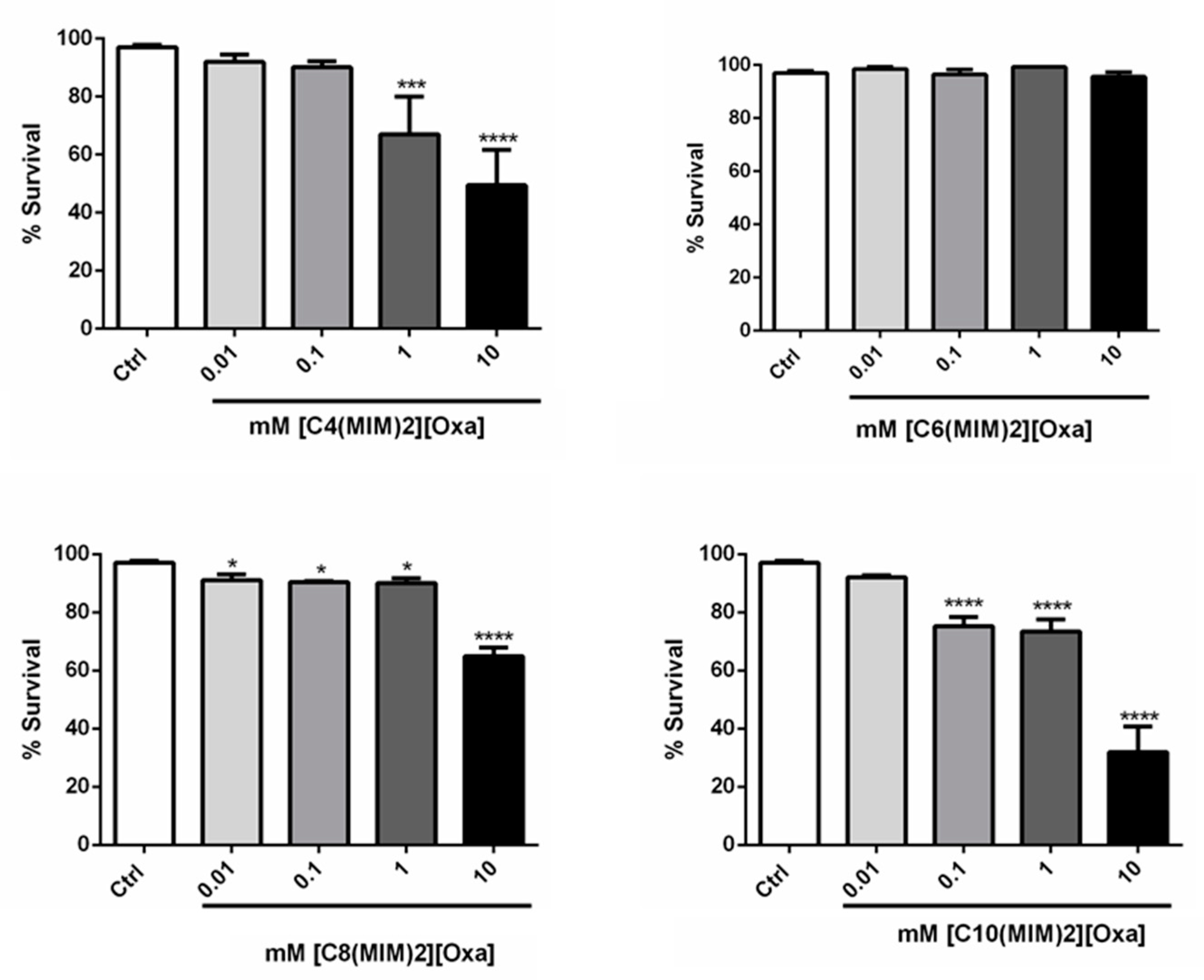
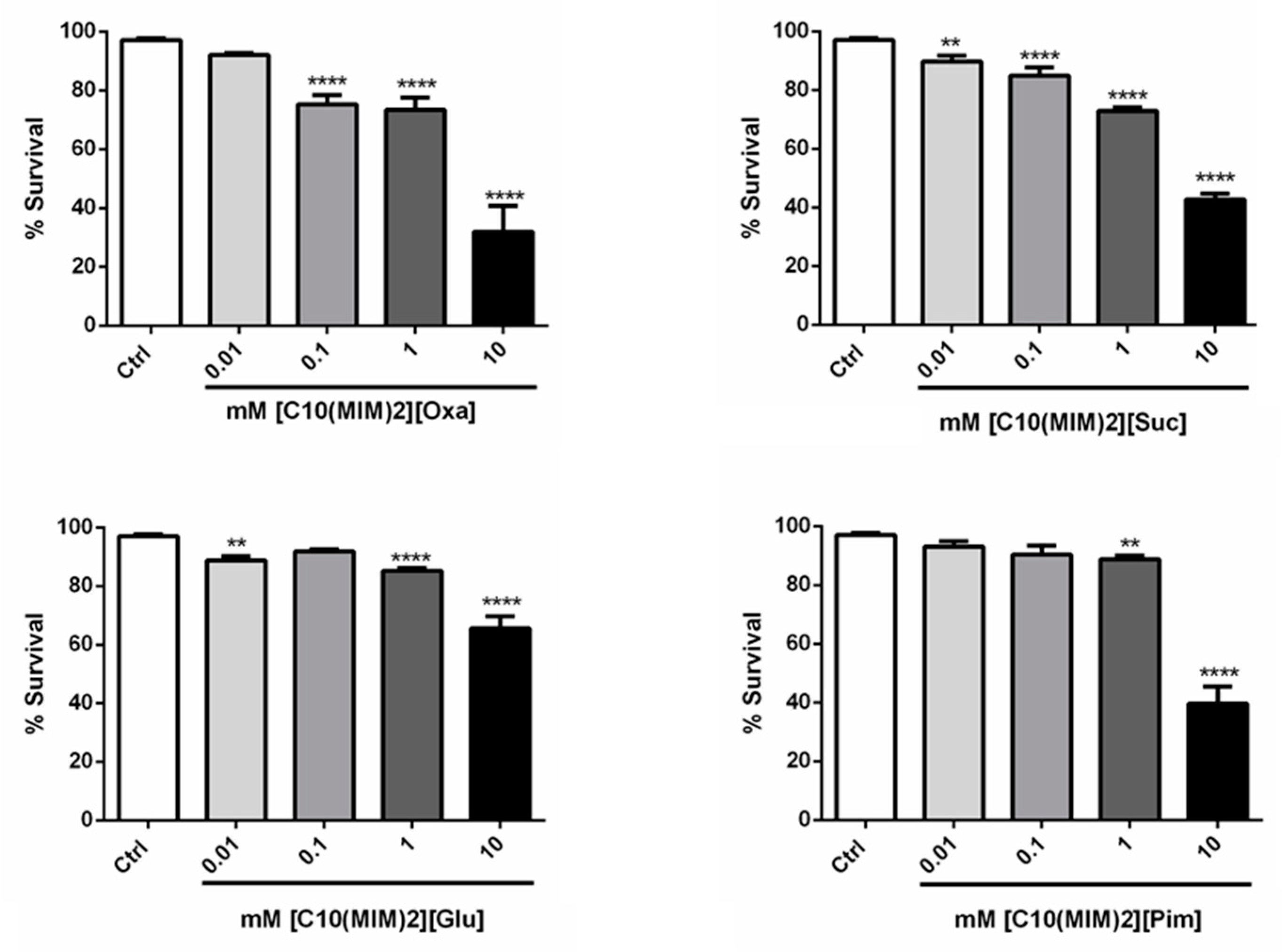
| IL | Concentration (mM) | |||
|---|---|---|---|---|
| 0.01 | 0.1 | 1 | 10 | |
| [C4(MIM)2][Oxa] | 92 ± 6 | 90± 4 | 67± 26 *** | 50 ± 27 **** |
| [C4(MIM)2][Mal] | 97 ± 2 | 98 ± 3 | 98 ± 2 | 94 ± 4 |
| [C4(MIM)2][Suc] | 92 ± 6 | 92 ± 7 | 92 ± 8 | 76 ± 10 **** |
| [C4(MIM)2][Glu] | 96 ± 5 | 92 ± 4 | 89 ± 8 | 82 ± 11 **** |
| [C4(MIM)2][Adi] | 91 ± 8 | 92 ± 9 | 90 ± 8 | 86 ± 4 ** |
| [C4(MIM)2][Pim] | 95 ± 5 | 89 ± 6 * | 84 ± 8 *** | 89 ± 8 * |
| [C6(MIM)2][Oxa] | 99 ± 2 | 97 ± 4 | 99 ± 2 | 96 ± 5 |
| [C6(MIM)2][Mal] | 99 ± 2 | 95 ± 4 | 96 ± 4 | 96 ± 4 |
| [C6(MIM)2][Suc] | 97 ± 4 | 98 ± 2 | 93± 5 | 95 ± 3 |
| [C6(MIM)2][Glu] | 100 ± 0 | 99 ± 2 | 97± 3 | 98± 2 |
| [C6(MIM)2][Adi] | 95 ± 2 | 100 ± 0 | 99 ± 3 | 96 ± 3 |
| [C6(MIM)2][Pim] | 98 ± 2 | 96 ± 4 | 98 ± 1.72 | 97 ± 3 |
| [C8(MIM)2][Oxa] | 91 ± 4 * | 90 ± 1 * | 90± 3 * | 65 ± 5 *** |
| [C8(MIM)2][Mal] | 92 ± 4 | 78 ± 8 **** | 64 ± 12 **** | 42 ± 11 **** |
| [C8(MIM)2][Suc] | 93 ± 2 | 92 ± 3 | 85 ± 2 * | 19 ± 15 **** |
| [C8(MIM)2][Glu] | 93 ± 1 | 92 ± 3 | 95 ± 2 | 90 ± 4 |
| [C8(MIM)2][Adi] | 92 ± 3 | 79 ± 7 **** | 74 ± 8 **** | 30 ± 9 **** |
| [C8(MIM)2][Pim] | 92 ± 4 | 91 ± 6 | 75 ± 5 **** | 18 ± 17 **** |
| [C10(MIM)2][Oxa] | 92 ± 1 | 75 ± 6 **** | 74 ±7 **** | 32 ± 15 **** |
| [C10(MIM)2][Suc] | 90 ± 4 ** | 85 ± 5 **** | 73 ± 2 **** | 43 ± 4 **** |
| [C10(MIM)2][Glu] | 89 ± 3 ** | 92 ± 1 | 85 ± 2**** | 66 ± 7 **** |
| [C10(MIM)2][Pim] | 93 ± 3 | 90 ± 5 | 89 ± 2 ** | 40 ± 10 **** |
Publisher’s Note: MDPI stays neutral with regard to jurisdictional claims in published maps and institutional affiliations. |
© 2021 by the authors. Licensee MDPI, Basel, Switzerland. This article is an open access article distributed under the terms and conditions of the Creative Commons Attribution (CC BY) license (https://creativecommons.org/licenses/by/4.0/).
Share and Cite
Kuhn, B.L.; Kaminski, T.F.A.; Carvalho, Â.R.; Fuentefria, A.M.; Johann, B.M.B.C.; da Silva, E.E.; Silveira, G.P.; da Silveira, T.L.; Soares, F.A.A.; Zanatta, N.; et al. Antimicrobial and Toxicity Evaluation of Imidazolium-Based Dicationic Ionic Liquids with Dicarboxylate Anions. Pharmaceutics 2021, 13, 639. https://0-doi-org.brum.beds.ac.uk/10.3390/pharmaceutics13050639
Kuhn BL, Kaminski TFA, Carvalho ÂR, Fuentefria AM, Johann BMBC, da Silva EE, Silveira GP, da Silveira TL, Soares FAA, Zanatta N, et al. Antimicrobial and Toxicity Evaluation of Imidazolium-Based Dicationic Ionic Liquids with Dicarboxylate Anions. Pharmaceutics. 2021; 13(5):639. https://0-doi-org.brum.beds.ac.uk/10.3390/pharmaceutics13050639
Chicago/Turabian StyleKuhn, Bruna L., Taís F. A. Kaminski, Ânderson R. Carvalho, Alexandre M. Fuentefria, Bianca M. B. C. Johann, Edilma E. da Silva, Gustavo P. Silveira, Tássia L. da Silveira, Félix A. A. Soares, Nilo Zanatta, and et al. 2021. "Antimicrobial and Toxicity Evaluation of Imidazolium-Based Dicationic Ionic Liquids with Dicarboxylate Anions" Pharmaceutics 13, no. 5: 639. https://0-doi-org.brum.beds.ac.uk/10.3390/pharmaceutics13050639