
A β–Sitosterol Encapsulated Biocompatible Alginate/Chitosan Polymer Nanocomposite for the Treatment of Breast Cancer

 ,
,  , , , , , , , and
Abstract
:1. Introduction
2. Materials and Methods
2.1. Reagents
2.2. Optimization
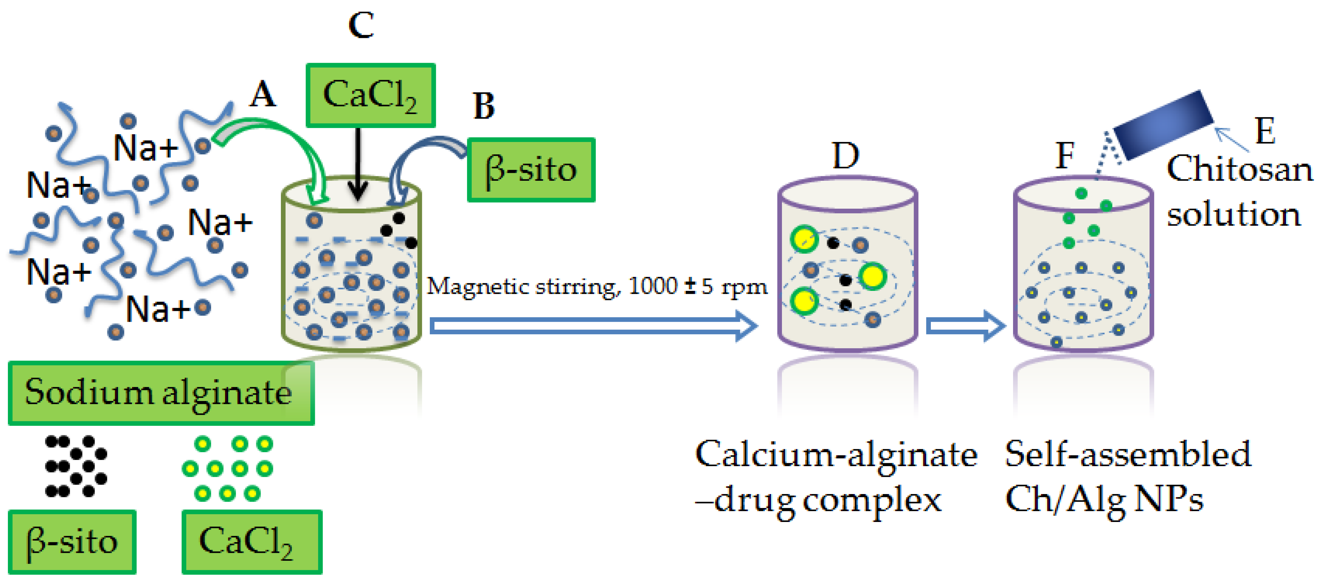
2.3. Alg/Ch/NPs Preparation
2.4. Formulation Characterization
2.4.1. Nanoparticle Size, Distribution Pattern, Zeta Potential and TEM
2.4.2. Entrapment Efficiency (EE) and Drug Loading (DL)
2.4.3. Differential Scanning Calorimetry
2.4.4. Thermogravimetric Analysis (TGA)
2.4.5. Fourier Transform Infrared Spectroscopy (FT-IR)
2.4.6. X-ray Diffraction Study
2.4.7. Drug Release Study
2.4.8. Ex Vivo Drug Permeation Study
2.4.9. MTT Assay
2.4.10. Pharmacokinetic Assessment
2.4.11. Radical Scavenging Assay
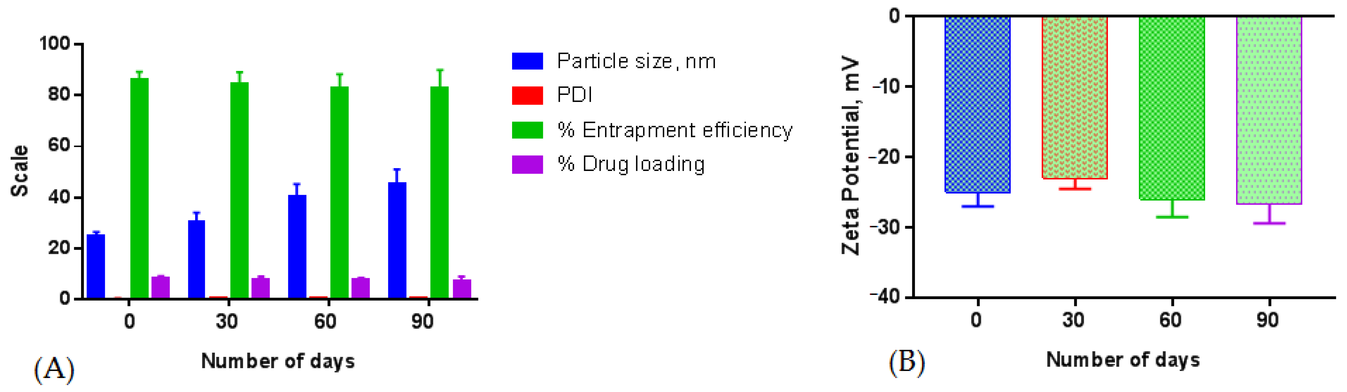
3. Formulation Stability
4. Data Analysis
5. Results and Discussion
5.1. Optimum Formulation
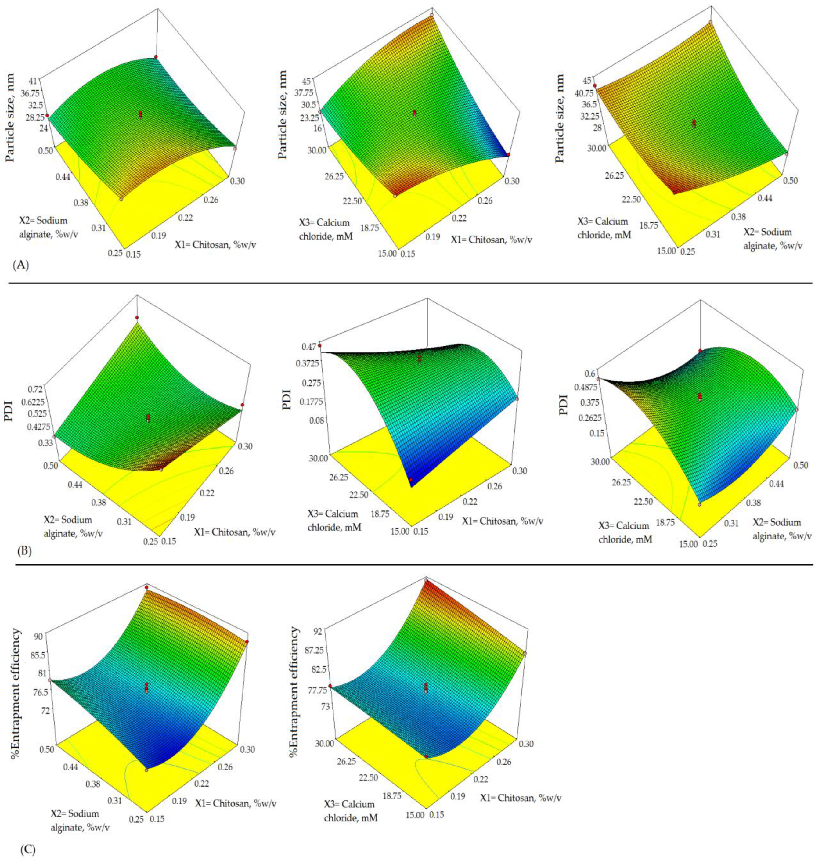
5.2. Impact of Formulation Variables on Y1
5.3. Impact of Formulation Variables on Y2
5.4. Impact of Formulation Variables on Y3
5.5. Validation and Optimum Checkpoint Analysis
5.6. Physico-Chemical Characterization of Formulation
5.6.1. % Entrapment and Loading Efficiency, Particle Size, PDI, Zeta Potential, and TEM
5.6.2. Thermal Analysis
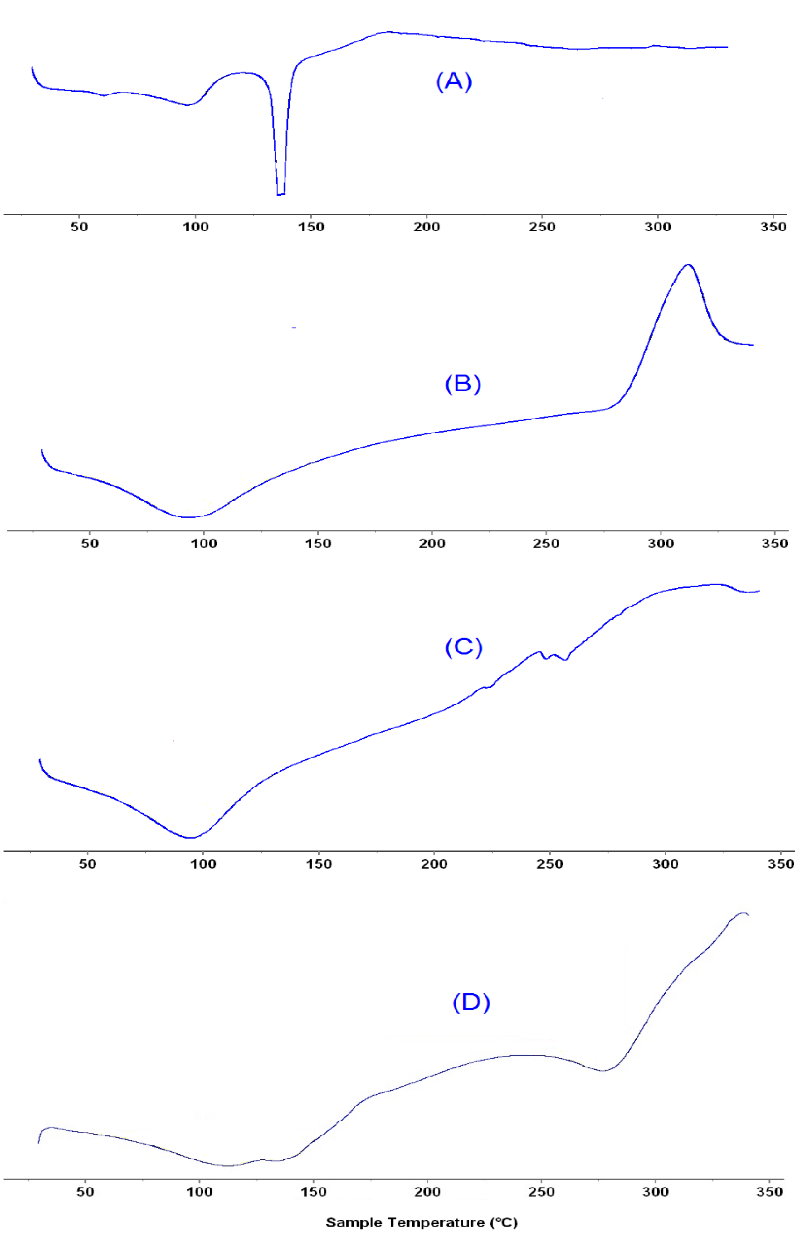
Differential Scanning Calorimetry
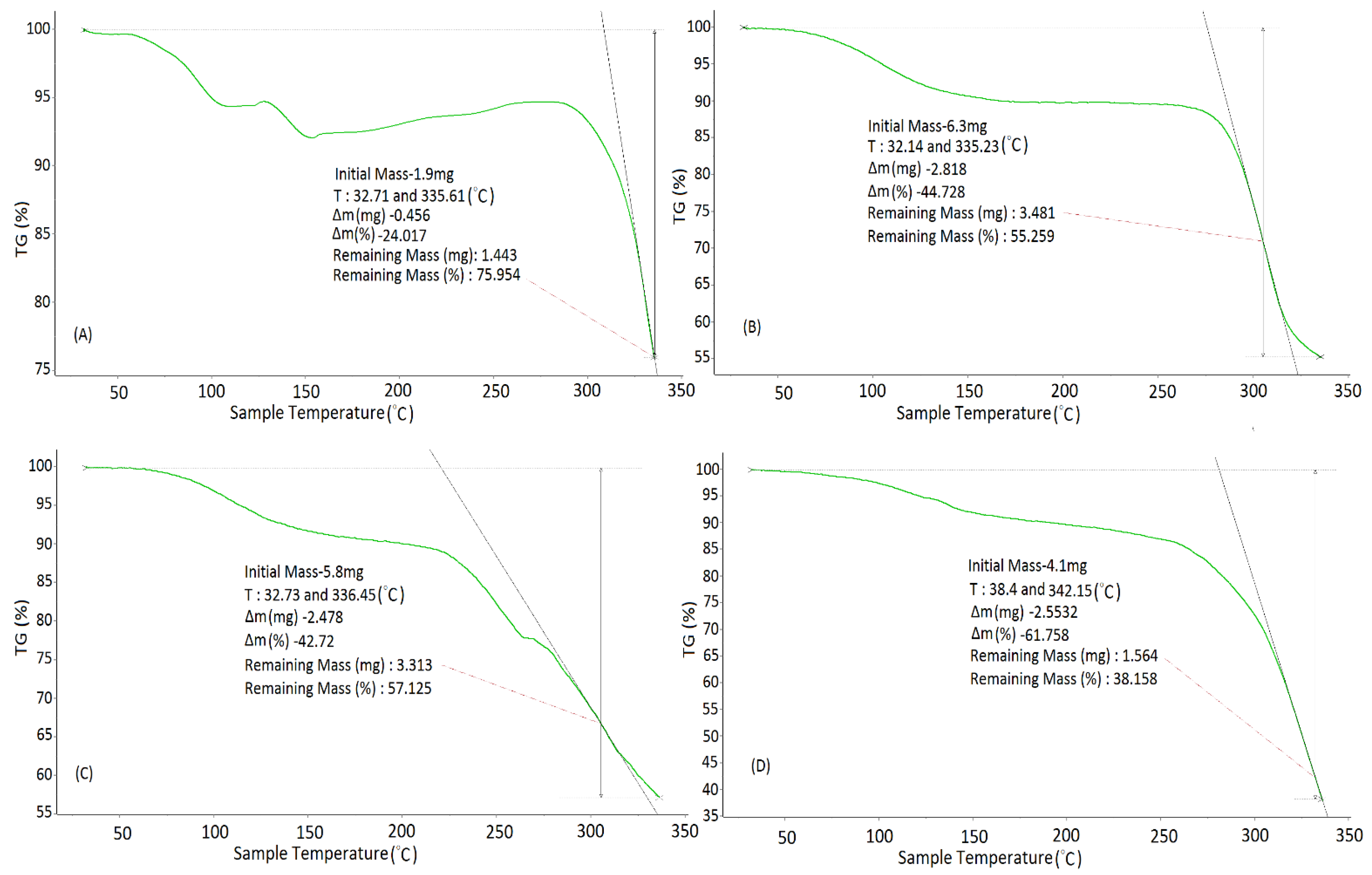
Thermogravimetric Analysis (TGA)
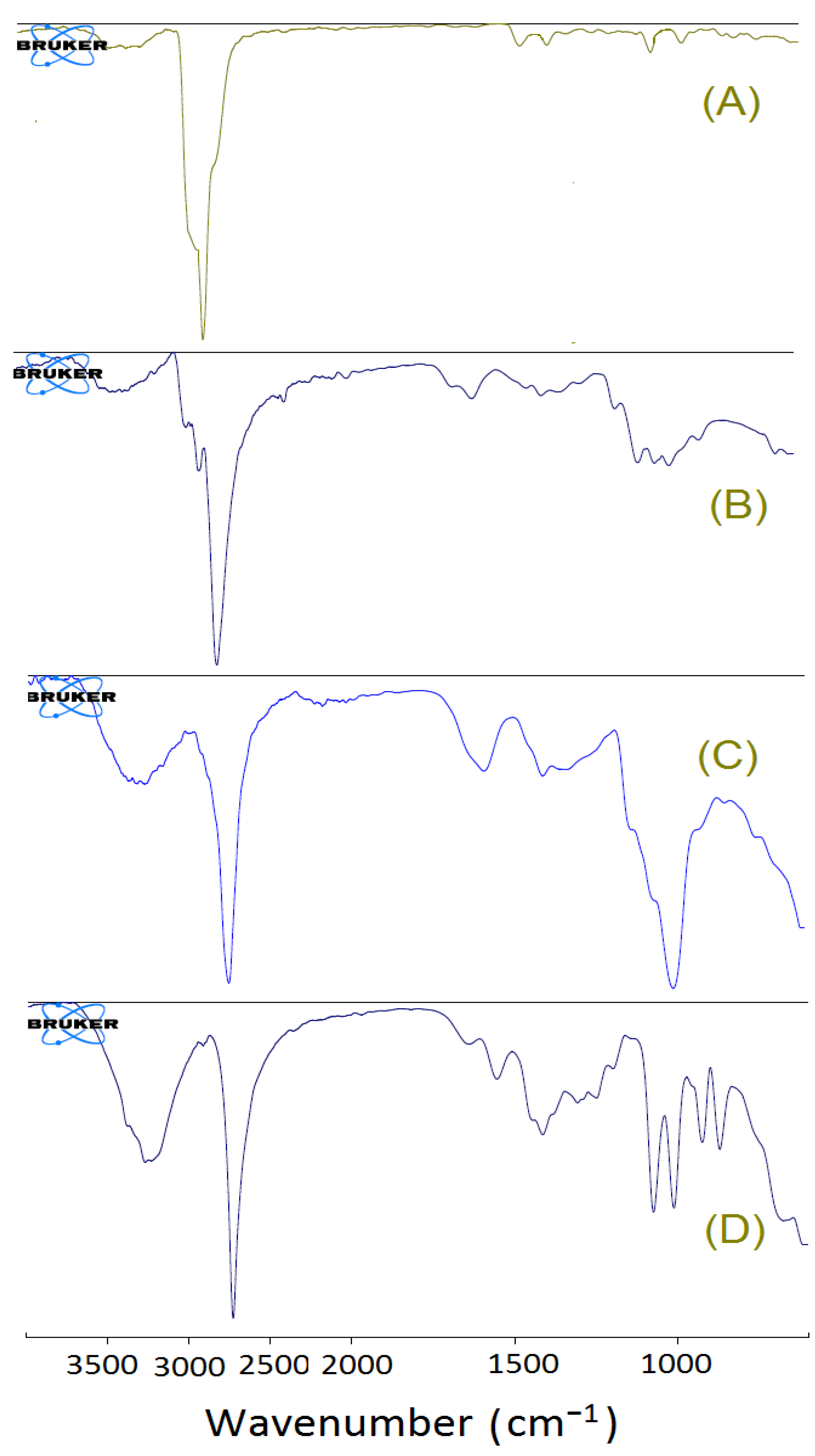
5.6.3. FT-IR Spectral Analysis
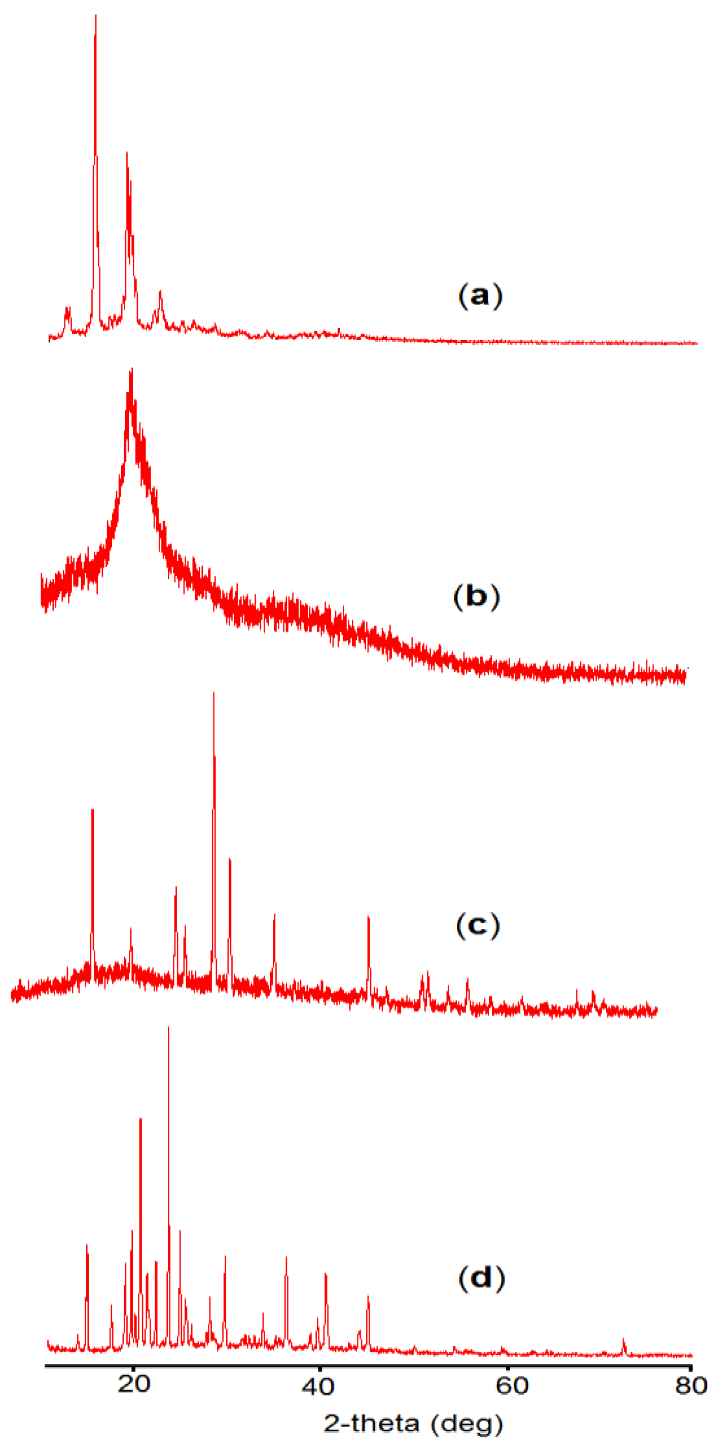
5.6.4. X-ray Diffraction Study
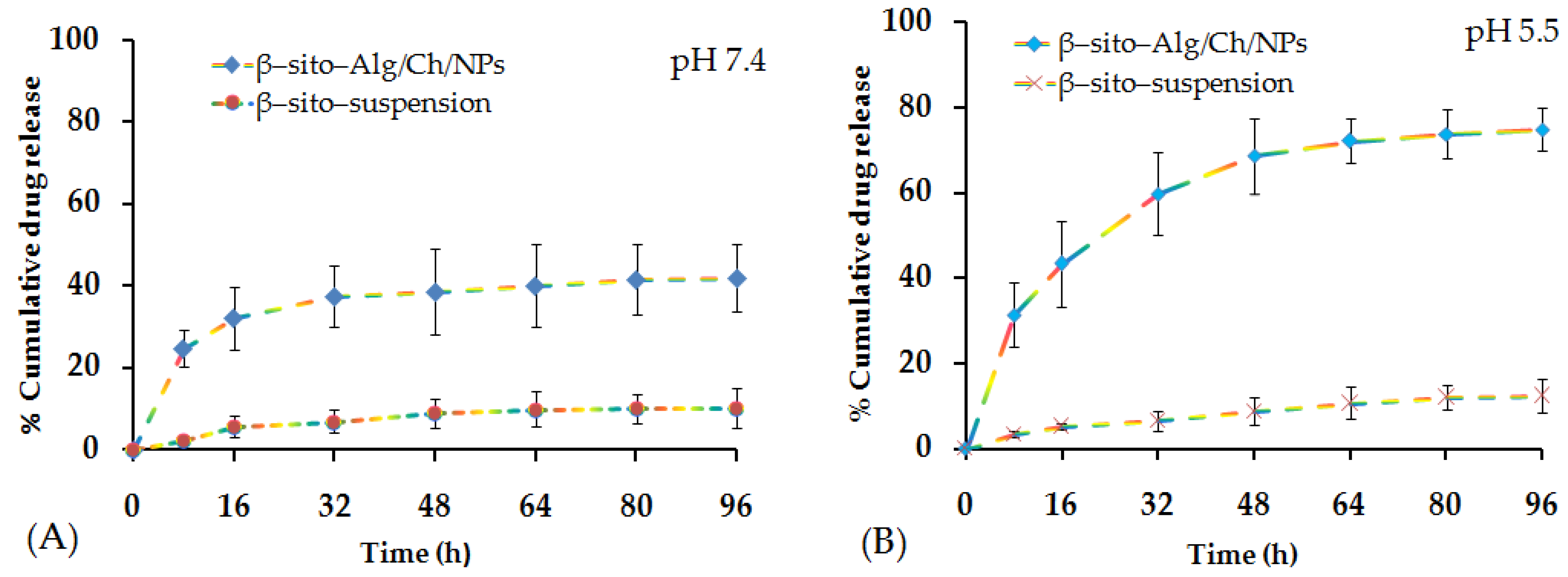
5.6.5. Release Studies and Kinetic Model
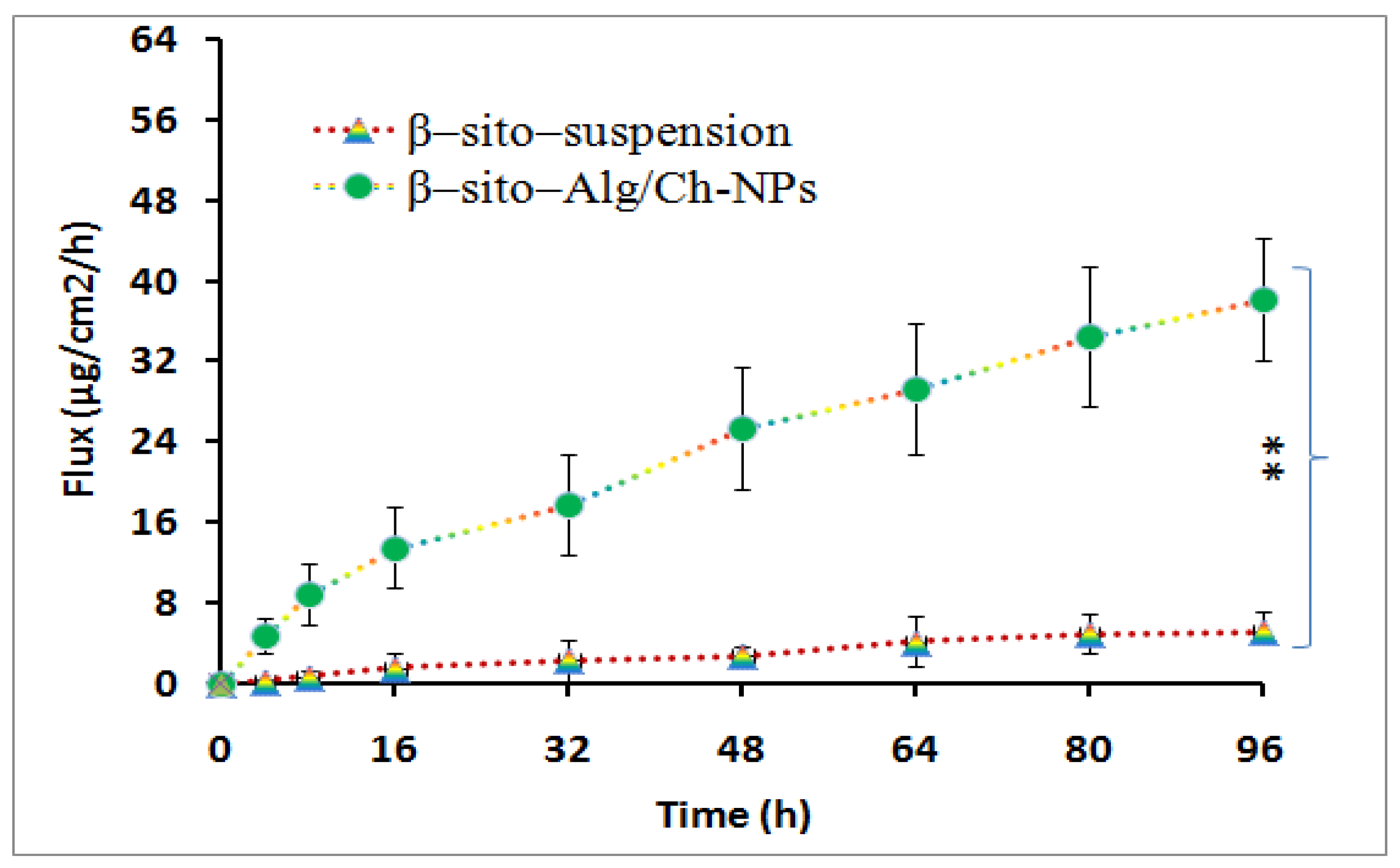
5.6.6. Intestinal Permeation Study
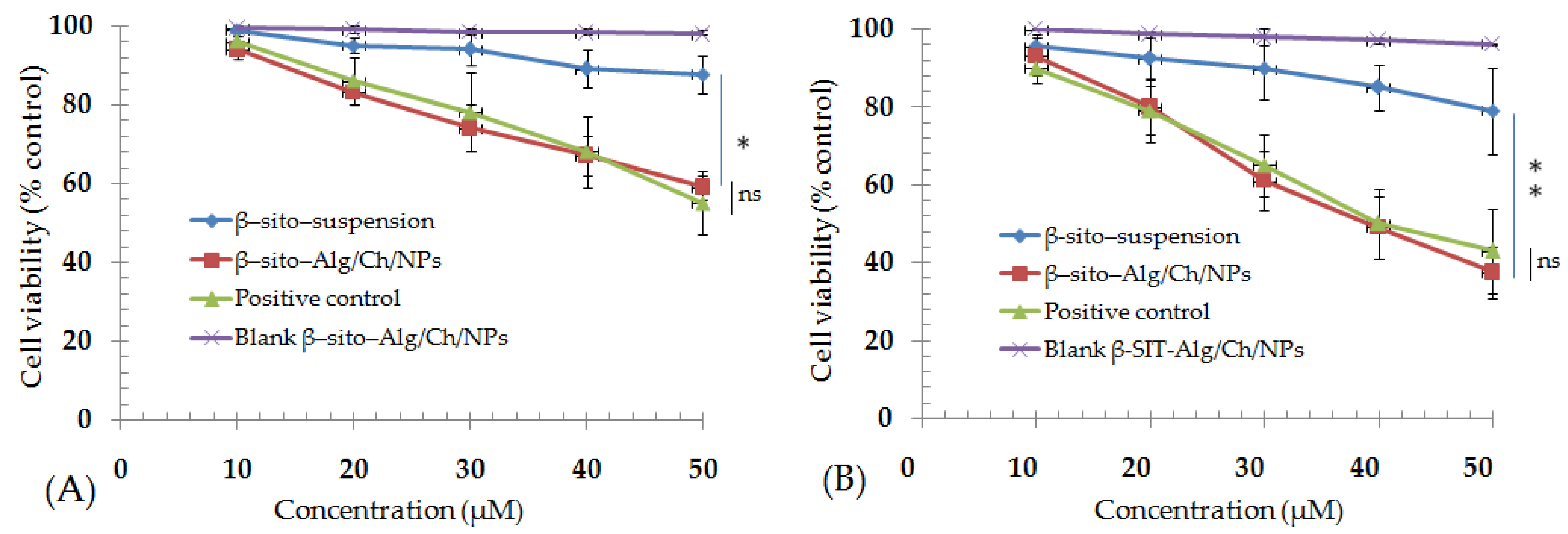
5.6.7. MTT Assay
5.6.8. Pharmacokinetic Assessment
5.6.9. Radical Scavenging Assay
5.6.10. Formulation Stability
6. Conclusions
Author Contributions
Funding
Institutional Review Board Statement
Informed Consent Statement
Data Availability Statement
Acknowledgments
Conflicts of Interest
References
- Zaheed, O.; Samson, J.; Dean, K. A bioinformatics approach to identify novel long, non-coding RNAs in breast cancer cell lines from an existing RNA-sequencing dataset. Noncoding RNA Res. 2020, 5, 48–59. [Google Scholar] [CrossRef] [PubMed]
- Siegel, R.L.; Miller, K.D.; Fuchs, H.E.; Jemal, A. Cancer statistics 2021. CA A Cancer J. Clin. 2021, 71, 7–33. [Google Scholar] [CrossRef] [PubMed]
- Ji, X.; Lu, Y.; Tian, H.; Meng, X.; Wei, M.; Cho, W.C. Chemoresistance mechanisms of breast cancer and their countermeasures. Biomed. Pharm. 2019, 114, 108800. [Google Scholar] [CrossRef] [PubMed]
- Bae, Y.H. Drug targeting and tumor heterogeneity. J. Control. Release 2009, 133, 2–3. [Google Scholar] [CrossRef] [PubMed]
- Patra, J.K.; Das, G.; Fraceto, L.F.; Campos, E.V.R.; Rodriguez-Torres, M.D.P.; Acosta-Torres, L.S.; Diaz-Torres, L.A.; Grillo, R.; Swamy, M.K.; Sharma, S.; et al. Nano based drug delivery systems: Recent developments and future prospects. J. Nanobiotechnol. 2018, 16, 71. [Google Scholar] [CrossRef] [PubMed]
- Din, F.U.; Aman, W.; Ullah, I.; Qureshi, O.S.; Mustapha, O.; Shafique, S.; Zeb, A. Effective use of nanocarriers as drug delivery systems for the treatment of selected tumors. Int. J. Nanomed. 2017, 12, 7291–7309. [Google Scholar] [CrossRef] [PubMed]
- Huda, S.; Alam, A.; Sharma, P.K. Smart nanocarriers-based drug delivery for cancer therapy: An innovative and developing strategy. J. Drug Deliv. Sci. Technol. 2020, 60, 102018. [Google Scholar] [CrossRef]
- Akhter, M.H.; Rizwanullah, M.; Ahmad, J.; Ahsan, M.J.; Mujtaba, A.; Amin, S. Nano carriers in advanced drug targeting: Setting novel paradigm in cancer therapeutics. Artif. Cells Nanomed. Biotechnol. 2018, 46, 873–884. [Google Scholar] [CrossRef]
- Rosenblum, D.; Joshi, N.; Tao, W.; Karp, J.M.; Peer, D. Progress and challenges towards targeted delivery of cancer therapeutics. Nat. Commun. 2018, 9, 1410. [Google Scholar] [CrossRef] [PubMed]
- Prabhakar, U.; Maeda, H.; Jain, R.K.; Sevick-Muraca, E.M.; Zamboni, W.; Farokhzad, O.C.; Barry, S.T.; Gabizon, A.; Grodzinski, P.; Blakey, D.C. Challenges and key considerations of the enhanced permeability and retention effect for nanomedicine drug delivery in oncology. Cancer Res. 2013, 73, 2412–2417. [Google Scholar] [CrossRef] [PubMed]
- Herdiana, Y.; Wathoni, N.; Shamsuddin, S.; Joni, I.M.; Muchtaridi, M. Chitosan-Based Nanoparticles of Targeted Drug Delivery System in Breast Cancer Treatment. Polymers 2017, 13, 1717. [Google Scholar] [CrossRef] [PubMed]
- Niang, P.M.; Huang, Z.; Dulong, V.; Souguir, Z.; Cerf, D.L.; Picton, L. Thermo-controlled rheology of electro-assembled polyanionic polysaccharide (alginate) and polycationic thermo-sensitive polymers. Carbohydr. Polym. 2017, 139, 67–74. [Google Scholar] [CrossRef] [PubMed]
- Patel, M.A.; AbouGhaly, M.H.H.; Schryer-Praga, J.V.; Chadwick, K. The effect of ionotropic gelation residence time on alginate cross-linking and properties. Carbohydr. Polym. 2017, 155, 362–371. [Google Scholar] [CrossRef]
- Karim, S.; Akhter, M.H.; Burzangi, A.S.; Alkreathy, H.; Alharthy, B.; Kotta, S.; Md, S.; Rashid, M.A.; Afzal, O.; Altamimi, A.S.A.; et al. Phytosterol-Loaded Surface-Tailored Bioactive-Polymer Nanoparticles for Cancer Treatment: Optimization, In Vitro Cell Viability, Antioxidant Activity, and Stability Studies. Gels 2022, 8, 219. [Google Scholar] [CrossRef]
- Akhter, M.H.; Kumar, S.; Nomani, S. Sonication tailored enhance cytotoxicity of naringenin nanoparticle in pancreatic cancer: Design, optimization, and in vitro studies. Drug Dev. Ind. Pharm. 2020, 46, 659–672. [Google Scholar] [CrossRef]
- Soni, K.; Mujtaba, A.; Akhter, M.H.; Zafar, A.; Kohli, K. Optimisation of ethosomal nanogel for topical nano-CUR and sulphoraphane delivery in effective skin cancer therapy. J. Microencapsul. 2019, 37, 91–108. [Google Scholar] [CrossRef]
- Md, S.; Alhakamy, N.A.; Aldawsari, H.M.; Husain, M.; Khan, N.; Alfaleh, M.A.; Asfour, H.Z.; Riadi, Y.; Bilgrami, A.L.; Akhter, M.H. Plumbagin-Loaded Glycerosome Gel as Topical Delivery System for Skin Cancer Therapy. Polymers 2021, 13, 923. [Google Scholar] [CrossRef] [PubMed]
- Md, S.; Alhakamy, N.A.; Neamatallah, T.; Alshehri, S.; Mujtaba, M.A.; Riadi, Y.; Radhakrishnan, A.K.; Khalilullah, H.; Gupta, M.; Akhter, M.H. Development, Characterization, and Evaluation of α-Mangostin-Loaded Polymeric Nanoparticle Gel for Topical Therapy in Skin Cancer. Gels 2021, 7, 230. [Google Scholar] [CrossRef] [PubMed]
- Takayasu, B.S.; Martins, I.R.; Garnique, A.M.B.; Miyamoto, S.; Machado-Santelli, G.M.; Uemi, M.; Onuki, J. Biological effects of an oxyphytosterol generated by β-Sitosterol ozonization. Arch. Biochem. Biophys. 2020, 696, 108654. [Google Scholar] [CrossRef] [PubMed]
- Christiansen, L.; Lähteenmäki, P.L.; Mannelin, M.R.; Seppänen-Laakso, T.E.; Hiltunen, R.V.; Yliruusi, J.K. Cholesterol-lowering effect of spreads enriched with microcrystalline plant sterols in hypercholesterolemic subjects. Eur. J. Nutr. 2001, 40, 66–73. [Google Scholar] [CrossRef] [PubMed]
- Moreau, R.A.; Nystrom, L.; Whitaker, B.D.; Winkler-Moser, J.K.; Baer, D.J.; Gebauer, S.K.; Hicks, K.B. Phytosterols and their Derivatives: Structural Diversity, Distribution, Metabolism, Analysis, and Health-Promoting Uses. Prog. Lipid Res. 2018, 70, 35–61. [Google Scholar] [CrossRef] [PubMed]
- Cheung, C.-L.; Ho, D.K.-C.; Sing, C.-W.; Tsoi, M.-F.; Cheng, V.K.-F.; Lee, G.K.-Y.; Ho, Y.-N.; Cheung, B.M.Y. Randomized Controlled Trial of the Effect of Phytosterols-Enriched Low-Fat Milk on Lipid Profile in Chinese. Sci. Rep. 2017, 7, 41084. [Google Scholar] [CrossRef] [PubMed]
- Blom, W.A.M.; Koppenol, W.P.; Hiemstra, H.; Stojakovic, T.; Scharnagl, H.; Trautwein, E.A. A Low-Fat Spread with Added Plant Sterols and Fish Omega-3 Fatty Acids Lowers Serum Triglyceride and LDL-Cholesterol Concentrations in Individuals with Modest Hypercholesterolaemia and Hypertriglyceridaemia. Eur. J. Nutr. 2019, 58, 1615–1624. [Google Scholar] [CrossRef] [PubMed]
- Paniagua-Pérez, R.; Flores-Mondragón, G.; Reyes-Legorreta, C.; Herrera-López, B.; Cervantes-Hernández, I.; Madrigal-Santillán, O.; Morales-González, J.A.; Álvarez-González, I.; Madrigal-Bujaidar, E. Evaluation of the anti-inflammatory capacity of beta-sitosterol in rodent assays. Afr. J. Tradit. Complement. Altern. Med. 2016, 14, 23–130. [Google Scholar] [CrossRef]
- Gupta, R.; Sharma, A.K.; Dobhal, M.P.; Sharma, M.C.; Gupta, R.S. Antidiabetic and antioxidant potential of β-sitosterol in streptozotocin-induced experimental hyperglycemia. J. Diabetes 2011, 3, 29–37. [Google Scholar] [CrossRef]
- Yin, Y.; Liu, X.; Liu, J.; Cai, E.; Zhao, Y.; Lic, H.; Zhanga, L.; Lic, P.; Gao, Y. The effect of beta-sitosterol and its derivatives on depression by the modification of 5-HT, DA and GABA-ergic systems in mice. RSC Adv. 2018, 8, 671–680. [Google Scholar] [CrossRef]
- Subramaniam, S.; Keerthiraja, M.; Sivasubramanian, A. Synergistic antibacterial action of β-sitosterol-d-glucopyranoside isolated from Desmostachya bipinnata leaves with antibiotics against common human pathogens. Rev. Bras. Farmacogn. 2014, 24, 44–50. [Google Scholar] [CrossRef]
- Olaiya, C.O.; Esan, A.M.; Alabi, T.D. Ameliorative effects of β-sitosterol on some biochemical indices of hypertension in wistar albino rats. Afr. J. Med. Med. Sci. 2014, 43, 157–166. [Google Scholar]
- Sayeed, M.S.B.; Ameen, S.S. Beta-Sitosterol: A Promising but Orphan Nutraceutical to Fight Against Cancer. Nut. Cancer 2015, 67, 1216–1222. [Google Scholar] [CrossRef]
- Fraile, L.; Crisci, E.; Córdoba, L.; Navarro, M.A.; Osada, J.; Montoya, M. Immunomodulatory properties of Beta-sitosterol in pig immune responses. Int. Immunopharmacol. 2012, 13, 316–321. [Google Scholar] [CrossRef]
- Moosavi, B.; Liu, S.; Wang, N.N.; Zhu, X.L.; Yang, G.F. The anti-fungal β-sitosterol targets the yeast oxysterol-binding protein Osh4. Pest. Manag. Sci. 2020, 76, 704–711. [Google Scholar] [CrossRef] [PubMed]
- Abbas, M.M.; Al-Rawi, N.; Abbas, M.A.; Al-Khateeb, I. Naringenin potentiated β-sitosterol healing effect on the scratch wound assay. Res. Pharm. Sci. 2019, 14, 566–573. [Google Scholar] [CrossRef] [PubMed]
- Kausar, H.; Mujeeb, M.; Ahad, A.; Moolakkadath, T.; Aqil, M.; Ahmad, A.; Akhter, M.H. Optimization of ethosomes for topical thymoquinone delivery for the treatment of skin acne. J. Drug Deliv. Sci. Technol. 2019, 49, 177–187. [Google Scholar] [CrossRef]
- Thai, H.; Nguyen, C.T.; Thach, L.T.; Tran, M.T.; Mai, H.D.; Nguyen, T.T.T.; Le, G.D.; Can, M.V.; Tran, L.D.; Bach, G.L.; et al. Characterization of chitosan/alginate/lovastatin nanoparticles and investigation of their toxic effects in vitro and in vivo. Sci. Rep. 2020, 10, 909. [Google Scholar] [CrossRef]
- Katuwavila, N.P.; Perera, A.D.L.C.; Samarakoon, S.R.; Soysa, P.; Karunaratne, V.; Amaratunga, G.A.J.; Karunaratne, D.N. Chitosan-alginate nanoparticle system efficiently delivers doxorubicin to MCF-7 Cells. J. Nanomater. 2016, 2016, 3178904. [Google Scholar] [CrossRef]
- Li, P.; Dai, Y.N.; Zhang, J.P.; Wang, A.Q.; Wei, Q. Chitosan-alginate nanoparticles as a Q. novel drug delivery system for nifedipine. Int. J. Biomed. Sci. 2008, 4, 221–228. [Google Scholar]
- Ayaz, M.; Junaid, M.; Ullah, U.F.; Subhan, F.; Sadiq, A.; Ali, G.; Ovais, M.; Shahid, M.; Ahmad, A.; Wadood, A.; et al. Anti-Alzheimer′s Studies on β-Sitosterol Isolated from Polygonum hydropiper L. Front. Pharmacol. 2017, 8, 697. [Google Scholar] [CrossRef]
- ICH Q1A(R2). Stability Testing Guidelines: Stability Testing of New Drug Substances and Products; ICH Steering Committee: Amsterdam, The Netherlands, 2003. [Google Scholar]
- Akhter, M.H.; Ahmad, A.; Ali, J.; Mohan, G. Formulation and Development of CoQ10-Loaded s-SNEDDS for Enhancement of Oral Bioavailability. J. Pharm. Innov. 2014, 9, 121–131. [Google Scholar] [CrossRef]
- Ruktanonchai, U.; Bejrapha, P.; Sakulkhu, U.; Opanasopit, P.; Bunyapraphatsara, N.; Junyaprasert, V.; Puttipipatkhachorn, S. Physicochemical Characteristics, Cytotoxicity, and Antioxidant Activity of Three Lipid Nanoparticulate Formulations of Alpha-lipoic Acid. AAPS PharmSciTech 2009, 10, 227. [Google Scholar] [CrossRef]
- Daemi, H.; Barikani, M. Synthesis and Characterization of Calcium Alginate Nanoparticles, Sodium Homopolymannuronate Salt and Its Calcium Nanoparticles. Sci. Iran. 2012, 19, 2023–2028. [Google Scholar] [CrossRef]
- Borges, O.; Cordeiro-Da-Silva, A.; Romeijn, S.G.; Amidi, M.; de Sousa, A.; Borchard, G.; Junginger, H.E. Uptake studies in rat Peyer’s patches, cytotoxicity and release studies of alginate coated chitosan nanoparticles for mucosal vaccination. J. Control. Release 2006, 114, 348–358. [Google Scholar] [CrossRef] [PubMed]
- Caetano, L.A.; Almeida, A.J.; Gonçalves, L.M. Effect of Experimental Parameters on Alginate/Chitosan Microparticles for BCG Encapsulation. Mar. Drugs 2016, 14, 90. [Google Scholar] [CrossRef] [PubMed]
- Sarmento, B.; Ribeiro, A.J.; Veiga, F.; Ferreira, D.C.; Neufeld, R.J. Insulin-loaded nanoparticles are prepared by alginate iono-tropic pre-gelation followed by chitosan polyelectrolyte complexation. J. Nanosci. Nanotechnol. 2007, 7, 2833–2841. [Google Scholar] [CrossRef]
- Motwari, S.K.; Chopra, S.; Talegaonkar, S.; Kohli, K.; Ahmad, F.J.; Khar, R.K. Chitosan–sodium alginate nanoparticles as submicroscopic reservoirs for ocular delivery: Formulation, optimisation and in vitro characterisation. Eur. J. Pharm. Biopharm. 2008, 68, 513–525. [Google Scholar]
- Szekalska, M.; Sosnowska, K.; Zakrzeska, A.; Kasacka, I.; Lewandowska, A.; Winnicka, K. The Influence of Chitosan Cross-linking on the Properties of Alginate Microparticles with Metformin Hydrochloride—In Vitro and In Vivo Evaluation. Molecules 2017, 22, 182. [Google Scholar] [CrossRef] [PubMed]
- Azevedo, M.A.; Bourbon, A.I.; Vicente, A.A.; Cerqueira, M.A. Alginate/chitosan nanoparticles for encapsulation and controlled release of vitamin B2. Int. J. Biol. Macromol. 2014, 71, 141–146. [Google Scholar] [CrossRef]
- Tummino, M.L.; Magnacca, G.; Cimino, D.; Laurenti, E.; Nisticò, R. The Innovation Comes from the Sea: Chitosan and Alginate Hybrid Gels and Films as Sustainable Materials for Wastewater Remediation. Int. J. Mol. Sci. 2020, 21, 550. [Google Scholar] [CrossRef]
- Leonardi, M.; Caruso, G.M.; Carroccio, S.C.; Boninelli, S.; Curcuruto, G.; Zimbone, M.; Allegra, M.; Torrisi, B.; Ferlitoe, F.; Miritello, M. Smart nanocomposites of chitosan/alginate nanoparticles loaded with copper oxide as alternative nanofertilizers. Environ. Sci. Nano 2021, 8, 174–187. [Google Scholar] [CrossRef]
- Kavithaa, K.; Paulpandi, M.; Ramya, S.; Ramesh, M.; Balachandar, V.; Ramasamy, K.; Narayanasamy, A. Sitosterol-fabricated chitosan nanocomplex induces apoptotic cell death through mitochondrial dysfunction in lung cancer animal model: An enhanced synergetic drug delivery system for lung cancer therapy. New J. Chem. 2021, 45, 9251–9263. [Google Scholar] [CrossRef]
- Li, L.; Li, J.; Si, S.; Wang, L.; Shi, C.; Sun, Y.; Liang, Z.; Mao, S. Efect of formulation variables on in vitro release of a water-soluble drug from chitosan-sodium alginate matrix tablets. Asia J. Pharm. Sci. 2015, 10, 314–321. [Google Scholar]
- Sorasitthiyanukarn, F.N.; Muangnoi, C.; Bhuket, P.R.N.; Rojsitthisak, P.; Rojsitthisak, P. Chitosan/alginate nanoparticles as a promising approach for oral delivery of curcumin diglutaric acid for cancer treatment. Mat. Sci. Eng. C 2018, 93, 178–190. [Google Scholar] [CrossRef]
- Zhang, F.; Liu, Z.; He, X.; Li, Z.; Shi, B.; Cai, F. β–sitosterol-loaded solid lipid nanoparticles ameliorate complete Freund’s adjuvant-induced arthritis in rats: Involvement of NF-κB and HO-1/Nrf-2 pathway. Drug Deliv. 2020, 27, 1329–1341. [Google Scholar] [CrossRef]
- Zheng, G.; Zheng, M.; Yang, B.; Fu, H.; Li, Y. Improving breast cancer therapy using doxorubicin loaded solid lipid nanoparticles: Synthesis of a novel arginine-glycine-aspartic tripeptide conjugated, pH sensitive lipid and evaluation of the nanomedicine in vitro and in vivo. Biomed. Pharmacoth. 2019, 116, 109006. [Google Scholar] [CrossRef]
- Akhter, M.H.; Beg, S.; Tarique, M.; Malik, A.; Afaq, S.; Choudhry, H.; Hosawi, S. Receptor-based targeting of engineered nanocarrier against solid tumors: Recent progress and challenges ahead. Biochim. Biophys. Acta (BBA)-Gen. Subj. 2021, 1865, 129777. [Google Scholar] [CrossRef]
- Akhter, M.H.; Amin, S. An Investigative Approach to Treatment Modalities for Squamous Cell Carcinoma of Skin. Curr. Drug Deliv. 2017, 14, 597–612. [Google Scholar] [CrossRef] [PubMed]
- Akhter, M.H.; Khalilullah, H.; Gupta, M.; Alfaleh, M.A.; Alhakamy, N.A.; Riadi, Y.; Md, S. Impact of Protein Corona on the Biological Identity of Nanomedicine: Understanding the Fate of Nanomaterials in the Biological Milieu. Biomedicines 2021, 9, 1496. [Google Scholar] [CrossRef]
- Mohammad, N.A.; Zaidel, D.N.A.; Muhamad, I.I.; Hamid, M.A.; Yaakob, H.; Jusoh, Y.M.M. Optimization of the antioxidant-rich xanthone extract from mangosteen (Garcinia mangostana L.) pericarp via microwave-assisted extraction. Heliyon 2019, 5, e02571. [Google Scholar] [CrossRef]


| Factors | Levels Used | ||
|---|---|---|---|
| Low (−1) | Medium (0) | High (+1) | |
| X1: Chitosan, %w/v | 0.15 | 0.22 | 0.30 |
| X2: Sodium alginate, %w/v | 0.25 | 0.38 | 0.50 |
| X3: Calcium chloride, mM | 15 | 22.5 | 30 |
| Responses | |||
| Y1: Particle size, nm | Minimize | ||
| Y2: PDI | Minimize | ||
| Y3: Entrapment efficiency, % | Maximize | ||
| Runs | Independent Variables | Responses | ||||
|---|---|---|---|---|---|---|
| X1, %w/v | X2, %w/v | X3, mM | Y1, nm | Y2 | Y3, % | |
| * 1 | 0.15 | 0.38 | 15.00 | 21.00 | 0.120 | 77.00 |
| 2 | 0.22 | 0.50 | 30.00 | 42.00 | 0.310 | 78.00 |
| 3 | 0.30 | 0.38 | 30.00 | 40.00 | 0.61 | 91.00 |
| * 4 | 0.22 | 0.25 | 15.00 | 45.00 | 0.204 | 72.00 |
| 5 | 0.22 | 0.38 | 22.00 | 34.00 | 0.380 | 75.00 |
| * 6 | 0.22 | 0.38 | 22.00 | 34.90 | 0.420 | 76.00 |
| 7 | 0.30 | 0.50 | 22.00 | 31.00 | 0.570 | 89.00 |
| 8 | 0.30 | 0.38 | 15.00 | 42.00 | 0.510 | 86.00 |
| 9 | 0.22 | 0.25 | 30.00 | 43.00 | 0.530 | 76.00 |
| 10 | 0.15 | 0.25 | 22.50 | 23.00 | 0.260 | 73.00 |
| * 11 | 0.15 | 0.50 | 22.50 | 16.00 | 0.330 | 79.00 |
| 12 | 0.22 | 0.38 | 22.50 | 32.00 | 0.340 | 78.00 |
| 13 | 0.22 | 0.38 | 22.50 | 33.00 | 0.360 | 76.30 |
| * 14 | 0.22 | 0.38 | 22.50 | 33.60 | 0.402 | 77.00 |
| 15 | 0.22 | 0.50 | 15.00 | 35.00 | 0.310 | 76.00 |
| 16 | 0.30 | 0.25 | 22.00 | 38.00 | 0.730 | 88.00 |
| 17 | 0.15 | 0.38 | 30.00 | 27.00 | 0.350 | 77.60 |
| Model | R2 | Adjusted R2 | Predicted R2 | SD | CV% | Desirability |
|---|---|---|---|---|---|---|
| Response: Y1 | 0.851 | |||||
| Quadratic | 0.9857 | 0.9674 | 0.8510 | 1.41 | 4.21 | |
| 2FI | 0.8012 | 0.6819 | 0.0006 | 4.39 | - | |
| Linear | 0.2448 | 0.0705 | −0.5756 | 7.50 | - | |
| Cubic | ||||||
| Response: Y2 | 0.851 | |||||
| Quadratic | 0.9772 | 0.9480 | 0.7845 | 0.036 | 9.96 | |
| 2FI | 0.5503 | 0.2805 | −1.2548 | 0.13 | - | |
| Linear | 0.1480 | −0.0486 | −0.7686 | 0.16 | - | |
| Cubic | 0.032 | 0.9897 | 0.9588 | - | - | |
| Response: Y3 | 0.851 | |||||
| Quadratic | 0.9884 | 0.9736 | 0.9545 | 0.93 | 1.17 | |
| 2FI | 0.6350 | 0.4161 | −0.5658 | 4.36 | - | |
| Linear | 0.6118 | 0.5223 | 0.2619 | 3.94 | - | |
| Cubic |
| Independent Variables | Optimized Composition | Predicted Response | Observed Response | % Error | ||||
|---|---|---|---|---|---|---|---|---|
| Y1, nm | Y2 (%) | Y3 (%) | Y1, nm | Y2 | Y3 (%) | |||
| X1:X2:X3 | 0.15%:0.50%:15.07 mM | 17.14 | 0.240 | 79.19 | 25 ± 1 | 0.231 | 86 ± 3 | Y1 = 4.7 Y2 = 3.8 Y3 = 8.7 |
| Formulation | AUC0−t (μg × h/mL) | Tmax (h) | Cmax | t1/2 (h) | ke (h−1) |
|---|---|---|---|---|---|
| β–sito–Alg/Ch/NPs | 1080 ± 1 | 4 | 180 ± 0.02 | 5 ± 0.1 | 0.2 ± 0.01 |
| β–sito–suspension | 317 ± 1 | 4 | 56 ± 0.2 | 2 ± 0.3 | 0.3 ± 0.02 |
Publisher’s Note: MDPI stays neutral with regard to jurisdictional claims in published maps and institutional affiliations. |
© 2022 by the authors. Licensee MDPI, Basel, Switzerland. This article is an open access article distributed under the terms and conditions of the Creative Commons Attribution (CC BY) license (https://creativecommons.org/licenses/by/4.0/).
Share and Cite
Afzal, O.; Akhter, M.H.; Ahmad, I.; Muzammil, K.; Dawria, A.; Zeyaullah, M.; Altamimi, A.S.A.; Khalilullah, H.; Mir Najib Ullah, S.N.; Rahman, M.A.; et al. A β–Sitosterol Encapsulated Biocompatible Alginate/Chitosan Polymer Nanocomposite for the Treatment of Breast Cancer. Pharmaceutics 2022, 14, 1711. https://0-doi-org.brum.beds.ac.uk/10.3390/pharmaceutics14081711
Afzal O, Akhter MH, Ahmad I, Muzammil K, Dawria A, Zeyaullah M, Altamimi ASA, Khalilullah H, Mir Najib Ullah SN, Rahman MA, et al. A β–Sitosterol Encapsulated Biocompatible Alginate/Chitosan Polymer Nanocomposite for the Treatment of Breast Cancer. Pharmaceutics. 2022; 14(8):1711. https://0-doi-org.brum.beds.ac.uk/10.3390/pharmaceutics14081711
Chicago/Turabian StyleAfzal, Obaid, Md Habban Akhter, Irfan Ahmad, Khursheed Muzammil, Adam Dawria, Mohammad Zeyaullah, Abdulmalik S. A. Altamimi, Habibullah Khalilullah, Shehla Nasar Mir Najib Ullah, Mohammad Akhlaquer Rahman, and et al. 2022. "A β–Sitosterol Encapsulated Biocompatible Alginate/Chitosan Polymer Nanocomposite for the Treatment of Breast Cancer" Pharmaceutics 14, no. 8: 1711. https://0-doi-org.brum.beds.ac.uk/10.3390/pharmaceutics14081711