Review

Selenium-Containing Agents Acting on Cancer—A New Hope?

by
Sabrina Garbo
1,†,
Silvia Di Giacomo
2,†,
Dorota Łażewska
3,
Ewelina Honkisz-Orzechowska
3,
Antonella Di Sotto
2,
Rossella Fioravanti
4,
Clemens Zwergel
4,*,† and
Cecilia Battistelli
1,*,†
1
Department of Molecular Medicine, Sapienza University of Rome, Viale Regina Elena 324, 00161 Rome, Italy
2
Department of Physiology and Pharmacology “V. Erspamer”, Sapienza University of Rome, Piazzale Aldo Moro 5, 00185 Rome, Italy
3
Department of Technology and Biotechnology of Drugs, Faculty of Pharmacy, Jagiellonian University Medical College in Kraków, Medyczna 9, 30-688 Kraków, Poland
4
Department of Drug Chemistry and Technologies, Sapienza University of Rome, Piazzale Aldo Moro 5, 00185 Rome, Italy
*
Authors to whom correspondence should be addressed.
These authors contributed equally to this work.
Submission received: 27 November 2022 / Revised: 18 December 2022 / Accepted: 24 December 2022 / Published: 28 December 2022

Abstract

:
Selenium-containing agents are more and more considered as an innovative potential treatment option for cancer. Light is shed not only on the considerable advancements made in understanding the complex biology and chemistry related to selenium-containing small molecules but also on Se-nanoparticles. Numerous Se-containing agents have been widely investigated in recent years in cancer therapy in relation to tumour development and dissemination, drug delivery, multidrug resistance (MDR) and immune system-related (anti)cancer effects. Despite numerous efforts, Se-agents apart from selenocysteine and selenomethionine have not yet reached clinical trials for cancer therapy. The purpose of this review is to provide a concise critical overview of the current state of the art in the development of highly potent target-specific Se-containing agents.

1. Introduction

In the early 19th century, Jöns Jacob Berzelius discovered selenium (Se), a chalcogen that belongs to group 16 along with oxygen, sulphur, tellurium, and polonium [1]. It is widely believed that elementary Se and compounds containing Se can cause mental illness, cancer, and other diseases [2]. In the 1970s, D. Forst discussed the, at that time, confusing effects of Selenium in cancer biology in his critical paper, coining the term “Selenophobia” [3]. However, today, it is well accepted that Se has both protective and toxic effects on organisms, such as the nervous system and the heart [2,4]. Se can safely be incorporated into a variety of organic compounds for the treatment of various diseases [2,4].
Chemistry-wise, Se has several oxidation states and can be oxidized and reduced by redox agents more easily than sulphur (S). Thus, Se can act as a nucleophilic or electrophilic agent by donating or accepting electrons [1]. Since Se behaves in this way, it is suitable for use in chemistry and biology. Medicinal chemistry, biochemistry, and biology are all interconnected areas where selenium and, specifically, seleno-compounds are considered promising candidates for drug discovery [4]. Since the 1980s, a variety of selenium-containing compounds have been described to exhibit chemopreventive and antioxidant properties with a number of positive outcomes [5]. As a result, considerable advancements have been made in understanding this element’s complex biology and chemistry, showing that Se plays a crucial role in various biological processes [2,6]. It can also exert toxic effects [2,6].
Selenoproteins (SePs) are particularly interesting, as they impact the health of many organs by maintaining broader homeostasis through regulation of the thyroid hormone level, endoplasmic reticulum stress and antioxidant defence [7], but also immune and inflammatory responses. The unique nature of selenium is that it is folded into its protein while the protein molecule is still being made [8]. SePs carry the rare selenium-containing selenocysteine 1 residue. This is of great importance, as it means that the incorporation of this 21st amino acid, selenocysteine 1, is genetically regulated, and through this process, it is possible to modify the activity of different SePs. Among the roughly 40 Se-dependent proteins discovered thus far with a variety of biological functions, glutathione peroxidase (GPx) [9,10] is the most studied and explored.
The purpose of this review is to provide a concise overview of selenium-containing small molecules as well as Se-nanoparticles, which have been widely investigated in recent years in cancer therapy, spanning from tumour development and dissemination, drug delivery to multidrug resistance (MDR) and immune system-related (anti)cancer effects.

2. Therapeutic Properties of Selenium as a Trace Element

Selenium is a solid non-metal with acidic properties (chemically resembling sulphur) that occurs essentially in numerous allotropic varieties. It occurs in nature and in organisms in two forms (inorganic and organic) and in four oxidation states: −II, 0, +IV and +VI. The most important inorganic and organic forms of selenium are shown in Figure 1 [11]. Inorganic selenium can be found in minerals in the forms of selenides (most commonly), selenates and selenites [12]. Selenium minerals are extremely rare. Only 123 such species have been described thus far [12]. In nature, organically bound selenium mainly occurs in selenoamino acids (mainly), selenopeptides and SePs [11]. Other organoselenium compounds may belong to various chemical groups. The most important forms are shown in Figure 2 [11,13]. Such compounds are usually characterised by higher bioavailability and lower toxicity than inorganic compounds.
The incorporation of selenium into various organoselenium compounds (synthetic and natural) often results in beneficial pharmacological activity. The type of selenium linkage in an organic molecule significantly influences the biological activity of these compounds, i.e., whether they exhibit a beneficial effect (reactive oxygen species (ROS) uptake, oxidative capacity) or a toxic effect (ROS induction or release of elemental Se). The biological activity of selenium compounds is mainly related to antioxidant properties, i.e., the ability to scavenge ROS, and depends on the ability of compounds to convert into the corresponding selenols and on the rate at which this process takes place. Selenols show the most potent antioxidant properties among selenium derivatives. They readily dissociate to form the RSe anion at physiological pH and are active in a GPx-like catalytic cycle [13]. The cytotoxic effect of organic selenium compounds relates to their ability to release elemental Se (0) that can interact with lipoproteins to form cytotoxic cysteine●selenium conjugates. This effect is usually missing in selenium heterocycles with selenium in sp2 hybridisation, as such compounds are stable and do not show the ability to release Se (0) [13].
Selenium is an essential micronutrient needed for the proper functioning of the body. It is a component of two key amino acids: selenocysteine 1 and selenomethionine 2, which, in turn, build up essential enzymes for the human body: glutathione peroxidases (GPxs), thioredoxin reductases, iodothyronine deionidases or SePs such as selenoprotein O, selenoprotein P or selenoprotein R [14,15]. SePs are involved in many metabolic and functional pathways, and most of them catalyse oxidation–reduction reactions. SePs P, mainly present in plasma, are also highly expressed in the brain. They are thought to be transporters of selenium from the liver to the brain [16], and recent studies have shown their involvement in the pathogenesis of diseases such as Alzheimer’s and Parkinson’s [17,18].
Selenium can be classified as a hermetic element because it is beneficial at low doses and harmful at high doses. Both of these conditions (deficiency and excess) are bad and can eventually lead to health problems, causing certain diseases and even death, especially when they occur over a long period. The main health problems that can arise from selenium deficiency or excess are shown in Table 1 [19,20,21]. In some cases, both selenium deficiency and excess can lead to the same health problems, i.e., increased mortality, type 2 diabetes or prostate cancer [19]. Selenium deficiency may affect more than one billion people worldwide and especially affects the expression of SePs in the body [22]. Thus, selenium deficiency can affect life expectancy, accelerating the ageing process or increasing susceptibility to immunological or cancerous diseases [21] and causing various cardiovascular diseases, including cardiomyopathies [7]. Moreover, lower (than in healthy) selenium levels were observed in patients with a more severe course of COVID-19 (SARS-CoV-2) [23]. Plasma selenium amount is thought to be the most reliable measurement of selenium levels in the body. It is considered that the optimum selenium concentration should be between 90 and 120 µg/L [24]. It is difficult to clearly determine what level of selenium is toxic, as the threshold of selenium toxicity may depend on genetic and environmental conditions and, above all, on the duration of the condition.
According to the WHO, the recommended daily intake of selenium should be between 55 and 70 µg/day for adults and 25 µg/day for children [25]. The amount also depends on (despite age and gender) the area of residence. In selenium-deficient areas (e.g., South-East China, the northern-east part of Europe [25,26]), much higher daily supplementation is recommended. The upper tolerable nutrient intake level recommended in many countries is 400 µg/day [27].
Selenium is mainly supplied to the body through food of plant origin but also from animal and fungal sources. The foods most rich in selenium are yeast, Brazil nuts, Shiitake mushrooms, garlic, onion, red meats, eggs, seafood or broccoli [21]. In the tissues of plants and animals, selenium occurs as organic selenium in the form of selenocysteine 1 and selenomethionine 2 [24]. The bioavailability of selenium present in food also depends on dietary factors such as fat, protein and heavy metal content.
Research in recent years has shown that selenium has beneficial and protective effects in several disorders and disease states, but often the exact mechanism of these effects is not known and requires further investigations to clarify. The therapeutic utility of selenium has been studied in many directions, both in vitro and in vivo studies and even in clinical trials. Figure 3 shows the most important systems and organs that are beneficially affected by adequate levels of selenium and which adverse effects it can mitigate.

3. Selenium-Containing Compounds as Therapeutic Agents

Selenium (Se)-containing compounds has attracted great attention in the past years due to their promising bioactivities, including antioxidant, antimicrobial, antiviral, antiparasitic, anti-inflammatory, neuroprotective, and anticancer ones [39,40]. Selenites, especially selenite 3 and its sodium salt 3a, are known to undergo oxidation and reduction reactions, leading to the generation of their divalent cations (Se2+), endowed with oxidant properties [41]. Sodium selenite 3a is also considered a broad-spectrum anti-bacterial agent and a promising adjuvant strategy to overcome antibiotic resistance [42]. Moreover, selenomethionine 1 has been considered as a potential candidate for the treatment of coronavirus infections owing to its antioxidant and immunomodulatory properties [43,44]. Lastly, ebselen 4 and its derivatives exhibited neuroprotective effects against different toxicants, thus suggesting a possible future interest in managing mental disorders and degenerative diseases [45,46,47].
In the years, special attention has been devoted to the potential usefulness of Se-containing compounds in the battle against cancer, acting as anticancer and chemopreventive agents, inducing antioxidant and pro-oxidant effects at low and high doses, respectively [48,49]. Inorganic compounds, especially selenite 3, showed chemopreventive properties higher than the organic ones, despite their higher toxicity [50]. Conversely, diverse organic compounds belonging to the selenide, diselenide, cyanate, urea and ester groups [51,52] were low-toxic and endowed with pleiotropic anticancer properties [51,52]. Apoptosis represents the primary mechanism by which Se-containing compounds exploit their anticancer activity in cells. However, other non-apoptotic cell death mechanisms, including cell cycle arrest, necrosis, autophagy, ferroptosis, necroptosis, entosis, anoikis, NETosis, or mitotic catastrophe, have also been reported.
Se-containing compounds can affect gene expression, cell signal pathways, DNA repair/damage, as well as angiogenesis and metastasis [52] through the formation of ROS and the oxidation of protein thiol groups. Evidence revealed that at low levels, these compounds induce antioxidant effects when selenium is incorporated into SePs, while pro-oxidant and anticancer effects occur at high doses [52]. Some compounds have also been found endowed with chemosensitising properties, potentiating the efficacy of anticancer drugs [53], likely through the inhibition of the efflux pumps [54]. Details about the most investigated compounds (Figure 4) and the significant findings collected in the last five years about their anticancer properties and efficacy are described in the following sections.

3.1. Selenites and Sodium Selenite

Selenite 3 and its sodium salt, sodium selenite 3a, represent the most investigated inorganic Se-containing compounds. These molecules have been shown to possess anticancer activity in several cancer cell lines, including prostate, breast, lung, hepatoma, bladder, and osteosarcoma [52]. Recently, the anticancer effects of sodium selenite 3a were also displayed in cervical cancer cells with IC50 values ranging from 6.26 to 8.00 μM and in the relative xenograft model [57]. Moreover, the antiproliferative activity of sodium selenite 3a was reported in breast cancer MCF-7 cells (IC50 = 5.92 μM) [58] and in drug-resistant pancreatic cancer [62].
Previous studies have already highlighted that resistant cells are more susceptible to selenite than sensitive ones due to their high metabolic activity and redox imbalance, which led them to be more vulnerable to redox-active selenium [62]. Induction of oxidative stress plays a central role in the antitumor effect of 3 and 3a, responsible for the damage at the mitochondria and endoplasmic reticulum levels, which determine the triggering of apoptosis [52]. Other types of cell death are involved in selenite 3 anticancer activity; the induction of necrosis has been observed in breast cancer MCF-7 cells, autophagy and necrosis in chemoresistant human bladder cancer RT-112/D21 cells [52] and ferroptosis in human breast, prostate and glioma cells [63].
Combination experiments to evaluate the chemosensitising properties of selenite have been carried out as well. The compound synergistically increased the effectiveness of imatinib, cisplatin, ethaselen 5, and auranofin against colorectal, breast, lung and ovarian cancer cells, respectively [52,64]. Moreover, it has been shown that the antiproliferative activity of 5-fluorouracil (5-FU), oxaliplatin, and irinotecan in HCT116 colon cancer cells was increased by selenite 3 of about 1.1-fold, 2.7-fold, and 2.6-fold, respectively. Conversely, in SW620 colon cancer cells, 1.5-fold and 4.3-fold increases in 5-FU and oxaliplatin cytotoxicity were observed in combination with selenite [64,65]. Furthermore, it reduced the side effects of some cytostatic drugs, including 5-FU and docetaxel [64]. These interesting findings have allowed for the design of several clinical trials, most of which are ongoing.

3.2. Amino Acid-Derived Selenium Compounds

Selenocysteine 1, methylselenocysteine 1a and selenomethionine 2, which contain a selenium atom in the place of sulphur, are the most studied selenoaminoacids as anticancer agents. Selenocysteine 1 is a naturally occurring amino acid carrying a Se–Se bond, whose oxidation leads to the generation of selenocysteine 1; it is incorporated in SePs, such as GPx, which deal with cellular redox state maintenance [66], while selenomethionine 2 is unspecifically included into proteins instead of methionine amino acid and undergoes oxidative decomposition to its active metabolite methylseleninic acid 7 by an L-methioninase enzyme [67]. Available evidence highlighted that 1, 1a and 2 are endowed with antiproliferative activities mediated by an increased apoptotic rate in different cancer cells (IC50 > 100 µM) [52,68]. Selenocysteine 1 has been reported to induce cell death in different cell lines (e.g., melanoma, cervical, breast, liver, or lung cancer; IC50 values ranging from 3.6 to 37.0 µM) and in vivo xenograft models (10 mg/g body weight per day; ten-day treatment) [52]. It also impairs the viability of human hepatocellular carcinoma cells (IC50 > 80 µM) by inducing oxidative-mediated DNA damage [69]. Recently, it has been demonstrated to inhibit the viability of JEG-3 choriocarcinoma cells (IC50~20 µM) through the activation of oxidative stress, DNA damage, S-phase arrest and apoptosis [70]. In summary, this evidence suggests an interest in compound 1 with broad-spectrum anticancer activity and encourages further studies for confirmation.
Accordingly, Zhang et al. [71] highlighted that L-selenocysteine 1 decreased the viability of HepG2 cells (IC50~4 µM) and induced apoptosis. Korbut et al. [72] showed that selenomethionine 2 suppressed the growth of HT-29 colorectal cancer cells (IC50 values of 283 and 630 μM in HT-29 and HaCaT colorectal cancer cells, respectively) by affecting the Wnt/β-catenin cascade; moreover, the Se-containing compound induced cytotoxic effects at concentrations higher than 10 μM and affected the redox state of MCF-7 breast cancer cells, although at a lower extent than selenocysteine 1 [73].
Methylselenocysteine 1a has been highlighted to affect the proliferation of A549 lung adenocarcinoma cells (IC50 > 200 μ), likely through promoting oxidative stress, despite low cytotoxicity in normal cells; moreover, it synergised the cytotoxic effects of different anticancer drugs [55]. It increased the etoposide cytotoxicity in HeLa cervical cancer cells by enhancing the gap junction activity through a GSH-dependent mechanism [74], and both methylselenocysteine 1a and methylseleninic acid 7 induced cytotoxicity (IC50 > 400 μM), apoptosis and cell cycle arrest in the G0/G1 phase in oesophagus cancer cells [56].
The promising preclinical evidence led to the study of methylselenocysteine 1a and selenomethionine 2 in clinical trials, especially as chemopreventive treatments, while no studies are reported for selenocysteine 1 [52]. Particularly, different trials explored the role of selenomethionine 2 as both chemopreventive and chemosensitising agents in prostate (NCT00736164, NCT00736645, NCT00030901, NCT00217516, NCT01497431, NCT00006392), colorectal (NCT00706121, NCT01211561, NCT00625183), lung (NCT00526890, NCT00008385), neck (NCT01682031) and thyroid (NCT04683575) cancers [75]. Likewise, methylselenocysteine 1a has been assessed as a chemopreventive intervention for breast and prostate cancers (NCT01611038, NCT01497431) and as a chemosensitiser in patients with diffuse large B-cell lymphoma that has relapsed or not responded to treatment (NCT00829205) [75].

3.3. Methylseleninic Acid

Methylseleninic acid 6 is an organo-selenium compound obtained by the oxidative decomposition of methylselenocysteine 1a [76]. Several in vitro studies demonstrated its chemopreventive and anticancer properties in different cancer cell lines, including prostate, head and neck, leukaemia, breast, lung, ovarian, pancreatic, and oesophageal squamous ones (IC50 values ranging from 1 to 40 µM) [59]; particularly, antiangiogenic, cell cycle blocking, and antiproliferative properties were reported [40]. Its anticancer effects were also displayed in a xenograft model of colon cancer [52]. The induction of oxidative stress, mediated by enhanced ROS generation and GSH depletion, seems to be the primary mechanism responsible for the anticancer effects of this Se-containing compound.
Recently, the induction of entosis in pancreatic cancer cells by methylseleninic acid 6 has been also reported [77], and other studies have highlighted that this compound has modulated some cellular pathways (e.g., JAK-STAT3) involved in tumour initiation, proliferation, progression and metastasis [78,79]. Lastly, methylseleninic acid 6 has also shown chemosensitising properties, increasing the effectiveness of radiation and of several anticancer drugs (e.g., cisplatin, paclitaxel, etoposide, and doxorubicin) when used in combination [52,80,81]. Particularly, 6 was able to increase the proapoptotic effects of SN38, etoposide, or paclitaxel by about four-fold with respect to the drug alone, in prostate cancer DU145 cells [65]; the modulation of JNK-dependent molecular targets seemed responsible for these chemosensitising effects [52]. Recently, methylseleninic acid 6 has also been reported to sensitize head and neck squamous cell carcinoma (HNSCC) to radiotherapy by inducing lipid peroxidation [65]. Noteworthy, normal skin cells were not affected by it. Based on the present evidence, methylseleninic acid 6 can be considered a promising candidate for cancer treatment.

3.4. Ebselen

Ebselen 4 is a synthetic Se-containing heterocyclic compound with chemopreventive properties due to its antioxidant, anti-inflammatory, and GPx-mimicking activities [4]. Many studies have investigated the pharmacological properties of compound 4, highlighting its promising application for treating several disorders (e.g., bipolar disorders, hearing loss, malaria, and tuberculosis), among which include cancer [4,82]. Compound 4 exhibited antiproliferative properties: it increased the apoptosis process in human hepatoma and multiple myeloma cells through oxidative stress induction [52]. In HepG2 cells, 50 and 75 µM of ebselen 4 determined an increase in the apoptosis rate through the depletion of thiol-containing molecules [52], and 40 µM ebselen, for 4 h in human multiple myeloma cells, increased apoptosis. The antiproliferative effect was ascribed to the high increase in ROS levels, which determined the translocation of Bax to the mitochondria, the release of cytochrome c into the cytoplasm and the initiation of apoptosis [52]. A 58% reduction of tumour development with respect to the control group was observed in a pancreatic cancer xenograft model after treatment for 28 days with a dose of ebselen 4 equal to 160 µg [83]. Recently, it has also been reported as the first inhibitor of the m6A readers YTHDF proteins, which play a key role in regulating genes involved in pathological conditions [84].
Some studies have also highlighted that ebselen 4 increases the apoptosis rate of human breast cancer and glioblastoma cell lines when assessed in combination with γ-ray or TNF-α, respectively [39,52], and its ability to destabilise the 14-3-3 proteins in the SHSY5Y neuroblastoma cell line [85]. These proteins are overexpressed in several cancers and are associated with poor prognosis and aggressive tumour growth; therefore, targeting 14-3-3 proteins could be a new strategy by which ebselen 4 makes cancer cells more susceptible to chemotherapy treatment, thus overcoming drug resistance.
Currently, no clinical trials have been carried out to prove the efficacy of ebselen 4 in cancer therapy. However, some studies have tested its safety, tolerability, and pharmacokinetics in humans (NCT03013400, NCT01452607, and NCT02603081). Moreover, another trial (NCT number: NCT01451853, phase 2) has been designed to evaluate its efficacy and safety in preventing hearing loss induced by platinum-based cytostatic drugs (e.g., cisplatin, carboplatin) [75].

3.5. Ethaselen

Ethaselen is a close analogue of ebselen 4 and seems to be a promising molecule with potential anticancer properties. In vitro and in vivo experiments highlighted its antitumor properties against several cancer models, including lung, tongue, stomach, liver, colon, prostate, cervix, nasopharyngeal cavity, and leukaemia [39]. Particularly, ethaselen 5 showed cytotoxic effects in PC-3 and DU145 cell lines with IC50 values of 17.81 and 15.45 μM, respectively [39]. Moreover, it showed potent antiproliferative effects on A549, Bel-7402, BGC823, HeLa and KB cell lines, with IC50 values ranging approximately from 6 μM to 40 μM [39]. The inhibition of mammalian thioredoxin reductase (TrxR), which in turn blocks cell proliferation and induces apoptosis, seems to be the mechanism responsible for the anticancer effects of ethaselen [4,39,52].
In addition, it has been highlighted that ethaselen 5 can synergistically increase the efficacy of chemotherapeutic drugs. It increases the sensitivity of leukemic-resistant and colon cancer cells to cisplatin and sunitinib treatment by about 20- and 2-fold, respectively [39,86]; similarly, it enhanced cisplatin toxicity in lung xenografts models [83]. Lastly, ethaselen 5 sensitised non-small cell lung cancer cells to radiotherapy [53]. At present, 5 is in a phase 1c clinical trial (NCT02166242) for treating patients with non-small cell lung cancer (NSCLC) overexpressing TrxR [75].

3.6. Diselenides

Diselenides are organic selenium compounds containing a Se–Se group with a low bond energy, which makes the compound easy to oxidize or reduce [87]. Some compounds, such as dimethyl diselenide 7 and diphenyl diselenide 8, have been found to possess antioxidant properties, although others (e.g., dipropyl diselenide) exhibited pro-oxidant effects [52]. Diselenides, selenocystamine 9 (IC50 > 10 µM in HepG2 cells) and diphenyl diselenide 8 (IC50~15–30 µM) have promising anticancer activities [39,51]; along with the antiproliferative properties in different cancer cells, likely mediated by the apoptosis pathway [52]. Diphenyl diselenide 8 was mainly studied for its antioxidant and genoprotective properties in combination with conventional anticancer drugs [52,88]. However, it showed a toxicity risk in vivo, depending on the route of administration and dosage; therefore, low toxic derivatives have been developed [52,89]. Among the diselenides, a diarylseleno derivative from diphenyl diselenide 8 was found to be cytotoxic against different human cancer cell lines (IC50 values ranging from 0.04 to 30 μM), especially in acute lymphoblastic leukaemia CCRF-CEM cells [60]; similarly, 4,4′-dimethoxy- (8b) and 3,3′-ditrifluoromethyl- (8c) diphenyl diselenide induced apoptosis and cell cycle arrest via a caspase-dependent pathway at 20 μM, while 3′,5′,3,5-tetratrifluoromethyl-diphenyl diselenide 8d exhibited marked cytotoxicity in vitro in different cancer cell lines (i.e., HL-60, MCF-7, PC-3, MIA-PA-Ca-2 and HCT-116; IC50~8–30 μM) [52].
Lastly, DSBA (2,2′-diselenyldibenzoicacid) 8a was found to retain the in vitro pharmacological properties of diphenyl diselenide but with lower toxicity (IC50 > 50 μM); it also produced chemopreventive properties in vivo by preventing the suppression of bone marrow hematopoietic cells induced by ionising radiation exposure: this effect was ascribed to the activation of the Nrf2 pathway [61]. Altogether, the present findings suggest an interest in diphenyl diselenide nontoxic derivatives as anticancer agents, although further studies and clinical trials are required to confirm their effectiveness.

3.7. Polysaccharide-Derived Selenium Compounds
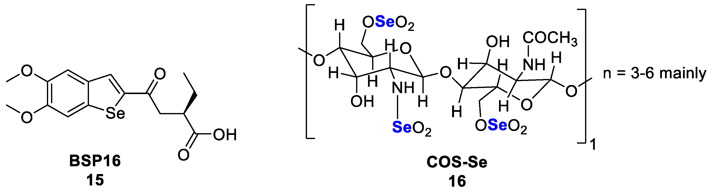
Selenium polysaccharides are natural compounds generated by the combination of inorganic selenium and polysaccharides and are expected to join the properties of both molecules. Some compounds have been isolated from nature and evaluated for their antiproliferative properties in different cancer models. Remarkably, the selenium-polysaccharide Se-GFP-22, isolated from the fungus Grifola frondosa (Fr.) S.F. Gray, was shown to counteract the immunosuppression induced by cyclophosphamide in mice, likely boosting the immune system by the upregulation of MAPKs signalling [90]. This suggested a possible role of Se-GFP-22 as an adjuvant strategy to relieve chemotherapy-induced immunosuppression.
Moreover, the selenium polysaccharide fraction Se-POP-3, obtained from Pleurotus ostreatus (Jacq.) Kummr, was found to moderately affect the viability of human gastric cancer MGC-803, colon cancer HCT-116 cells, and normal intestinal epithelial NCM460 cells (IC50 > > 600 μg/mL), to induce apoptosis and to block cell migration by disrupting the Bax/Bcl-2 ratio and by inhibiting the epithelial-to-mesenchymal transition (EMT); it is noteworthy that Se-POP-3 did not affect the growth of normal cells [91,92]. This evidence suggested a possible interest in Se-POP-3 as a novel anticancer strategy; however, further studies are required to confirm the antitumor effects in vivo and in clinical trials.

3.8. Selol

This compound was obtained by reacting triglycerides from sunflower oil with selenic acid; however, the precise structure is not yet known [93]. The activity of selol is strictly dependent on its Se content, based on the number of dioxaselenolane rings in the molecule. While selol 2% has been reported to exert antioxidant activity, selol 7% showed cytotoxic effects [52]. The compound was found to inhibit cell proliferation and induce apoptosis in leukemic cells, being more effective in resistant ones. The following IC50 values were highlighted: 25 µg Se/mL in HL-60, 20 µg Se/mL in vincristine-resistant HL-60/Vinc, and 15 µg Se/mL in doxorubicin-resistant HL-60/Dox cells [94]. Similar effects were also observed in androgen-dependent prostate cancer cells but not in normal cells [52].
The chemosensitising properties of selol have been highlighted since it increased the doxorubicin antiproliferative effects by reducing the expression of MYC and BCL2 genes [95]; moreover, it enhanced the analgesic effects of fentanyl, buprenorphine and morphine in an animal model of vincristine-induced hyperalgesia [52]. The pro-oxidant effect of selol and its ability to inhibit TrxR could be involved [52,96]; however, the precise mechanism and the molecular targets of selol still have to be determined.

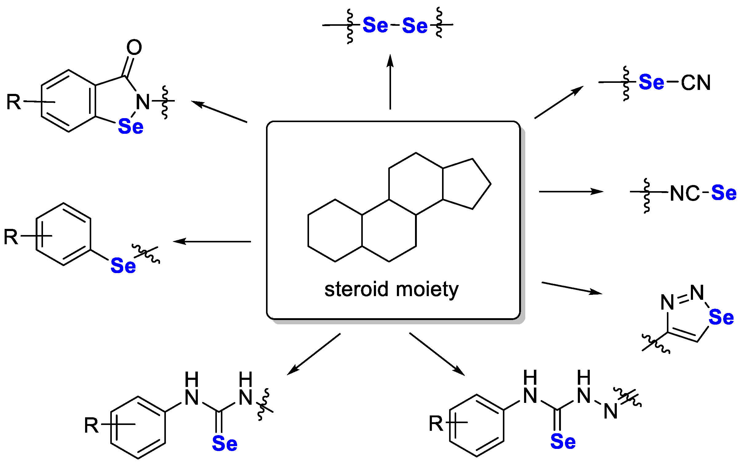
3.9. Selenium in Hybrid Compounds

As cancer is a complex disease, hybrid compounds that act on several biological targets simultaneously may be more effective in treatment than selective drugs. Recently, Singh et al. [97] reviewed such compounds, of which the general structures are shown in Figure 5.
Similarly, a fascinating review paper on selenosteroid compounds was recently described by Jastrzębska et al. [98]. In these compounds, selenium (in an organoselenium moiety) was attached directly to the steroid molecule or by linking to a selenium-containing group (i.e., selenourea, selenocyanine or isoselenocyanine moiety). Such compounds have shown promising antioxidant and/or anticancer activity in in vitro studies. The general structure of such compounds with possible modifications is shown in Figure 6.
Here, we focus on hybrid selenium compounds that have not been described in earlier mentioned publications and discuss their anticancer activity.

3.9.1. Isocombretastatin A and Phenstatin-Based Hybrids

Pang et al. [99] described hybrid compounds formed by combining methyl(phenyl)selane with tubulin polymerisation inhibitors (isocombretastatin A and phenstatin) in a single molecule. Antitubulin compounds are drugs used in the treatment of solid tumours, as well as in haematological malignancies such as lymphomas [100,101]. Thus, such dual-targeting compounds with synergic effects could show higher therapeutic efficacy in cancer. Pang et al. synthesised 20 compounds and tested them in cancer cell lines (A549, MDAMB231, HEPG2, LOVO and RKO) for antiproliferative activity after 48 h. Most compounds were active in the nanomolar range (<1000 nM) in all the tested cells. The most active compound, 10 (Figure 7), caused proliferation block with IC50 values in the range from 2.2 nM (in MDA-MB-231 cells) to 11 nM (in RKO cells). This compound, further tested on other cancer cell lines (MCF7, HELA, HCT116, MGC803 and A2780), also exhibited auspicious activity (IC50 = 3–27 nM). Moreover, 10 was tested in the cisplatin-resistant (A549/CDDP) and the doxorubicin-resistant (HEPG2/DOX) cell lines and showed antiproliferative activity with IC50 values of 34.4 and 20.3 nM and resistance factor values of 8.8 and 6.8, respectively. Additional studies confirmed the ability of 10 to inhibit tubulin polymerisation with an IC50 of 1.38 nM. Furthermore, in vitro efficacy of 10 was confirmed later by in vivo studies in the A549 xenograft model in nude mice. To enhance biodegradability, disodium phosphate salt of 10 was used as a prodrug for this study. The compound was administered intraperitoneally at a dose of 30 mg/kg body weight for the tested period, and a 73% reduction in tumour weight was observed in comparison to the control group.

3.9.2. Phenylselenoethers

Ali et al. [102] described 13 phenylselenoethers as P-glycoprotein (ABCB1) MDR efflux pump modulators. The hybrids were obtained by attaching a phenyl ring via a selenoether bond with aryl-hydantoins (three compounds), (aryl)piperazines (six compounds) and 1,3,5-triazines (four compounds). Cytotoxic and antiproliferative activity of compounds were evaluated in the resistant (MDR) and in the sensitive (PAR) mouse T-lymphoma cells. The highest cytotoxic effect was shown for compound 11 (Figure 7) with IC50 of 0.67 µM for PAR and IC50 of 0.90 µM for MDR. This compound also exhibited an interesting antiproliferative effect in tested cell lines (IC50 = 3.84 µM for PAR; IC50 = 1.34 µM for MDR). In further tests, in JURKAT leukaemia cells, the effect of compound 11 alone or in combination with the chemotherapeutic doxorubicin was evaluated after 24 and 72 h, indicating a positive effect of co-treatment of compound 11 with doxorubicin against cell proliferation, especially after 72 h. Next, it was demonstrated that anticancer activity relates to the ability of compound 11 to inhibit the expression of cyclin D1 and induce the p53 protein expression level.

3.9.3. Coumarin-Based Hybrids

The coumarin ring is present in several compounds that have shown a variety of anticancer activities (e.g., inhibition of angiogenesis), kinases or enzymes (e.g., telomerase, aromatase, carbonic anhydrase or steroid sulfatase) [103,104]. Yildirim et al. [105] described a small series (four compounds) of coumarin hybrids with 2-aminoselenophene-3-carbonitrile (Figure 7). The compounds were tested against prostate cancer on DU145 cells and showed antiproliferative activity with IC50 ranging from 20 to 44.0 µM (higher than for the corresponding coumarin analogues). The highest activity was observed for compound 12 (IC50 = 20 µM; Figure 7). Next, an attempt was made to clarify which apoptosis pathway was affected by the tested compound (at 20 µM concentration) by measuring the levels of caspases 3, 8 and 9 in DU145 cells by ELISA. The results showed that compound 12 caused an increase in the expression of the tested caspases, with caspase 8 being not statistically significant.

3.9.4. Non-Steroidal Anti-Inflammatory Drug-Based Hybrids

Ramos-Inza et al. [106] attached the selenoester moiety to known non-steroidal anti-inflammatory drugs (NSAIDs) such as aspirin, salicylic acid, naproxen, indomethacin and ketoprofen. Such drugs (especially aspirin) might have a beneficial effect on breast cancer patients, acting as a prophylactic or reducing mortality [107,108]. The synthesised 25 compounds were preliminarily tested for cytotoxic effects in the MTT assay in a panel of four cancer lines (HTB54, HT29, DU145 and MCF7) and two non-cancer lines (184B5 and BEAS-2B). The selected five most active compounds were then tested in other cancer lines (T47D, MDA-MB-231, H1299) and in a panel of 60 lines in the NCI Development Therapeutics Program (US). These studies showed that two compounds, derivatives of indomethacin, had very strong cytotoxic effects, especially in breast cancer cells. Inhibition of cancer cell proliferation by these compounds was further confirmed in the Trypan Blue assay in MCF7 and MDA-MB-231 cells. In addition, Annexin V, Cell death and Caspase3/7 assays were performed to measure cell death by apoptosis and showed that compound 13 (Figure 7) effectively induced apoptosis and increased caspase 3/7 activity.

3.9.5. Isoxazole-Based Hybrids

Five benzo[1,2,3]selenodiazole hybrids with an isoxazole moiety were obtained by Oubella et al. [109]. Isoxazole derivatives (e.g., luminespid) [110] are described as potent anticancer compounds [111]; thus, combining this moiety with selenium-containing derivatives may allow for compounds with greater anticancer activity. The final five benzoselenadizole-isoxazoles were estimated by MTT assay on four tumour lines (HT1080, A549, MCF-7 and MDA-MB-231) [109]. Results (after 24 h) showed lower cytotoxic activity of these compounds (IC50 from 18.7 to >100 µM) in comparison with their semicarbazone precursors (IC50 from 10.9 to 21.6 µM) with the most promising compound in this series represented by 14 (Figure 7). The test on healthy cells (MRC5) showed their moderate survival after 14 treatment; furthermore, the study of apoptosis induction by 14 in HT1080 and A549 cell lines in the Annexin-V binding assay showed a 7- and 17-fold increase in apoptosis, respectively, compared to control cells. Further studies confirmed that this process occurs through caspase 3/7 induction.

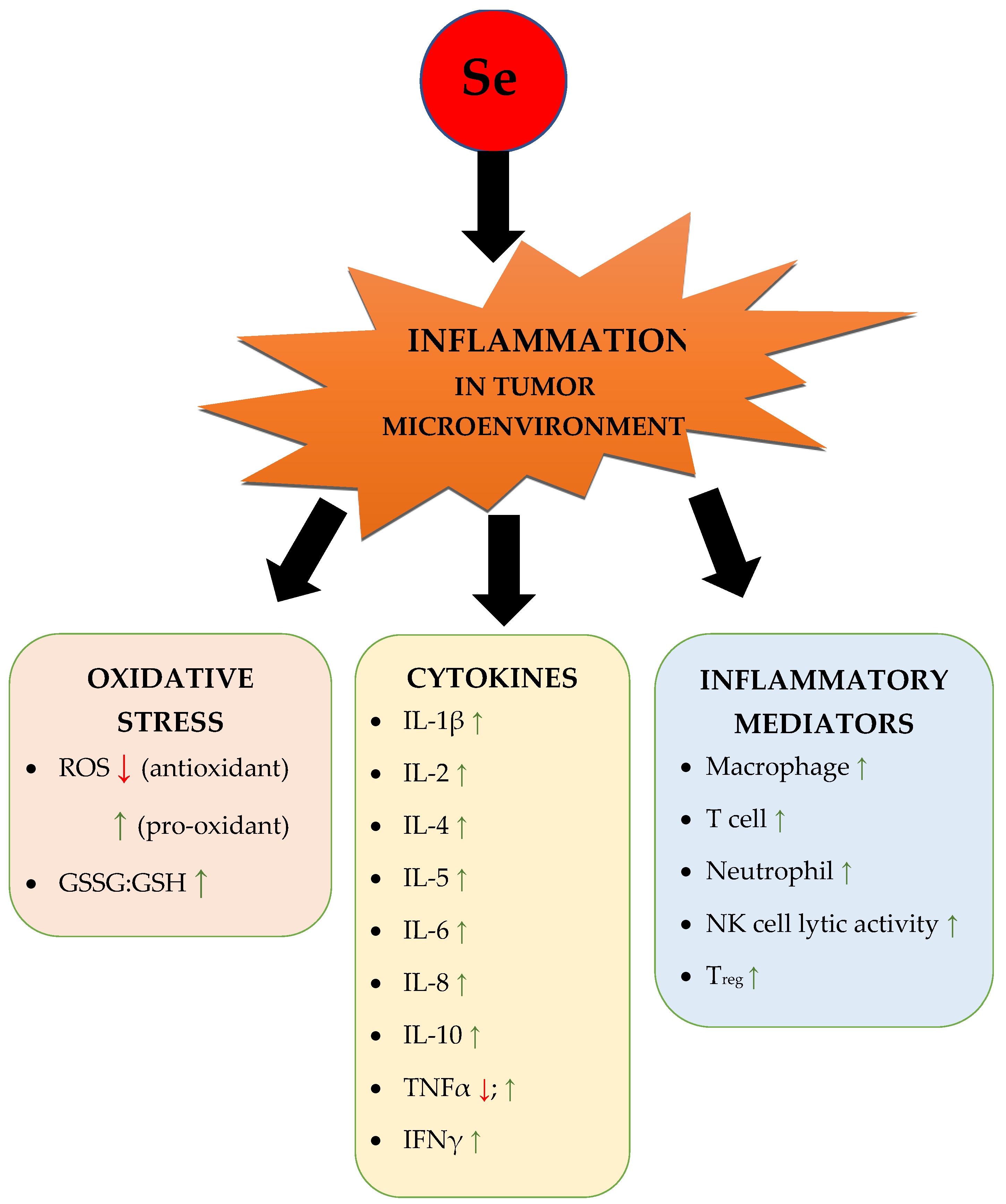
4. Role of Selenium in Inflammation and Immunity

Selenoproteins regulate the health of many organs by maintaining broader homeostasis through regulation of the thyroid hormone level, endoplasmic reticulum stress and antioxidant defence [7], but also immune and inflammatory responses. Many members of the selenoprotein family function as enzymes related to immune functions. The most important are GPXs, thioredoxin reductases (TXNRDs), iodothyronine deiodinases (DIOs), methionine-R-sulfoxide reductase B1 (MSRB1), and selenophosphate synthetase 2 (SPS2).
The concept of linking inflammation and cancer was extensively developed, and today, we have a reasonably broad understanding of processes that occur in microenvironments rich in inflammatory cells, which produce pro-survival and pro-migration factors, thus promoting cancer development and metastasis. Consideration of the role of selenium compounds in the inflammatory process is most warranted. It is well known that the balance of REDOX is regulated by ROS, which, being both toxic and signalling molecules, play an essential role in the inflammatory process [112]. In line with this, the knowledge of the effect of selenium on the immune system has dramatically improved, and at present, selenium is considered a micronutrient belonging to the immunoceuticals, which are immune-modulating compounds that tune up the immune system.

4.1. Cytokines and Inflammatory Signals

Cytokines affect the growth of all blood cells and other cells that help the body’s immune and inflammation responses. They also help increase the anti-cancer activity by sending signals that help to kill abnormal cells as reported in a recent paper, indicating that TNF-α and IFN-γ together induce cell death in 13 distinct human cancer cell lines derived from colon and lung cancer, melanoma, and leukaemia [113] and enable normal cells to live longer. Epidemiological studies have shown a relationship between the level of selenium and the risk of cancer in people with low levels of selenium [114]; additional Se supplementation might result in upregulating the key pro-inflammatory signals. In the anti-tumour effect, B-cells have also contributed as cells presenting antigens which help tumour-infiltrating T cells to proliferate [115] while natural killer cells (NK) carry out immune surveillance via different receptors on their surface which can programme cell death in cancer cells without major histocompatibility class I [116].
Recently, an attractive approach in anticancer immunity is the activation of the stimulator of the interferon genes pathway (STING). STING is a protein expressed in the endoplasmic reticulum, mainly in immune cells. It is activated by DNA released from phagocyted tumour cells and produces the necessary cytokines to induce strong anti-tumour T-cell reactions [117]. BSP16 (15, LF250) (Figure 8), a selenium-containing STING agonist, was reported as an antitumor agent that caused tumour regression and induced antitumor immunity. BSP16 15 (0.1–100 μM) can selectively stimulate the STING pathway in human leukaemia monocytic cell line (ISG-THP1) and mouse macrophage cell line (ISGRAW264.7) with EC50 values of 9.24 and 5.71 μM, respectively. In an in vivo study, BSP16 15 showed an excellent pharmacokinetic profile as an oral drug (oral, 15 and 30 mg/kg, q3d; oral, 20 mg/kg, q5d) with regression of tumour after 21 days in a colon carcinoma tumour model [118]. This direct action on STING is crucial when mutagen-induced inflammation-driven epithelial cancer is developed. Another interesting study demonstrated that Se-enriched mushroom extract (SME) regulated the expression profile of the cancer cell proliferation factor Raf-1 and of the pro-apoptotic-related factors P53 and caspase-3 as well as the production of inflammatory cytokines IL-6 and IL-10 [119]. Dietary consumption of selenium to prevent cancer development is a promising approach. The efficient delivery of selenium is crucial in this case, which is why improved forms of organic selenium are developed. Chitosan oligosaccharide-conjugated selenium (COS-Se, 16, Figure 8) is a new chemical structure similar to natural selenium oligosaccharide. In vitro studies demonstrated that COS-Se 16 increased immunologic mediators such as TNF-α and IL-1β in peritoneal macrophages at concentrations of 50, 100, 200, and 500 μg/mL. Equally interesting results described in this publication relate to in vivo studies showing that COS-Se 16 supplementation at doses of 50 and 100 mg/kg effectively inhibited the growth of transplanted gastric adenocarcinoma in Balb/c nu mice with inhibitory rates of 29% and 33%, respectively. Two mechanisms were behind this tumour suppression: the reduction of MMP-9 and VEGF serum levels in mice, which means that tumour metastasis and angiogenesis were blocked [120].
An inorganic form of selenium, such as selenium selenite 3, has also been shown to decrease oxidative stress in cancer tissues [121]. Adiponectin knock-out mice were under a Se-enhanced diet, which contained 0.75 ppm (200 μg per day) of added Se for 194 days; chronic inflammation-induced colon cancer (CICC) was initiated with the colon cancer agent 1,2 dimethyl hydrazine (DMH) along with dextran sodium sulphate (DSS). The results presented by Saxena et al. demonstrated a significant reduction in tumour number and in common clinical symptoms of colon cancer. The oxidation products from proteins and lipids, nitrotyrosine and 4-hydroxynonenal, respectively, were also significantly decreased in colon cancer tissue of knockout mice. DSS + DMH administered animals on a Se-enriched diet presented a lower degree of inflammation, immune cell infiltration and tumour growth compared with the same animal model on a regular diet.

4.2. Selenoproteins and Inflammation

Selenium is the integral compound of selenoproteins in the human body. In this review, we briefly introduced some selenoproteins and their role in inflammation. SeP is mainly synthesised in the liver and, in human plasma, is the main selenium-containing protein. It is unique since it has as many as 132 selenocysteines in its molecule, making it energetically costly to produce; however, it supplies tissues (such as the brain, kidney and testis) with selenium [122]. SeP also serves in antioxidant protection, which explains the fact that its reduced expression is observed in several types of tumours, including prostate, renal, hepatocellular or colorectal cancer [123]. Particularly noteworthy is the fact that during tumour development, a certain activity of the cells of the immune system is observed. It is also noted that SELENOP mRNA expression might be related to some immune functions, as it was positively correlated with immune marker genes for macrophage subtypes, including tumour-associated macrophages (CD80, CD86 and HLA-G), M1 macrophages (CD36, IL-6 and NOS2), and M2 macrophages (TGFβ, STAT6 and IL-10) in the following cancers: kidney clear cell carcinoma, brain lower grade glioma, lung adenocarcinoma and skin cutaneous melanoma [123]. SeP also reflects the selenium nutritional status in brain tissues, where it is the only selenium donor. It was shown that selenium supplementation in BTBR mice significantly decreased the mRNA and protein levels of inflammatory cytokines IL-6, IL-1β and TNF-α in the hippocampal tissue, pointing out the protective role of selenium [124].

4.3. Selenium Deficiency Impaired Immune Function

Studies have found that selenium deficiency might impair immune function and lead to inflammation-related diseases. Selenium deficiency blocked the glutathione and thioredoxin antioxidant systems and led to redox imbalance. The activation of NF-κB and HIF-1α transcription factors are observed, which results in increasing of pro-inflammatory cytokines (IL-1β, IL-6, IL-8, IL-17, and TNF-α), decreasing anti-inflammatory cytokines (IL-10, IL-13, and TGF-β) and increased expression of the downstream genes COX-2 and iNOS, which in turn induced inflammation [125]. Other structural studies using transmission electron microscopy revealed damages in the jejunum epithelial microvilli of broilers, mitochondrial swelling and vacuolation due to the lack of selenium. In the same study at the molecular level, intestinal inflammatory factors (NF-κB P65, IKKα, IKKβ, IL-1β, IL-6, IFN-γ, and TNF-α) were upregulated, while anti-inflammatory IL-10 was significantly downregulated [126]. Only a few studies focused on the epithelial barrier integrity following enteric infection; in a paper on this issue, the mice fed with a selenium-supplemented or a selenium-deficient diet were infected with C. rodentium strain ICC 169 (nalidixic acid resistant), and the analysis of ILC3 and Th17 cells was performed. In the group of mice with selenium supplementation, this cell population was much higher compared to the selenium-deficient group, which would suggest a greater capacity for the clearance of the enteric pathogen and for maintaining the epithelial barrier integrity of the colon during infection. Moreover, if mice fed with the selenium diet were treated with CAY10397 (inhibitor of 15-hydroxy prostaglandin dehydrogenase), this treatment diminished the protective effect of selenium, suggesting that the oxidation of lipid mediator PGE2 is a key protective mechanism that is associated with selenium levels [127].

4.4. Selenium on Immune Cells Activity

The reported therapeutic role of Selenium in stimulating immune response is due to its ability to target different populations of immune cells (Figure 9).
It has been reported that the knockdown of selenoproteins significantly reduces the number of mature T-cells in lymphoid tissues [128]; for this reason, a significant change in tissue inflammation is observed.
Concerning T cells, it has been reported that knockout for the gene TSRP has an important readout in immunomodulatory effects. TSRP is a gene involved in the synthesis of selenoproteins affecting not only T-cell proliferation, differentiation and activation but also in controlling the number of T-helper cells with a substantial impact on B-cell activity and immunoglobulin production [128].
In Crohn’s disease, a well-characterized chronic inflammatory intestinal disease, it was observed that there is a deficiency in some metabolites, including selenium. Notably, the supplementation of selenium reduces the symptoms of this disease and regulates the differentiation of Th1 cells; this happens through the involvement of selenoprotein W-mediated (SELW-mediated) oxygen-reacting species scavenging [129].
Concerning cancer, it has been proven that Selenium is able to increase the proliferation and activation of CD8-positive cells, a population of immune cells found able to fight tumours in preclinical and clinical studies [129].
Molecularly, CD8+ cytotoxic T-cells play an important role in anticancer immune response since they are able to recognize cancer cells and induce cell death via the induction of the apoptotic pathway [129].
In addition, macrophage activity is regulated by selenium presence in the serum; this reflects a significant impact on the inflammatory signalling and the anti-pathogen response efficiency. Specifically, it seems that selenium was able to induce the switch of macrophages from the M1 pro-inflammatory phenotype to the M2 anti-inflammatory phenotype [130], regulating macrophage phagocytosis and migration properties.
Concerning neutrophils, endogenous oxidative stress is defeated by selenium supplementation [131] while in natural killer cells, selenium supplementation relates to NK activation and to a consequent increase in TNFα and IFNγ secretion and an augmentation of lytic activity.
Figure 9. Overview of the influence of Se in the inflammatory tumour microenvironment. A green upward-facing arrow () indicates that Se has a stimulating effect, while a red downward-facing arrow () indicates that Se has an inhibiting effect. Abbreviations: ROS—reactive oxygen species; GSSG:GSH—the ratio of oxidized to reduced glutathione; IL—interleukin; TNFα—tumour necrosis factor alpha; IFNγ—interferon gamma; NK—natural killer [52,119,120,123,132,133,134,135,136,137,138,139,140,141,142,143,144,145,146,147,148,149,150,151,152].
Pharmaceutics 15 00104 g009

4.5. Selenium and Selenium-Containing Compounds Enhance Cancer Immunotherapy

Concerning cancer immunotherapy, selenium has been proposed and validated as a potent agent. It has been proven that the selenoprotein SELENBP1 in colorectal cancer (CRC) has a lower expression compared to normal conditions. Notably, its expression is strongly correlated to the activation of immune cells and to the release of chemokines, and thus, it plays a central role and strongly influences immune infiltration in this kind of cancer [153].
Moreover, there are some initial observations about the cytotoxicity of selenium-immunoconjugates against triple-negative breast cancer [154]. In this work, IgG1 antibodies Bevacizumab and Trastuzumab were conjugated with the redox form of selenium in order to form Selenobevacizumab and Selenotrastuzumab and were tested in different cell lines with a strong arrest of cancer cells growth.

4.6. Selenium on Infection Diseases and Vaccination

Due to the pleiotropic effects that selenium can have on cellular pathways and cellular responses, it is quite challenging to circumscribe the individual molecular mechanism through which selenium is able to potentiate cellular immunity. However, there are some theories based on specific selenoproteins to take into consideration. One of these proteins is represented by the selenoprotein MSRB1, which is able to regulate actin polymerization, promoting phagocytosis and cytokine secretion [155]. Another important factor is the selenoprotein SELENOK, which is able to activate calcium reflux during immune cell activation [156].
Selenium supplementation is also able to enhance GPX4 in T-cells. In addition to the previously described effect of selenium in regulating GPX4, the major lipid peroxidase scavenger, its selenium-dependent increase enhances follicular helper T-cells (Tfh) number, viability and antibody response in vivo in immunized mice after influenza vaccination. Moreover, the Selenium–GPX4 axis protects follicular helper T-cells from ferroptosis [157].
Besides the role of selenium and selenoprotein in inducing an immune response in cancer contexts, they can also directly affect viruses’ proliferation and infection. In particular, it has been described that the Coxsackie virus shows higher toxicity when the selenium level is low [158]. The low selenium level can cause an oxidative condition through which oxidative pressure can enhance the probability of accumulating mutations, allowing for viral evolution [159].
Other observations indicate that selenium levels decrease with the development of AIDS [160], and some selenoproteins such as GPX1, GPX4, and TRX1 decrease with HIV infection [161]; notably, TRX1 selenoprotein can inhibit in vitro HIV replication targeting Tat protein [162].
In addition, vaccine efficacy seems to be improved with the supplementation of selenium; a selenium-based approach against poliovirus infection has been proposed and validated; it seems that the vaccine’s efficacy against this specific infectious agent is significantly improved when it is administrated with selenium [163].
Selenium deficiency in the diet is also correlated to COVID-19 mortality. The selenium levels in patients who survived COVID-19 were significantly higher than those observed in dead patients [164]. Selenium shows an antiviral effect due to the regulation of CD4+ response through stimulation of proliferation, differentiation and activation of these cells; moreover, it is crucial for the cytotoxic effect of CD8+ cells and natural killer cells (NK) [165].
Finally, in recent years, great interest has been given to selenium nanoparticles (better described before) as a powerful strategy for cancer immunotherapy [133,134].
In a study, the efficacy of a selenium (Se)-bearing ruthenium (Ru) complex (RuSe) for immune-sensitising natural killer cells for immunotherapy purposes against prostate cancer was demonstrated [132]. Pre-treatment of prostate cancer cells with RuSe potentiated the lysis potency of NK cells and increased the release of IFNγ and IL-2 [132]. IFNγ, in turn, has cytostatic and pro-apoptotic functions and is useful for adjuvant immunotherapy for different types of cancer, as it can stimulate an anti-tumour immune response.

5. Selenium-Containing Nanoparticles (SeNPs) as An Innovative Therapeutic Strategy

Nanomaterials represent a heterogeneous family of particles, including liposomes, micelles and organic or inorganic nanoparticles whose potential roles as anticancer drugs or small molecules or RNA therapeutics carriers have been recently proposed to enhance their effective delivery in target cells [166,167]. The interest in nanomaterials grew in biomedicines due to the advantages related to their use; specifically, they are characterised by solubility and stability, they can be engineered to increase their specificity to target cells, and they show high biocompatibility, a low grade of immunogenicity and low side effects [167].
SeNPs show numerous advantages, such as high degradability, low toxicity and anticancer, antimicrobial and antiviral activities [168]. With respect to the low toxicity, it has been widely reported that this selectively protects normal cells from the cytotoxic effects of the treatment against cancer [169,170]; moreover, SeNPs can be rapidly excreted without long-term toxicity [171]. Their efficacy has been reported against oxidative stress and inflammation-mediated disorders (e.g., cancer, arthritis, diabetes and nephropathy [172]). These particles are also capable of targeting macrophages to activate innate immunity for antimicrobial inhibition through the production of cytokines [173], and these abilities have been translated into the field of cancer treatment since these nanoparticles can regulate tumour-associated macrophages and T cells (Figure 10) [140].

5.1. Preparation of SeNPs

The preparation of SeNPs can be performed through different approaches. The chemical reduction method (using vitamin C, sodium sulphite, sodium thiosulfate and hydrazine as reducing agents) is the most convenient, but other strategies have been developed. Hydrothermal synthesis is cheap, simple, efficient and produces functional nanoparticles with different shapes and diameters [174]. However, this preparation strategy requires high pressure and temperature in a specific hydrothermal autoclave reactor to produce small particles, SeNPs can show some agglomeration due to high surface energy and electrochemical properties; the process is difficult to control, and there is a low reliability and reproducibility [175,176]. Moreover, SeNPs can be formed using some chemical templates (e.g., PEG200, SPS, folate, hyaluronic acid, PEI) as stabilisers. Another approach to produce SeNPs is laser irradiation, also known as pulsed laser ablation (PLA) or deposition, exploited to inhibit bacterial growth [177]. Finally, in light of the so-called green chemistry, a large spectrum of plants, bacteria, and fungi have been reported as able to synthesise selenium-containing highly stable nanoparticles based on their ability to reduce selenite [178]. This last approach is highly controllable [179], produces highly pure SeNPs, displays great potential for anti-bacterial and anti-cancer treatments and is extremely cheap and fast, non-toxic, and overall, it is completely biocompatible.
Based on the oral administration of NPs and in order to limit the reduced selenium absorption and to improve the availability of Se, the production of SeNPs was implemented using the Eudragit® RL and Eudragit® RS polymers [180]. Recently, a new method for the production of SeNPs with high stability, good biocompatibility and narrow size that is environmentally friendly and economical has been proposed. This method uses the yeast as a bio-reducing agent, and the derived nanoparticles are stable and have fewer side effects in mice when added to their feed, protecting the liver, spleen, and kidney from oxidative stress, stimulating the humoral immune potential and significantly increasing the levels of IgM, IgA, and IgG [181], thus representing a potential substitute for antibiotics.

5.2. Therapeutic Potential of SeNPs as Drug Carriers

Moreover, since nanomaterials can be synthesised in order to contain selected elements [182], the incorporation of selenium into polymers has been described as a good strategy for the delivery of drugs, warranting low toxicity if used at low doses (less than 50 mg for adult people) and sensitivity to oxidative/reductive and radiation stimuli. Since redox conditions significantly differ between cancer and non-cancer cells, selenium-containing nanomaterials can be considered good candidates for the delivery of anticancer drugs. Moreover, SeNPs induce cell cycle arrest at the S phase [183] by deregulating the eIF3 protein complex [184] with a high selectivity against cancer cells (higher than selenium Se + IV at similar concentrations) [185].
Besides the effects of SeNPs per se, these particles can be used for the delivery of specific drugs. The size of these materials allows for efficient uptake by target cells and drug accumulation at target sites [186]; moreover, they can be decorated to enhance anticancer efficacy through increased uptake [187,188].
In this regard, the chemotherapeutic agent doxorubicin has been loaded into diselenide polymeric micelles to be released in cancer cells [189], and hydrogel and metal-organic frameworks (MOFs) have also been developed. In this frame, hydrogels responsive to radiation applied as radiotherapy that produces ROS (gamma radiation), cleaving diselenide bonds, have been projected to load anticancer drugs. Upon the exposure of both micelles and hydrogels to irradiation, the nanoparticles disassemble (due to the cleavage of the diselenide bond), and the chemotherapeutic drug is released.
Concerning monoselenide, also in this case, micelles formed by selenium-containing polymers can be loaded with drugs (e.g., doxorubicin) and are sensitive to oxidation stimuli [190], such as treatment with hydrogen peroxide or exposure to near-infrared light, and can revert and reassemble under reductive stimuli.
Selenium-containing nanomaterials can be used as drug carriers, but they show an anticancer effect due to the presence of selenium per se.
Selenium shows antioxidant properties at low levels, while it becomes a pro-oxidant at high doses, increasing ROS production. Although cancer cells show high levels of ROS and reducing agents, they are sensitive to additional ROS; this reflects an ability of selenium to impair cancer cell survival. Moreover, the diselenide-containing polymer has been proposed as a drug carrier able to include hydrophobic anticancer compounds, while monoselenide-containing polymers possess anticancer activity independent of the ability to encapsulate drugs [191]. Recently, great interest was given to developing selenium-containing nanomaterials to be applied both for the delivery of anticancer drugs and for showing anticancer activity per se. In a work by Zhang and collaborators, the authors describe a very innovative approach for the delivery of the chemotherapeutic agent paclitaxel, taking advantage of spherical nanoparticles containing diselenide-containing small molecules, and they show the effectiveness of these chimeric particles in inducing apoptosis selectively in cancer cells (e.g., HeLa and MCF-7) through the generation of ROS [135]. Coherently, diselenide-containing nanocapsules have been reported to exert anticancer effects due to the activity of both selenium and encapsulated drugs [192].

5.3. Therapeutic Potential of SeNPs Per Se

Notably, other interesting publications report that coordination complexes (e.g., coordinating platinum) containing selenium have anticancer effects being highly toxic against cancer cells (both in vitro and in vivo) in a selective way due to the induction of ROS and above all to the presence of selenium [136,137]. The selenium-containing selenomethionine (2)-based self-assemblies exert anticancer abilities [193] and can be combined with drugs to deliver specific compounds in cancer cells. In addition to platinum, selenium can interact with other metals (e.g., gold, zinc, copper, nickel) to constitute nanoparticles with anticancer activity through the induction of ROS in cancer cells, stimulating their apoptosis [194]. Selenium-containing molecules can also be combined with porphyrins [195,196], can be activated by light (photodynamic therapy) and can activate cell apoptosis. Moreover, selenium-containing polymers activate the immune response by NK cells, thus representing a synergic strategy with low side effects to apply for tumour management in combination with conventional therapies [138]. In order to increase SeNPs’ solubility, stability, cellular uptake (also increased by the nanoparticles’ decoration with conventional chemotherapeutic agents such as 5-fluorouracil [187]) and efficacy against cancer cells in vivo and in vitro, chemical strategies have been developed, such as the addition of polysaccharides (e.g., spirulina [188,197]), proteins (e.g., transferrin [198]), folic acid [199] or hydrogels [200] coating them. Despite the described role of SeNPs to facilitate drug delivery, they can be considered as shuttles for other small molecules such as siRNAs targeting specific cancer-related mRNAs [201] or viral RNAs [202].

5.4. Therapeutic Potential of SeNPs in Non-Cancer Diseases

SeNPs have been applied not only to cancer but also to other diseases of different origins. One of the recent examples of the application of SeNPs is the treatment of Vibrio cholerae infection. Notably, selenium particles increase the immune responses in vivo in a mouse cholera model due to their cytotoxic activity and to a significant increase in V. cholerae-specific IgG and IgA responses in serum and saliva. Moreover, the levels of both IL-4 and IL-5 are significantly increased, indicating that SeNPs induce immune cell effectors [139].
In this regard, apoptosis is a common pathway for destroying intracellular bacteria [203] that prevents pathogens from exiting the host cells for further infection spread. However, some pathogens possess highly stable cell walls and the ability to inhibit host cell apoptosis as a critical way for their immune escape. Notably, a recent work on Mtb infection reported the ability of SeNPs to induce infected macrophage apoptosis, which is useful for intracellular clearance and antimicrobial immunity [173].
Moreover, SeNPs can be used to counteract multidrug-resistant bacterial infections [204] and viral infections (e.g., type-1 dengue virus [205]), both alone and in combination with lysozyme [206].
Gram-negative, Gram-positive, multiresistant bacteria, C. albicans, different protozoa and parasites (e.g., Leishmania, Toxoplasma gondii and Schistosoma mansoni), and several viral infections (e.g., influenza, hepatitis, HIV, H1N1) have been reported as sensitive targets of SeNPs [207].
This approach is also helpful in tumour cells where SeNPs activate the early phase of autophagy while the late one is blocked [208].
Moreover, SeNPs display a protective role against the cytotoxic effects of several chemotherapeutic agents such as cisplatin [209]. In this regard, the co-treatment with both cisplatin and SeNPs increases serum testosterone, sperm quality, and spermatogenesis and limits nephrotoxicity [210].

5.5. Advantages of SeNPs in Cancer Treatment

The high bioavailability (depending overall on particle size [211]) together with the low toxicity (lower than selenomethionine [212]) make the nanoform of selenium more attractive with respect to the inorganic and organic forms [212,213,214]. Moreover, SeNPs can resist adverse conditions such as digestion and enzymatic cleavage and use the selenium in a zero oxidation state, which is low toxic, bioavailable [211,212] and stable when encapsulated inside particles such as chitosan (CS) [215]. In this regard, the incorporation of selenium into CS nanoparticles improves the availability of this element, facilitates the expression of selenoproteins and can be translated into therapy as a chemopreventive agent and chemotherapeutic drug delivery approach [184,187,216,217].
Many literature reports demonstrate that selenium can both prevent/reduce cancer incidence [218,219] and can act in synergy with chemotherapeutics [220,221].
The dual role of SeNPs as anticancer agents and drug carriers has been extensively investigated [188,222,223,224,225,226] highlighting their ability to impair cancer cell growth [216,227]. Moreover, SeNPs are able to enter cancer cells and show high ability in chemoprevention, inducing glutathione S-transferase (GST) more than SeMet and selenite [212,228].
Notably, a large spectrum of experimental evidence obtained in different tumour cells highlights the promising role of SeNPs in inducing mitochondria-mediated apoptosis [185] in melanoma cells, cell cycle progression inhibition in pancreatic cancer cells, antiproliferative effects in human cervical carcinoma and breast cancer cells, and anticancer activity in hepatocellular carcinoma cells [183,229,230]. Focusing on cell proliferation and other typical features of tumour cells such as viability, cell cycle arrest, migration and invasion, it has been reported in an in vitro model of hepatocellular carcinoma that the effects of SeNPs and selenocysteine were similar, while Se + IV was more toxic and cells treated with Se + IV, selenomethionine and seleno-metylselenocysteine did not show significant difference with respect to not-treated ones.
These direct effects of SeNPs on tumour cells parallel with their potential as chemotherapeutic delivery carriers [183,187,231,232,233,234,235] since they passively accumulate in cancer cells, and their decoration on the surface enhances their specific uptake [188,199]. The SeNPs activate ROS and, in turn, regulate the apoptotic signal mediated by p53 and MAPK phosphorylation, also preventing DNA repair in cancer cells [168]. Among the therapeutic cargo described for SeNPs, 5-fluorouracil and cisplatin are the best-known drugs [187].
In addition to cell proliferation/invasion/migration, SeNPs are also effective against cancer cells by stimulating the immune response [236] due to the increase in chemotactic activity and respiratory burst response of neutrophils [237]; this effect was not evident after the treatment with inorganic selenium.
Despite the growing interest of scientists in SeNPs and the high number of reports on their wide range of positive effects, there are some concerns about their toxicity and objections to their use in clinical practice. Based on the described formulations of selenium to be applied in therapy, it seems evident that not only the concentration of this element but also the chemical form is crucial for its effectiveness against several diseases, including cancer [187]. Specifically, with respect to the SeNPs, they clearly show a notable advantage due to their low grade of toxicity, higher efficiency and biosafety [212,238,239] compared with selenite (LD50 around 18-fold higher), chemopreventive potential [187] and selectivity against cancer cells.

6. Safety of Selenium-Containing Compounds

Despite the encouraging results about the role of Se-containing compounds as chemopreventive and anticancer agents, the clinical application is sometimes limited owing to their possible side effects [82]. Mainly, inorganic compounds are known to be metabolised into redox-active products, such as hydrogen selenide, which arises from selenite 3, whose accumulation into cells can lead to genotoxic effects (single strand DNA breaks), likely as a consequence of the oxidative stress induction [52]. For this reason, inorganic Se-containing compounds are considered more toxic than organic ones [52]. Furthermore, sodium selenite 3a resulted in being well tolerated in short-term toxicity studies [52].
In the last few years, significant attention has been devoted to Se-containing organic molecules to retain the pharmacological properties of inorganic compounds while lowering their toxicity risk. Notably, selenoaminoacids, such as selenomethionine 2 (L-selenomethionine) and methylselenocysteine 1, exhibited a very low toxicity and lack genotoxicity [52,240]; moreover, methylseleninic acid 6 was found to be nongenotoxic with respect to selenite 3 and nontoxic to normal tissues [40].
Diphenyl diselenide 8 showed a toxicity risk in vivo, depending on the route of administration and dosage, but its derivatives, among which includes DSBA 8a, could overcome this limit [51]. Accordingly, selol resulted to be nontoxic and nonmutagenic only when administered parenterally; by contrast, it was toxic after oral administration, likely due to the formation of harmful metabolites during digestion [52].
Overall, the mechanisms involved in selenium toxicity are not entirely understood yet; however, the oxidation of thiols (e.g., glutathione and cysteine) by both inorganic and organic selenium-containing compounds seems to play a central role in their toxicity as well as in their biological activities [241]. Considering that even compounds with low toxicity in vivo (e.g., ebselen 4 and diphenyl diselenide 8) can determine toxic effects at high doses, depending on the species, exposure time, and route of administration, it is of utmost importance to support the pharmacological findings with toxicological studies in order to allow for future clinical application of these bioactive compounds.

7. Conclusions

In this review, we illustrate the most promising Se-containing compounds and SeNPs that have emerged as promising cancer treatments in recent years.
In comparison to conventional chemotherapeutic agents, organoselenium compounds such as 10–14 have excellent cytotoxic properties with low side effects [99,102,105,106,109]. Recent studies have also pointed to selenium-based nanomedicines, suggesting the design of selenium nanoparticles as an efficient strategy to improve drug delivery and thus cancer treatment [190].
Over the last decade, significant progress has been made in understanding the complex biology and chemistry of Se. It was long believed that Se and Se-containing compounds maintain the redox status of cells and prevent oxidative damage caused by ROS. Recent studies indicate, however, that Se plays a dual role in oxidative stress, exhibiting a pro-oxidant and an antioxidant function depending on the type of incorporation of Se in the relative compound. The uncertainty about the anticancer and immunostimulatory properties of Se agents is often also reflected in their unclear mechanism [2]; thus, more research is needed.
As discussed above, some compounds are rather promiscuous binders and, as such, can only be considered as hit compounds needing further optimization. An excellent example of a rather advanced Se-containing compound is compound BSP16 (15, LF250), a selenium-containing STING agonist, with excellent anticancer properties in vitro and in vivo better than its sulphur containing analogue. Furthermore, it possesses an excellent pharmacokinetic profile as an oral drug leading to tumour regression after 21 days in a colon carcinoma tumour model [118].
However, most other Se-containing agents in our collection are not (yet) very potent when it comes to in vivo efficacy.
Besides a direct anti-cancer action of Se-containing agents, it might also be interesting to apply a hybrid approach as shown by Ali et al. [102,242]. The phenylselenoethers exhibited P-glycoprotein (ABCB1) MDR efflux pump inhibitory activity and possessed interesting cytotoxic and antiproliferative MDR mouse T-Lymphoma cells reaching, with compound 11, the submicromolar range. Notably, compound 11 was also more active when co-administered with the well-known chemotherapeutic agent doxorubicin, underlining the fact that a combination therapy approach is worthwhile to investigate.
Due to the small number of researchers and companies working on these promising agents, the optimization process is relatively slow [4]. An interdisciplinary effort is urgently required to dissect the biological effects of the different Se incorporations in small organic molecules in order to improve the situation, thereby opening the door to the development of more highly potent and target-specific Se-containing agents. These molecules seem to possess essential advantages: high specificity and the high effectiveness, above all, in relation to their formulation that can include the Se element in low-toxic molecules or can insert Se inside NPs. In our view, researchers should try more often to replace oxygen or sulphur with Se in their design strategies for novel anti-cancer small molecules. In addition, the approach to create SeNPs has promising first data in cancer, but intensive research is necessary from a toxicity point of view prior to thinking of an application in therapy.
To sum up, Se-based therapy approaches in cancer might have a bright future, but a lot of research efforts are still necessary prior to seeing a clinical candidate or even an approved drug.

Author Contributions

Conceptualization of the review with a strong focus on the years 2016–2022, C.B. and C.Z.; literature research via Scopus and PubMed as well as writing of the original draft, S.G., S.D.G., D.Ł., E.H.-O., A.D.S., R.F., C.Z. and C.B.; editing, S.G., S.D.G., A.D.S., C.B. and C.Z.; supervision, C.B. and C.Z. All authors have read and agreed to the published version of the manuscript.

Funding

This research was funded by Sapienza University of Rome SEED PNR 2021 to C.B. and by Kohr Aerospace GmbH to C.Z., C.Z. and S.D.G., who are thankful for the generous financial support of the FSE REACT-EU within the program PON “Research and Innovation” 2014–2020, Action IV.6 “Contratti di ricerca su tematiche Green”. S.D.G. and A.D.S. thank the “Enrico and Enrica Sovena” Foundation (Italy) for supporting the present work.

Institutional Review Board Statement

Not applicable.

Informed Consent Statement

Not applicable.

Data Availability Statement

No additional data has been created.

Acknowledgments

All authors would like to thank C. Jacob, J. Handzlik, A. Mai, M. Tripodi and S. Valente for the fruitful discussions. Furthermore, they are grateful for the technical assistance of Nick Hinderlaad.

Conflicts of Interest

The authors declare no conflict of interest.

References

  1. Greenwood, N.N.; Earnshaw, A. Chemistry of the Elements; Elsevier: Amsterdam, The Netherlands, 2012. [Google Scholar]
  2. Lenardão, E.J.; Santi, C.; Sancineto, L. Bioactive Organoselenium Compounds and Therapeutic Perspectives. In New Frontiers in Organoselenium Compounds; Lenardão, E.J., Santi, C., Sancineto, L., Eds.; Springer International Publishing: Cham, Switzerland, 2018; pp. 99–143. [Google Scholar]
  3. Frost, D.V. The two faces of selenium—Can selenophobia be cured? CRC Crit. Rev. Toxicol. 1972, 1, 467–514. [Google Scholar] [CrossRef] [PubMed]
  4. Ali, W.; Benedetti, R.; Handzlik, J.; Zwergel, C.; Battistelli, C. The innovative potential of selenium-containing agents for fighting cancer and viral infections. Drug Discov. Today 2021, 26, 256–263. [Google Scholar] [CrossRef] [PubMed]
  5. Santi, C.; Bagnoli, L. Celebrating Two Centuries of Research in Selenium Chemistry: State of the Art and New Prospective. Molecules 2017, 22. [Google Scholar] [CrossRef] [Green Version]
  6. Vinceti, M.; Filippini, T.; Cilloni, S.; Bargellini, A.; Vergoni, A.V.; Tsatsakis, A.; Ferrante, M. Health risk assessment of environmental selenium: Emerging evidence and challenges (Review). Mol. Med. Rep. 2017, 15, 3323–3335. [Google Scholar] [CrossRef] [PubMed] [Green Version]
  7. Ye, R.; Huang, J.; Wang, Z.; Chen, Y.; Dong, Y. The Role and Mechanism of Essential Selenoproteins for Homeostasis. Antioxidants 2022, 11, 973. [Google Scholar] [CrossRef] [PubMed]
  8. Zinoni, F.; Birkmann, A.; Stadtman, T.C.; Bock, A. Nucleotide sequence and expression of the selenocysteine-containing polypeptide of formate dehydrogenase (formate-hydrogen-lyase-linked) from Escherichia coli. Proc. Natl. Acad. Sci. USA 1986, 83, 4650–4654. [Google Scholar] [CrossRef] [Green Version]
  9. Mukherjee, A.J.; Zade, S.S.; Singh, H.B.; Sunoj, R.B. Organoselenium chemistry: Role of intramolecular interactions. Chem. Rev. 2010, 110, 4357–4416. [Google Scholar] [CrossRef]
  10. Reich, H.J.; Hondal, R.J. Why Nature Chose Selenium. ACS Chem. Biol. 2016, 11, 821–841. [Google Scholar] [CrossRef]
  11. Perrone, D.; Monteiro, M.; Nunes, J.C. CHAPTER 1. The Chemistry of Selenium. In Selenium; Food and Nutritional Components in Focus; Royal Society of Chemistry: London, UK, 2015; pp. 3–15. [Google Scholar]
  12. Krivovichev, V.G.; Krivovichev, S.V.; Charykova, M.V. Selenium Minerals: Structural and Chemical Diversity and Complexity. Minerals 2019, 9, 455. [Google Scholar] [CrossRef] [Green Version]
  13. Hou, W.; Dong, H.; Zhang, X.; Wang, Y.; Su, L.; Xu, H. Selenium as an emerging versatile player in heterocycles and natural products modification. Drug Discov. Today 2022, 27, 2268–2277. [Google Scholar] [CrossRef]
  14. Zoidis, E.; Seremelis, I.; Kontopoulos, N.; Danezis, G.P. Selenium-Dependent Antioxidant Enzymes: Actions and Properties of Selenoproteins. Antioxidants 2018, 7, 66. [Google Scholar] [CrossRef] [PubMed] [Green Version]
  15. Zhang, Y.; Roh, Y.J.; Han, S.J.; Park, I.; Lee, H.M.; Ok, Y.S.; Lee, B.C.; Lee, S.R. Role of Selenoproteins in Redox Regulation of Signaling and the Antioxidant System: A Review. Antioxidants 2020, 9, 383. [Google Scholar] [CrossRef] [PubMed]
  16. Yakubov, E.; Buchfelder, M.; Eyupoglu, I.Y.; Savaskan, N.E. Selenium action in neuro-oncology. Biol. Trace Elem. Res. 2014, 161, 246–254. [Google Scholar] [CrossRef] [PubMed]
  17. Zhang, X.; Liu, R.P.; Cheng, W.H.; Zhu, J.H. Prioritized brain selenium retention and selenoprotein expression: Nutritional insights into Parkinson’s disease. Mech. Ageing Dev. 2019, 180, 89–96. [Google Scholar] [CrossRef]
  18. Zhang, Z.H.; Song, G.L. Roles of Selenoproteins in Brain Function and the Potential Mechanism of Selenium in Alzheimer’s Disease. Front. Neurosci. 2021, 15, 646518. [Google Scholar] [CrossRef]
  19. Rayman, M.P. Selenium intake, status, and health: A complex relationship. Hormones 2020, 19, 9–14. [Google Scholar] [CrossRef] [Green Version]
  20. Guillin, O.M.; Vindry, C.; Ohlmann, T.; Chavatte, L. Selenium, Selenoproteins and Viral Infection. Nutrients 2019, 11, 2101. [Google Scholar] [CrossRef] [Green Version]
  21. Bjorklund, G.; Shanaida, M.; Lysiuk, R.; Antonyak, H.; Klishch, I.; Shanaida, V.; Peana, M. Selenium: An Antioxidant with a Critical Role in Anti-Aging. Molecules 2022, 27, 6613. [Google Scholar] [CrossRef]
  22. Shreenath, A.P.; Ameer, M.A.; Dooley, J. Selenium Deficiency. In StatPearls; StatPearls Publishing: Treasure Island, FL, USA, 2022. [Google Scholar]
  23. Fakhrolmobasheri, M.; Mazaheri-Tehrani, S.; Kieliszek, M.; Zeinalian, M.; Abbasi, M.; Karimi, F.; Mozafari, A.M. COVID-19 and Selenium Deficiency: A Systematic Review. Biol. Trace Elem. Res. 2022, 200, 3945–3956. [Google Scholar] [CrossRef]
  24. Radomska, D.; Czarnomysy, R.; Radomski, D.; Bielawska, A.; Bielawski, K. Selenium as a Bioactive Micronutrient in the Human Diet and Its Cancer Chemopreventive Activity. Nutrients 2021, 13, 1649. [Google Scholar] [CrossRef]
  25. Kieliszek, M. Selenium(-)Fascinating Microelement, Properties and Sources in Food. Molecules 2019, 24, 1298. [Google Scholar] [CrossRef] [PubMed] [Green Version]
  26. Kaur, N.; Sharma, S.; Kaur, S.; Nayyar, H. Selenium in agriculture: A nutrient or contaminant for crops? Arch. Agron. Soil Sci. 2014, 60, 1593–1624. [Google Scholar] [CrossRef]
  27. Chen, N.; Zhao, C.; Zhang, T. Selenium transformation and selenium-rich foods. Food Biosci. 2021, 40, 100875. [Google Scholar] [CrossRef]
  28. Yang, T.; Lee, S.Y.; Park, K.C.; Park, S.H.; Chung, J.; Lee, S. The Effects of Selenium on Bone Health: From Element to Therapeutics. Molecules 2022, 27, 392. [Google Scholar] [CrossRef] [PubMed]
  29. Ye, R.; Huang, J.; Wang, Z.; Chen, Y.; Dong, Y. Trace Element Selenium Effectively Alleviates Intestinal Diseases. Int. J. Mol. Sci. 2021, 22, 11708. [Google Scholar] [CrossRef] [PubMed]
  30. Gorini, F.; Sabatino, L.; Pingitore, A.; Vassalle, C. Selenium: An Element of Life Essential for Thyroid Function. Molecules 2021, 26, 7084. [Google Scholar] [CrossRef]
  31. Avery, J.C.; Hoffmann, P.R. Selenium, Selenoproteins, and Immunity. Nutrients 2018, 10, 1203. [Google Scholar] [CrossRef] [Green Version]
  32. Shalihat, A.; Hasanah, A.N.; Mutakin; Lesmana, R.; Budiman, A.; Gozali, D. The role of selenium in cell survival and its correlation with protective effects against cardiovascular disease: A literature review. Biomed Pharm. 2021, 134, 111125. [Google Scholar] [CrossRef]
  33. Lima, L.G.; Santos, A.; Gueiber, T.D.; Gomes, R.Z.; Martins, C.M.; Chaikoski, A.C. Relation between Selenium and Female Fertility: A Systematic Review. Rev. Bras. Ginecol. Obstet. 2022, 44, 701–709. [Google Scholar] [CrossRef]
  34. Mintziori, G.; Mousiolis, A.; Duntas, L.H.; Goulis, D.G. Evidence for a manifold role of selenium in infertility. Hormones 2020, 19, 55–59. [Google Scholar] [CrossRef]
  35. Schweizer, U.; Fabiano, M. Selenoproteins in brain development and function. Free Radic. Biol. Med. 2022, 190, 105–115. [Google Scholar] [CrossRef] [PubMed]
  36. Bartolini, D.; Tew, K.D.; Marinelli, R.; Galli, F.; Wang, G.Y. Nrf2-modulation by seleno-hormetic agents and its potential for radiation protection. Biofactors 2020, 46, 239–245. [Google Scholar] [CrossRef] [PubMed]
  37. Kielczykowska, M.; Kocot, J.; Pazdzior, M.; Musik, I. Selenium—A fascinating antioxidant of protective properties. Adv. Clin. Exp. Med. 2018, 27, 245–255. [Google Scholar] [CrossRef] [PubMed]
  38. Tran, P.; Kopel, J.; Ristic, B.; Marsh, H.; Fralick, J.; Reid, T. Antimicrobial seleno-organic coatings and compounds acting primarily on the plasma membrane: A review. Adv. Redox Res. 2022, 4, 100031. [Google Scholar] [CrossRef]
  39. Chuai, H.; Zhang, S.Q.; Bai, H.; Li, J.; Wang, Y.; Sun, J.; Wen, E.; Zhang, J.; Xin, M. Small molecule selenium-containing compounds: Recent development and therapeutic applications. Eur. J. Med. Chem. 2021, 223, 113621. [Google Scholar] [CrossRef]
  40. Moran-Serradilla, C.; Angulo-Elizari, E.; Henriquez-Figuereo, A.; Sanmartin, C.; Sharma, A.K.; Plano, D. Seleno-Metabolites and Their Precursors: A New Dawn for Several Illnesses? Metabolites 2022, 12, 874. [Google Scholar] [CrossRef]
  41. Barchielli, G.; Capperucci, A.; Tanini, D. The Role of Selenium in Pathologies: An Updated Review. Antioxidants 2022, 11, 251. [Google Scholar] [CrossRef]
  42. Mosolygo, T.; Kincses, A.; Csonka, A.; Tonki, A.S.; Witek, K.; Sanmartin, C.; Marc, M.A.; Handzlik, J.; Kiec-Kononowicz, K.; Dominguez-Alvarez, E.; et al. Selenocompounds as Novel Antibacterial Agents and Bacterial Efflux Pump Inhibitors. Molecules 2019, 24, 1487. [Google Scholar] [CrossRef] [Green Version]
  43. Mal’tseva, V.N.; Goltyaev, M.V.; Turovsky, E.A.; Varlamova, E.G. Immunomodulatory and Anti-Inflammatory Properties of Selenium-Containing Agents: Their Role in the Regulation of Defense Mechanisms against COVID-19. Int. J. Mol. Sci. 2022, 23, 2360. [Google Scholar] [CrossRef]
  44. Ren, Z.; Ding, T.; He, H.; Wei, Z.; Shi, R.; Deng, J. Mechanism of selenomethionine inhibiting of PDCoV replication in LLC-PK1 cells based on STAT3/miR-125b-5p-1/HK2 signaling. Front. Immunol. 2022, 13, 952852. [Google Scholar] [CrossRef]
  45. Qu, L.; Ji, L.; Wang, C.; Luo, H.; Li, S.; Peng, W.; Yin, F.; Lu, D.; Liu, X.; Kong, L.; et al. Synthesis and evaluation of multi-target-directed ligands with BACE-1 inhibitory and Nrf2 agonist activities as potential agents against Alzheimer’s disease. Eur. J. Med. Chem. 2021, 219, 113441. [Google Scholar] [CrossRef] [PubMed]
  46. De Luca, S.N.; Brassington, K.; Chan, S.M.H.; Dobric, A.; Mou, K.; Seow, H.J.; Vlahos, R. Ebselen prevents cigarette smoke-induced cognitive dysfunction in mice by preserving hippocampal synaptophysin expression. J. Neuroinflamm. 2022, 19, 72. [Google Scholar] [CrossRef] [PubMed]
  47. Birmann, P.T.; Casaril, A.M.; Abenante, L.; Penteado, F.; Bruning, C.A.; Savegnago, L.; Lenardao, E.J. Neuropharmacology of Organoselenium Compounds in Mental Disorders and Degenerative Diseases. Curr. Med. Chem. 2022; 29, ahead of print. [Google Scholar] [CrossRef] [PubMed]
  48. Brozmanova, J.; Manikova, D.; Vlckova, V.; Chovanec, M. Selenium: A double-edged sword for defense and offence in cancer. Arch. Toxicol. 2010, 84, 919–938. [Google Scholar] [CrossRef] [PubMed]
  49. Ahsan, A.; Liu, Z.; Su, R.; Liu, C.; Liao, X.; Su, M. Potential Chemotherapeutic Effect of Selenium for Improved Canceration of Esophageal Cancer. Int. J. Mol. Sci. 2022, 23, 5509. [Google Scholar] [CrossRef] [PubMed]
  50. Ronai, Z.; Tillotson, J.K.; Traganos, F.; Darzynkiewicz, Z.; Conaway, C.C.; Upadhyaya, P.; el-Bayoumy, K. Effects of organic and inorganic selenium compounds on rat mammary tumor cells. Int. J. Cancer 1995, 63, 428–434. [Google Scholar] [CrossRef] [PubMed]
  51. Alvarez-Perez, M.; Ali, W.; Marc, M.A.; Handzlik, J.; Dominguez-Alvarez, E. Selenides and Diselenides: A Review of Their Anticancer and Chemopreventive Activity. Molecules 2018, 23, 628. [Google Scholar] [CrossRef] [PubMed] [Green Version]
  52. Radomska, D.; Czarnomysy, R.; Radomski, D.; Bielawski, K. Selenium Compounds as Novel Potential Anticancer Agents. Int. J. Mol. Sci. 2021, 22, 1009. [Google Scholar] [CrossRef] [PubMed]
  53. Chen, Z.; Lai, H.; Hou, L.; Chen, T. Rational design and action mechanisms of chemically innovative organoselenium in cancer therapy. Chem. Commun. 2019, 56, 179–196. [Google Scholar] [CrossRef]
  54. Gajdacs, M.; Spengler, G.; Sanmartin, C.; Marc, M.A.; Handzlik, J.; Dominguez-Alvarez, E. Selenoesters and selenoanhydrides as novel multidrug resistance reversing agents: A confirmation study in a colon cancer MDR cell line. Bioorg. Med. Chem. Lett. 2017, 27, 797–802. [Google Scholar] [CrossRef]
  55. Ma, J.; Huang, J.; Sun, J.; Zhou, Y.; Ji, X.; Guo, D.; Liu, C.; Li, J.; Zhang, J.; Song, H. L-Se-methylselenocysteine sensitizes lung carcinoma to chemotherapy. Cell Prolif. 2021, 54, e13038. [Google Scholar] [CrossRef]
  56. Liu, T.; Sun, Y.; Yang, S.; Liang, X. Inhibitory Effect of Selenium on Esophagus Cancer Cells and the Related Mechanism. J. Nutr. Sci. Vitaminol. 2020, 66, 456–461. [Google Scholar] [CrossRef] [PubMed]
  57. Qi, L.; Wang, Y.; Su, S.; Wang, M.; Jablonska, E.; Jia, Y.; Wang, R.; Hao, S.; Feng, C.; Li, G.; et al. Sodium selenite inhibits cervical cancer growth via ROS mediated AMPK/FOXO3a/GADD45a axis. Chem. Biol. Interact. 2022, 367, 110171. [Google Scholar] [CrossRef] [PubMed]
  58. Cao, L.; Zhang, J.; Du, Y.; Sun, M.; Xiang, Y.; Sheng, Y.; Ren, X.; Shao, J. Selenite induced breast cancer MCF7 cells apoptosis through endoplasmic reticulum stress and oxidative stress pathway. Chem. Biol. Interact. 2021, 349, 109651. [Google Scholar] [CrossRef] [PubMed]
  59. Varlamova, E.G.; Turovsky, E.A. The Main Cytotoxic Effects of Methylseleninic Acid on Various Cancer Cells. Int. J. Mol. Sci. 2021, 22, 6614. [Google Scholar] [CrossRef]
  60. Diaz, M.; Gonzalez, R.; Plano, D.; Palop, J.A.; Sanmartin, C.; Encio, I. A diphenyldiselenide derivative induces autophagy via JNK in HTB-54 lung cancer cells. J. Cell Mol. Med. 2018, 22, 289–301. [Google Scholar] [CrossRef]
  61. Bartolini, D.; Wang, Y.; Zhang, J.; Giustarini, D.; Rossi, R.; Wang, G.Y.; Torquato, P.; Townsend, D.M.; Tew, K.D.; Galli, F. A seleno-hormetine protects bone marrow hematopoietic cells against ionizing radiation-induced toxicities. PLoS ONE 2019, 14, e0205626. [Google Scholar] [CrossRef] [Green Version]
  62. Moro, C.F.; Selvam, A.K.; Ghaderi, M.; Pimenoff, V.N.; Gerling, M.; Bozoky, B.; Elduayen, S.P.; Dillner, J.; Bjornstedt, M. Drug-induced tumor-specific cytotoxicity in a whole tissue ex vivo model of human pancreatic ductal adenocarcinoma. Front. Oncol. 2022, 12, 965182. [Google Scholar] [CrossRef]
  63. Subburayan, K.; Thayyullathil, F.; Pallichankandy, S.; Cheratta, A.R.; Galadari, S. Superoxide-mediated ferroptosis in human cancer cells induced by sodium selenite. Transl. Oncol. 2020, 13, 100843. [Google Scholar] [CrossRef]
  64. Tan, H.W.; Mo, H.Y.; Lau, A.T.Y.; Xu, Y.M. Selenium Species: Current Status and Potentials in Cancer Prevention and Therapy. Int. J. Mol. Sci. 2018, 20, 75. [Google Scholar] [CrossRef]
  65. Hu, H.; Jiang, C.; Ip, C.; Rustum, Y.M.; Lu, J. Methylseleninic acid potentiates apoptosis induced by chemotherapeutic drugs in androgen-independent prostate cancer cells. Clin. Cancer Res. 2005, 11, 2379–2388. [Google Scholar] [CrossRef] [PubMed] [Green Version]
  66. Labunskyy, V.M.; Hatfield, D.L.; Gladyshev, V.N. Selenoproteins: Molecular pathways and physiological roles. Physiol. Rev. 2014, 94, 739–777. [Google Scholar] [CrossRef] [PubMed] [Green Version]
  67. Kumar, M.S.; Pollok, K.E.; Smith, M.L. Selenomethionine or methylseleninic acid inhibits mutagenesis of a reporter gene in mouse bone marrow. Anticancer Res. 2010, 30, 291–293. [Google Scholar] [PubMed]
  68. Kalimuthu, K.; Keerthana, C.K.; Mohan, M.; Arivalagan, J.; Christyraj, J.; Firer, M.A.; Choudry, M.H.A.; Anto, R.J.; Lee, Y.J. The emerging role of selenium metabolic pathways in cancer: New therapeutic targets for cancer. J. Cell Biochem. 2022, 123, 532–542. [Google Scholar] [CrossRef]
  69. Wahyuni, E.A.; Yii, C.Y.; Liang, H.L.; Luo, Y.H.; Yang, S.H.; Wu, P.Y.; Hsu, W.L.; Nien, C.Y.; Chen, S.C. Selenocystine induces oxidative-mediated DNA damage via impairing homologous recombination repair of DNA double-strand breaks in human hepatoma cells. Chem. Biol. Interact. 2022, 365, 110046. [Google Scholar] [CrossRef]
  70. Zhao, M.; Hou, Y.; Fu, X.; Li, D.; Sun, J.; Fu, X.; Wei, Z. Selenocystine inhibits JEG-3 cell growth in vitro and in vivo by triggering oxidative damage-mediated S-phase arrest and apoptosis. J. Cancer Res. Ther. 2018, 14, 1540–1548. [Google Scholar] [CrossRef]
  71. Zhang, K.; Su, J.; Chen, D.; Lin, B.; Wu, Y.; Wang, Y.; Lei, J.; Zheng, R.; Zhu, B.; Li, Y. L-Selenocysteine induced HepG-2 cells apoptosis through reactive oxygen species-mediated signaling pathway. Mol. Biol. Rep. 2022, 49, 8381–8390. [Google Scholar] [CrossRef]
  72. Korbut, E.; Ptak-Belowska, A.; Brzozowski, T. Inhibitory effect of selenomethionine on carcinogenesis in the model of human colorectal cancer in vitro and its link to the Wnt/beta-catenin pathway. Acta Biochim. Pol. 2018, 65, 359–366. [Google Scholar] [CrossRef] [Green Version]
  73. Pons, D.G.; Moran, C.; Alorda-Clara, M.; Oliver, J.; Roca, P.; Sastre-Serra, J. Micronutrients Selenomethionine and Selenocysteine Modulate the Redox Status of MCF-7 Breast Cancer Cells. Nutrients 2020, 12, 865. [Google Scholar] [CrossRef] [Green Version]
  74. Zhou, X.; Li, M.; Cheng, Q.; Shao, Y.; Wang, W.; Du, Q.; Liu, J.; Yang, Y. Methylselenocysteine Potentiates Etoposide-Induced Cytotoxicity by Enhancing Gap Junction Activity. Biol. Pharm. Bull. 2022, 45, 467–476. [Google Scholar] [CrossRef]
  75. Clinical Trials. Available online: http://www.clinicaltrials.gov (accessed on 21 November 2022).
  76. Hariharan, S.; Dharmaraj, S. Selenium and selenoproteins: It’s role in regulation of inflammation. Inflammopharmacology 2020, 28, 667–695. [Google Scholar] [CrossRef]
  77. Khalkar, P.; Diaz-Argelich, N.; Antonio Palop, J.; Sanmartin, C.; Fernandes, A.P. Novel Methylselenoesters Induce Programed Cell Death via Entosis in Pancreatic Cancer Cells. Int. J. Mol. Sci. 2018, 19, 2849. [Google Scholar] [CrossRef] [PubMed] [Green Version]
  78. Dinakar, Y.H.; Kumar, H.; Mudavath, S.L.; Jain, R.; Ajmeer, R.; Jain, V. Role of STAT3 in the initiation, progression, proliferation and metastasis of breast cancer and strategies to deliver JAK and STAT3 inhibitors. Life Sci. 2022, 309, 120996. [Google Scholar] [CrossRef] [PubMed]
  79. Hu, W.; Ma, Y.; Zhao, C.; Yin, S.; Hu, H. Methylseleninic acid overcomes programmed death-ligand 1-mediated resistance of prostate cancer and lung cancer. Mol. Carcinog. 2021, 60, 746–757. [Google Scholar] [CrossRef]
  80. Lobb, R.J.; Jacobson, G.M.; Cursons, R.T.; Jameson, M.B. The Interaction of Selenium with Chemotherapy and Radiation on Normal and Malignant Human Mononuclear Blood Cells. Int. J. Mol. Sci. 2018, 19, 3167. [Google Scholar] [CrossRef] [PubMed] [Green Version]
  81. Poluboyarinov, P.A.; Elistratov, D.G.; Moiseeva, I.J. Antitumor Activity of Selenium and Search Parameters for Its New Potentially Active Derivatives. Russ. J. Bioorg. Chem. 2020, 46, 989–1003. [Google Scholar] [CrossRef]
  82. Bartolini, D.; Sancineto, L.; Fabro de Bem, A.; Tew, K.D.; Santi, C.; Radi, R.; Toquato, P.; Galli, F. Selenocompounds in Cancer Therapy: An Overview. In Selenium and Selenoproteins in Cancer; Advances in Cancer Research; Elsevier: Amsterdam, The Netherlands, 2017; pp. 259–302. [Google Scholar]
  83. Gandin, V.; Khalkar, P.; Braude, J.; Fernandes, A.P. Organic selenium compounds as potential chemotherapeutic agents for improved cancer treatment. Free Radic. Biol. Med. 2018, 127, 80–97. [Google Scholar] [CrossRef]
  84. Micaelli, M.; Dalle Vedove, A.; Cerofolini, L.; Vigna, J.; Sighel, D.; Zaccara, S.; Bonomo, I.; Poulentzas, G.; Rosatti, E.F.; Cazzanelli, G.; et al. Small-Molecule Ebselen Binds to YTHDF Proteins Interfering with the Recognition of N (6)-Methyladenosine-Modified RNAs. ACS Pharmacol. Transl. Sci. 2022, 5, 872–891. [Google Scholar] [CrossRef]
  85. Waloen, K.; Jung-Kc, K.; Vecchia, E.D.; Pandey, S.; Gasparik, N.; Doskeland, A.; Patil, S.; Kleppe, R.; Hritz, J.; Norton, W.H.J.; et al. Cysteine Modification by Ebselen Reduces the Stability and Cellular Levels of 14-3-3 Proteins. Mol. Pharmacol. 2021, 100, 155–169. [Google Scholar] [CrossRef]
  86. Zhao, F.; Yan, J.; Deng, S.; Lan, L.; He, F.; Kuang, B.; Zeng, H. A thioredoxin reductase inhibitor induces growth inhibition and apoptosis in five cultured human carcinoma cell lines. Cancer Lett. 2006, 236, 46–53. [Google Scholar] [CrossRef]
  87. Ji, S.; Xia, J.; Xu, H. Dynamic Chemistry of Selenium: Se-N and Se-Se Dynamic Covalent Bonds in Polymeric Systems. ACS Macro Lett. 2016, 5, 78–82. [Google Scholar] [CrossRef] [PubMed] [Green Version]
  88. Fulco, B.C.W.; Jung, J.T.K.; Brum, L.O.B.; Zborowski, V.A.; Goulart, T.A.C.; Nogueira, C.W. Similar hepatoprotective effectiveness of Diphenyl diselenide and Ebselen against cisplatin-induced disruption of metabolic homeostasis and redox balance in juvenile rats. Chem. Biol. Interact 2020, 330, 109234. [Google Scholar] [CrossRef] [PubMed]
  89. Krasowska, D.; Iraci, N.; Santi, C.; Drabowicz, J.; Cieslak, M.; Kazmierczak-Baranska, J.; Palomba, M.; Krolewska-Golinska, K.; Magiera, J.; Sancineto, L. Diselenides and Benzisoselenazolones as Antiproliferative Agents and Glutathione-S-Transferase Inhibitors. Molecules 2019, 24, 2914. [Google Scholar] [CrossRef] [PubMed] [Green Version]
  90. Li, Q.; Chen, G.; Chen, H.; Zhang, W.; Ding, Y.; Yu, P.; Zhao, T.; Mao, G.; Feng, W.; Yang, L.; et al. Se-enriched G. frondosa polysaccharide protects against immunosuppression in cyclophosphamide-induced mice via MAPKs signal transduction pathway. Carbohydr. Polym. 2018, 196, 445–456. [Google Scholar] [CrossRef] [PubMed]
  91. Zhang, Y.; Zhang, Z.; Liu, H.; Wang, J.; Deng, Z.; Li, T.; He, Y.; Yang, Y.; Zhong, S. Physicochemical characterization and antitumor activity in vitro of a selenium polysaccharide from Pleurotus ostreatus. Int. J. Biol. Macromol. 2020, 165, 2934–2946. [Google Scholar] [CrossRef]
  92. Zhang, Y.; Zhang, Z.; Liu, H.; Wang, J.; Liu, M.; Yang, Y.; Zhong, S. A natural selenium polysaccharide from Pleurotus ostreatus: Structural elucidation, anti-gastric cancer and anti-colon cancer activity in vitro. Int. J. Biol. Macromol. 2022, 201, 630–640. [Google Scholar] [CrossRef]
  93. Ślusarczyk, J.; Dudek, M.; Wierzbicka, M.; Suchocki, P.; Kuraś, M. Antimitotic effect of Selol and sodium selenate (IV) onAlliumtest cells. Caryologia 2014, 67, 250–259. [Google Scholar] [CrossRef]
  94. Suchocki, P.; Misiewicz, I.; Skupinska, K.; Waclawek, K.; Fijalek, Z.; Kasprzycka-Guttman, T. The activity of Selol in multidrug-resistant and sensitive human leukemia cells. Oncol. Rep. 2007, 18, 893–899. [Google Scholar] [CrossRef] [Green Version]
  95. Dudkiewicz-Wilczynska, J.; Grabowska, A.; Ksiazek, I.; Sitarz, K.; Suchocki, P.; Anuszewska, E. Comparison of selected gene expression profiles in sensitive and resistant cancer cells treated with doxorubicin and Selol. Contemp. Oncol. 2014, 18, 90–94. [Google Scholar] [CrossRef]
  96. Grosicka-Maciag, E.; Kurpios-Piec, D.; Wozniak, K.; Kowalewski, C.; Szumilo, M.; Drela, N.; Kiernozek, E.; Suchocki, P.; Rahden-Staron, I. Selol (Se IV) modulates adhesive molecules in control and TNF-alpha-stimulated HMEC-1 cells. J. Trace Elem. Med. Biol. 2019, 51, 106–114. [Google Scholar] [CrossRef]
  97. Singh, A.K.; Kumar, A.; Singh, H.; Sonawane, P.; Paliwal, H.; Thareja, S.; Pathak, P.; Grishina, M.; Jaremko, M.; Emwas, A.H.; et al. Concept of Hybrid Drugs and Recent Advancements in Anticancer Hybrids. Pharmaceuticals 2022, 15, 1071. [Google Scholar] [CrossRef] [PubMed]
  98. Jastrzebska, I.; Grzes, P.A.; Niemirowicz-Laskowska, K.; Car, H. Selenosteroids—Promising hybrid compounds with pleiotropic biological activity: Synthesis and biological aspects. J. Steroid Biochem. Mol. Biol. 2021, 213, 105975. [Google Scholar] [CrossRef]
  99. Pang, Y.; An, B.; Lou, L.; Zhang, J.; Yan, J.; Huang, L.; Li, X.; Yin, S. Design, Synthesis, and Biological Evaluation of Novel Selenium-Containing Isocombretastatins and Phenstatins as Antitumor Agents. J. Med. Chem. 2017, 60, 7300–7314. [Google Scholar] [CrossRef] [PubMed]
  100. Shuai, W.; Wang, G.; Zhang, Y.; Bu, F.; Zhang, S.; Miller, D.D.; Li, W.; Ouyang, L.; Wang, Y. Recent Progress on Tubulin Inhibitors with Dual Targeting Capabilities for Cancer Therapy. J. Med. Chem. 2021, 64, 7963–7990. [Google Scholar] [CrossRef]
  101. Barreca, M.; Stathis, A.; Barraja, P.; Bertoni, F. An overview on anti-tubulin agents for the treatment of lymphoma patients. Pharmacol. Ther. 2020, 211, 107552. [Google Scholar] [CrossRef] [PubMed]
  102. Ali, W.; Spengler, G.; Kincses, A.; Nove, M.; Battistelli, C.; Latacz, G.; Starek, M.; Dabrowska, M.; Honkisz-Orzechowska, E.; Romanelli, A.; et al. Discovery of phenylselenoether-hydantoin hybrids as ABCB1 efflux pump modulating agents with cytotoxic and antiproliferative actions in resistant T-lymphoma. Eur. J. Med. Chem. 2020, 200, 112435. [Google Scholar] [CrossRef]
  103. Thakur, A.; Singla, R.; Jaitak, V. Coumarins as anticancer agents: A review on synthetic strategies, mechanism of action and SAR studies. Eur. J. Med. Chem. 2015, 101, 476–495. [Google Scholar] [CrossRef]
  104. Rawat, A.; Vijaya Bhaskar Reddy, A. Recent advances on anticancer activity of coumarin derivatives. Eur. J. Med. Chem. Rep. 2022, 5, 100038. [Google Scholar] [CrossRef]
  105. Yildirim, M.; Ersatir, M.; Arslan, B.; ES, G.I. Cytotoxic and apoptotic potential of some coumarin and 2-amino-3-carbonitrile selenophene derivatives in prostate cancer. Turk. J. Chem. 2021, 45, 192–198. [Google Scholar] [CrossRef]
  106. Ramos-Inza, S.; Encio, I.; Raza, A.; Sharma, A.K.; Sanmartin, C.; Plano, D. Design, synthesis and anticancer evaluation of novel Se-NSAID hybrid molecules: Identification of a Se-indomethacin analog as a potential therapeutic for breast cancer. Eur. J. Med. Chem. 2022, 244, 114839. [Google Scholar] [CrossRef]
  107. Lu, L.; Shi, L.; Zeng, J.; Wen, Z. Aspirin as a potential modality for the chemoprevention of breast cancer: A dose-response meta-analysis of cohort studies from 857,831 participants. Oncotarget 2017, 8, 40389–40401. [Google Scholar] [CrossRef] [Green Version]
  108. Liu, J.; Zheng, F.; Yang, M.; Wu, X.; Liu, A. Effect of aspirin use on survival benefits of breast cancer patients: A meta-analysis. Medicine 2021, 100, e26870. [Google Scholar] [CrossRef] [PubMed]
  109. Oubella, A.; Fawzi, M.; Bimoussa, A.; Ousidi, A.N.; Auhmani, A.; Riahi, A.; Robert, A.; El Firdoussi, L.; Morjani, H.; Itto, M.Y.A. Convenient route to benzo[1,2,3]selenadiazole-isoxazole hybrids and evaluation of their in vitro cytotoxicity. Chem. Pap. 2022, 76, 2935–2946. [Google Scholar] [CrossRef]
  110. Wendel, T.; Zhen, Y.; Suo, Z.; Bruheim, S.; Wiedlocha, A. The novel HSP90 inhibitor NVP-AUY922 shows synergistic anti-leukemic activity with cytarabine in vivo. Exp. Cell Res. 2016, 340, 220–226. [Google Scholar] [CrossRef] [PubMed]
  111. Arya, G.C.; Kaur, K.; Jaitak, V. Isoxazole derivatives as anticancer agent: A review on synthetic strategies, mechanism of action and SAR studies. Eur. J. Med. Chem. 2021, 221, 113511. [Google Scholar] [CrossRef] [PubMed]
  112. D’Autreaux, B.; Toledano, M.B. ROS as signalling molecules: Mechanisms that generate specificity in ROS homeostasis. Nat. Rev. Mol. Cell Biol. 2007, 8, 813–824. [Google Scholar] [CrossRef]
  113. Malireddi, R.K.S.; Karki, R.; Sundaram, B.; Kancharana, B.; Lee, S.; Samir, P.; Kanneganti, T.D. Inflammatory Cell Death, PANoptosis, Mediated by Cytokines in Diverse Cancer Lineages Inhibits Tumor Growth. Immunohorizons 2021, 5, 568–580. [Google Scholar] [CrossRef]
  114. Narod, S.A.; Huzarski, T.; Jakubowska, A.; Gronwald, J.; Cybulski, C.; Oszurek, O.; Debniak, T.; Jaworska-Bieniek, K.; Lener, M.; Bialkowska, K.; et al. Serum selenium level and cancer risk: A nested case-control study. Hered Cancer Clin. Pract. 2019, 17, 33. [Google Scholar] [CrossRef] [Green Version]
  115. Rubtsov, A.V.; Rubtsova, K.; Kappler, J.W.; Jacobelli, J.; Friedman, R.S.; Marrack, P. CD11c-Expressing B Cells Are Located at the T Cell/B Cell Border in Spleen and Are Potent APCs. J. Immunol. 2015, 195, 71–79. [Google Scholar] [CrossRef] [Green Version]
  116. Marcus, A.; Gowen, B.G.; Thompson, T.W.; Iannello, A.; Ardolino, M.; Deng, W.; Wang, L.; Shifrin, N.; Raulet, D.H. Recognition of tumors by the innate immune system and natural killer cells. Adv. Immunol. 2014, 122, 91–128. [Google Scholar] [CrossRef]
  117. Barber, G.N. STING: Infection, inflammation and cancer. Nat. Rev. Immunol. 2015, 15, 760–770. [Google Scholar] [CrossRef] [PubMed] [Green Version]
  118. Feng, X.; Pan, L.; Qian, Z.; Liu, D.; Guan, X.; Feng, L.; Song, B.; Xu, X.; Tan, N.; Ma, Y.; et al. Discovery of Selenium-Containing STING Agonists as Orally Available Antitumor Agents. J. Med. Chem. 2022, 65, 15048–15065. [Google Scholar] [CrossRef] [PubMed]
  119. Fekry, T.; Salem, M.F.; Abd-Elaziz, A.A.; Muawia, S.; Naguib, Y.M.; Khalil, H. Anticancer Properties of Selenium-Enriched Oyster Culinary-Medicinal Mushroom, Pleurotus ostreatus (Agaricomycetes), in Colon Cancer In Vitro. Int. J. Med. Mushrooms 2022, 24, 1–20. [Google Scholar] [CrossRef] [PubMed]
  120. Jiang, Z.; Chi, J.; Li, H.; Wang, Y.; Liu, W.; Han, B. Effect of chitosan oligosaccharide-conjugated selenium on improving immune function and blocking gastric cancer growth. Eur. J. Pharmacol. 2021, 891, 173673. [Google Scholar] [CrossRef]
  121. Saxena, A.; Fayad, R.; Kaur, K.; Truman, S.; Greer, J.; Carson, J.A.; Chanda, A. Dietary selenium protects adiponectin knockout mice against chronic inflammation induced colon cancer. Cancer Biol. Ther. 2017, 18, 257–267. [Google Scholar] [CrossRef] [Green Version]
  122. Lu, J.; Holmgren, A. Selenoproteins. J. Biol. Chem. 2009, 284, 723–727. [Google Scholar] [CrossRef] [Green Version]
  123. Yang, Y.; Li, D.; Wu, W.; Huang, D.; Zheng, H.; Aihaiti, Y. A Pan-Cancer Analysis of the Role of Selenoprotein P mRNA in Tumorigenesis. Int. J. Gen. Med. 2021, 14, 7471–7485. [Google Scholar] [CrossRef]
  124. Wu, H.; Zhao, G.; Liu, S.; Zhang, Q.; Wang, P.; Cao, Y.; Wu, L. Supplementation with selenium attenuates autism-like behaviors and improves oxidative stress, inflammation and related gene expression in an autism disease model. J. Nutr. Biochem. 2022, 107, 109034. [Google Scholar] [CrossRef]
  125. Li, S.; Sun, W.; Zhang, K.; Zhu, J.; Jia, X.; Guo, X.; Zhao, Q.; Tang, C.; Yin, J.; Zhang, J. Selenium deficiency induces spleen pathological changes in pigs by decreasing selenoprotein expression, evoking oxidative stress, and activating inflammation and apoptosis. J. Anim. Sci. Biotechnol. 2021, 12, 65. [Google Scholar] [CrossRef]
  126. He, Y.; Peng, L.; Zhao, X.; Fan, X.; Tang, X.; Shi, G.; Li, S. Selenium Deficiency Induces Inflammatory Response and Decreased Antimicrobial Peptide Expression in Chicken Jejunum Through Oxidative Stress. Biol. Trace Elem. Res. 2022; ahead of print. [Google Scholar] [CrossRef]
  127. Nettleford, S.K.; Zhao, L.; Qian, F.; Herold, M.; Arner, B.; Desai, D.; Amin, S.; Xiong, N.; Singh, V.; Carlson, B.A.; et al. The Essential Role of Selenoproteins in the Resolution of Citrobacter rodentium-Induced Intestinal Inflammation. Front. Nutr. 2020, 7, 96. [Google Scholar] [CrossRef] [PubMed]
  128. Shrimali, R.K.; Irons, R.D.; Carlson, B.A.; Sano, Y.; Gladyshev, V.N.; Park, J.M.; Hatfield, D.L. Selenoproteins mediate T cell immunity through an antioxidant mechanism. J. Biol. Chem. 2008, 283, 20181–20185. [Google Scholar] [CrossRef] [PubMed] [Green Version]
  129. Huang, L.J.; Mao, X.T.; Li, Y.Y.; Liu, D.D.; Fan, K.Q.; Liu, R.B.; Wu, T.T.; Wang, H.L.; Zhang, Y.; Yang, B.; et al. Multiomics analyses reveal a critical role of selenium in controlling T cell differentiation in Crohn’s disease. Immunity 2021, 54, 1728–1744.e1727. [Google Scholar] [CrossRef] [PubMed]
  130. Nelson, S.M.; Lei, X.; Prabhu, K.S. Selenium levels affect the IL-4-induced expression of alternative activation markers in murine macrophages. J. Nutr. 2011, 141, 1754–1761. [Google Scholar] [CrossRef] [PubMed] [Green Version]
  131. Kose, S.A.; Naziroglu, M. Selenium reduces oxidative stress and calcium entry through TRPV1 channels in the neutrophils of patients with polycystic ovary syndrome. Biol. Trace Elem. Res. 2014, 158, 136–142. [Google Scholar] [CrossRef]
  132. Lai, H.; Zeng, D.; Liu, C.; Zhang, Q.; Wang, X.; Chen, T. Selenium-containing ruthenium complex synergizes with natural killer cells to enhance immunotherapy against prostate cancer via activating TRAIL/FasL signaling. Biomaterials 2019, 219, 119377. [Google Scholar] [CrossRef]
  133. Gao, S.; Li, T.; Guo, Y.; Sun, C.; Xianyu, B.; Xu, H. Selenium-Containing Nanoparticles Combine the NK Cells Mediated Immunotherapy with Radiotherapy and Chemotherapy. Adv. Mater. 2020, 32, e1907568. [Google Scholar] [CrossRef]
  134. Pan, S.; Li, T.; Tan, Y.; Xu, H. Selenium-containing nanoparticles synergistically enhance Pemetrexed&NK cell-based chemoimmunotherapy. Biomaterials 2022, 280, 121321. [Google Scholar] [CrossRef]
  135. Zhang, W.; Lin, W.H.; Pei, Q.; Hu, X.L.; Xie, Z.G.; Jing, X.B. Redox-Hypersensitive Organic Nanoparticles for Selective Treatment of Cancer Cells. Chem. Mater. 2016, 28, 4440–4446. [Google Scholar] [CrossRef]
  136. Zeng, L.; Li, Y.; Li, T.; Cao, W.; Yi, Y.; Geng, W.; Sun, Z.; Xu, H. Selenium-platinum coordination compounds as novel anticancer drugs: Selectively killing cancer cells via a reactive oxygen species (ROS)-mediated apoptosis route. Chem. Asian J. 2014, 9, 2295–2302. [Google Scholar] [CrossRef]
  137. Li, T.Y.; Yi, Y.; Xu, H.P. Selenium-containing Coordinating Assemblies with Selective Anti-cancer Activity: The Control of Reactive Oxygen Species. Acta Chim. Sinica 2014, 72, 1079–1084. [Google Scholar] [CrossRef] [Green Version]
  138. Li, T.; Pan, S.; Gao, S.; Xiang, W.; Sun, C.; Cao, W.; Xu, H. Diselenide-Pemetrexed Assemblies for Combined Cancer Immuno-, Radio-, and Chemotherapies. Angew. Chem. Int. Ed. Engl. 2020, 59, 2700–2704. [Google Scholar] [CrossRef] [PubMed]
  139. Raahati, Z.; Bakhshi, B.; Najar-Peerayeh, S. Selenium Nanoparticles Induce Potent Protective Immune Responses against Vibrio cholerae WC Vaccine in a Mouse Model. J. Immunol. Res. 2020, 2020, 8874288. [Google Scholar] [CrossRef] [PubMed]
  140. Hu, Y.; Liu, T.; Li, J.; Mai, F.; Li, J.; Chen, Y.; Jing, Y.; Dong, X.; Lin, L.; He, J.; et al. Selenium nanoparticles as new strategy to potentiate gammadelta T cell anti-tumor cytotoxicity through upregulation of tubulin-alpha acetylation. Biomaterials 2019, 222, 119397. [Google Scholar] [CrossRef] [PubMed]
  141. Bruntz, R.C.; Belshoff, A.C.; Zhang, Y.; Macedo, J.K.A.; Higashi, R.M.; Lane, A.N.; Fan, T.W. Inhibition of Anaplerotic Glutaminolysis Underlies Selenite Toxicity in Human Lung Cancer. Proteomics 2019, 19, e1800486. [Google Scholar] [CrossRef] [PubMed]
  142. Chi, X.; Liu, Z.; Wei, W.; Hu, X.; Wang, Y.; Wang, H.; Xu, B. Selenium-rich royal jelly inhibits hepatocellular carcinoma through PI3K/AKT and VEGF pathways in H22 tumor-bearing mice. Food Funct. 2021, 12, 9111–9127. [Google Scholar] [CrossRef]
  143. Dehghani, M.; Shokrgozar, N.; Ramzi, M.; Kalani, M.; Golmoghaddam, H.; Arandi, N. The impact of selenium on regulatory T cell frequency and immune checkpoint receptor expression in patients with diffuse large B cell lymphoma (DLBCL). Cancer Immunol. Immunother. 2021, 70, 2961–2969. [Google Scholar] [CrossRef]
  144. Ertilav, K.; Naziroglu, M.; Ataizi, Z.S.; Braidy, N. Selenium Enhances the Apoptotic Efficacy of Docetaxel Through Activation of TRPM2 Channel in DBTRG Glioblastoma Cells. Neurotox Res. 2019, 35, 797–808. [Google Scholar] [CrossRef]
  145. Faghfuri, E.; Yazdi, M.H.; Mahdavi, M.; Sepehrizadeh, Z.; Faramarzi, M.A.; Mavandadnejad, F.; Shahverdi, A.R. Dose-response relationship study of selenium nanoparticles as an immunostimulatory agent in cancer-bearing mice. Arch. Med. Res. 2015, 46, 31–37. [Google Scholar] [CrossRef]
  146. He, X.; Nie, Y.; Zhong, M.; Li, S.; Li, X.; Guo, Y.; Liu, Z.; Gao, Y.; Ding, F.; Wen, D.; et al. New organoselenides (NSAIDs-Se derivatives) as potential anticancer agents: Synthesis, biological evaluation and in silico calculations. Eur. J. Med. Chem. 2021, 218, 113384. [Google Scholar] [CrossRef]
  147. He, X.; Zhong, M.; Li, S.; Li, X.; Li, Y.; Li, Z.; Gao, Y.; Ding, F.; Wen, D.; Lei, Y.; et al. Synthesis and biological evaluation of organoselenium (NSAIDs-SeCN and SeCF(3)) derivatives as potential anticancer agents. Eur. J. Med. Chem. 2020, 208, 112864. [Google Scholar] [CrossRef]
  148. Meplan, C.; Johnson, I.T.; Polley, A.C.; Cockell, S.; Bradburn, D.M.; Commane, D.M.; Arasaradnam, R.P.; Mulholland, F.; Zupanic, A.; Mathers, J.C.; et al. Transcriptomics and proteomics show that selenium affects inflammation, cytoskeleton, and cancer pathways in human rectal biopsies. FASEB J. 2016, 30, 2812–2825. [Google Scholar] [CrossRef] [PubMed] [Green Version]
  149. Liang, P.; Qian, D.; Zhenfeng, G.; Yongyi, C.; Yi, S.; Liang, L. Biological Selenium Nano-particles Modify Immune Responses of Macrophages Exposed to Bladder Tumor Antigens. J. Clust. Sci. 2020, 32, 1623–1633. [Google Scholar] [CrossRef]
  150. Rocha, K.C.; Vieira, M.L.; Beltrame, R.L.; Cartum, J.; Alves, S.I.; Azzalis, L.A.; Junqueira, V.B.; Pereira, E.C.; Fonseca, F.L. Impact of Selenium Supplementation in Neutropenia and Immunoglobulin Production in Childhood Cancer Patients. J. Med. Food 2016, 19, 560–568. [Google Scholar] [CrossRef] [PubMed]
  151. Spyridopoulou, K.; Aindelis, G.; Pappa, A.; Chlichlia, K. Anticancer Activity of Biogenic Selenium Nanoparticles: Apoptotic and Immunogenic Cell Death Markers in Colon Cancer Cells. Cancers 2021, 13, 5335. [Google Scholar] [CrossRef]
  152. Zhang, C.; Lin, J.; Ge, J.; Wang, L.L.; Li, N.; Sun, X.T.; Cao, H.B.; Li, J.L. Selenium triggers Nrf2-mediated protection against cadmium-induced chicken hepatocyte autophagy and apoptosis. Toxicol. In Vitro 2017, 44, 349–356. [Google Scholar] [CrossRef] [PubMed]
  153. Zhu, C.; Wang, S.; Du, Y.; Dai, Y.; Huai, Q.; Li, X.; Du, Y.; Dai, H.; Yuan, W.; Yin, S.; et al. Tumor microenvironment-related gene selenium-binding protein 1 (SELENBP1) is associated with immunotherapy efficacy and survival in colorectal cancer. BMC Gastroenterol. 2022, 22, 437. [Google Scholar] [CrossRef]
  154. Khandelwal, S.; Boylan, M.; Spallholz, J.E.; Gollahon, L. Cytotoxicity of Selenium Immunoconjugates against Triple Negative Breast Cancer Cells. Int. J. Mol. Sci. 2018, 19, 3352. [Google Scholar] [CrossRef] [Green Version]
  155. Lee, B.C.; Peterfi, Z.; Hoffmann, F.W.; Moore, R.E.; Kaya, A.; Avanesov, A.; Tarrago, L.; Zhou, Y.; Weerapana, E.; Fomenko, D.E.; et al. MsrB1 and MICALs regulate actin assembly and macrophage function via reversible stereoselective methionine oxidation. Mol. Cell 2013, 51, 397–404. [Google Scholar] [CrossRef] [Green Version]
  156. Verma, S.; Hoffmann, F.W.; Kumar, M.; Huang, Z.; Roe, K.; Nguyen-Wu, E.; Hashimoto, A.S.; Hoffmann, P.R. Selenoprotein K knockout mice exhibit deficient calcium flux in immune cells and impaired immune responses. J. Immunol. 2011, 186, 2127–2137. [Google Scholar] [CrossRef]
  157. Yao, Y.; Chen, Z.; Zhang, H.; Chen, C.; Zeng, M.; Yunis, J.; Wei, Y.; Wan, Y.; Wang, N.; Zhou, M.; et al. Selenium-GPX4 axis protects follicular helper T cells from ferroptosis. Nat. Immunol. 2021, 22, 1127–1139. [Google Scholar] [CrossRef] [PubMed]
  158. Beck, M.A.; Kolbeck, P.C.; Rohr, L.H.; Shi, Q.; Morris, V.C.; Levander, O.A. Benign human enterovirus becomes virulent in selenium-deficient mice. J. Med. Virol. 1994, 43, 166–170. [Google Scholar] [CrossRef] [PubMed]
  159. Beck, M.A.; Handy, J.; Levander, O.A. Host nutritional status: The neglected virulence factor. Trends Microbiol. 2004, 12, 417–423. [Google Scholar] [CrossRef] [PubMed]
  160. Xu, X.M.; Carlson, B.A.; Grimm, T.A.; Kutza, J.; Berry, M.J.; Arreola, R.; Fields, K.H.; Shanmugam, I.; Jeang, K.T.; Oroszlan, S.; et al. Rhesus monkey simian immunodeficiency virus infection as a model for assessing the role of selenium in AIDS. J. Acquir. Immune Defic. Syndr. 2002, 31, 453–463. [Google Scholar] [CrossRef] [PubMed]
  161. Gladyshev, V.N.; Stadtman, T.C.; Hatfield, D.L.; Jeang, K.T. Levels of major selenoproteins in T cells decrease during HIV infection and low molecular mass selenium compounds increase. Proc. Natl. Acad. Sci. USA 1999, 96, 835–839. [Google Scholar] [CrossRef] [PubMed] [Green Version]
  162. Kalantari, P.; Narayan, V.; Natarajan, S.K.; Muralidhar, K.; Gandhi, U.H.; Vunta, H.; Henderson, A.J.; Prabhu, K.S. Thioredoxin reductase-1 negatively regulates HIV-1 transactivating protein Tat-dependent transcription in human macrophages. J. Biol. Chem. 2008, 283, 33183–33190. [Google Scholar] [CrossRef] [Green Version]
  163. Broome, C.S.; McArdle, F.; Kyle, J.A.; Andrews, F.; Lowe, N.M.; Hart, C.A.; Arthur, J.R.; Jackson, M.J. An increase in selenium intake improves immune function and poliovirus handling in adults with marginal selenium status. Am. J. Clin. Nutr. 2004, 80, 154–162. [Google Scholar] [CrossRef] [Green Version]
  164. Moghaddam, A.; Heller, R.A.; Sun, Q.; Seelig, J.; Cherkezov, A.; Seibert, L.; Hackler, J.; Seemann, P.; Diegmann, J.; Pilz, M.; et al. Selenium Deficiency Is Associated with Mortality Risk from COVID-19. Nutrients 2020, 12, 2098. [Google Scholar] [CrossRef]
  165. Bae, M.; Kim, H. Mini-Review on the Roles of Vitamin C, Vitamin D, and Selenium in the Immune System against COVID-19. Molecules 2020, 25, 5346. [Google Scholar] [CrossRef]
  166. Mitchell, M.J.; Billingsley, M.M.; Haley, R.M.; Wechsler, M.E.; Peppas, N.A.; Langer, R. Engineering precision nanoparticles for drug delivery. Nat. Rev. Drug Discov. 2021, 20, 101–124. [Google Scholar] [CrossRef]
  167. Garbo, S.; Maione, R.; Tripodi, M.; Battistelli, C. Next RNA Therapeutics: The Mine of Non-Coding. Int. J. Mol. Sci. 2022, 23, 7471. [Google Scholar] [CrossRef]
  168. Hosnedlova, B.; Kepinska, M.; Skalickova, S.; Fernandez, C.; Ruttkay-Nedecky, B.; Peng, Q.; Baron, M.; Melcova, M.; Opatrilova, R.; Zidkova, J.; et al. Nano-selenium and its nanomedicine applications: A critical review. Int. J. Nanomed. 2018, 13, 2107–2128. [Google Scholar] [CrossRef] [PubMed] [Green Version]
  169. Liu, X.; Deng, G.; Wang, Y.; Wang, Q.; Gao, Z.; Sun, Y.; Zhang, W.; Lu, J.; Hu, J. A novel and facile synthesis of porous SiO2-coated ultrasmall Se particles as a drug delivery nanoplatform for efficient synergistic treatment of cancer cells. Nanoscale 2016, 8, 8536–8541. [Google Scholar] [CrossRef]
  170. Liu, X.; Wang, Y.; Yu, Q.; Deng, G.; Wang, Q.; Ma, X.; Wang, Q.; Lu, J. Selenium nanocomposites as multifunctional nanoplatform for imaging guiding synergistic chemo-photothermal therapy. Colloids Surf. B Biointerfaces 2018, 166, 161–169. [Google Scholar] [CrossRef]
  171. Zheng, N.; Wang, Q.; Li, C.; Wang, X.; Liu, X.; Wang, X.; Deng, G.; Wang, J.; Zhao, L.; Lu, J. Responsive Degradable Theranostic Agents Enable Controlled Selenium Delivery to Enhance Photothermal Radiotherapy and Reduce Side Effects. Adv. Healthc. Mater. 2021, 10, e2002024. [Google Scholar] [CrossRef]
  172. Khurana, A.; Tekula, S.; Saifi, M.A.; Venkatesh, P.; Godugu, C. Therapeutic applications of selenium nanoparticles. Biomed. Pharmacother. 2019, 111, 802–812. [Google Scholar] [CrossRef] [PubMed]
  173. Pi, J.; Shen, L.; Yang, E.; Shen, H.; Huang, D.; Wang, R.; Hu, C.; Jin, H.; Cai, H.; Cai, J.; et al. Macrophage-Targeted Isoniazid-Selenium Nanoparticles Promote Antimicrobial Immunity and Synergize Bactericidal Destruction of Tuberculosis Bacilli. Angew. Chem. Int. Ed. Engl. 2020, 59, 3226–3234. [Google Scholar] [CrossRef]
  174. Gan, Y.X.; Jayatissa, A.H.; Yu, Z.; Chen, X.; Li, M. Hydrothermal Synthesis of Nanomaterials. J. Nanomater. 2020, 2020, 8917013. [Google Scholar] [CrossRef] [Green Version]
  175. Tavakoli, A.; Sohrabi, M.; Kargari, A. A review of methods for synthesis of nanostructured metals with emphasis on iron compounds. Chem. Pap. 2007, 61, 151–170. [Google Scholar] [CrossRef]
  176. Jamkhande, P.G.; Ghule, N.W.; Bamer, A.H.; Kalaskar, M.G. Metal nanoparticles synthesis: An overview on methods of preparation, advantages and disadvantages, and applications. J. Drug Deliv. Sci. Technol. 2019, 53, 101174. [Google Scholar] [CrossRef]
  177. Guisbiers, G.; Wang, Q.; Khachatryan, E.; Mimun, L.C.; Mendoza-Cruz, R.; Larese-Casanova, P.; Webster, T.J.; Nash, K.L. Inhibition of E. coli and S. aureus with selenium nanoparticles synthesized by pulsed laser ablation in deionized water. Int. J. Nanomed. 2016, 11, 3731–3736. [Google Scholar] [CrossRef] [PubMed] [Green Version]
  178. Ingale, A.G.; Chaudhari, A.N. Biogenic Synthesis of Nanoparticles and Potential Applications: An Eco- Friendly Approach. J. Nanomed. Nanotechnol. 2013, 4, 1–7. [Google Scholar] [CrossRef]
  179. Eszenyi, P.; Sztrik, A.; Babka, B.; Prokisch, J. Elemental, Nano-Sized (100–500 nm) Selenium Production by Probiotic Lactic Acid Bacteria. Int. J. Biosci. Biochem. Bioinform. 2011, 1, 148–152. [Google Scholar] [CrossRef] [Green Version]
  180. Romero-Perez, A.; Garcia-Garcia, E.; Zavaleta-Mancera, A.; Ramirez-Bribiesca, J.E.; Revilla-Vazquez, A.; Hernandez-Calva, L.M.; Lopez-Arellano, R.; Cruz-Monterrosa, R.G. Designing and evaluation of sodium selenite nanoparticles in vitro to improve selenium absorption in ruminants. Vet. Res. Commun. 2010, 34, 71–79. [Google Scholar] [CrossRef] [Green Version]
  181. Wu, Z.; Ren, Y.; Liang, Y.; Huang, L.; Yang, Y.; Zafar, A.; Hasan, M.; Yang, F.; Shu, X. Synthesis, Characterization, Immune Regulation, and Antioxidative Assessment of Yeast-Derived Selenium Nanoparticles in Cyclophosphamide-Induced Rats. ACS Omega 2021, 6, 24585–24594. [Google Scholar] [CrossRef]
  182. Ma, N.; Li, Y.; Xu, H.; Wang, Z.; Zhang, X. Dual redox responsive assemblies formed from diselenide block copolymers. J. Am. Chem. Soc. 2010, 132, 442–443. [Google Scholar] [CrossRef]
  183. Luo, H.; Wang, F.; Bai, Y.; Chen, T.; Zheng, W. Selenium nanoparticles inhibit the growth of HeLa and MDA-MB-231 cells through induction of S phase arrest. Colloids Surf. B Biointerfaces 2012, 94, 304–308. [Google Scholar] [CrossRef]
  184. Estevez, H.; Garcia-Lidon, J.C.; Luque-Garcia, J.L.; Camara, C. Effects of chitosan-stabilized selenium nanoparticles on cell proliferation, apoptosis and cell cycle pattern in HepG2 cells: Comparison with other selenospecies. Colloids Surf. B Biointerfaces 2014, 122, 184–193. [Google Scholar] [CrossRef]
  185. Chen, T.; Wong, Y.S.; Zheng, W.; Bai, Y.; Huang, L. Selenium nanoparticles fabricated in Undaria pinnatifida polysaccharide solutions induce mitochondria-mediated apoptosis in A375 human melanoma cells. Colloids Surf. B Biointerfaces 2008, 67, 26–31. [Google Scholar] [CrossRef]
  186. Desai, M.P.; Labhasetwar, V.; Walter, E.; Levy, R.J.; Amidon, G.L. The mechanism of uptake of biodegradable microparticles in Caco-2 cells is size dependent. Pharm. Res. 1997, 14, 1568–1573. [Google Scholar] [CrossRef]
  187. Liu, W.; Li, X.; Wong, Y.S.; Zheng, W.; Zhang, Y.; Cao, W.; Chen, T. Selenium nanoparticles as a carrier of 5-fluorouracil to achieve anticancer synergism. ACS Nano 2012, 6, 6578–6591. [Google Scholar] [CrossRef] [PubMed]
  188. Yang, F.; Tang, Q.; Zhong, X.; Bai, Y.; Chen, T.; Zhang, Y.; Li, Y.; Zheng, W. Surface decoration by Spirulina polysaccharide enhances the cellular uptake and anticancer efficacy of selenium nanoparticles. Int. J. Nanomed. 2012, 7, 835–844. [Google Scholar] [CrossRef] [Green Version]
  189. Birhan, Y.S.; Darge, H.F.; Hanurry, E.Y.; Andrgie, A.T.; Mekonnen, T.W.; Chou, H.Y.; Lai, J.Y.; Tsai, H.C. Fabrication of Core Crosslinked Polymeric Micelles as Nanocarriers for Doxorubicin Delivery: Self-Assembly, In Situ Diselenide Metathesis and Redox-Responsive Drug Release. Pharmaceutics 2020, 12, 580. [Google Scholar] [CrossRef] [PubMed]
  190. Wang, Y.; Deng, Y.; Luo, H.; Zhu, A.; Ke, H.; Yang, H.; Chen, H. Light-Responsive Nanoparticles for Highly Efficient Cytoplasmic Delivery of Anticancer Agents. ACS Nano 2017, 11, 12134–12144. [Google Scholar] [CrossRef]
  191. Liu, J.; Pang, Y.; Zhu, Z.; Wang, D.; Li, C.; Huang, W.; Zhu, X.; Yan, D. Therapeutic nanocarriers with hydrogen peroxide-triggered drug release for cancer treatment. Biomacromolecules 2013, 14, 1627–1636. [Google Scholar] [CrossRef] [PubMed]
  192. Wang, Y.; Li, W.; Yang, Y.; Zeng, Q.; Wong, K.H.; Li, X.; Chen, T. An integrin-targeting nanosystem as a carrier of the selenadiazole derivative to induce ROS-mediated apoptosis in bladder cancer cells, from rational design to action mechanisms. J. Mater. Chem. B 2015, 3, 9374–9382. [Google Scholar] [CrossRef]
  193. Li, T.; Xiang, W.; Li, F.; Xu, H. Self-assembly regulated anticancer activity of platinum coordinated selenomethionine. Biomaterials 2018, 157, 17–25. [Google Scholar] [CrossRef]
  194. Li, N.K.; Zhang, J.Q.; Sun, B.B.; Li, H.Y.; Wang, X.W. Chiral Diphosphine-Palladium-Catalyzed Sequential Asymmetric Double-Friedel-Crafts Alkylation and N-Hemiketalization for Spiro-polycyclic Indole Derivatives. Org. Lett. 2017, 19, 1954–1957. [Google Scholar] [CrossRef]
  195. Sun, C.; Ji, S.; Li, F.; Xu, H. Diselenide-Containing Hyperbranched Polymer with Light-Induced Cytotoxicity. ACS Appl. Mater. Interfaces 2017, 9, 12924–12929. [Google Scholar] [CrossRef]
  196. Sun, C.; Wang, L.; Xianyu, B.; Li, T.; Gao, S.; Xu, H. Selenoxide elimination manipulate the oxidative stress to improve the antitumor efficacy. Biomaterials 2019, 225, 119514. [Google Scholar] [CrossRef]
  197. Zeng, D.; Zhao, J.; Luk, K.H.; Cheung, S.T.; Wong, K.H.; Chen, T. Potentiation of in Vivo Anticancer Efficacy of Selenium Nanoparticles by Mushroom Polysaccharides Surface Decoration. J. Agric. Food Chem. 2019, 67, 2865–2876. [Google Scholar] [CrossRef] [PubMed]
  198. Huang, Y.; He, L.; Liu, W.; Fan, C.; Zheng, W.; Wong, Y.S.; Chen, T. Selective cellular uptake and induction of apoptosis of cancer-targeted selenium nanoparticles. Biomaterials 2013, 34, 7106–7116. [Google Scholar] [CrossRef] [PubMed]
  199. Yang, Y.; Xie, Q.; Zhao, Z.; He, L.; Chan, L.; Liu, Y.; Chen, Y.; Bai, M.; Pan, T.; Qu, Y.; et al. Functionalized Selenium Nanosystem as Radiation Sensitizer of (125)I Seeds for Precise Cancer Therapy. ACS Appl. Mater. Interfaces 2017, 9, 25857–25869. [Google Scholar] [CrossRef] [PubMed]
  200. Zheng, L.; Li, C.; Huang, X.; Lin, X.; Lin, W.; Yang, F.; Chen, T. Thermosensitive hydrogels for sustained-release of sorafenib and selenium nanoparticles for localized synergistic chemoradiotherapy. Biomaterials 2019, 216, 119220. [Google Scholar] [CrossRef] [PubMed]
  201. Xia, Y.; Xu, T.; Wang, C.; Li, Y.; Lin, Z.; Zhao, M.; Zhu, B. Novel functionalized nanoparticles for tumor-targeting co-delivery of doxorubicin and siRNA to enhance cancer therapy. Int. J. Nanomed. 2018, 13, 143–159. [Google Scholar] [CrossRef] [Green Version]
  202. Lin, Z.; Li, Y.; Xu, T.; Guo, M.; Wang, C.; Zhao, M.; Chen, H.; Kuang, J.; Li, W.; Zhang, Y.; et al. Inhibition of Enterovirus 71 by Selenium Nanoparticles Loaded with siRNA through Bax Signaling Pathways. ACS Omega 2020, 5, 12495–12500. [Google Scholar] [CrossRef]
  203. Behar, S.M.; Briken, V. Apoptosis inhibition by intracellular bacteria and its consequence on host immunity. Curr. Opin. Immunol. 2019, 60, 103–110. [Google Scholar] [CrossRef]
  204. Lin, A.; Liu, Y.; Zhu, X.; Chen, X.; Liu, J.; Zhou, Y.; Qin, X.; Liu, J. Bacteria-Responsive Biomimetic Selenium Nanosystem for Multidrug-Resistant Bacterial Infection Detection and Inhibition. ACS Nano 2019, 13, 13965–13984. [Google Scholar] [CrossRef]
  205. Ramya, S.; Shanmugasundaram, T.; Balagurunathan, R. Biomedical potential of actinobacterially synthesized selenium nanoparticles with special reference to anti-biofilm, anti-oxidant, wound healing, cytotoxic and anti-viral activities. J. Trace Elem. Med. Biol. 2015, 32, 30–39. [Google Scholar] [CrossRef]
  206. Vahdati, M.; Tohidi Moghadam, T. Synthesis and Characterization of Selenium Nanoparticles-Lysozyme Nanohybrid System with Synergistic Antibacterial Properties. Sci. Rep. 2020, 10, 510. [Google Scholar] [CrossRef]
  207. Lin, W.; Zhang, J.; Xu, J.F.; Pi, J. The Advancing of Selenium Nanoparticles Against Infectious Diseases. Front. Pharmacol. 2021, 12, 682284. [Google Scholar] [CrossRef] [PubMed]
  208. Cui, D.; Ma, J.; Liang, T.; Sun, L.; Meng, L.; Liang, T.; Li, Q. Selenium nanoparticles fabricated in laminarin polysaccharides solutions exert their cytotoxicities in HepG2 cells by inhibiting autophagy and promoting apoptosis. Int. J. Biol. Macromol. 2019, 137, 829–835. [Google Scholar] [CrossRef] [PubMed]
  209. Rezvanfar, M.A.; Rezvanfar, M.A.; Shahverdi, A.R.; Ahmadi, A.; Baeeri, M.; Mohammadirad, A.; Abdollahi, M. Protection of cisplatin-induced spermatotoxicity, DNA damage and chromatin abnormality by selenium nano-particles. Toxicol. Appl. Pharmacol. 2013, 266, 356–365. [Google Scholar] [CrossRef] [PubMed]
  210. Li, Y.; Li, X.; Wong, Y.S.; Chen, T.; Zhang, H.; Liu, C.; Zheng, W. The reversal of cisplatin-induced nephrotoxicity by selenium nanoparticles functionalized with 11-mercapto-1-undecanol by inhibition of ROS-mediated apoptosis. Biomaterials 2011, 32, 9068–9076. [Google Scholar] [CrossRef]
  211. Torres, S.K.; Campos, V.L.; León, C.G.; Rodríguez-Llamazares, S.M.; Rojas, S.M.; González, M.; Smith, C.; Mondaca, M.A. Biosynthesis of selenium nanoparticles by Pantoea agglomerans and their antioxidant activity. J. Nanopart. Res. 2012, 14, 1236. [Google Scholar] [CrossRef]
  212. Wang, H.; Zhang, J.; Yu, H. Elemental selenium at nano size possesses lower toxicity without compromising the fundamental effect on selenoenzymes: Comparison with selenomethionine in mice. Free Radic. Biol. Med. 2007, 42, 1524–1533. [Google Scholar] [CrossRef] [PubMed]
  213. Shi, L.; Xun, W.; Yue, W.; Zhang, C.; Ren, Y.; Shi, L.; Wang, Q.; Yang, R.; Lei, F. Effect of sodium selenite, Se-yeast and nano-elemental selenium on growth performance, Se concentration and antioxidant status in growing male goats. Small Rumin. Res. 2011, 96, 49–52. [Google Scholar] [CrossRef]
  214. Zhang, J.; Wang, X.; Xu, T. Elemental selenium at nano size (Nano-Se) as a potential chemopreventive agent with reduced risk of selenium toxicity: Comparison with se-methylselenocysteine in mice. Toxicol. Sci. 2008, 101, 22–31. [Google Scholar] [CrossRef] [Green Version]
  215. Zhai, X.; Zhang, C.; Zhao, G.; Stoll, S.; Ren, F.; Leng, X. Antioxidant capacities of the selenium nanoparticles stabilized by chitosan. J. Nanobiotechnol. 2017, 15, 4. [Google Scholar] [CrossRef] [Green Version]
  216. Luo, Y.; Teng, Z.; Wang, Q. Development of zein nanoparticles coated with carboxymethyl chitosan for encapsulation and controlled release of vitamin D3. J. Agric. Food Chem. 2012, 60, 836–843. [Google Scholar] [CrossRef]
  217. Sonkusre, P. Improved Extraction of Intracellular Biogenic Selenium Nanoparticles and their Specificity for Cancer Chemoprevention. J. Nanomed. Nanotechnol. 2014, 5, 1. [Google Scholar] [CrossRef] [Green Version]
  218. Popova, N.V. Perinatal selenium exposure decreases spontaneous liver tumorogenesis in CBA mice. Cancer Lett. 2002, 179, 39–42. [Google Scholar] [CrossRef] [PubMed]
  219. Prokopczyk, B.; Rosa, J.G.; Desai, D.; Amin, S.; Sohn, O.S.; Fiala, E.S.; El-Bayoumy, K. Chemoprevention of lung tumorigenesis induced by a mixture of benzo(a)pyrene and 4-(methylnitrosamino)-1-(3-pyridyl)-1-butanone by the organoselenium compound 1,4-phenylenebis(methylene)selenocyanate. Cancer Lett. 2000, 161, 35–46. [Google Scholar] [CrossRef] [PubMed]
  220. Hu, H.; Li, G.X.; Wang, L.; Watts, J.; Combs, G.F., Jr.; Lu, J. Methylseleninic acid enhances taxane drug efficacy against human prostate cancer and down-regulates antiapoptotic proteins Bcl-XL and survivin. Clin. Cancer Res. 2008, 14, 1150–1158. [Google Scholar] [CrossRef] [PubMed] [Green Version]
  221. Li, S.; Zhou, Y.; Wang, R.; Zhang, H.; Dong, Y.; Ip, C. Selenium sensitizes MCF-7 breast cancer cells to doxorubicin-induced apoptosis through modulation of phospho-Akt and its downstream substrates. Mol. Cancer Ther. 2007, 6, 1031–1038. [Google Scholar] [CrossRef] [PubMed] [Green Version]
  222. Yang, X.; Zhang, W.; Zhao, Z.; Li, N.; Mou, Z.; Sun, D.; Cai, Y.; Wang, W.; Lin, Y. Quercetin loading CdSe/ZnS nanoparticles as efficient antibacterial and anticancer materials. J. Inorg. Biochem. 2017, 167, 36–48. [Google Scholar] [CrossRef]
  223. Jia, X.; Liu, Q.; Zou, S.; Xu, X.; Zhang, L. Construction of selenium nanoparticles/beta-glucan composites for enhancement of the antitumor activity. Carbohydr. Polym. 2015, 117, 434–442. [Google Scholar] [CrossRef]
  224. Liao, W.; Yu, Z.; Lin, Z.; Lei, Z.; Ning, Z.; Regenstein, J.M.; Yang, J.; Ren, J. Biofunctionalization of Selenium Nanoparticle with Dictyophora Indusiata Polysaccharide and Its Antiproliferative Activity through Death-Receptor and Mitochondria-Mediated Apoptotic Pathways. Sci. Rep. 2015, 5, 18629. [Google Scholar] [CrossRef] [Green Version]
  225. Liao, W.; Zhang, R.; Dong, C.; Yu, Z.; Ren, J. Novel walnut peptide-selenium hybrids with enhanced anticancer synergism: Facile synthesis and mechanistic investigation of anticancer activity. Int. J. Nanomed. 2016, 11, 1305–1321. [Google Scholar] [CrossRef] [Green Version]
  226. Bao, P.; Chen, Z.; Tai, R.Z.; Shen, H.M.; Martin, F.L.; Zhu, Y.G. Selenite-induced toxicity in cancer cells is mediated by metabolic generation of endogenous selenium nanoparticles. J. Proteome Res. 2015, 14, 1127–1136. [Google Scholar] [CrossRef]
  227. Lopez Heras, I. Effect of Chitosan-Stabilized Selenium Nanoparticles on Cell Cycle Arrest and Invasiveness in Hepatocarcinoma Cells Revealed by Quantitative Proteomics. J. Nanomed. Nanotechnol. 2014, 5, 1. [Google Scholar] [CrossRef] [Green Version]
  228. Li, H.; Zhang, J.; Wang, T.; Luo, W.; Zhou, Q.; Jiang, G. Elemental selenium particles at nano-size (Nano-Se) are more toxic to Medaka (Oryzias latipes) as a consequence of hyper-accumulation of selenium: A comparison with sodium selenite. Aquat. Toxicol. 2008, 89, 251–256. [Google Scholar] [CrossRef] [PubMed]
  229. Yanhua, W.; Hao, H.; Li, Y.; Zhang, S. Selenium-substituted hydroxyapatite nanoparticles and their in vivo antitumor effect on hepatocellular carcinoma. Colloids Surf. B Biointerfaces 2016, 140, 297–306. [Google Scholar] [CrossRef] [PubMed]
  230. Anal, A.K.; Singh, H. Recent advances in microencapsulation of probiotics for industrial applications and targeted delivery. Trends Food Sci. Technol. 2007, 18, 240–251. [Google Scholar] [CrossRef]
  231. Sanpui, P.; Chattopadhyay, A.; Ghosh, S.S. Induction of apoptosis in cancer cells at low silver nanoparticle concentrations using chitosan nanocarrier. ACS Appl. Mater. Interfaces 2011, 3, 218–228. [Google Scholar] [CrossRef]
  232. Kano, M.R.; Bae, Y.; Iwata, C.; Morishita, Y.; Yashiro, M.; Oka, M.; Fujii, T.; Komuro, A.; Kiyono, K.; Kaminishi, M.; et al. Improvement of cancer-targeting therapy, using nanocarriers for intractable solid tumors by inhibition of TGF-beta signaling. Proc. Natl. Acad. Sci. USA 2007, 104, 3460–3465. [Google Scholar] [CrossRef] [PubMed] [Green Version]
  233. Choi, K.Y.; Yoon, H.Y.; Kim, J.H.; Bae, S.M.; Park, R.W.; Kang, Y.M.; Kim, I.S.; Kwon, I.C.; Choi, K.; Jeong, S.Y.; et al. Smart nanocarrier based on PEGylated hyaluronic acid for cancer therapy. ACS Nano 2011, 5, 8591–8599. [Google Scholar] [CrossRef] [PubMed]
  234. Cho, H.S.; Dong, Z.; Pauletti, G.M.; Zhang, J.; Xu, H.; Gu, H.; Wang, L.; Ewing, R.C.; Huth, C.; Wang, F.; et al. Fluorescent, superparamagnetic nanospheres for drug storage, targeting, and imaging: A multifunctional nanocarrier system for cancer diagnosis and treatment. ACS Nano 2010, 4, 5398–5404. [Google Scholar] [CrossRef]
  235. Fan, X.; Jiao, G.; Zhao, W.; Jin, P.; Li, X. Magnetic Fe3O4-graphene composites as targeted drug nanocarriers for pH-activated release. Nanoscale 2013, 5, 1143–1152. [Google Scholar] [CrossRef]
  236. Yazdi, M.H.; Mahdavi, M.; Setayesh, N.; Esfandyar, M.; Shahverdi, A.R. Selenium nanoparticle-enriched Lactobacillus brevis causes more efficient immune responses in vivo and reduces the liver metastasis in metastatic form of mouse breast cancer. DARU J. Pharm. Sci. 2013, 21, 33. [Google Scholar] [CrossRef]
  237. Kojouri, G.A.; Sadeghian, S.; Mohebbi, A.; Mokhber Dezfouli, M.R. The effects of oral consumption of selenium nanoparticles on chemotactic and respiratory burst activities of neutrophils in comparison with sodium selenite in sheep. Biol. Trace Elem. Res. 2012, 146, 160–166. [Google Scholar] [CrossRef] [PubMed] [Green Version]
  238. Zhang, J.; Wang, H.; Yan, X.; Zhang, L. Comparison of short-term toxicity between Nano-Se and selenite in mice. Life Sci. 2005, 76, 1099–1109. [Google Scholar] [CrossRef] [PubMed]
  239. Bai, K.; Hong, B.; He, J.; Hong, Z.; Tan, R. Preparation and antioxidant properties of selenium nanoparticles-loaded chitosan microspheres. Int. J. Nanomed. 2017, 12, 4527–4539. [Google Scholar] [CrossRef] [Green Version]
  240. Yang, H.; Jia, X. Safety evaluation of Se-methylselenocysteine as nutritional selenium supplement: Acute toxicity, genotoxicity and subchronic toxicity. Regul. Toxicol. Pharmacol. 2014, 70, 720–727. [Google Scholar] [CrossRef]
  241. Nogueira, C.W.; Barbosa, N.V.; Rocha, J.B.T. Toxicology and pharmacology of synthetic organoselenium compounds: An update. Arch. Toxicol. 2021, 95, 1179–1226. [Google Scholar] [CrossRef] [PubMed]
  242. Ali, W.; Garbo, S.; Kincses, A.; Nove, M.; Spengler, G.; Di Bello, E.; Honkisz-Orzechowska, E.; Karcz, T.; Szymanska, E.; Zeslawska, E.; et al. Seleno-vs. thioether triazine derivatives in search for new anticancer agents overcoming multidrug resistance in lymphoma. Eur. J. Med. Chem. 2022, 243, 114761. [Google Scholar] [CrossRef]
Figure 1. The most important inorganic and organic forms of selenium [11].
Pharmaceutics 15 00104 g001
Figure 2. General formulas of major organoselenium compounds [11,13].
Pharmaceutics 15 00104 g002
Figure 3. Effect of selenium on various organs and functions. (IBD, inflammatory bowel disease; based on references: [20,24,28,29,30,31,32,33,34,35,36,37,38]).
Pharmaceutics 15 00104 g003
Figure 4. Chemical structures of representative inorganic and organic selenium compounds 1-8 discussed in this review. Data from: a [55], b [56], c [57], d [58], e [39], f [59], g [60], h [61], i [52].
Pharmaceutics 15 00104 g004aPharmaceutics 15 00104 g004b
Figure 5. General structures of selenium–based hybrids described in the review of Singh et al. [97].
Pharmaceutics 15 00104 g005
Figure 6. General structure of selenium-based hybrids of steroid derivatives described in the review of Jastrzębska et al. [98].
Pharmaceutics 15 00104 g006
Figure 7. Structures and activity of the most potent selenium-based hybrids 10-14. Data from: a [99], b [102], c [105], d [106], e [109].
Pharmaceutics 15 00104 g007
Figure 8. Se-containing compounds 15 and 16 with an immunomodulatory effect in cancer [118,120].
Pharmaceutics 15 00104 g008
Figure 10. Overview of the properties and effects of SeNPs; in green are highlighted the SeNPs synthesis approaches, in blue their target diseases and in purple the advantages of SeNPs-based therapies. Relative references are reported in the text.
Pharmaceutics 15 00104 g010
Table 1. The main health problems connected with selenium deficiency or excess [19,20,21].
ConditionHealth Problem Concentration in Plasma
deficiencyKeshan and Kashin–Beck disease
immune disorders
decrease fertility (in men)
thyroid autoimmune diseases
cognitive decline/dementia (e.g., AD, PD)
type 2 diabetes
cancer (e.g., prostate cancer)
weaker defence against infectious diseases (e.g., HIV, hepatitis C, influenza A, Ebola or SARS-CoV-2)
increased mortality
<85 µg/L
optimal levelGood health90–120 µg/L
excessselenosis
alopecia
dermatitis
increased mortality
type 2 diabetes
prostate cancer risk
>125 µg/L *
* potentially toxic dose taken as a dose above the optimal dose; in italics—health problems related to both the excess and the deficit of Se.
Disclaimer/Publisher’s Note: The statements, opinions and data contained in all publications are solely those of the individual author(s) and contributor(s) and not of MDPI and/or the editor(s). MDPI and/or the editor(s) disclaim responsibility for any injury to people or property resulting from any ideas, methods, instructions or products referred to in the content.

