Next Issue
Volume 13, June
Previous Issue
Volume 12, December


Surg. Tech. Dev., Volume 13, Issue 1 (March 2024) – 6 articles

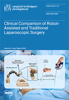
Cover Story (view full-size image): New future proof medical technology is developed at the Delft University of Technology in collaboration with the University of Malta that addresses relevant sustainability concerns. The picture shows the development of a versatile “waste free” robot platform that allows users to combine modules into their own solution for a specific surgical procedure. This solution can range from a simple reusable steerable handheld set towards a fully functional multi arm robot platform all based on the same unique new instrument driving technology. As Robot and Conventional laparoscopy merge into a single platform, the discussion about what is better will change in the future. View this paper
  • Issues are regarded as officially published after their release is announced to the table of contents alert mailing list.
  • You may sign up for e-mail alerts to receive table of contents of newly released issues.
  • PDF is the official format for papers published in both, html and pdf forms. To view the papers in pdf format, click on the "PDF Full-text" link, and use the free Adobe Reader to open them.
Order results
Result details
Select all
Export citation of selected articles as:
10 pages, 507 KiB  
Article
What Are the Risk Factors for Mechanical Failure in Spinal Arthrodesis? An Observational Study
by Vincenzo Peccerillo, Antonio Culcasi, Riccardo Ruisi, Francesca Amaducci, Maria Grazia Benedetti, Marco Girolami, Andrea Evangelista and Mattia Morri
Surg. Tech. Dev. 2024, 13(1), 87-96; https://0-doi-org.brum.beds.ac.uk/10.3390/std13010006 - 07 Mar 2024
Viewed by 340
Abstract
Background: The aim of this study was to identify the incidence of early mechanical failure in the first post-surgical year in patients who had undergone spinal surgery and to assess the related risk factors. Methods: A retrospective observational study was conducted examining all [...] Read more.
Background: The aim of this study was to identify the incidence of early mechanical failure in the first post-surgical year in patients who had undergone spinal surgery and to assess the related risk factors. Methods: A retrospective observational study was conducted examining all patients who consecutively underwent arthrodesis surgery. The incidence of postoperative mechanical failure during the first year was calculated as the primary outcome. Results: A total of 237 patients were identified for statistical analysis. The median age of the group of patients was 47 years (IQR of 44), and 66.6% were female. The incidence of mechanical failure in the first postoperative year was 5.1% overall, with 12 events, and the median time between surgery and the need for revision surgery was 5 months (IQR = 7.75). ASA score (OR = 2.39; p = 0.134), duration of the surgical procedure (OR = 1.27; p = 0.118), and inability to walk at discharge (OR = 7.86; p = 0.007) were independent risk factors associated with the mechanical failure. Conclusions: A higher ASA score and longer duration of surgery were risk factors for mechanical failure in the first year in patients who had undergone spinal surgery and must be carefully considered when planning spinal surgery. Early recovery of ambulation must be encouraged to prevent mechanical failure. Full article
Show Figures

Graphical abstract

11 pages, 2308 KiB  
Technical Note
A Detailed Exploration of the Ex Utero Intrapartum Treatment Procedure with Center-Specific Advancements
by Marta Domínguez-Moreno, Ángel Chimenea, María Remedios Viegas-González, Clara Morales-Muñoz, Lutgardo García-Díaz and Guillermo Antiñolo
Surg. Tech. Dev. 2024, 13(1), 76-86; https://0-doi-org.brum.beds.ac.uk/10.3390/std13010005 - 23 Feb 2024
Viewed by 562
Abstract
The Ex Utero Intrapartum Treatment (EXIT) procedure has long been an invaluable tool in managing complex fetal conditions requiring airway interventions during the transition from intrauterine to extrauterine life. This technical note offers an in-depth examination of the EXIT procedure, emphasizing the refinements [...] Read more.
The Ex Utero Intrapartum Treatment (EXIT) procedure has long been an invaluable tool in managing complex fetal conditions requiring airway interventions during the transition from intrauterine to extrauterine life. This technical note offers an in-depth examination of the EXIT procedure, emphasizing the refinements and innovations introduced at our center. The technique focuses on meticulous preoperative assessment and uses distinctive techniques and anesthetic methodologies. A multidisciplinary team assembles to plan the EXIT procedure, emphasizing patient communication and risk discussion. Our technique involves atraumatic access to the uterine cavity, achieved through the application of a uterine progressive distractor developed for this purpose. Following the use of this distractor, vascular clamps and a stapling device (Premium Poly Cs-57 Autosuture®, Medtronic) are employed. Our anesthetic approach employs general anesthesia with epidural catheter placement. Maternal operation involves low transverse laparotomy and intraoperative ultrasonography-guided hysterotomy. Fetal exposure includes gentle extraction or external version, ensuring airway access. After securing fetal airway access, umbilical cord clamping and maternal abdominal closure conclude the procedure. By revisiting the core principles of EXIT and incorporating center-specific advancements, we enhance our understanding and technical expertise. To our knowledge, this is the first time a detailed description of the technique has been published. Full article
Show Figures

Figure 1

18 pages, 1720 KiB  
Article
Objective Gait Analysis Using a Single-Point Wearable Sensor to Assess Lumbar Spine Patients Pre- and Postoperatively
by R Dineth Fonseka, Pragadesh Natarajan, Monish Movin Maharaj, Lianne Koinis, Luke Sy and Ralph Jasper Mobbs
Surg. Tech. Dev. 2024, 13(1), 58-75; https://0-doi-org.brum.beds.ac.uk/10.3390/std13010004 - 14 Feb 2024
Viewed by 722
Abstract
Background: Outcome measurement in lumbar surgery is traditionally performed using patient questionnaires that may be limited by subjectivity. Objective gait analysis may supplement patient assessment but must be clinically viable. We assessed gait metrics in lumbar spine patients pre- and postoperatively using a [...] Read more.
Background: Outcome measurement in lumbar surgery is traditionally performed using patient questionnaires that may be limited by subjectivity. Objective gait analysis may supplement patient assessment but must be clinically viable. We assessed gait metrics in lumbar spine patients pre- and postoperatively using a small and lightweight wearable sensor. Methods: This was a prospective observational study with intervention including 12 patients undergoing lumbar spine surgery and 24 healthy controls matched based on age and sex. All the subjects underwent gait analysis using the single-point wearable MetaMotionC sensor. The lumbar spine patients also completed traditional patient questionnaires including the Oswestry Disability Index (ODI). Results: The ODI score significantly improved in the patients from the baseline to six weeks postoperatively (42.4 to 22.8; p = 0.01). Simultaneously, the patients demonstrated significant improvements in gait asymmetry (asymmetry in step length, swing time, single support time, and double support time, by 17.4–60.3%; p ≤ 0.039) and variability (variability in gait velocity, step time, step length, stance time, swing time, single support time, and double support time, by 21.0–65.8%; p ≤ 0.023). After surgery, changes in most spatiotemporal (gait velocity, step length, stance time, swing time, and single limb support time) and asymmetry (asymmetry in step time, stance time, swing time, and single limb support time) metrics correlated strongly (magnitude of r = 0.581–0.914) and significantly (p ≤ 0.037) with changes in the ODI. Conclusions: Gait analysis using a single-point wearable sensor can demonstrate objective evidence of recovery in lumbar spine patients after surgery. This may be used as a routine pre- and postoperative assessment during scheduled visits to the clinic. Full article
Show Figures

Figure 1

36 pages, 2063 KiB  
Systematic Review
A Comparison of Clinical Outcomes of Robot-Assisted and Conventional Laparoscopic Surgery
by Storm Chabot, Jean Calleja-Agius and Tim Horeman
Surg. Tech. Dev. 2024, 13(1), 22-57; https://0-doi-org.brum.beds.ac.uk/10.3390/std13010003 - 31 Jan 2024
Viewed by 887
Abstract
Background: Although robot-assisted laparoscopic surgery has become more in popular, it remains unclear what clinical advantages it offers over conventional laparoscopic surgery. Objective: This (systematic) umbrella review aims to synthesize and compare the clinical outcomes of robot-assisted laparoscopic surgery versus conventional laparoscopic surgery. [...] Read more.
Background: Although robot-assisted laparoscopic surgery has become more in popular, it remains unclear what clinical advantages it offers over conventional laparoscopic surgery. Objective: This (systematic) umbrella review aims to synthesize and compare the clinical outcomes of robot-assisted laparoscopic surgery versus conventional laparoscopic surgery. Methods: A systematic literature search was conducted in PubMed and Scopus. All systematic reviews and meta-analyses published in the past five years that compared the clinical outcomes for cholecystectomy, colectomy, hysterectomy, nephrectomy, and/or prostatectomy were included. The quality of all included reviews was assessed with the AMSTAR 2 quality assessment tool. Each review’s study characteristics and primary sources were extracted, along with the quantitative and qualitative data for blood loss, rate of conversion to open surgery, hospitalization costs, incisional hernia rate, intraoperative complication rate, postoperative complication rate, length of hospital stay, operative time, readmission rate, and wound infection. Results: Fifty-two systematic reviews and (network) meta-analyses were included in this umbrella review, covering more than 1,288,425 patients from 1046 primary sources published between 1996 and 2022. The overall quality of the included reviews was assessed to be low or critically low. Robot-assisted laparoscopic surgery yielded comparable results to conventional laparoscopic surgery in terms of blood loss, conversion to open surgery rate, intraoperative complication rate, postoperative complication rate, readmission rate, and wound infection rate for most surgical procedures. While the hospitalization costs of robot-assisted laparoscopic surgery were higher and the operative times of robot-assisted laparoscopic surgery were longer than conventional laparoscopic surgery, robot-assisted laparoscopic surgery reduced the length of hospital stay of patients in nearly all cases. Conclusion: Robot-assisted laparoscopic surgery achieved comparable results with conventional laparoscopic surgery for cholecystectomy, colectomy, hysterectomy, nephrectomy, and prostatectomy based on ten clinical outcomes. Full article
Show Figures

Figure 1

13 pages, 640 KiB  
Systematic Review
Postoperative Cast Immobilization Might Be Unnecessary after Pelvic Osteotomy for Children with Developmental Hip Dysplasia: A Systematic Review
by Mohamed Mai, Renée A. van Stralen, Sophie Moerman and Christiaan J. A. van Bergen
Surg. Tech. Dev. 2024, 13(1), 9-21; https://0-doi-org.brum.beds.ac.uk/10.3390/std13010002 - 15 Jan 2024
Viewed by 736
Abstract
Background: Developmental dysplasia of the hip (DDH) is a common disorder of atypical hip development. Pelvic osteotomy (e.g., according to Salter, Pemberton or Dega) may be indicated for children with DDH at walking age. The most popular postoperative treatment is a hip spica [...] Read more.
Background: Developmental dysplasia of the hip (DDH) is a common disorder of atypical hip development. Pelvic osteotomy (e.g., according to Salter, Pemberton or Dega) may be indicated for children with DDH at walking age. The most popular postoperative treatment is a hip spica cast. Alternative postoperative options include abduction braces and non-weightbearing protocols combined with physical therapy. The aim of this systematic review was to determine the most effective form of postoperative treatment after unilateral pelvic osteotomy in children with DDH in terms of clinical and radiological outcomes and complications. Methods: A systematic review was conducted and reported according to Preferred Reporting Items for Systematic Reviews and Meta-Analysis 2020 guidelines and registered in the international prospective register of systematic reviews. Articles were selected from PubMed, Embase and Cochrane databases. The quality of all (non-)randomized included studies was assessed using the Methodological Index for Non-Randomized Studies (MINORS) criteria. Results: The search strategy yielded 3524 articles. Fourteen articles with 367 total hips were included in this review. A total of 312 hips were treated with spica casts, 49 with abduction braces and 6 with non-weightbearing protocols. The quality of evidence was moderate (MINORS, 3–12 points). All types of postoperative treatments had good clinical outcomes overall, without secondary displacement of the osteotomy. Clinical outcomes for spica casts were reported according to McKay’s criteria in 135 hips, with 123 excellent and 12 good results. Clinical outcomes for abduction braces showed satisfaction for all parents (49 of 49). The radiological outcome was overall well preserved with any postoperative treatment. There was a higher complication rate with the use of hip spica casts, including avascular necrosis, pain complaints and superficial infections. Conclusion: This systematic review showed no benefit of postoperative spica casts compared with abduction braces and avoidance of weightbearing after simple pelvic osteotomy for residual DDH. Full article
Show Figures

Figure 1

8 pages, 4343 KiB  
Case Report
Ultrasound-Assisted Removal of a Wooden Foreign Body Embedded in the Neck
by Daniele Vitali, Pietro Orlando, Giandomenico Maggiore, Oreste Gallo and Ilaria Bindi
Surg. Tech. Dev. 2024, 13(1), 1-8; https://0-doi-org.brum.beds.ac.uk/10.3390/std13010001 - 19 Dec 2023
Viewed by 595
Abstract
Objectives: The deep submucosal migration of ingested foreign bodies into the pharyngolaryngeal mucosa is a sporadic event, and its management can be very challenging. In the case of the failure of endoscopic retrieval, open surgical techniques are usually required, and intraoperative ultrasonography can [...] Read more.
Objectives: The deep submucosal migration of ingested foreign bodies into the pharyngolaryngeal mucosa is a sporadic event, and its management can be very challenging. In the case of the failure of endoscopic retrieval, open surgical techniques are usually required, and intraoperative ultrasonography can become a useful adjunct for identifying their precise localization. Methods: An 84-year-old woman presented with new-onset dysphagia and odynophagia after the accidental ingestion of a fragment of a toothpick a few hours before in the absence of hoarseness or respiratory distress. Ultrasonography and an unenhanced CT scan of the neck revealed a 3 cm linear foreign body embedded into the neck between the left pyriform sinus and the esophageal wall. Results: We report the removal of a fragment of a wooden toothpick deeply lodged between the left pyriform sinus and the esophageal wall, which was managed via an open transcervical approach with the aid of intraoperative ultrasound guidance. Conclusions: We suggest that both preoperative and intraoperative ultrasonography should represent the first-line imaging technique for deeply embedded neck foreign bodies. Full article
Show Figures

Figure 1

Previous Issue
Next Issue
Back to TopTop