Article

Insulin Controls Triacylglycerol Synthesis through Control of Glycerol Metabolism and Despite Increased Lipogenesis

by
Ana Cecilia Ho-Palma
1,2,
Pau Toro
1,
Floriana Rotondo
1,
María del Mar Romero
1,3,4,
Marià Alemany
1,3,4,
Xavier Remesar
1,3,4 and
José Antonio Fernández-López
1,3,4,*
1
Department of Biochemistry and Molecular Biomedicine, Faculty of Biology, University of Barcelona, 08028 Barcelona, Spain
2
Faculty of Medicine, Universidad Nacional del Centro del Perú, 12006 Huancayo, Perú
3
Institute of Biomedicine, University of Barcelona, 08028 Barcelona, Spain
4
Centro de Investigación Biomédica en Red Fisiopatología de la Obesidad y Nutrición (CIBER-OBN), 08028 Barcelona, Spain
*
Author to whom correspondence should be addressed.
Submission received: 7 February 2019 / Revised: 22 February 2019 / Accepted: 22 February 2019 / Published: 28 February 2019
(This article belongs to the Special Issue Dietary Fat and Human Health)

Abstract

:
Under normoxic conditions, adipocytes in primary culture convert huge amounts of glucose to lactate and glycerol. This “wasting” of glucose may help to diminish hyperglycemia. Given the importance of insulin in the metabolism, we have studied how it affects adipocyte response to varying glucose levels, and whether the high basal conversion of glucose to 3-carbon fragments is affected by insulin. Rat fat cells were incubated for 24 h in the presence or absence of 175 nM insulin and 3.5, 7, or 14 mM glucose; half of the wells contained 14C-glucose. We analyzed glucose label fate, medium metabolites, and the expression of key genes controlling glucose and lipid metabolism. Insulin increased both glucose uptake and the flow of carbon through glycolysis and lipogenesis. Lactate excretion was related to medium glucose levels, which agrees with the purported role of disposing excess (circulating) glucose. When medium glucose was low, most basal glycerol came from lipolysis, but when glucose was high, release of glycerol via breakup of glycerol-3P was predominant. Although insulin promotes lipogenesis, it also limited the synthesis of glycerol-3P from glucose and its incorporation into acyl-glycerols. We assume that this is a mechanism of adipose tissue defense to avoid crippling fat accumulation which has not yet been described.

1. Introduction

White adipose tissue (WAT) is a disperse organ [1], distributed in a number of locations in which its basic energy storage activity [2] is complemented by many other physiological functions [3,4,5]. In any case, its main role is to contribute to the defense of energy homoeostasis helping to control glucose [6], lipid [7], and amino acid [8] metabolism.
Most studies on WAT metabolism have been focused on adipocytes, since they are considered the characteristic and defining cells of WAT. However, adipose tissue structure is far more complex, containing a large number of cell types, not only mature adipocytes, in spite of them occupying most of the space because of their larger size [5]. Between 60 and 80% of the adipose tissue nucleated cells constitute the heterogeneous “stromal vascular fraction”, including stem cells, preadipocytes, endothelial cells, and macrophages [9]. Adipose tissue also contains other cell types such as fibroblasts, histiocytes, mast cells, lymphocytes, granulocytes, blood cells, and nerve terminals.
An excessive intake of energy may trigger the process that results in chronic insulin resistance and the proinflammatory state that characterizes metabolic syndrome [10]. In all this succession of events, proinflammatory adipokines, released by adipose tissue, play a significant role, as suggested by the fact that the adipose tissue of obese individuals expresses proinflammatory adipokines in a higher proportion than in those with normal weight [11]. Proinflammatory effects are not limited to increased circulating adipokines released by adipose tissue; excess nutrients such as circulating lipids or glucose may become akin to “toxic”, enhancing oxidative stress and promoting inflammatory responses, whereas food restriction to normal levels reduces both [12].
We recently found that, under normoxic conditions, adipocytes in primary cultures were able to convert huge amounts of the medium glucose to lactate and glycerol, fulfilling most of their energy needs through anaerobic glycolysis [13,14]. We postulated that this “wasting” of glucose might help to diminish hyperglycemia because of the large combined mass of WAT. In ex vivo studies, we also observed the accumulation of lactate in WAT masses [15], in agreement with the results observed in cultured cells and the low in vivo WAT oxygen consumption [16,17,18] and high WAT lactate production [18,19] observed in humans.
The “unnecessary” anaerobic use of glucose by adipocytes (and WAT as a whole) producing large amounts of 3-carbon fragments seems to be intrinsic to the tissue, because the high production of lactate occurred in the absence of external stimuli, and independent of the availability of oxygen. Given the assumed importance of insulin in the metabolism of adipose tissue, and because of its anabolic role, we wanted to discern whether insulin could indeed favor the accumulation of triacylglycerol (TAG) reserves in the adipocyte, since this way it would hinder the breakup of excess glucose to 3C fragments. The consequence could be the loss of the potential protective action of the tissue modulating hyperglycemia. Consequently, we investigated how insulin intervened on glucose disposal via glycolysis to 3C fragments, in contrast to its effect on fatty acid synthesis and increased TAG storage. We quantitatively analyzed the fate of labelled glucose and the metabolic changes induced by insulin on primary cultures of adipocytes. Quantitative analyses of metabolites and key gene expressions allowed us to obtain a wider picture of what is in fact the actual role of insulin in the handling of glucose loads by mature adipocytes.

2. Materials and Methods

2.1. Rats and Sampling

All animal handling procedures and the experimental setup were in accordance with the animal handling guidelines of the corresponding European and Catalan Authorities. The Committee on Animal Experimentation of the University of Barcelona specifically authorized the specific procedures used (# DAAM 6911).
Healthy adult male Wistar rats (Janvier, Le Genest-Saint Isle, France), weighing 399 ± 64 g were used. The animals were kept in two-rat cages with wood shards as bedding material, at 21–22 °C, and 50–60% relative humidity; lights were on from 08:00 to 20:00. The rats had unrestricted access to water and standard rat chow (#2014, Teklad Diets, Madison, WI, USA).
The rats were sacrificed, under isoflurane anesthesia, by exsanguination from the exposed aorta. They were dissected and samples of epididymal WAT were extracted and minced with scissors before further processing.

2.2. WAT Adipocyte Isolation and Incubation Procedures

Cells were isolated [20] at 37 °C for 1 h in a shaking bath using collagenase (LS004196, type I, from Worthington Biomedical, Lakewood, NJ, USA) in 2.5 volumes of modified Krebs–Henseleit buffer [21]. The cell suspension was filtered through a double layer of nylon hose, transferred to vertical syringes, and left standing for 5–6 min at room temperature. Adipocytes formed an upper loose cake, floating over a liquid phase that was slowly drained from the syringe; the adipocyte layer was gently re-suspended in fresh buffer (free of collagenase) and the process of mixing and draining was repeated twice, discarding the washing fluids. Aliquots of the adipocyte layer were used for incubation immediately after the final washing. All cell preparations were maintained at room temperature (approximately 22 °C), and manipulated within a time as short as possible. Cells were counted and their spherical (when free) diameters measured using ImageJ software (http://imagej.nih.gov/ij/).
The complete cell incubation procedure was previously described by us [14,22]. Briefly, cell incubations were carried out using 12-well plates (#734-2324VWR International BVBA/Sprl., Leuven, Belgium) filled with 1.7 mL of DMEM (#11966-DMEM-no glucose; Gibco, Thermo-Fisher Scientific, Waltham, MA, USA), supplemented with 30 mL/L fetal bovine serum (FBS, Gibco). The medium also contained 25 mM HEPES (Sigma-Aldrich, St Louis, MO, USA), 2mM glutamine (Lonza Biowhittaker, Radnor, PA, USA), 30 mg/mL delipidated bovine serum albumin (Millipore Calbiochem, Bedford, MA, USA), and 100 nM adenosine, 100 U/mL penicillin plus 100 mg/L streptomycin (Sigma-Aldrich). Half of the wells were supplemented with bovine insulin (Sigma-Aldrich): final concentration 175 nM.
The incubation medium was also supplemented with 14C-(U)-D glucose, (#ARC0122B, American Radiolabeled Chemicals, St. Louis, MO, USA; specific radioactivity 11 GBq/mmol). Final glucose concentrations in the wells were, nominally, 3.5, 7, or 14 mM. In the labelled samples, the amount of label added per well was approximately 394 Bq/mmol of glucose.
A “parallel” series of wells was developed, containing the same adipocytes’ suspension and identical medium composition and other conditions than those described above, but in which no label was added. These wells were used for cell gene transcription and medium metabolite analyses.
Each well received 400 μL of the corresponding cell suspension; after initial sampling (100 µL), the final incubation volume was 2.0 mL. The cells were incubated at 37 °C in a chamber ventilated with air supplemented with 5% CO2, which gave a theoretical pO2 of 20 kPa [23]. The cells were incubated for 24 h without any further intervention.

2.3. Processing of the Incubation Media: Label Distribution

The label-containing samples were used to fraction the label distribution applying a protocol previously described by us [13]. Lactate (including pyruvate) label was determined using centrifuge microcolumns made up with sieve-filter type centrifugation inserts (Ultrafree-MC, Millipore, Bedford, MA, USA) containing 250 mg of hydrated, spin-dried cationic-form Dowex 1 × 2 ion exchange resin (Sigma-Aldrich) [13]. The retained lactate was eluted with acid and counted.
The medium free of lactate was used in part to convert all glucose to gluconate by incubation with glucose oxidase (type VII from Aspergillus niger, Sigma-Aldrich); as well as catalase (from bovine liver, Sigma-Aldrich). Catalase was added to destroy H2O2 and to help maintain O2 availability. The change of nonionic glucose to gluconate allowed its retention (and acidic elution) using microcolumns as described for lactate. The label retained was that of the unaltered glucose remaining in the medium after incubation [13,24].
A second aliquot of the label-containing medium free of lactate was treated with glycerol kinase (from Escherichia coli, #G6278, Sigma-Aldrich) and ATP for the conversion of glycerol to glycerol-3P. The change in ionization was used to remove the glycerol (as glycerol-3P) from the medium, eluting it with acid and thus counting the label retained in the glycerol moiety [13,25].
Combination of “cold” metabolite measurements and their radioactivity allowed us to calculate the fate of the initial glucose label under all conditions tested and to estimate the specific-C radioactivity for all of them.
Carbon dioxide production along the lipogenic process was estimated from the label present in the TAG fatty acids. We considered that for every 3C from glucose, one is lost in the conversion to 2C fragments (from pyruvate to acetyl), and assumed that 1 mole of CO2 was produced in the pentose-P pathway for each 2 moles of NADPH generated (explained in more detail in Ho-Palma et al. [13]). This amount of CO2 represents the minimum possible values, since the CO2 that could have been produced in oxidative processes is not included.

2.4. Processing of Labelled Cell Components

The procedure for measuring label distribution in the different cell fractions and media has been previously developed, tested, and quantified [13]. Briefly, the cells incubated with labelled glucose were weighed, frozen with liquid nitrogen, transferred to glass tubes, and immediately extracted with chilled peroxide-free diethyl ether [26]. The aqueous fraction (and interface) was wholly used to estimate radioactivity. It contained mainly glycogen, but also cell proteins, metabolites, membrane microsomes, and non-lipophilic debris. The organic phase, containing essentially TAG, was dried, weighed, re-dissolved in ethyl ether, and saponified with KOH in ethanol in the cold. The ether-insoluble potassium soaps were extracted and counted. The aqueous phase, containing all glycerides-glycerol, was also removed and counted [13]. Soap label was that of TAG fatty acids.

2.5. Analysis of Metabolites in the Medium

Medium glucose was measured using a glucose oxidase-peroxidase kit (#11504, Biosystems, Barcelona, Spain) to which we added 740 nkat/mL mutarotase (porcine kidney, 136A5000, Calzyme, St. Louis, MO, USA) [27]. Lactate was measured with kit 1001330 (Spinreact, Sant Esteve d’en Bas, Spain); glycerol was estimated with kit #F6428 (Sigma-Aldrich). Non-esterified fatty acids (NEFA) were measured using kit NEFA-HR (Wako Life Sciences, Mountain View, CA, USA).

2.6. Gene Expression Analysis

Total cell RNA was extracted from all the harvested cells (parallel, non-labelled plates) using the Tripure reagent (Roche Applied Science, Indianapolis, IN, USA). RNA content was quantified in an ND-1000 spectrophotometer (NanoDrop Technologies, Wilmington, DE, USA). RNA samples were reverse transcribed using oligo-dT primers (Gene Link, Westchester, NY, USA) and the MMLV reverse transcriptase (Promega, Madison, WI, USA) system.
Real-time PCR amplification was carried out using 10 μL amplification mixtures containing Power SYBR Green PCR Master Mix (Applied Biosystems, Foster City, CA, USA), 4 ng of reverse-transcribed RNA, and 150 nmol of primers. Reactions were run on an ABI PRISM 7900 HT detection system (Applied Biosystems) using a fluorescent threshold manually set to 0.5 for all runs.
A semi-quantitative approach for the estimation of the concentration of specific gene mRNAs per unit of tissue weight was used [28]. Arbp was the charge control gene [29]. We expressed the data as the number of transcript copies per adipocyte. The genes analyzed and a list of primers used are presented in Table 1.

2.7. Statistical Procedures
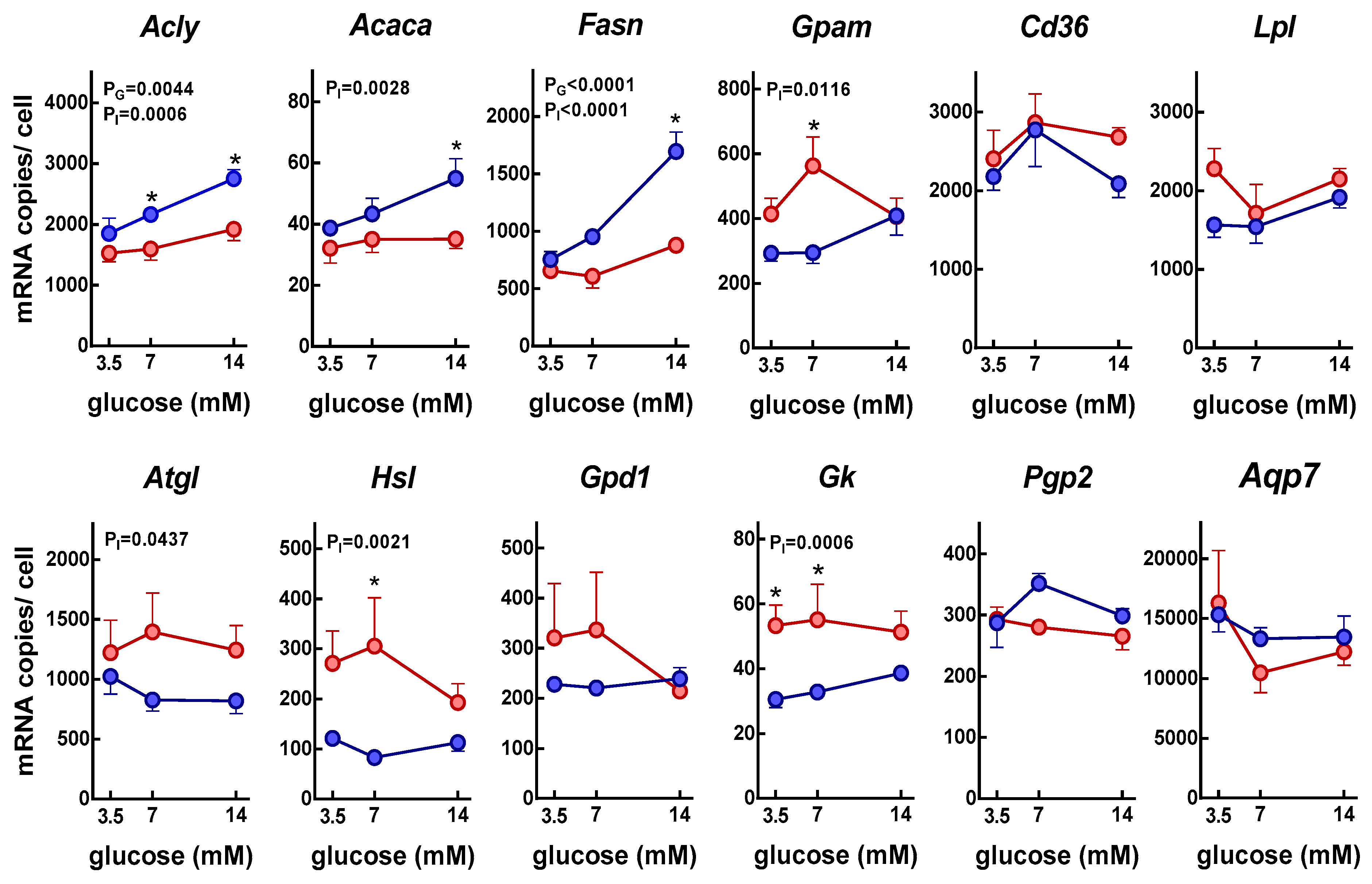
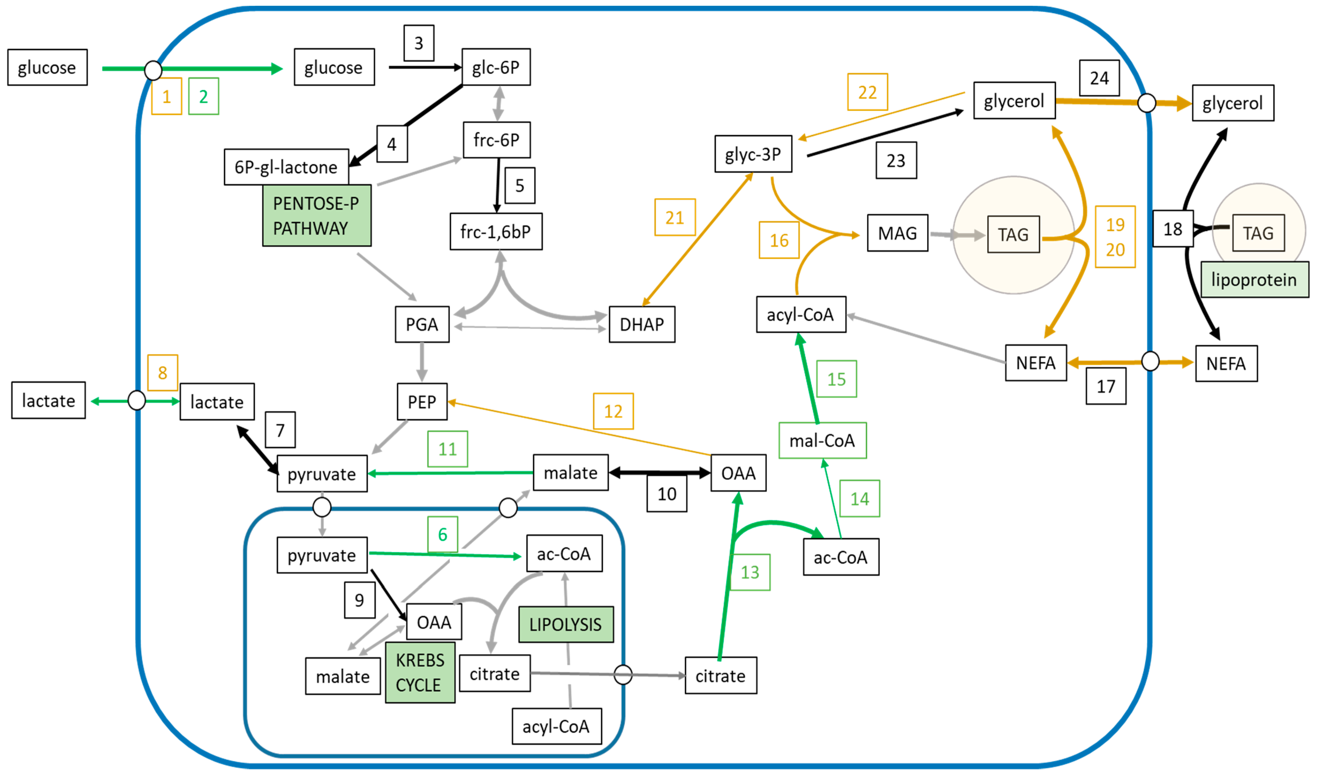
Statistical analyses and the establishment of significant differences between groups (two-way ANOVAs and post-hoc Sidak test) were done with the GraphPad Prism 6 program (GraphPad Software, La Jolla, CA, USA).

3. Results

Figure 1 shows the rates of glucose uptake and other metabolite efflux after the incubation of adipocytes for 24 h with 3.5, 7, or 14 mM glucose, in the presence or absence of insulin. Glucose uptake by adipocytes was slightly affected by initial glucose concentration showing a peak with 7 mM initial glucose. Lactate efflux was also affected by initial glucose concentration, being its efflux greater at 7 and 14 mM glucose; conversely, NEFA efflux decreased with glucose concentration. Insulin, increased lactate efflux, slightly decreased that of glycerol, and almost abolished NEFA output in adipocytes.
Figure 2 shows the specific carbon radioactivities of the fractions in which direct measurement of cold and labelled compounds were done. The data are shown as percentages of the initial glucose carbon specific activity. As expected, glucose specific activity was maintained independently of insulin or initial glucose concentration; that of lactate was also maintained on the same range than glucose. In adipocytes, specific radioactivities of free glycerol increased with the amount of glucose in the medium: with elevated concentrations of glucose (14 mM initial glucose), most of the glycerol was obtained from glucose; with lower glucose concentrations, a progressively smaller proportion of glycerol had its origin from glucose.
In adipocytes, the specific activity of glycerides-glycerol was low, and was unaffected by the initial glucose concentration or the presence of insulin. The specific radioactivity of the glycerides-fatty acids was very low, close to one order of magnitude lower than that of glycerol in glycerides, although in this case the presence of insulin increased specific radioactivity.
Figure 3 shows the main metabolites obtained from glucose metabolism. The major products, at least with high glucose concentrations, were lactate and glycerol. In the case of glycerol, the results were different from those obtained in Figure 1, since glycerol production from glucose and its release to the medium increased with the concentration of glucose; however, glycerol (from glucose) incorporated into TAG was constant and independent of both glucose concentration and insulin. On the contrary, insulin stimulated the synthesis of lactate and fatty acids (and consequently CO2) from glucose. Glucose incorporation into glycogen was small and unaffected by insulin.
Figure 4 presents the changes in gene expression of key enzymes and transporters implicated in the glycolytic utilization of glucose by adipocytes. Data were expressed as the number of copies of the corresponding mRNA per cell. Each well contained 6.32 ± 0.14 × 10⁵ adipocytes (i.e., 0.34 g of adipose tissue). The glucose transporter gene Glut1, showed a similar number of copies of its mRNA per cell for all glucose concentration groups, but insulin decreased its expression. The pattern for glucose transporter gene Glut4 was different, since both insulin and glucose increased its expression; in the case of glucose, the increase only occurred in the presence of insulin. Expression levels of Glut4 were lower than those of Glut1.
Expressions of hexokinase (Hk1), 6-phosphofructokinase liver type (Pfkl), lactate dehydrogenase (Ldha), pyruvate carboxylase (Pc), and malate dehydrogenase (Mdh1) were unaffected by glucose concentration and insulin. The monocarboxylate transporter gene (Mct1), responsible for lactate (and pyruvate) efflux, slightly decreased its expression in the presence of insulin.
The expression of phospho-enolpyruvate carboxykinase (Pck1), a main control point for the regulation of gluconeogenesis in liver, was lower in the presence of insulin, but only at 7 mM glucose.
The data suggest that insulin increased the activity of pyruvate dehydrogenase, the enzyme responsible for the conversion of pyruvate to acetyl-CoA, by decreasing the expression of its main inhibitor, pyruvate dehydrogenase kinase 4 (Pdk4).
The main providers of NADPH in the cytoplasm to sustain lipogenesis are malic enzyme and the reductive part of the pentose-phosphate cycle. There was a tendency to increase the expression of Me1 but not G6pd by insulin.
Figure 5 shows the changes in gene expression of key enzymes and transporters implicated in the glycerol and lipid metabolism of adipocytes. When we analyzed the expression of three key points of control of lipogenesis, ATP: citrate lyase (Acly), acetyl-CoA carboxylase (Acaca), and fatty acid synthase (Fas) genes, a similar profile was observed, with higher expressions in the presence of insulin and higher glucose concentrations. This latter effect was only apparent in the presence of both glucose and insulin. Nevertheless, the expression of glycerol-3P acyl-transferase (Gpam), a critical enzyme for TAG synthesis, was inhibited by insulin.
The expression of the main enzymes responsible for endogenous lipolysis (triacylglycerol lipase, Atgl, and hormone-sensitive lipase, Hsl) was inhibited by insulin. In the case of lipoprotein lipase (Lpl) responsible for the degradation of triacylglycerols of exogenous origin, it was not affected by glucose or insulin.
The expression of the enzymes responsible for the synthesis of glycerol 3-phosphate in the adipocyte tended to be inhibited by the presence of insulin. This was the case of the glyceraldehyde-3-phosphate dehydrogenase (Gdp1; although the inhibition was not statistically significant, p = 0.0628), responsible for the synthesis of glycerol 3-phosphate from dihydroxyacetone-phosphate, and that of glycerokinase (Gk) responsible for the phosphorylation of glycerol. The expression of this last enzyme was very low. In the case of the enzymes that use glycerol-3-phosphate as substrate, the expression of phosphoglycolate phosphatase (Pgp2), which catalyzes the hydrolysis of glycerol 3-phosphate to glycerol, was not affected.
Finally, the expression of aquaporin 7, a transporter playing an important role in glycerol transport, and Cd36, that imports fatty acids inside cells, was unaffected by the conditions tested in our study.

4. Discussion

Adipose tissue is a complex organ, made up of different tissue masses. One of the limitations of the study was that only adipocytes of epididymal origin were used. Previous studies indicate the existence of differences in the amount of lactate and glycerol produced by adipocytes from different locations, as well as in the expression of genes related to both carbohydrate and lipid metabolism, an aspect mainly affected by sex [30,31]. In any case, adipose tissues from all sites seem to share a similar metabolic profile, only showing differences that are mainly quantitative.
Under basal conditions, and in the absence of insulin, isolated adipocytes and intact WAT use significant amounts of glucose and secrete high amounts of 3C metabolites as lactate and glycerol. These 3C compounds may be used for hepatic gluconeogenesis [32] or lipogenesis [33], or for energy purposes in other tissues.
WAT limits excessive TAG accumulation (excess energy) in the tissue, reducing substrate availability by decreasing blood flow [34]. Additionally, with this high production of lactate and glycerol release (i.e., glucose breakup), WAT defends itself from excess glucose, preventing an inordinate enlargement of its TAG stores [23]. The utilization of 3C metabolites by tissues is unaffected by insulin resistance and circumvents the strict regulation of glycolysis, providing partially metabolized and directly usable energy substrates. The results presented here agree with this interpretation, confirming that most of the glucose taken up by adipocytes is just returned to the medium as 3C metabolites, essentially lactate and glycerol [35], thus helping to lower circulating glucose availability.
As expected, insulin elicited an anabolic response in WAT, increasing glucose uptake [36], as well as the flow of carbon through glycolysis and lipogenesis [37]. Figure 6 shows the actions of insulin on these main metabolic pathways in the context of the glucose-fatty acid metabolism in the adipocyte.
Basal glucose uptake was high, even in the absence of insulin, mainly due to the high expression of Glut1, a widely distributed insulin-independent glucose transporter. Insulin increased glucose uptake, despite a lower expression of Glut1, compensated by a higher expression of Glut4, which facilitates glucose transport into insulin-sensitive cells. Glut4 is responsible for the insulin-regulated glucose transport into muscle and adipose cells [38], but a large proportion of glucose enters most body cells by means of Glut1. This is the most widely expressed hexose transporter, whose main role is assumed to maintain basal glucose transport in most types of cells [39,40]. Glut1 is regulated through the control of gene expression [41,42,43], and only in part via regulative control, which is the main control system for Glut4 [44].
It has been described that hypoxia increases monosaccharide uptake capacity in adipocytes, contributing to adipose tissue dysregulation in obesity [45]. However, the effects on glucose transporters are the opposite of those induced by insulin: exposure of adipocytes to hypoxia and increased Glut1 expression, in contrast with no changes in Glut4 [45].
Although there were no changes in the expression of genes related to the mainstream glycolysis (such as hexokinase and phosphofructokinases), there was a clear increase in the flow of carbon along the glycolytic pathway, as shown by the increased glucose uptake of cultured adipocytes and higher lactate efflux to the medium. The absence of changes in the expression of phosphofructokinases, one of the key regulatory and rate limiting steps of glycolysis, may be explained in part because phosphofructokinase regulation of glycolysis is essentially done through allosteric mechanisms [46].
Lactate and glycerol production (from glucose) helps sustain the basic energy needs of the cell through a fully anaerobic (albeit facultative, since it occurs under normoxic conditions) process [23]. The sheer size of adipocytes, and thin cytoplasm layer around its huge lipid vacuole, hamper the intracellular circulation of substrates, thus limiting and compartmentalizing most metabolic activities. In adipose tissue, glucose can be taken up easily from the circulation by adipocytes and converted anaerobically to lactate, pyruvate, or glycerol, covering the minimal needs of ATP. However, the production of acetyl-CoA requires access to mitochondria, few and sparsely distributed in large adipocytes [47]. Additionally, glucose utilization helps lower glycaemia, breaking up 6C to 3C molecules. This way, a portion of circulating glucose was substituted in large proportions by less-regulated 3C substrates used elsewhere for energy but also for splanchnic lipogenesis or gluconeogenesis. Therefore, WAT lactate efflux may be considered primarily a normal consequence of the need to use glycolytic (anaerobic) ATP and to eliminate excess circulating glucose. Consequently, it should not be considered a specific indicator of hypoxia, despite its generalized association with lactate [48]. This is reinforced by the fact that the high production of lactate (and that of glycerol) from glucose only occurs in the presence of high concentrations of glucose (in our case, 7 and 14 mM), although glucose uptake by the adipocyte is similar in all cases. Glucose is used almost entirely anaerobically at 14 mM glucose concentrations. Accordingly, higher circulating levels of lactate and glycerol are observed in type 2 diabetes [49]; likewise, obese women with higher glucose levels after an oral glucose tolerance test, release more glycerol and lactate from their subcutaneous adipose tissue, both in the postabsorptive state and after the oral glucose tolerance test [50].
On the other hand, the global production of glycerol was preserved and sustained, remaining practically constant regardless of the medium initial glucose concentration. This maintained production of glycerol by adipocytes in amounts similar or even greater than those of lactate, together with the different origin of glycerol according to the availability of glucose in the medium, suggest that glycerol release represents a constitutive process rather independent of external factors. Insulin only slightly decreased glycerol release to the medium, probably a consequence of the inhibition of lipolysis by insulin. In any case, the limitation of lipolysis by insulin was minimal (at the limit of significance in Figure 1C, and absent in Figure 2B) under the in vitro conditions tested. This may be due to the absence of any previous stimulation of lipolysis and/or to the presence of adenosine (a known inhibitor of lipolysis [51]) in the medium. Glycerol-3P, derived from glucose via trioses-P, is hydrolyzed to glycerol by a glycerophosphatase, as previously postulated by us [52]. This phosphatase, recently described in liver, is also present in WAT [53]. The expression of this enzyme (Pgp2) was unaffected by both glucose and insulin and was also not correlated with the lower efflux of glycerol observed in insulin-treated cells. The removal of excess glycerol-3-P under heavy glycolytic pressure (high glucose levels, insulin activation of Glut4) may help lower its cell pool, thus helping to maintain a lower glycerol esterification with acyl-CoA and thus downregulate TAG synthesis.
The equilibrium between these pathways—(a) incorporation into the acyl-glycerol pool or (b) direct generation of glycerol from glycerol-3P—seems to be a critical point in the regulation of TAG synthesis, regulated by insulin, which lowers the expression of glycerol-P acyl-transferase despite a marked increase in gene expressions along the lipogenic pathway. Glycerol-3-phosphate acyltransferase is the rate-limiting enzyme in the glycerolipid synthesis pathway [54]. Thus, this critically different regulation of glycerol-P acyl-transferase expression, with respect to the other lipogenic enzymes, can be tentatively explained because the induction of glycerol-P acyl-transferase mRNA by insulin is mediated through the expression of SREBP-1, independently of ChREBP [55,56]. Insulin increases SREBP-1 expression in liver and adipose tissue, but only increases the post-translational activation of SREBP-1c in liver, decreasing it in adipose tissue [57]; consequently, it inhibits the expression of adipose glycerol-P acyl-transferase.
The production of glycerol is more complex than that of lactate. Although its contribution to 3C fragment production from glucose and lowering glycaemia may also be accomplished by lactate, glycerol synthesis is nevertheless preferred. These data agree with our previous findings indicating that adipocytes’ glycerol production is a definite objective in addition to being a 3C substrate [14].
The lower production of glycerol and, therefore, the attempt to limit the accumulation of lipid in the adipocyte, has an obvious protective effect for the adipocyte itself. It can be speculated that this action, in contrast, could be counterproductive for the organism, since it may favor the ectopic deposition of lipid in other tissues. However, fat content in all WAT depots change in a proportion similar to that of the lipids in the rest of organs and tissues [2], which makes this possibility highly improbable. Similarly, although adipocytes may potentially decrease blood glucose levels by releasing lactate, it has been described that high hepatic lactate levels favor the appearance of NASH [58]. Again, a protective effect in the tissue could have unwanted consequences favoring the development of metabolic syndrome-related pathologies.
The higher incorporation of the glucose carbons into fatty acids of the TAG in the presence of insulin, but without changes in the incorporation of glycerol into the TAG, suggests that insulin induces a higher turnover of TAG. However, we cannot distinguish whether the origin of these TAG is endogenous or exogenous, from the action of the LPL on the adipocyte debris present in the medium. The activation of TAG turnover [14], when glycerogenic flow decreases, is contrary to the assumed focus of insulin-stimulated adipocytes, where lipogenesis and inhibition of lipolysis are trends clearly established through gene expression patterns.

5. Conclusions

In conclusion, under conditions of high availability of glucose and energy, WAT (at least the adipocytes) defend themselves to prevent the pathologic accumulation of excess energy. Part of the panoply of adipose tissue defense systems to prevent this damage include the following: (a) lower blood flow through the tissue, to decrease the arrival of nutrients [34]; (b) the adipocyte massively metabolizes excess glucose to fragments of three carbons that can be used for energy elsewhere, but also helping to decrease hyperglycemia; (c) WAT, especially under conditions of obesity, is able to inactivate large amounts of insulin [59], thus limiting its lipogenic effect; and (d) last, but not least, as we have shown here, insulin priority rests on the derivation of excess C to the secretion of glycerol rather than on a higher incorporation of glycerol-3P into the synthesis of TAG. In all cases, the measures are short term, and may have only a limited success since, in any case, the presence of insulin induces a higher incorporation of glucose carbon into the adipocyte TAG pool.

Author Contributions

Conceptualization, M.A., X.R., and J.A.F.-L.; Data curation, A.C.H.-P.; Formal analysis, J.A.F.-L.; Funding acquisition, M.A. and X.R.; Investigation, A.C.H.-P., P.T., F.R., and M.d.M.R.; Methodology, A.C.H.-P., F.R., and M.A.; Resources, X.R.; Supervision, J.A.F.-L.; Validation, X.R. and J.A.F.-L.; Visualization, J.A.F.-L.; Writing—original draft, J.A.F.-L.; Writing—review and editing, A.C.H.-P., P.T., F.R., M.d.M.R., M.A., X.R., and J.A.F.-L.

Funding

This study has not been specifically funded by any financing agency, although the University of Barcelona provided funds for complementary work and the Centro de Investigación Biomédica en Red de la Fisiopatología de la Obesidad y Nutrición (CIBER-OBN) contributed partially to this study.

Conflicts of Interest

The authors declare no conflict of interest. The funders had no role in the design of the study; in the collection, analyses, or interpretation of data; in the writing of the manuscript; or in the decision to publish the results.

References

  1. Cinti, S. The adipose organ. Prostaglandins Leukot. Essent. Fat. Acids 2005, 73, 9–15. [Google Scholar] [CrossRef] [PubMed]
  2. Romero, M.M.; Roy, S.; Pouillot, K.; Feito, M.; Esteve, M.; Grasa, M.M.; Fernández-López, J.A.; Alemany, M.; Remesar, X. Treatment of rats with a self-selected hyperlipidic diet, increases the lipid content of the main adipose tissue sites in a proportion similar to that of the lipids in the rest of organs and tissues. PLoS ONE 2014, 9, e90995. [Google Scholar] [CrossRef] [PubMed]
  3. Trayhurn, P. Endocrine and signalling role of adipose tissue: New perspectives on fat. Acta Physiol. Scand. 2005, 184, 285–293. [Google Scholar] [CrossRef] [PubMed]
  4. Hauner, H. The new concept of adipose tissue function. Physiol. Behav. 2004, 83, 653–658. [Google Scholar] [CrossRef] [PubMed]
  5. Alemany, M.; Fernández-López, J.A. Adipose tissue: Something more than just adipocytes. Curr. Nutr. Food Sci. 2006, 2, 141–150. [Google Scholar] [CrossRef]
  6. Rosen, E.D.; Spiegelman, B.M. Adipocytes as regulators of energy balance and glucose homeostasis. Nature 2006, 444, 847–853. [Google Scholar] [CrossRef] [PubMed] [Green Version]
  7. Arner, P.; Bernard, S.; Salehpour, M.; Possnert, G.; Liebl, J.; Steier, P.; Buchholz, B.A.; Eriksson, M.; Arner, E.; Hauner, H.; et al. Dynamics of human adipose lipid turnover in health and metabolic disease. Nature 2011, 478, 110–113. [Google Scholar] [CrossRef] [PubMed] [Green Version]
  8. Arriarán, S.; Agnelli, S.; Remesar, X.; Fernández-López, J.A.; Alemany, M. Effects of sex and site on amino acid metabolism enzyme gene expression and activity in rat white adipose tissue. PeerJ 2015, 3, e1399. [Google Scholar] [CrossRef] [PubMed] [Green Version]
  9. Tchoukalova, Y.D.; Sarr, M.G.; Jensen, M.D. Measuring committed preadipocytes in human adipose tissue from severely obese patients by using adipocyte fatty acid binding protein. Am. J. Physiol. Regul. Integr. Comp. Physiol. 2004, 287, R1132–R1140. [Google Scholar] [CrossRef] [PubMed] [Green Version]
  10. Johnson, A.R.; Milner, J.J.; Makowski, L. The inflammation highway: Metabolism accelerates inflammatory traffic in obesity. Immunol. Rev. 2012, 249, 218–238. [Google Scholar] [CrossRef] [PubMed]
  11. Makki, K.; Froguel, P.; Wolowczuk, I. Adipose tissue in obesity-related inflammation and insulin resistance: Cells, cytokines, and chemokines. ISRN Inflamm. 2013, 139239. [Google Scholar] [CrossRef] [PubMed]
  12. Dandona, P.; Aljada, A.; Chaudhuri, A.; Mohanty, P.; Garg, R. Metabolic syndrome: A comprehensive perspective based on interactions between obesity, diabetes, and inflammation. Circulation 2005, 111, 1448–1454. [Google Scholar] [CrossRef] [PubMed]
  13. Ho-Palma, A.C.; Rotondo, F.; Romero, M.M.; Memmolo, S.; Remesar, X.; Fernández-López, J.A.; Alemany, M. A method for the measurement of lactate, glycerol and fatty acid production from 14C-glucose in primary cultures of rat epididymal adipocytes. Anal. Methods 2016, 8, 7873–7885. [Google Scholar] [CrossRef]
  14. Rotondo, F.; Ho-Palma, A.C.; Remesar, X.; Fernández-López, J.A.; Romero, M.M.; Alemany, M. Glycerol is synthesized and secreted by adipocytes to dispose of excess glucose, via glycerogenesis and increased acyl-glycerol turnover. Sci. Rep. 2017, 7, 8983. [Google Scholar] [CrossRef] [PubMed] [Green Version]
  15. Arriarán, S.; Agnelli, S.; Sabater, D.; Remesar, X.; Fernández-López, J.A.; Alemany, M. Evidences of Basal lactate production in the main white adipose tissue sites of rats. Effects of sex and a cafeteria diet. PLoS ONE 2015, 10, e0119572. [Google Scholar] [CrossRef] [PubMed]
  16. Hodson, L. Adipose tissue oxygenation: Effects on metabolic function. Adipocyte 2014, 3, 75–80. [Google Scholar] [CrossRef] [PubMed]
  17. Goossens, G.H.; Bizzarri, A.; Venteclef, N.; Essers, Y.; Cleutjens, J.P.; Konings, E.; Jocken, J.W.; Cajlakovic, M.; Ribitsch, V.; Clément, K.; et al. Increased adipose tissue oxygen tension in obese compared with lean men is accompanied by insulin resistance, impaired adipose tissue capillarization, and inflammation. Circulation 2011, 124, 67–76. [Google Scholar] [CrossRef] [PubMed]
  18. Hodson, L.; Humphreys, S.M.; Karpe, F.; Frayn, K.N. Metabolic signatures of human adipose tissue hypoxia in obesity. Diabetes 2013, 62, 1417–1425. [Google Scholar] [CrossRef] [PubMed]
  19. DiGirolamo, M.; Newby, F.D.; Lovejoy, J. Lactate production in adipose tissue: A regulated function with extra-adipose implications. FASEB J. 1992, 6, 2405–2412. [Google Scholar] [CrossRef] [PubMed]
  20. Rodbell, M. Metabolism of Isolated Fat Cells: I. Effects of hormones on glucose metabolism and lipolysis. J. Biol. Chem. 1964, 239, 375–380. [Google Scholar] [PubMed]
  21. Krebs, H.A.; Henseleit, K. Untersuchungen über die Harnstoffbildung im Tierkörper. Klin. Wochenschr. 1932, 11, 757–759. [Google Scholar] [CrossRef]
  22. Rotondo, F.; Romero, M.M.; Ho-Palma, A.C.; Remesar, X.; Fernández-López, J.A.; Alemany, M. Quantitative analysis of rat adipose tissue cell recovery, and non-fat cell volume, in primary cell cultures. PeerJ 2016, 4, e2725. [Google Scholar] [CrossRef] [PubMed] [Green Version]
  23. Sabater, D.; Arriarán, S.; Romero, M.M.; Agnelli, S.; Remesar, X.; Fernández-López, J.A.; Alemany, M. Cultured 3T3L1 adipocytes dispose of excess medium glucose as lactate under abundant oxygen availability. Sci. Rep. 2014, 4, 3663. [Google Scholar] [CrossRef] [PubMed] [Green Version]
  24. Azain, M.J.; Kasser, T.R.; Baile, C.A. A method for the determination of glucose synthesis in isolated bovine hepatocytes. J. Nutr. Biochem. 1999, 10, 205–209. [Google Scholar] [CrossRef]
  25. Crespi, C.; Gastalver, G.; Palou, A.; Roca, P. Enzymatic determination of carbon (14C)-labeled glycerol in biological samples. J. Biochem. Biophys. Methods 1995, 30, 179–183. [Google Scholar] [CrossRef]
  26. Ferraz, T.P.L.; Fiúza, M.C.; Dos Santos, M.L.A.; Pontes de Carvalho, L.; Soares, N.M. Comparison of six methods for the extraction of lipids from serum in terms of effectiveness and protein preservation. J. Biochem. Biophys. Methods 2004, 58, 187–193. [Google Scholar] [CrossRef] [PubMed]
  27. Oliva, L.; Baron, C.; Fernández-López, J.A.; Remesar, X.; Alemany, M. Marked increase in rat red blood cell membrane protein glycosylation by one-month treatment with a cafeteria diet. PeerJ 2015, 3, e1101. [Google Scholar] [CrossRef] [PubMed] [Green Version]
  28. Romero, M.M.; Grasa, M.M.; Esteve, M.; Fernández-López, J.A.; Alemany, M. Semiquantitative RT-PCR measurement of gene expression in rat tissues including a correction for varying cell size and number. Nutr. Metab. 2007, 4, 26. [Google Scholar] [CrossRef] [PubMed] [Green Version]
  29. Bamias, G.; Goukos, D.; Laoudi, E.; Balla, I.G.; Siakavellas, S.I.; Daikos, G.L.; Ladas, S.D. Comparative study of candidate housekeeping genes for quantification of target gene messenger RNA expression by real-time PCR in patients with inflammatory bowel disease. Inflamm. Bowel Dis. 2013, 19, 2840–2847. [Google Scholar] [CrossRef] [PubMed]
  30. Rotondo, F.; Ho-Palma, A.C.; Remesar, X.; Fernández-López, J.A.; Romero, M.M.; Alemany, M. Effect of sex on glucose handling by adipocytes isolated from rat subcutaneous, mesenteric and perigonadal adipose tissue. PeerJ 2018, 6, e5440. [Google Scholar] [CrossRef] [PubMed]
  31. Rotondo, F.; Ho-Palmaa, A.C.; Romero, M.M.; Remesar, X.; Fernández-López, J.A.; Alemany, M. Higher lactate production from glucose in cultured adipose nucleated stromal cells than for rat adipocytes. Adipocyte 2019. [Google Scholar] [CrossRef] [PubMed]
  32. Ross, B.D.; Hems, R.; Krebs, H.A. The rate of gluconeogenesis from various precursors in the perfused rat liver. Biochem. J. 1967, 102, 942–951. [Google Scholar] [CrossRef] [PubMed] [Green Version]
  33. Katz, J.; Wals, P.A. Lipogenesis from lactate in rat adipose tissue. Biochim. Biophys. Acta 1974, 348, 344–356. [Google Scholar] [CrossRef]
  34. Alemany, M. Regulation of adipose tissue energy availability through blood flow control in the metabolic syndrome. Free Radic. Biol. Med. 2012, 52, 2108–2119. [Google Scholar] [CrossRef] [PubMed]
  35. Jansson, P.A.; Larsson, A.; Smith, U.; Lonnroth, P. Glycerol production in subcutaneous adipose tissue in lean and obese humans. J. Clin. Investig. 1992, 89, 1610–1617. [Google Scholar] [CrossRef] [PubMed]
  36. Watson, R.T.; Kanzaki, M.; Pessin, J.E. Regulated membrane trafficking of the insulin-responsive glucose transporter 4 in adipocytes. Endocr. Rev. 2004, 25, 177–204. [Google Scholar] [CrossRef] [PubMed]
  37. Saltiel, A.R.; Kahn, C.R. Insulin signalling and the regulation of glucose and lipid metabolism. Nature 2001, 414, 799–806. [Google Scholar] [CrossRef] [PubMed]
  38. Huang, S.; Czech, M.P. The GLUT4 glucose transporter. Cell Metab. 2007, 5, 237–252. [Google Scholar] [CrossRef] [PubMed]
  39. Manolescu, A.R.; Witkowska, K.; Kinnaird, A.; Cessford, T.; Cheeseman, C. Facilitated hexose transporters: New perspectives on form and function. Physiology 2007, 22, 234–240. [Google Scholar] [CrossRef] [PubMed]
  40. Kaestner, K.H.; Christy, R.J.; McLenithan, J.C.; Braiterman, L.T.; Cornelius, P.; Pekala, P.H.; Lane, M.D. Sequence, tissue distribution, and differential expression of mRNA for a putative insulin-responsive glucose transporter in mouse 3T3-L1 adipocytes. Proc. Natl. Acad. Sci. USA 1989, 86, 3150–3154. [Google Scholar] [CrossRef] [PubMed]
  41. Behrooz, A.; Ismail-Beigi, F. Dual control of glut1 glucose transporter gene expression by hypoxia and by inhibition of oxidative phosphorylation. J. Biol. Chem. 1997, 272, 5555–5562. [Google Scholar] [CrossRef] [PubMed]
  42. Fandos, C.; Sánchez-Feutrie, M.; Santalucía, T.; Viñals, F.; Cadefau, J.; Gumà, A.; Cussó, R.; Kaliman, P.; Canicio, J.; Palacín, M.; et al. GLUT1 glucose transporter gene transcription is repressed by Sp3. Evidence for a regulatory role of Sp3 during myogenesis. J. Mol. Biol. 1999, 294, 103–119. [Google Scholar] [CrossRef] [PubMed]
  43. Hwang, D.Y.; Ismail-Beigi, F. Control of Glut1 promoter activity under basal conditions and in response to hyperosmolarity: Role of Sp1. Am. J. Physiol. Cell Physiol. 2006, 290, C337–C344. [Google Scholar] [CrossRef] [PubMed]
  44. Leto, D.; Saltiel, A.R. Regulation of glucose transport by insulin: Traffic control of GLUT4. Nat. Rev. Mol. Cell Biol. 2012, 13, 383–396. [Google Scholar] [CrossRef] [PubMed]
  45. Wood, I.S.; Wang, B.; Lorente-Cebrián, S.; Trayhurn, P. Hypoxia increases expression of selective facilitative glucose transporters (GLUT) and 2-deoxy-d-glucose uptake in human adipocytes. Biochem. Biophys. Res. Commun. 2007, 361, 468–473. [Google Scholar] [CrossRef] [PubMed] [Green Version]
  46. Mansour, T.E. Phosphofructokinase. Curr. Top. Cell. Regul. 1972, 5, 1–46. [Google Scholar] [PubMed]
  47. Zhang, Y.; Zeng, X.; Jin, S. Autophagy in adipose tissue biology. Pharm. Res. 2012, 66, 505–512. [Google Scholar] [CrossRef] [PubMed]
  48. Trayhurn, P. Hypoxia and adipose tissue function and dysfunction in obesity. Physiol. Rev. 2013, 93, 1–21. [Google Scholar] [CrossRef] [PubMed]
  49. Chung, S.T.; Hsia, D.S.; Chacko, S.K.; Rodriguez, L.M.; Haymond, M.W. Increased gluconeogenesis in youth with newly diagnosed type 2 diabetes. Diabetologia 2015, 58, 596–603. [Google Scholar] [CrossRef] [PubMed]
  50. van der Merwe, M.T.; Crowther, N.J.; Schlaphoff, G.P.; Boyd, I.H.; Gray, I.P.; Joffe, B.I.; Lönnroth, P.N. Lactate and glycerol release from the subcutaneous adipose tissue of obese urban women from South Africa; important metabolic implications. J. Clin. Endocrinol. Metab. 1998, 83, 4084–4091. [Google Scholar] [CrossRef] [PubMed]
  51. Fredholm, B.B. Adenosine and lipolysis. Int. J. Obes. 1981, 5, 643–649. [Google Scholar] [PubMed]
  52. Romero, M.M.; Sabater, D.; Fernández-López, J.A.; Remesar, X.; Alemany, M. Glycerol production from glucose and fructose by 3T3-L1 Cells: A mechanism of adipocyte defense from excess substrate. PLoS ONE 2015, 10, e0139502. [Google Scholar] [CrossRef] [PubMed]
  53. Mugabo, Y.; Zhao, S.; Seifried, A.; Gezzar, S.; Al-Mass, A.; Zhang, D.; Lamontagne, J.; Attane, C.; Poursharifi, P.; Iglesias, J.; et al. Identification of a mammalian glycerol-3-phosphate phosphatase: Role in metabolism and signaling in pancreatic β-cells and hepatocytes. Proc. Natl. Acad. Sci. USA 2016, 113, E430–E439. [Google Scholar] [CrossRef] [PubMed] [Green Version]
  54. Yu, J.; Loh, K.; Song, Z.Y.; Yang, H.Q.; Zhang, Y.; Lin, S. Update on glycerol-3-phosphate acyltransferases: The roles in the development of insulin resistance. Nutr. Diabetes 2018, 8, 34. [Google Scholar] [CrossRef] [PubMed]
  55. Cha, J.Y.; Repa, J.J. The Liver X Receptor (LXR) and hepatic lipogenesis. The carbohydrate-response element-binding protein is a target gene of LXR. J. Biol. Chem. 2007, 282, 743–751. [Google Scholar] [CrossRef] [PubMed]
  56. Takeuchi, K.; Reue, K. Biochemistry, physiology, and genetics of GPAT, AGPAT, and lipin enzymes in triglyceride synthesis. Am. J. Physiol. Endocrinol. Metab. 2009, 296, E1195–E1209. [Google Scholar] [CrossRef] [PubMed] [Green Version]
  57. Boden, G.; Salehi, S.; Cheung, P.; Homko, C.; Song, W.; Loveland-Jones, C.; Jayarajan, S. Comparison of in vivo effects of insulin on SREBP-1c activation and INSIG-1/2 in rat liver and human and rat adipose tissue. Obesity 2013, 21, 1208–1214. [Google Scholar] [CrossRef] [PubMed] [Green Version]
  58. Carneiro, L.; Asrih, M.; Repond, C.; Sempoux, C.; Stehle, J.C.; Leloup, C.; Jornayvaz, F.R.; Pellerin, L. AMPK activation caused by reduced liver lactate metabolism protects against hepatic steatosis in MCT1 haploinsufficient mice. Mol. Metab. 2017, 6, 1625–1633. [Google Scholar] [CrossRef] [PubMed]
  59. Rafecas, I.; Fernández-López, J.A.; Salinas, I.; Formiguera, X.; Remesar, X.; Foz, M.; Alemany, M. Insulin degradation by adipose tissue is increased in human obesity. J. Clin. Endocrinol. Metab. 1995, 80, 693–695. [Google Scholar] [CrossRef] [PubMed]
Figure 1. Effect of insulin on the rates of glucose uptake and other metabolite efflux after the incubation of adipocytes for 24 h with 3.5, 7, or 14 mM glucose. The data are the mean ± SEM from 12 different animals. Values are expressed as µmol per well of (A) glucose taken, or (B) lactate, (C) glycerol, and (D) NEFA (non-esterified fatty acids) released to the medium after 24 h of incubation. Red circles: adipocytes incubated in absence of insulin; blue circles: adipocytes incubated in presence of insulin. Statistical significance of the differences between groups (2-way ANOVA): PG corresponds to the differences with respect to initial glucose, and PI corresponds to the differences with respect to the presence or not of insulin. Not significant values (p > 0.05) were not represented. *post-hoc significant differences between groups with and without insulin of identical glucose concentration.
Nutrients 11 00513 g001
Figure 2. Insulin effect in the carbon specific radioactivity of the main label fractions obtained from 14C-glucose after incubation of epididymal adipocytes in a primary culture. The data are presented as mean ± SEM from six different animals. Specific radioactivities (A) from label fractions (glucose, lactate, and glycerol) in the medium and (B) from label fractions (glycerol and fatty acids) in cell triacylglycerol (TAG). C-specific radioactivity corresponds to the quotient of label found in the fraction divided by the molar concentration and the number of carbons the compound contains. In this case, all data have been referred to initial glucose C-specific radioactivity, to which a value of 100 was given. Red circles: adipocytes incubated in absence of insulin; blue circles: adipocytes incubated in presence of insulin. The statistical significance data and conventions are the same as in Figure 1.
Nutrients 11 00513 g002
Figure 3. Final distribution of the medium glucose label calculated from the radioactivity incorporated into the different label-containing fractions studied. Lactate and glycerol data correspond to the amount of lactate and glycerol found in the medium; glycerides-glycerol, EFA, and glycogen data were obtained from the cell intracellular content; CO2 was calculated from EFA values as indicated in Materials and Methods. The data (calculated as glucose equivalents) are presented as mean ± SEM from six different animals. EFA = esterified fatty acids (in the cell lipid droplet). The statistical significance data are the same as in Figure 1. Since CO2 was directly calculated from the radioactivity found in the esterified fatty acids, the statistical data were similar to that of EFA.
Nutrients 11 00513 g003
Figure 4. Insulin effect on the gene expressions of proteins related to glucose metabolism in adipocytes incubated under varying glucose concentrations for 24 h. The data are presented as number of the corresponding mRNA copies per cell, and are mean ± SEM of data from six rats. The data were obtained from the “parallel” incubations (i.e., no label). Red circles: adipocytes incubated in absence of insulin; blue circles: adipocytes incubated in presence of insulin. The statistical significance data and conventions are the same as in Figure 1. *post-hoc significant differences between groups with and without insulin of identical glucose concentration. The correspondence between gene names and those of the proteins they code can be seen in Table 1.
Nutrients 11 00513 g004
Figure 5. Insulin effect in gene expression of proteins related to lipids and glycerol metabolism in adipocytes incubated under varying glucose concentrations for 24 h. The data are presented as number of the corresponding mRNA copies per cell, and are mean ± SEM of data from six rats. The data were obtained from the “parallel” incubations (i.e., no label). Red circles: adipocytes incubated in absence of insulin; blue circles: adipocytes incubated in presence of insulin. The statistical significance data and conventions are the same as in Figure 1 and Figure 4. The correspondence between gene names and those of the proteins they code can be seen in Table 1. *post-hoc significant differences between groups with and without insulin of identical glucose concentration.
Nutrients 11 00513 g005
Figure 6. Insulin regulation of the main metabolic pathways in the adipocyte during incubation with glucose. The graph presents the main intermediate metabolites and substrates. Green lines correspond to pathways activated by incubation with insulin, whereas brown lines represent inhibited pathways. The figure incorporates data from label fate, metabolite concentrations, specific radioactivity, and gene expression. The squares with numbers represent the genes whose expression has been measured. 1—Glut1 (glucose transporter type 1). 2—Glut4 (glucose transporter type 4). 3Hk1 (hexokinase 1). 4—G6pd (glucose-6-phosphate dehydrogenase). 5—Pfkl (phospho-fructokinase, liver, b-type). 6—Pdh (pyruvate dehydrogenase) (activation resulting from marked inhibition of the expression of Pdk4, pyruvate dehydrogenase kinase 4). 7—Ldha (L-lactate dehydrogenase a). 8—Mct1 (monocarboxylate transporter 1). 9—Pc (pyruvate carboxylase). 10—Mdh1 (malate dehydrogenase). 11—Me1 (malic enzyme). 12—Pck1 (phospho-enolpyruvate carboxykinase). 13—Acly (ATP: citrate lyase). 14—Acaca (acetyl-CoA carboxylase alpha). 15—Fas (fatty acid synthase). 16—Gpam (glycerol-3P acyl-transferase). 17—Cd36 (platelet glycoprotein 4) (fatty acid transporter). 18—Lpl (lipoprotein lipase). 19—Atgl (triacylglycerol lipase, adipose tissue). 20—Hsl (hormone-sensitive lipase). 21—Gdp1 (glycerol-3P dehydrogenase). 22—Gk (glycerol kinase). 23—Pgp2 (phospho-glycolate phosphatase). 24—Aqp7 (aquoporin 7). 6P-gl-lactone = 6-phosphogluconolactone; ac-CoA = acetyl coenzyme A; DHAP = dihydroxyacetone phosphate; frc-1,6bP = fructose 1,6-bisphosphate; frc-6P = fructose 6-phosphate; glc-6P =glucose 6-phosphate; glyc-3P = glycerol 3-phosphate; MAG = monoacylglycerol; mal-CoA = malonyl coenzyme A; NEFA = non-esterified fatty acids; PEP = phosphoenolpyruvate; PGA = 3-phosphoglycerate; OAA = oxaloacetate; TAG = triacylglycerol.
Nutrients 11 00513 g006
Table 1. Table 1. Primers used for the analysis of gene expression.
Protein5′ primer3′ primerbp
Glut1glucose transporter type 1GCTCGGGTATCGTCAACACGATGCCAGCCAGACCAATGAG97
Glut4glucose transporter type 4CTTGATGACGGTGGCTCTGCCACAATGAACCAGGGGATGG127
Hk1hexokinase 1TGGATGGGACGCTCTACAAAGACAGGAGGAAGGACACGGTA100
G6pdglucose-6-phosphate dehydrogenaseGACTGTGGGCAAGCTCCTCAAGCTAGTGTGGCTATGGGCAGGT77
PFkLphospho-fructokinase, liver, b-typeCAGCCACCATCAGCAACAATTGCGGTCACAACTCTCCATT90
Pdk4pyruvate dehydrogenase kinase 4CTGCTCCAACGCCTGTGATGCATCTGTCCCATAGCCTGA142
LdhaL-lactate dehydrogenase aAAAGGCTGGGAGTTCATCCACGGCGACATTCACACCACT96
Mct1monocarboxylate transporter 1CCCAGAGGTTCTCCAGTGCTACGCCACAAGCCCAGTATGT133
Pcpyruvate carboxylaseGCCAGAGGCAGGTGTTCTTTGTTTGGCCCTTCACATCCTTCA120
Mdh1malate dehydrogenase 1GCTGGCTCAAGGGAGAGTTCTCTCATGTGGTCCGAGATGG116
Me1NADP+-dependent malic enzymeGGAGTTGCTCTTGGGGTAGTGGCGGATGGTGTTCAAAGGAGGA143
Pck1phosphoenolpyruvate carboxykinase 1CGGGTGGAAAGTTGAATGTGAATGGCGTTCGGATTTGTCT142
AclyATP citrate lyaseTGTGCTGGGAAGGAGTATGGGCTGCTGGCTCGGTTACAT137
Acacaacetyl-coA carboxylase alphaAGGAAGATGGTGTCCGCTCTGGGGGAGATGTGCTGGGTCAT145
Fasnfatty acid synthaseCCCGTTGGAGGTGTCTTCAAAGGTTCAGGGTGCCATTGT117
Gpamglycerol-3P acyl-transferaseGGTGAGGAGCAGCGTGATTGTGGACAAAGATGGCAGCAG129
Cd36platelet glycoprotein 4 (fatty acid transporter)TGGTCCCAGTCTCATTTAGCCTTGGATGTGGAACCCATAACTG154
Lpllipoprotein lipase TGGCGTGGCAGGAAGTCTCCGCATCATCAGGAGAAAGG116
Atgltriacylglycerol lipase (adipose tissue)CACCAACACCAGCATCCAATCGAAGTCCATCTCGGTAGCC120
Hslhormone-sensitive lipaseTCCTCTGCTTCTCCCTCTCGATGGTCCTCCGTCTCTGTCC108
Gpd1glycerol-3P dehydrogenase (NAD+)CTGGAGAAAGAGATGCTGAACGGCGGTGAACAAGGGAAACTT113
Gkglycerol kinaseACTTGGCAGAGACAAACCTGTGACCAGCGGATTACAGCACCA74
Pgp2phosphoglycolate phosphatase (glycerophosphatase)CCTGGACACAGACATCCTCCTTTCCTGATTGCTCTTCACATCC100
Aqp7aquaporin 7 ACAGGTCCCAAATCCACTGCCCGTGATGGCGAAGATACAC127
Arbp0 S acidic ribosomal phosphoprotein PO (housekeeping gene)CCTTCTCCTTCGGGCTGATCACATTGCGGACACCCTCTA122

