Article

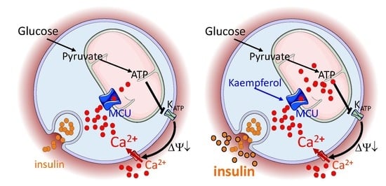
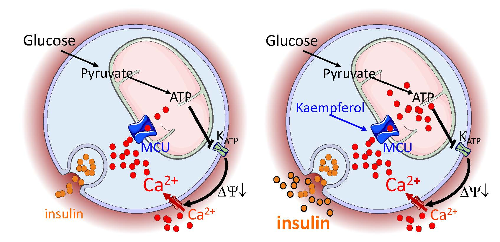
Targeting Mitochondrial Calcium Uptake with the Natural Flavonol Kaempferol, to Promote Metabolism/Secretion Coupling in Pancreatic β-cells

Nestlé Research—EPFL Innovation Park, CH-1015 Lausanne, Switzerland
*
Author to whom correspondence should be addressed.
These authors equally contributed to this work.
Submission received: 27 January 2020 / Accepted: 14 February 2020 / Published: 19 February 2020
(This article belongs to the Special Issue Effects of Dietary Intake on Pancreas)

Abstract

:
Pancreatic β-cells secrete insulin to lower blood glucose, following a meal. Maintenance of β-cell function is essential to preventing type 2 diabetes. In pancreatic β-cells, mitochondrial matrix calcium is an activating signal for insulin secretion. Recently, the molecular identity of the mitochondrial calcium uniporter (MCU), the transporter that mediates mitochondrial calcium uptake, was revealed. Its role in pancreatic β-cell signal transduction modulation was clarified, opening new perspectives for intervention. Here, we investigated the effects of a mitochondrial Ca2+-targeted nutritional intervention strategy on metabolism/secretion coupling, in a model of pancreatic insulin-secreting cells (INS-1E). Acute treatment of INS-1E cells with the natural plant flavonoid and MCU activator kaempferol, at a low micromolar range, increased mitochondrial calcium rise during glucose stimulation, without affecting the expression level of the MCU and with no cytotoxicity. Enhanced mitochondrial calcium rises potentiated glucose-induced insulin secretion. Conversely, the MCU inhibitor mitoxantrone inhibited mitochondrial Ca2+ uptake and prevented both glucose-induced insulin secretion and kaempferol-potentiated effects. The kaempferol-dependent potentiation of insulin secretion was finally validated in a model of a standardized pancreatic human islet. We conclude that the plant product kaempferol activates metabolism/secretion coupling in insulin-secreting cells by modulating mitochondrial calcium uptake.

Graphical Abstract

1. Introduction

Targeting pancreatic β-cells is a promising strategy to treat diabetes, due to the crucial role of the pancreatic β-cells in the pathogenesis of both type 1 and type 2 diabetes. Therefore, preservation, expansion, or improved function of β-cells are current approaches for targeting this cell type in the management of diabetes. The modulation of the biological pathways that regulate β-cell function represents the next stage of discovery in this field [1].
Within this framework, targeting mitochondrial Ca2+ in pancreatic β-cells is emerging as an innovative and promising strategy for diabetic patients. Indeed, mitochondrial matrix Ca2+ has been demonstrated to be an activating signal for insulin secretion [2]. The proposed mechanism of mitochondrial Ca2+-dependent pancreatic β-cell stimulation is based on the coordinated activation of two mitochondrial processes—oxidative metabolism and ATP-synthase-dependent respiration, which promotes robust insulin secretion [3]. In addition, mitochondrial Ca2+ rise has been suggested to contribute to the improvement of glucose-stimulated insulin secretion by sulfonylurea compounds in type 2 diabetic patients [4]. Finally, genetic evidences have recently demonstrated the high impact of mitochondrial Ca2+ uptake for pancreatic β-cell signal transduction and therefore insulin secretion [5,6,7]. Indeed, the molecular nature of the mitochondrial calcium uniporter (MCU), the transporter which mediates the mitochondrial Ca2+ influx, was recently disclosed [8,9], opening new perspectives for investigation and molecular intervention [10]. In mouse pancreatic β-cells, consistently with a role of mitochondrial Ca2+ on metabolism/secretion coupling, MCU knockdown displayed strong reduction of mitochondrial Ca2+ uptake, accompanied by impaired ATP production [7]. Similar results were obtained in pancreatic β-cell lines (INS-1 and INS-1E cells). The genetic depletion of two subunits of the MCU complex (Mcu and Micu1) were characterized by diminished mitochondrial Ca2+ transients, impaired respiration rate and ATP levels, and reduced insulin secretion, during glucose stimulation [5,6].
While the impacts of Ca2+ homeostasis [11,12] and mitochondrial function [13] on β-cell function are widely accepted, and despite the unquestionable role of mitochondrial Ca2+ for pancreatic β-cell signal transduction [5,6,7], very few therapies or interventions based on the modulation of mitochondrial Ca2+ have been developed until now. In this context, we recently demonstrated that natural bioactive quinic acid increases glucose-stimulated mitochondrial Ca2+ rise, enhancing insulin secretion in both INS-1E β-cells and mouse islets, and improves glucose tolerance in mice [14]. In that study, quinic acid was not a direct activator of MCU but it enhanced the release of Ca2+ from the endoplasmic reticulum, improving Ca2+ transmission between the endoplasmic reticulum and mitochondria. A more specific MCU-targeted pharmacology is expected to improve β-cell MCU activation, promoting beneficial effects in the context of diabetes treatment. Interestingly, some natural plant flavonoids often present in the diet, and especially kaempferol, have been previously reported to direct activate mitochondrial Ca2+ uptake in HeLa cells [15]. Moreover, several flavonoids, which are members of the natural bioactive polyphenol family, have been proposed to strengthen survival processes and insulin secretory capacity of β-cells (reviewed in [16]). The idea that flavonoids can preserve the function and survival of β-cells is based on the antioxidant and anti-inflammatory properties of these polyphenols, as well as their ability to stimulate pro-survival pathways and to improve bioenergetics function. However, little attention has been devoted to the possibility of targeting mitochondrial Ca2+ uptake with polyphenols to promote metabolism/secretion coupling in pancreatic β-cells.
To determine the impact of a mitochondrial Ca2+-targeted intervention on pancreatic β-cell metabolism/secretion coupling, we measured here the effect of the paradigmatic polyphenol putative MCU activator kaempferol on mitochondrial Ca2+ rise, insulin secretion, and cell death, during glucose stimulation, in pancreatic INS-1E β-cells. In addition, by taking advantage of the recently developed MCU pharmacology, we quantified the effect of the MCU inhibitor mitoxantrone on mitochondrial Ca2+ rise and granule exocytosis in both glucose-stimulated and kaempferol-potentiated pancreatic INS-1E cell function. Finally, we validated the effect of kaempferol on insulin secretion in a model of human pseudo-islets. We proposed to target mitochondrial Ca2+ with kaempferol as a nutritional non-cytotoxic intervention, to promote metabolism/secretion coupling in pancreatic β-cells.

2. Materials and Methods

2.1. Materials

Chemicals were from Sigma-Aldrich (Buchs, Switzerland), Invitrogen (Life Technologies, Zug, Switzerland), or VWR (Zug, Switzerland), unless otherwise indicated. Kaempferol was dissolved in DMSO at a concentration of 1 mM and then diluted at 10 µM in Krebs-Ringer bicarbonate Hepes buffer (KRBH), containing (in mM): 140 NaCl, 3.6 KCl, 0.5 NaH2PO4, 0.5 MgSO4, 1.5 CaCl2, 10 Hepes, and 5 NaHCO3; pH 7.4. Glucose was 2.5 mM unless otherwise specified. Drs. T. Pozzan and R. Rizzuto (University of Padova, Padova, Italy) kindly provided the plasmid containing the mitochondrial aequorin mutant to measure mitochondrial [Ca2+]. Some figures contain illustrations made by Servier Medical Art, http://www.servier.fr/servier-medical-art.

2.2. Cell Culture, Mitochondrial Isolation, and Western Blots

INS-1E cells [17] were cultured at 37 °C in a humidified atmosphere (5% CO2) in RPMI-1640 medium (Invitrogen) containing 11 mM glucose, supplemented with 10 mM Hepes (pH 7.3), 10% (v/v) heat-inactivated fetal calf serum (FCS; Brunschwig AG, Basel, Switzerland), 1 mM sodium pyruvate, 50 μM β-mercaptoethanol, 50 μg/mL penicillin, and 100 μg/mL streptomycin (INS-1 medium). Culturing HeLa cells has been previously described [18].
Mitochondria were isolated from INS1E and HeLa cell culture as previously described [19]. Western blots were previously described [20]. The membranes were probed with anti-MCU (Sigma-Aldrich, Buchs, Switzerland), anti-Tom20 (Santa Cruz Biotechnology, Dallas, TX, USA), and anti-ATP-5A (Abcam, Cambridge, United Kingdom) antibodies.

2.3. Mitochondrial Ca2+ Measurements

Mitochondrial Ca2+ was measured with the genetically encoded luminesce sensor mitochondrial-muted aequorin [15,21]. INS-1E cells were plated on 96-well plates and transfected with the mitochondrial mutant aequorin plasmid, using JetPRIME transfection reagent (Polyplus Transfection, Ilkirch, France). Two days after transfection, cells were washed with KRBH containing 145 mM NaCl, 5 mM KCl, 1 mM MgCl2, 10 mM HEPES, 1 mM CaCl2, and 10 mM glucose; pH 7.4. Then, the cells were incubated with 5 µM coelenterazine native (Biotium, Fremont, CA, USA), dissolved in aequorin buffer, for 2 h. After a second wash with KRBH aequorin buffer, each well was treated for 30 min with DMSO 1% (control) or 10 µM kaempferol or 50 µM mitoxantrone. Luminescence was measured with a Cytation3 plate reader (Biotek, Sursee, Switzerland). The basal luminescence was measured for 30 s, then cells were stimulated for 90 s with glucose (16.7 mM). The experiment was ended by the addition of a digitonin (25µM)/CaCl2 (40 mM) solution to calibrate the fluorescence signal, as described by Montero and collaborators [15]. The data per well were analyzed as previously described [22]. The analysis protocol first removed all unused wells than proceeded with a calibration of the data followed by a curve fitting and peak-finding algorithm. Calibration was calculated using the following equation:
Ca 2 +   =   L L m a x   ×   λ 1 n +   L L m a x   ×   λ 1 n   × K T R 1 K R L L m a x   ×   λ 1 n ×   K R
where L is the luminescence value at sampling time, Lmax is the total luminescence emitted up until the sampling time, and KR and KTR are the constants for calcium-bound state and the calcium-unbound state, respectively. Finally, λ is the rate constant for aequorin consumption at saturating Ca2+ concentration and n is the number of Ca2+-binding sites. The calibrated values were then fitted using a spline interpolation, and the area under the curve (AUC) was computed on the fitted curve to serve as the primary read-out for the calcium.

2.4. Static Insulin Secretion in INS-1E Cells

INS-1E cells were plated on a polyornithine-treated 24-well plate. After 48 h, cells were washed in Krebs-Ringer bicarbonate Hepes buffer (KRBH), containing (in mM) 140 NaCl, 3.6 KCl, 0.5 NaH2PO4, 0.5 MgSO4, 1.5 CaCl2, 10 Hepes, and 5 NaHCO3, pH 7.4 containing 2.5 mM glucose. Cells were stimulated for 30 min with 16.7 mM glucose and supernatants were collected. Cellular insulin content was extracted with a mixture of ethanol (75%) and HCl (1.5%) overnight at 4 °C. Insulin was measured using a Rat Insulin Enzyme Immunoassay kit (SpiBio-Bertin Pharma, Montigny le Brotonneux, France).

2.5. Cell Death

Kinetic experiments of apoptosis were performed with the IncuCyte ZOOM instrument (Essen Bioscience, Ann Arbor, MI, USA). INS-1E cells were seeded at 50% confluence in 96-well-plate format in RPMI-1640 medium. Then, 24 h later and just before the beginning of the acquisition, the cells were incubated with IncuCyte Annexin-V Green (4642), according to supplier’s instructions. Annexin-V is commonly used to detect apoptotic cells by its ability to bind to phosphatidylserine, an early marker of apoptosis, when it is on the outer leaflet of the plasma membrane (e.g., [23]). Four images per well were collected at the indicated time using a 10× objective and bandwidth filters (Ex: 440/80 nm; Em: 504/44 nm). For image segmentation and processing definition, the following constraints were applied—Parameter: Top-Hat (background subtraction); Radius: 50 µm; Threshold: 0.800 GCU; Edge: 0; Fill: 0; Area: 0; Eccentricity: 0; Mean intensity: 0; and Integrated intensity: 0. Data were exported as area (µm2) per well covered by Annexin-V positive objects.

2.6. Human Islets and Insulin Secretion

3D InSight™ Human Islet Microtissues were obtained from InSphero AG (Schlieren, Switzerland). Upon arrival, the pseudo-islets were treated following manufacturer’s instructions for measurement of glucose-stimulated insulin secretion. Briefly, the isolated pseudo-islets were carefully washed twice and incubated for 1 h at low glucose in modified Krebs-Ringer buffer (KRB) containing (in mM) 131 NaCl, 4.8 KCl, 1.3 CaCl2, 1.2 KH2PO4, 1.2 MgSO4, 5 NaHCO3, 25 HEPES, and 2.8 glucose, and 0.5% BSA. This solution is referred to as low glucose solution (LGS, 2.8 mM). Then, isolated islets were carefully washed with LGS and incubated for 2 h in fresh KRB containing 16.7 mM glucose and 0.5% BSA in presence of test substances at indicated concentrations. Measures of insulin levels in supernatants and cell extracts (extraction in acid ethanol (1.5% (v/v) HCl in 70% (v/v) ethanol) were performed using a sensitive chemiluminescence enzyme-linked immunosorbent assay (ALPCO, Salem, NH, USA). All experiments with human islets were approved by the Ethical commission of the Human Research Act (Bern, Switzerland).

2.7. Statistics

Data are expressed as means ± SEM. Statistically significant differences between means were determined using Student’s t-test for comparison between two means. ANOVA test was used when comparing mean values from multiple groups. Additionally, p-values < 0.05 were accepted as significant and represented as * in respective figure panels (NS, not significant, were indicated).

3. Results

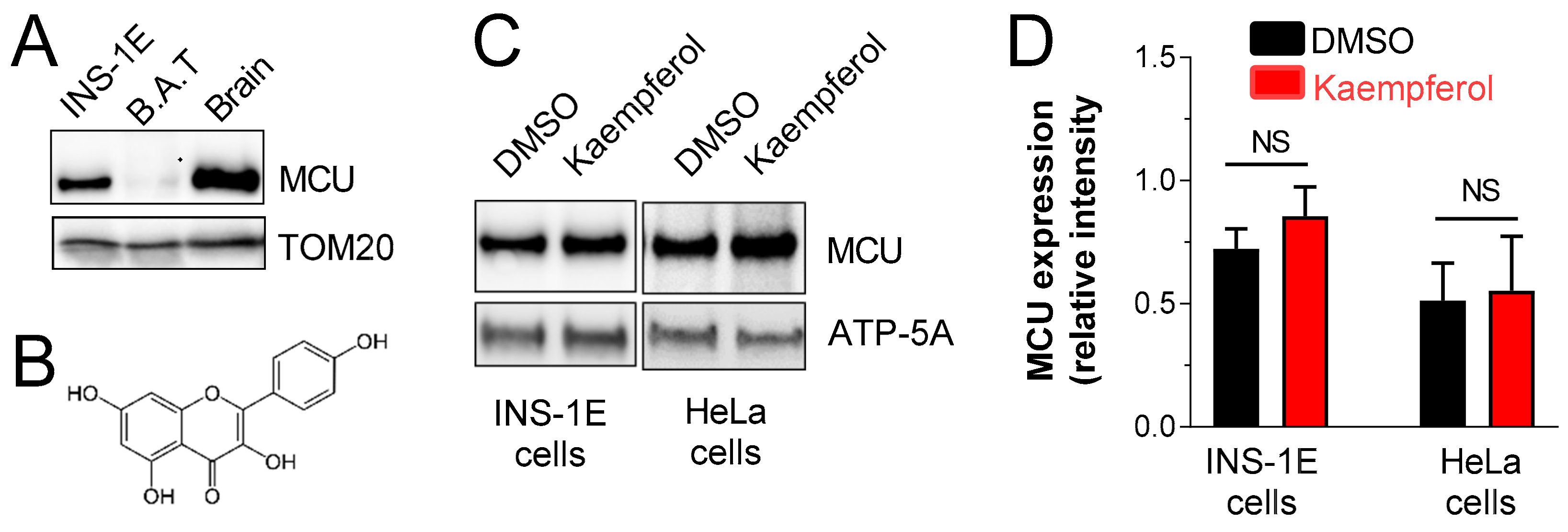
3.1. MCU Is Expressed in Pancreatic INS-1E β-Cells, Irrespectively of Acute Kaempferol Treatment

To assess the possibility of targeting the mitochondrial Ca2+ in pancreatic β-cell metabolism/secretion coupling, the expression level of MCU protein was investigated in a model of glucose-sensitive pancreatic β-cells, named INS-1E β-cells [17]. As shown in Figure 1A, MCU was highly expressed in mitochondria isolated from INS-1E cells. The MCU expression level was also detected in rat mitochondria isolated from brown adipose tissue and brain as control tissues. The impact of the flavonoid kaempferol (Figure 1B) on MCU expression was then quantified in INS1-1E β-cells (Figure 1C). In addition, we checked the effect of the acute treatment with kaempferol on HeLa cells, a system in which mitochondrial calcium uptake is activated by that polyphenol [10]. As shown in Figure 1D, MCU was expressed in both INS-1E and HeLa cells and the acute incubation with kaempferol did not influence the expression level of that mitochondrial transporter. These results validated INS-1E as a good system to study the effect of a mitochondrial Ca2+-targeted intervention in a cellular model of glucose-stimulated metabolism/secretion coupling activation.

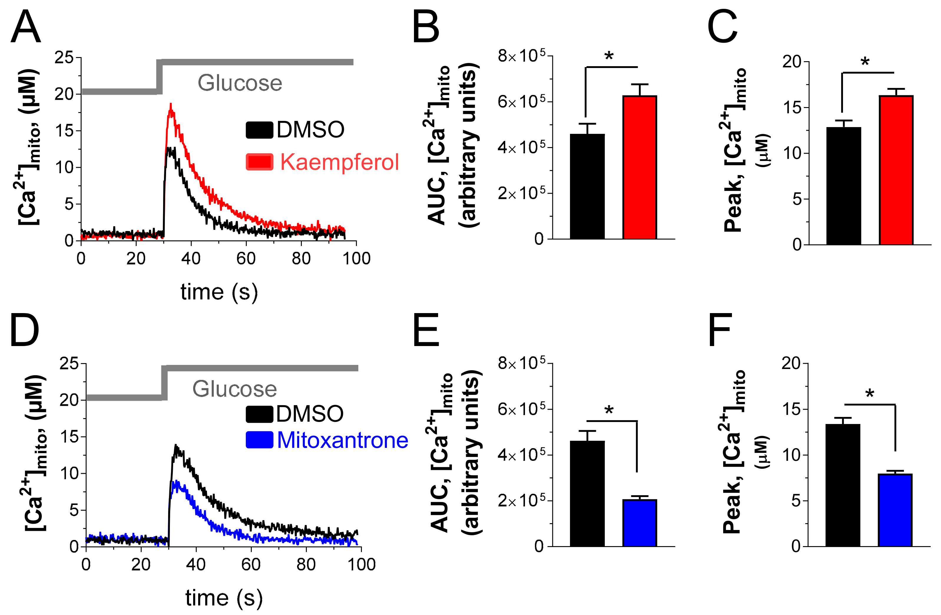
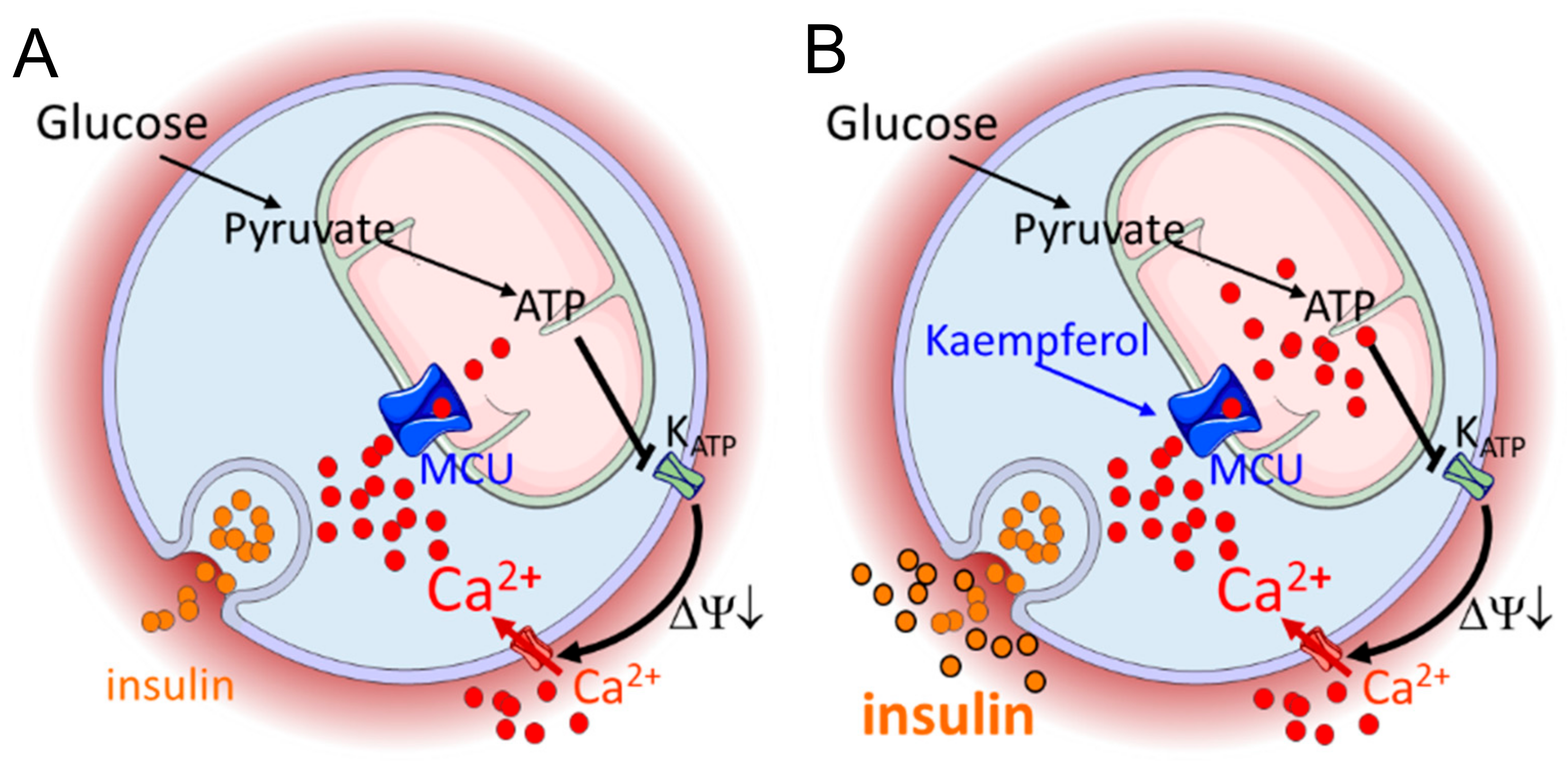
3.2. Kaempferol Enhances Glucose-Stimulated Mitochondrial Ca2+ Rise and Mitoxantrone Inhibits Matrix Ca2+ Elevation in Pancreatic Insulin-Secreting Cells

To investigate the effect of a mitochondrial Ca2+-targeted intervention on insulin-secreting cells, we measured the effect of kaempferol on mitochondrial Ca2+ rise in glucose-stimulated INS-1E cells, transfected with the genetically encoded sensor-mutated aequorin, targeted to mitochondria. As shown in Figure 2A, stimulation with 16.7 mM glucose promoted robust mitochondrial Ca2+ elevation in control INS-1E cells, treated with DMSO. Acute treatment with 10µM kaempferol (30 min incubation) potentiated mitochondrial Ca2+ rise. Therefore, this treatment increased the integrated mitochondrial Ca2+ elevation evoked by glucose stimulation by 37 % (Figure 2B) and the amplitude of mitochondrial Ca2+ signal by 27% (Figure 2C). Then, we used mitoxantrone (50 µM), which has been recently identified as a direct inhibitor of MCU [24]. As shown in Figure 2D, mitoxantrone strongly reduced the mitochondrial Ca2+ elevation evoked by glucose, the integrated mitochondrial Ca2+ response (Figure 2E) and the amplitude of mitochondrial Ca2+ signal (Figure 2F) being reduced by 55% and 40%, respectively. These data support the possibility of targeting the mitochondrial Ca2+ transport in pancreatic insulin-secreting cells with the natural bioactive kaempferol.

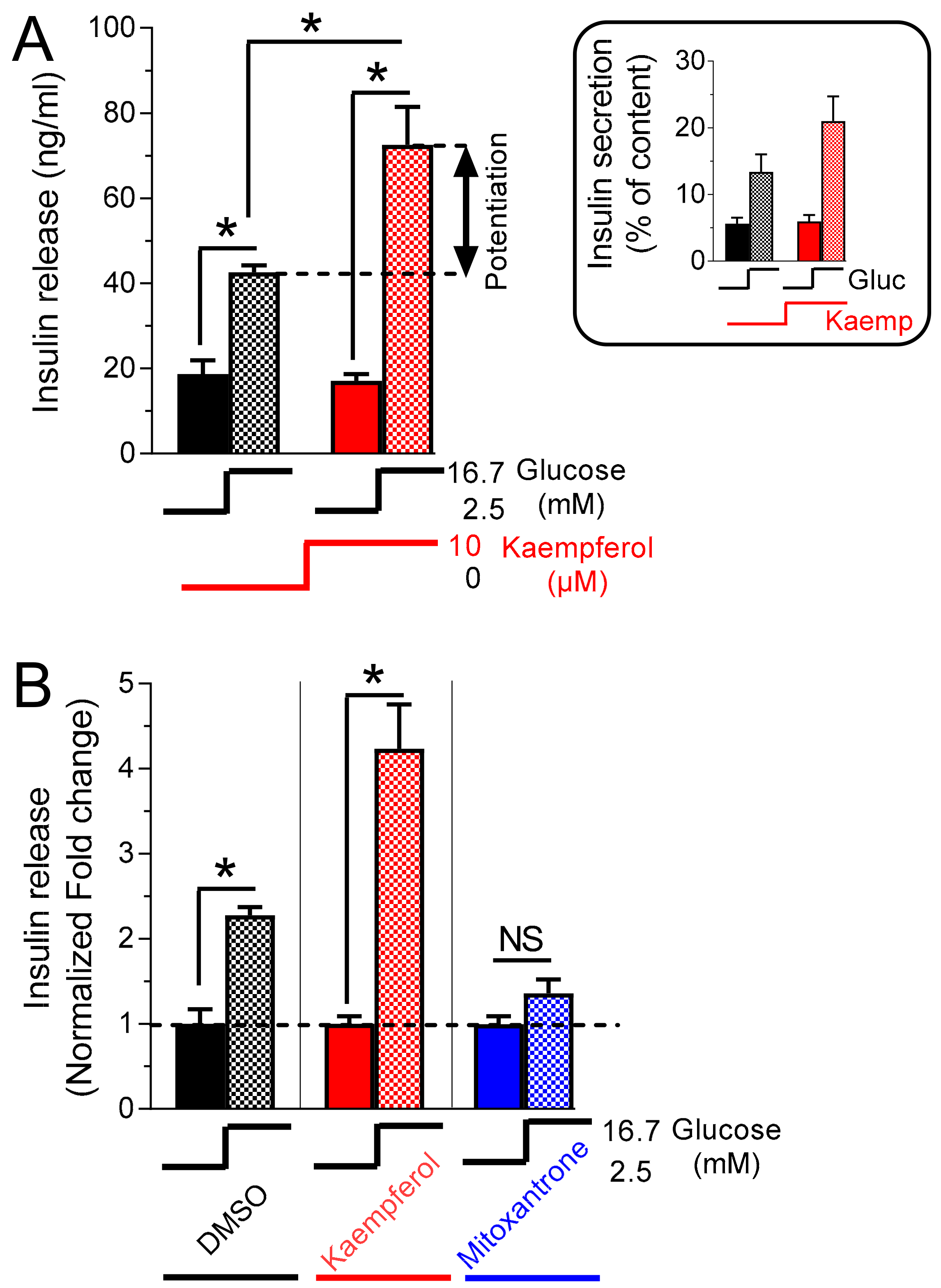
3.3. Kaempferol Potentiates Glucose-Stimulated Insulin Secretion

Mitochondrial Ca2+ activation has been demonstrated to be an activating signal for insulin secretion [2] via activation of mitochondrial Ca2+-dependent processes [3]. Given the positive effect of kaempferol on glucose-stimulated mitochondrial Ca2+ rise, we investigated the effect of this flavonoid on insulin secretion. We found that in control cells glucose stimulation promoted robust release of insulin (Figure 3A).
Kaempferol (10 µM) strongly potentiated glucose-induced hormone exocytosis, the insulin release being increased by 70% (in parallel with the increased percentage of the secreted insulin, inset Figure 3A). To confirm this relationship between mitochondrial calcium transport modulation and insulin secretion, we measured hormone exocytosis in the presence of the MCU inhibitor mitoxantrone. When INS-1E cells were stimulated with 16.7 mM glucose (Figure 3B), no additional insulin release was recorded, indicating that pharmacological inhibition of MCU prevents glucose-stimulated hormone exocytosis. Consistent with genetic manipulation of MCU, these data demonstrate that pharmacological/nutritional intervention on MCU can modulate metabolism/secretion coupling in insulin-secreting cells.

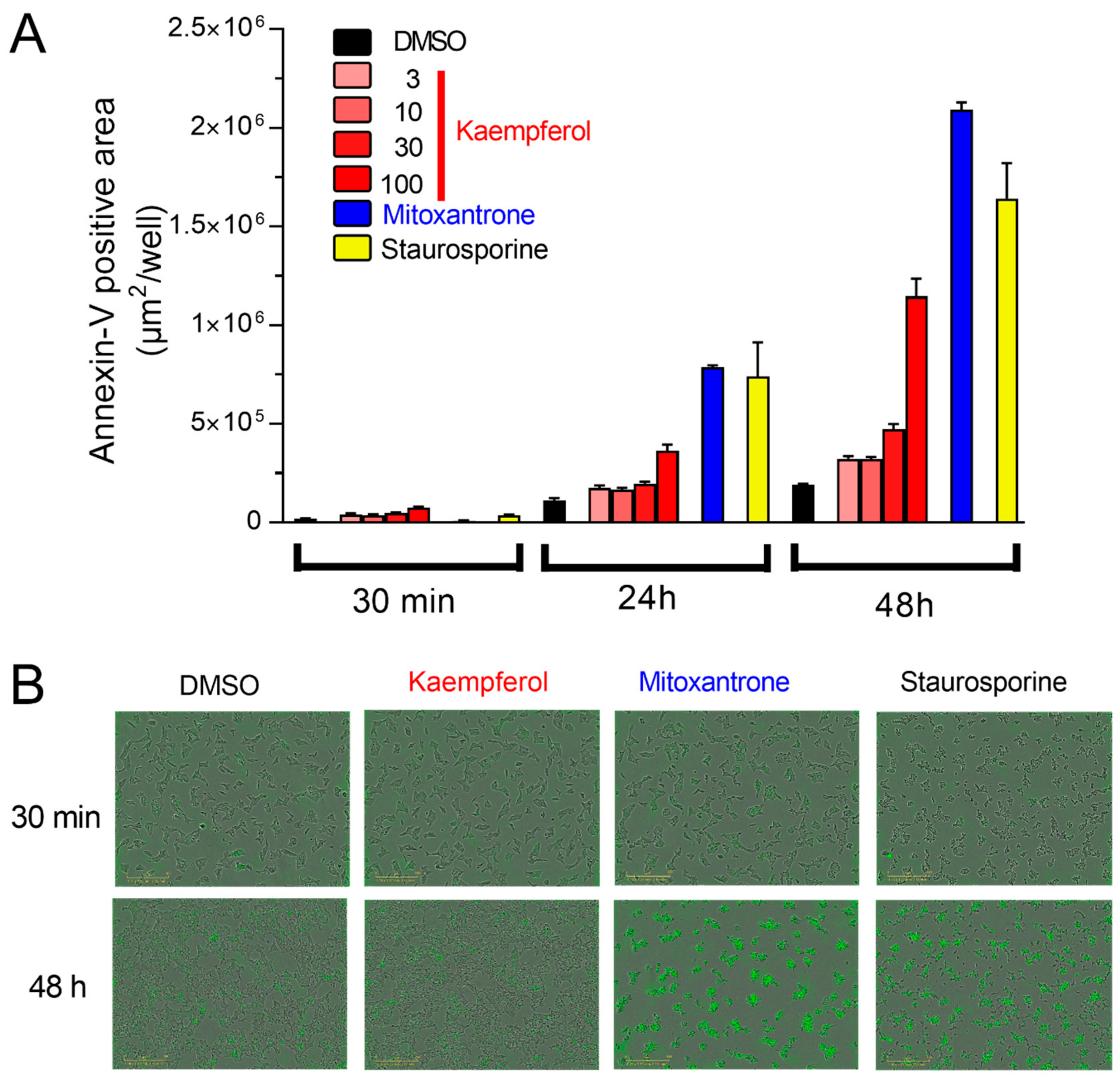
3.4. Acute Kaempferol-Dependent Mitochondrial Ca2+ Rise Is Not Cytotoxic

Transient increase of mitochondrial calcium uptake promotes cellular energetics [25] and modulates metabolism/secretion coupling [4] in physiological condition. However, a massive increase of mitochondrial calcium uptake regulates cell survival, promoting cell death [10,26]. To investigate if kaempferol-potentiated mitochondrial calcium uptake is associated with cell death in insulin-secreting cells, we measured apoptosis (Figure 4A).
Acute incubation of kaempferol (30 min) did not enhance phosphatidylserine exposure, an early marker of apoptosis (Figure 4B), indicating absence of cytotoxicity. Interestingly, absence of cytotoxicity was also recorded in cells acutely treated with mitoxantrone, suggesting that the mitoxantrone-induced insulin release is not linked to cell death but it depends on other unspecific effects of this compound. After 24 h, kaempferol (or its metabolites) slightly increased cell death, compared to control (DMSO), whereas mitoxantrone and staurosporine triggered apoptosis, as expected. These effects were amplified after chronic treatment (48 h, Figure 4A,B). These data reveal that acute treatment with kaempferol boosts physiological release of insulin secretion and that it is not cytotoxic in insulin-secreting cells.

3.5. Mitoxantrone Prevents Kaempferol-Dependent Mitochondrial Ca2+ Rise and Inhibits the Potentiation of Insulin Secretion

To confirm that an intervention with kaempferol potentiates mitochondrial calcium rise and therefore insulin secretion via MCU, we measured the effects of kaempferol in insulin-secreting cells treated with the MCU inhibitor mitoxantrone. In glucose-stimulated cells, mitoxantrone inhibited kaempferol-potentiated mitochondrial calcium rise (Figure 5A) and insulin secretion (Figure 5B).
No acute cytotoxic effect was recorded under these experimental conditions (Figure 5C). These experiments indicate that an intervention on MCU aimed to modulate β-cell function is not a simple trademark of kaempferol treatment; instead, this polyphenol enhances metabolism/secretion coupling via MCU activation (Figure 5D).

3.6. Kaempferol Potentiates Insulin Secretion in Hhuman islet Beta Cells

To validate the impact of kaempferol on the potentiation of insulin secretion in a relevant human model of pancreatic β-cells, we measured glucose-induced insulin secretion in standardized human single islet microtissues, derived from primary islet (Figure 6A, 3D InSight™ Human Islet Microtissues). We refer to these tissues as pseudo-islets. Pseudo-islets are uniform in size, cellular composition, and function. In the control experiment, single pseudo-islets were stimulated with glucose 16.7 mM (Figure 6B, left). As expected, glucose stimulation promoted robust release of insulin. Kaempferol (10 µM, Figure 6B, right) also strongly potentiated glucose-induced hormone secretion in pseudo-islets, the insulin release being increased by 57%. These data indicate that the effect of kaempferol in human tissue is consistent with the results obtained in INS-1E cells.

4. Discussion

Diabetes is a leading cause of morbidity and mortality worldwide. Targeting pancreatic β-cell signal transduction has been proposed as an effective therapeutic approach in both type 1 and type 2 diabetes [1]. Mitochondrial calcium uniporter represents a novel molecular target in this context, due to the ability of matrix Ca2+ to modulate metabolism/secretion coupling in the β-cell [2,3]. Genetic and pharmacological evidences [5,6,7] have already reproducibly demonstrated the impact of MCU for β-cell signal transduction. In addition, the mechanism of mitochondrial Ca2+-dependent β-cell activation has been previously investigated [2,3]. However, very few interventions based on the modulation of mitochondrial Ca2+ have been developed until now. By taking advantage of the recent molecular discovery of the MCU, the mitochondrial channel which mediates matrix calcium transport, and its pharmacology, here we efficiently demonstrated how an intervention on that channel improves glucose-stimulated signal transduction and therefore promotes hormone secretion, in a model of insulin-secreting β-cells (Figure 7). In addition, we validated the effect of the mitochondrial Ca2+ activator kaempferol on the potentiation of glucose-induced insulin secretion in a model of human islet beta cells.
Given the ability of the natural bioactive kaempferol to activate mitochondrial calcium uptake in HeLa cells [15], we investigated the efficacy of that polyphenol also in a model of pancreatic insulin-secreting cells. The binding site of the kaempferol to MCU complex is unknown. The composition and stoichiometry of the MCU components (MCU/MCUb) and of the associated proteins (MICU 1, 2, and 3, EMRE, and MCUR1) is tissue specific and a large variety of mitochondrial Ca2+ currents have been observed across different tissues and cell types [27,28]. The relative abundance of distinct MCU subunits and regulators has been demonstrated to affect mitochondrial Ca2+ transport in different tissues (e.g., [29]), suggesting a possible tissue-specific effect of kaempferol. Here, we demonstrated that this natural flavonol activates MCU also in insulin-secreting cells. In addition, kaempferol-evoked matrix Ca2+ rise was prevented by the pharmacological inhibition of the uniporter (e.g., with mitoxantrone), indicating that the use of kaempferol can be applied to modulate MCU-dependent function in the pancreatic β-cell.
The impact of kaempferol treatment on β-cell function results in a non-cytotoxic potentiation of glucose-stimulated insulin secretion. By targeting the basic molecular machinery of metabolism/secretion coupling, kaempferol promotes hormone secretion via MCU activation. On the other hand, mitochondrial Ca2+ overload is well known to also promote cell death pathways [10,26]. Therefore, a massive rise of mitochondrial matrix Ca2+ has been established to open the permeability transition pore (review in [30]), a high conductance inner membrane channel [31]. Persistent opening of the permeability transition pore was demonstrated to cause the remodeling of mitochondrial inner membrane cristae, triggering the release of pro-apoptotic proteins (e.g., cytochrome c) and initiation of cell death [32]. We verified the absence of β-cell cytotoxicity by acute treatment with kaempferol, by measuring an early marker of apoptosis (e.g., exposure of phosphatidylserine on the cell surface). The transient increase of kaempferol-dependent mitochondrial Ca2+ rise activates mitochondria without promoting cell death. Therefore, matrix Ca2+ was extruded immediately after the uptake (Figure 2). Interestingly, prolonged in vitro administration of that natural flavonol (24–48 h) promoted dose-dependent cell death. However, one has to keep in mind that the in vitro reactive polyphenols are antioxidant compounds that can act as or can be converted to prooxidant molecules [33,34], especially in cell culture media [35]. These conditions are not likely to occur in vivo upon consumption of flavon-containing foods, since the bioavailability of polyphenols in foodstuffs is low and metabolism by enterocytes and hepatocytes is very effective [34].
Kaempferol is a plant-derived compound. Plant products are very attractive molecules to target pancreatic β-cells for the treatment of diabetes [36]. These compounds are more attractive than synthetic drugs because of their diversity and minimal side effects. Several natural products and extracts and their bioactive compounds are known to promote beneficial effects on pancreatic beta-cell function [36] and growing amount of evidence supports their efficacy for prevention and attenuation of diabetes consequences [16]. However, in parallel with efficacy and safety evaluations, it is necessary to investigate their mechanism of action as an antidiabetic phytochemical. Kaempferol has previously been proposed to ameliorate streptozotocin-induced diabetes via promoting glucose metabolism in skeletal muscle and inhibiting gluconeogenesis in the liver [37]. In addition, kaempferol treatment has been demonstrated to promote cytoprotective effects in β-cell and human islets exposed to chronic high glucose [38]. These effects were associated with improved insulin secretory function and synthesis. More recently, kaempferol has been proposed to improve hyperglycemia, glucose tolerance, and blood insulin levels and to preserve pancreatic beta-cell mass in obese diabetic mice. Here, we demonstrated a direct effect of this flavonol on the basic molecular machinery of metabolism/secretion coupling of the pancreatic β-cell, via activating glucose-stimulated mitochondrial Ca2+ rise.
To validate an MCU-targeted intervention of kaempferol in pancreatic β-cells, we used mitoxantrone, which was recently identified as a direct inhibitor of MCU [24]. Our results demonstrate that the acute effect of this drug on glucose-stimulated cells and in kaempferol-treated cell cultures strongly supports a causality between mitochondrial Ca2+ modulation and pancreatic β-cell activation. Concerning the relevance of the kaempferol concentration used in this investigation, several studies have demonstrated different levels of kaempferol in plasma after treatment. Depending on the source and on the study design, the maximum plasma concentration of kaempferol was recorded in the range of 10/100 nanomolar in some studies (e.g., [39,40,41]). In another study [42], a single dose of 600 mg/kg of standardized kaempferol-containing Ginkgo biloba extract resulted in maximum plasma concentration of kaempferol in the micromolar range, which reflects the concentration range used in our experiments.
The flavonol kaempferol was selected in this study because it has been previously identified to activate mitochondrial Ca2+ uptake in HeLa cells [15]. However, in the same study, other natural flavonols were investigated and showed distinct efficacies to potentiate mitochondrial Ca2+ uptake. As previously mentioned, mitochondrial Ca2+ has been proposed to activate pancreatic β-cells by co-regulating oxidative metabolism (e.g., Nicotinamide adenine dinucleotide, NADH, production) and ATP-synthase-dependent respiration [3]. The oxidative metabolism is driven by the mitochondrial Ca2+-dependent activation of matrix dehydrogenases, which are involved in the direct supply of NADH and FADH (Flavin adenine dinucleotide) [43]. Given that the activity of mitochondrial dehydrogenases is sensitive of the mitochondrial Ca2+ level [43] and that different polyphenols promote distinct levels of mitochondrial Ca2+ uptakes, we speculate that different flavons could promote distinct levels of β-cell activations and therefore insulin secretion and might have similar or even better beneficial effects than kaempferol.

5. Conclusions

In summary, we provided evidence for the ability of kaempferol to boost metabolism/secretion coupling in a model of pancreatic β-cells, via potentiation of mitochondrial Ca2+ rise (Figure 7). These results, in combination with the non-cytotoxic acute effect of this natural bioactive and the potentiated glucose-induced insulin secretion in human pseudo-islets, indicate that the dietary intake of kaempferol may be developed as an adjuvant to support existing therapies for the treatment of diabetes. Conclusive preclinical and clinical evidences are the next step to validate the efficacy and safety of this phytochemical-based intervention.

Author Contributions

Conceptualization, supervision, and writing—review and editing, U.D.M.; investigation, formal analysis, and validation, F.B., A.H., R.B., G.J., and C.C.; methodology, writing—review and editing, J.S.-D.; supervision, review and editing, M.R.-C.K. and J.N.F. All authors have read and agree to the published version of the manuscript.

Acknowledgments

We thank T. Pozzan and R. Rizzuto (University of Padova, Italy) for providing the mitochondrial-targeted aequorin. We thank the University of Geneva and C. B. Wollheim for the INS-1E cells. We thank C. Canto (Nestlé Research) for providing control tissue lysates used for the Western blot in Figure 1A.

Conflicts of Interest

The authors are employees of Nestlé Research, which is part of the Société des Produits Nestlé SA.

References

  1. Vetere, A.; Choudhary, A.; Burns, S.M.; Wagner, B.K. Targeting the pancreatic beta-cell to treat diabetes. Nat. Rev. Drug Discov. 2014, 13, 278–289. [Google Scholar] [CrossRef] [PubMed]
  2. Wiederkehr, A.; Szanda, G.; Akhmedov, D.; Mataki, C.; Heizmann, C.W.; Schoonjans, K.; Pozzan, T.; Spat, A.; Wollheim, C.B. Mitochondrial matrix calcium is an activating signal for hormone secretion. Cell Metab. 2011, 13, 601–611. [Google Scholar] [CrossRef] [PubMed]
  3. De Marchi, U.; Thevenet, J.; Hermant, A.; Dioum, E.; Wiederkehr, A. Calcium co-regulates oxidative metabolism and ATP synthase-dependent respiration in pancreatic beta cells. J. Biol. Chem. 2014, 289, 9182–9194. [Google Scholar] [CrossRef] [PubMed] [Green Version]
  4. Wiederkehr, A.; Wollheim, C.B. Impact of mitochondrial calcium on the coupling of metabolism to insulin secretion in the pancreatic beta-cell. Cell Calcium 2008, 44, 64–76. [Google Scholar] [CrossRef]
  5. Alam, M.R.; Groschner, L.N.; Parichatikanond, W.; Kuo, L.; Bondarenko, A.I.; Rost, R.; Waldeck-Weiermair, M.; Malli, R.; Graier, W.F. Mitochondrial Ca2+ uptake 1 (MICU1) and mitochondrial Ca2+ uniporter (MCU) contribute to metabolism-secretion coupling in clonal pancreatic beta-cells. J. Biol. Chem. 2012, 287, 34445–34454. [Google Scholar] [CrossRef] [Green Version]
  6. Quan, X.; Nguyen, T.T.; Choi, S.K.; Xu, S.; Das, R.; Cha, S.K.; Kim, N.; Han, J.; Wiederkehr, A.; Wollheim, C.B.; et al. Essential role of mitochondrial Ca2+ uniporter in the generation of mitochondrial pH gradient and metabolism-secretion coupling in insulin-releasing cells. J. Biol. Chem. 2015, 290, 4086–4096. [Google Scholar] [CrossRef] [Green Version]
  7. Tarasov, A.I.; Semplici, F.; Ravier, M.A.; Bellomo, E.A.; Pullen, T.J.; Gilon, P.; Sekler, I.; Rizzuto, R.; Rutter, G.A. The mitochondrial Ca2+ uniporter MCU is essential for glucose-induced ATP increases in pancreatic beta-cells. PLoS ONE 2012, 7, e39722. [Google Scholar] [CrossRef] [Green Version]
  8. Baughman, J.M.; Perocchi, F.; Girgis, H.S.; Plovanich, M.; Belcher-Timme, C.A.; Sancak, Y.; Bao, X.R.; Strittmatter, L.; Goldberger, O.; Bogorad, R.L.; et al. Integrative genomics identifies MCU as an essential component of the mitochondrial calcium uniporter. Nature 2011, 476, 341–345. [Google Scholar] [CrossRef] [Green Version]
  9. De Stefani, D.; Raffaello, A.; Teardo, E.; Szabo, I.; Rizzuto, R. A forty-kilodalton protein of the inner membrane is the mitochondrial calcium uniporter. Nature 2011, 476, 336–340. [Google Scholar] [CrossRef]
  10. Rizzuto, R.; De Stefani, D.; Raffaello, A.; Mammucari, C. Mitochondria as sensors and regulators of calcium signalling. Nat. Rev. Mol. Cell Biol. 2012, 13, 566–578. [Google Scholar] [CrossRef]
  11. Gilon, P.; Chae, H.Y.; Rutter, G.A.; Ravier, M.A. Calcium signaling in pancreatic beta-cells in health and in Type 2 diabetes. Cell Calcium 2014, 56, 340–361. [Google Scholar] [CrossRef] [PubMed]
  12. Rorsman, P.; Braun, M.; Zhang, Q. Regulation of calcium in pancreatic alpha- and beta-cells in health and disease. Cell Calcium 2012, 51, 300–308. [Google Scholar] [CrossRef] [PubMed] [Green Version]
  13. Wiederkehr, A.; Wollheim, C.B. Mitochondrial signals drive insulin secretion in the pancreatic beta-cell. Mol. Cell. Endocrinol. 2012, 353, 128–137. [Google Scholar] [CrossRef] [PubMed]
  14. Heikkila, E.; Hermant, A.; Thevenet, J.; Bermont, F.; Kulkarni, S.S.; Ratajczak, J.; Santo-Domingo, J.; Dioum, E.H.; Canto, C.; Barron, D.; et al. The plant product quinic acid activates Ca(2+) -dependent mitochondrial function and promotes insulin secretion from pancreatic beta cells. Br. J. Pharmacol. 2019, 176, 3250–3263. [Google Scholar] [CrossRef] [PubMed]
  15. Montero, M.; Lobaton, C.D.; Hernandez-Sanmiguel, E.; Santodomingo, J.; Vay, L.; Moreno, A.; Alvarez, J. Direct activation of the mitochondrial calcium uniporter by natural plant flavonoids. Biochem. J. 2004, 384, 19–24. [Google Scholar] [CrossRef] [Green Version]
  16. Ghorbani, A.; Rashidi, R.; Shafiee-Nick, R. Flavonoids for preserving pancreatic beta cell survival and function: A mechanistic review. Biomed. Pharmacother. 2019, 111, 947–957. [Google Scholar] [CrossRef]
  17. Maechler, P.; Antinozzi, P.A.; Wollheim, C.B. Modulation of glutamate generation in mitochondria affects hormone secretion in INS-1E beta cells. IUBMB Life 2000, 50, 27–31. [Google Scholar] [CrossRef] [Green Version]
  18. Da Cruz, S.; De Marchi, U.; Frieden, M.; Parone, P.A.; Martinou, J.C.; Demaurex, N. SLP-2 negatively modulates mitochondrial sodium-calcium exchange. Cell Calcium 2010, 47, 11–18. [Google Scholar] [CrossRef] [Green Version]
  19. De Marchi, U.; Campello, S.; Szabo, I.; Tombola, F.; Martinou, J.C.; Zoratti, M. Bax does not directly participate in the Ca(2+)-induced permeability transition of isolated mitochondria. J. Biol. Chem. 2004, 279, 37415–37422. [Google Scholar] [CrossRef] [Green Version]
  20. De Marchi, U.; Galindo, A.N.; Thevenet, J.; Hermant, A.; Bermont, F.; Lassueur, S.; Domingo, J.S.; Kussmann, M.; Dayon, L.; Wiederkehr, A. Mitochondrial lysine deacetylation promotes energy metabolism and calcium signaling in insulin-secreting cells. FASEB J. 2019, 33, 4660–4674. [Google Scholar] [CrossRef]
  21. Montero, M.; Alonso, M.T.; Carnicero, E.; Cuchillo-Ibanez, I.; Albillos, A.; Garcia, A.G.; Garcia-Sancho, J.; Alvarez, J. Chromaffin-cell stimulation triggers fast millimolar mitochondrial Ca2+ transients that modulate secretion. Nat. Cell Biol. 2000, 2, 57–61. [Google Scholar] [CrossRef]
  22. Bonora, M.; Giorgi, C.; Bononi, A.; Marchi, S.; Patergnani, S.; Rimessi, A.; Rizzuto, R.; Pinton, P. Subcellular calcium measurements in mammalian cells using jellyfish photoprotein aequorin-based probes. Nat. Protoc. 2013, 8, 2105–2118. [Google Scholar] [CrossRef] [PubMed]
  23. Campello, S.; De Marchi, U.; Szabo, I.; Tombola, F.; Martinou, J.C.; Zoratti, M. The properties of the mitochondrial megachannel in mitoplasts from human colon carcinoma cells are not influenced by Bax. FEBS Lett. 2005, 579, 3695–3700. [Google Scholar] [CrossRef] [PubMed] [Green Version]
  24. Arduino, D.M.; Wettmarshausen, J.; Vais, H.; Navas-Navarro, P.; Cheng, Y.; Leimpek, A.; Ma, Z.; Delrio-Lorenzo, A.; Giordano, A.; Garcia-Perez, C.; et al. Systematic Identification of MCU Modulators by Orthogonal Interspecies Chemical Screening. Mol. Cell 2017, 67, 711–723.e717. [Google Scholar] [CrossRef] [PubMed]
  25. Glancy, B.; Balaban, R.S. Role of mitochondrial Ca2+ in the regulation of cellular energetics. Biochemistry 2012, 51, 2959–2973. [Google Scholar] [CrossRef] [PubMed] [Green Version]
  26. Penna, E.; Espino, J.; De Stefani, D.; Rizzuto, R. The MCU complex in cell death. Cell Calcium 2018, 69, 73–80. [Google Scholar] [CrossRef]
  27. Fieni, F.; Lee, S.B.; Jan, Y.N.; Kirichok, Y. Activity of the mitochondrial calcium uniporter varies greatly between tissues. Nat. Commun 2012, 3, 1317. [Google Scholar] [CrossRef] [Green Version]
  28. Santo-Domingo, J.; Wiederkehr, A.; De Marchi, U. Modulation of the matrix redox signaling by mitochondrial Ca2+. World J. Biol. Chem. 2015, 6, 310–323. [Google Scholar] [CrossRef]
  29. Paillard, M.; Csordas, G.; Szanda, G.; Golenar, T.; Debattisti, V.; Bartok, A.; Wang, N.; Moffat, C.; Seifert, E.L.; Spat, A.; et al. Tissue-Specific Mitochondrial Decoding of Cytoplasmic Ca(2+) Signals Is Controlled by the Stoichiometry of MICU1/2 and MCU. Cell Rep. 2017, 18, 2291–2300. [Google Scholar] [CrossRef] [Green Version]
  30. Rasola, A.; Bernardi, P. Mitochondrial permeability transition in Ca(2+)-dependent apoptosis and necrosis. Cell Calcium 2011, 50, 222–233. [Google Scholar] [CrossRef]
  31. Zoratti, M.; Szabo, I.; De Marchi, U. Mitochondrial permeability transitions: How many doors to the house? Biochim. Biophys. Acta 2005, 1706, 40–52. [Google Scholar] [CrossRef] [PubMed] [Green Version]
  32. Scorrano, L.; Ashiya, M.; Buttle, K.; Weiler, S.; Oakes, S.A.; Mannella, C.A.; Korsmeyer, S.J. A distinct pathway remodels mitochondrial cristae and mobilizes cytochrome c during apoptosis. Dev. Cell 2002, 2, 55–67. [Google Scholar] [CrossRef] [Green Version]
  33. Cao, G.; Sofic, E.; Prior, R.L. Antioxidant and prooxidant behavior of flavonoids: Structure-activity relationships. Free Radic. Biol. Med. 1997, 22, 749–760. [Google Scholar] [CrossRef]
  34. De Marchi, U.; Biasutto, L.; Garbisa, S.; Toninello, A.; Zoratti, M. Quercetin can act either as an inhibitor or an inducer of the mitochondrial permeability transition pore: A demonstration of the ambivalent redox character of polyphenols. Biochim. Biophys. Acta 2009, 1787, 1425–1432. [Google Scholar] [CrossRef] [PubMed] [Green Version]
  35. Halliwell, B. Are polyphenols antioxidants or pro-oxidants? What do we learn from cell culture and in vivo studies? Arch. Biochem. Biophys. 2008, 476, 107–112. [Google Scholar] [CrossRef] [PubMed]
  36. Oh, Y.S. Plant-Derived Compounds Targeting Pancreatic Beta Cells for the Treatment of Diabetes. Evid. Based Complement. Alternat. Med. 2015, 2015, 629863. [Google Scholar] [CrossRef] [PubMed] [Green Version]
  37. Alkhalidy, H.; Moore, W.; Wang, Y.; Luo, J.; McMillan, R.P.; Zhen, W.; Zhou, K.; Liu, D. The Flavonoid Kaempferol Ameliorates Streptozotocin-Induced Diabetes by Suppressing Hepatic Glucose Production. Molecules 2018, 23, 2338. [Google Scholar] [CrossRef] [Green Version]
  38. Zhang, Y.; Liu, D. Flavonol kaempferol improves chronic hyperglycemia-impaired pancreatic beta-cell viability and insulin secretory function. Eur. J. Pharmacol. 2011, 670, 325–332. [Google Scholar] [CrossRef]
  39. Calderon-Montano, J.M.; Burgos-Moron, E.; Perez-Guerrero, C.; Lopez-Lazaro, M. A review on the dietary flavonoid kaempferol. Mini Rev. Med. Chem. 2011, 11, 298–344. [Google Scholar] [CrossRef]
  40. DuPont, M.S.; Day, A.J.; Bennett, R.N.; Mellon, F.A.; Kroon, P.A. Absorption of kaempferol from endive, a source of kaempferol-3-glucuronide, in humans. Eur. J. Clin. Nutr. 2004, 58, 947–954. [Google Scholar] [CrossRef] [Green Version]
  41. Radtke, J.; Linseisen, J.; Wolfram, G. Fasting plasma concentrations of selected flavonoids as markers of their ordinary dietary intake. Eur. J. Nutr. 2002, 41, 203–209. [Google Scholar] [CrossRef] [PubMed]
  42. Rangel-Ordonez, L.; Noldner, M.; Schubert-Zsilavecz, M.; Wurglics, M. Plasma levels and distribution of flavonoids in rat brain after single and repeated doses of standardized Ginkgo biloba extract EGb 761(R). Planta Med. 2010, 76, 1683–1690. [Google Scholar] [CrossRef] [PubMed] [Green Version]
  43. Denton, R.M. Regulation of mitochondrial dehydrogenases by calcium ions. Biochim. Biophys. Acta 2009, 1787, 1309–1316. [Google Scholar] [CrossRef] [PubMed] [Green Version]
Figure 1. Mitochondrial calcium uniporter (MCU) is expressed in pancreatic INS-1E β-cells and its abundance is not modulated by acute treatment with kaempferol. Detection of MCU by Western blot. (A) Mitochondria were isolated from INS-1E cells, rat brown adipose tissue (BAT) or rat brain and 30 μg of proteins were analyzed. TOM20 was detected as control of mitochondrial fraction. (B) Structure of kaempferol. (C,D) Detection (C) and quantification (D) of the effect of kaempferol on MCU expression, in INS-1E and HeLa cells. Cells were incubated for 30 min with 10 μM kaempferol and then mitochondria were isolated and 30 μg of proteins were analyzed. (C) Representative blot from three independent experiments is shown. (D) The bars of MCU expression indicate optical density of the respective bands of panel C, which are normalized according the corresponding mitochondrial ATP-5A band. They are the mean ± S.E.M of 3 independent experiments (Student’s t-test). NS, not significant.
Nutrients 12 00538 g001
Figure 2. MCU activator kaempferol and MCU inhibitor mitoxantrone increases and inhibits, respectively, glucose-stimulated mitochondrial Ca2+ rise in INS-1E cells. (A,C) Mitochondrial-targeted mutated aequorin was reconstituted with native coelenterazine in INS-1E cells, as described. Then cells were treated for 30 min with 1% DMSO or 10 μM kaempferol (A), or 50 μM mitoxantrone (D), were placed in the plate reader and stimulated with 16.7 mM glucose, as indicated (Glucose). (B,C,E,F) Statistical evaluation of the effect of kaempferol (B,C) and mitoxantrone (E,F) on the integrated mitochondrial Ca2+ elevation (B,E) and on the amplitude of the mitochondrial Ca2+ signal (C,F), evoked by glucose stimulation. Data are representative (A,D) or the mean ± SEM (B,C,E,F) of 7 independent experiments (Student’s t-test. *, p < 0.05).
Nutrients 12 00538 g002
Figure 3. Kaempferol potentiates glucose-stimulated insulin secretion, whereas mitoxantrone inhibits glucose-stimulated hormone exocytosis. Static insulin secretion measurements. (A) Insulin release. Control (1% DMSO, black) and kaempferol (10 μM)-treated (red) INS-1E cells were incubated for 30 min in the presence of resting (2.5 mM) and stimulatory (16.7 mM) glucose (Glc) concentrations. Inset, secreted insulin expressed as a percentage of content. Shown is the average of 6 experiments (mean ± SEM; one-way ANOVA test. *, p < 0.05). (B) Effect of 30 min treatment of mitoxantrone (50 μM, blue) and kaempferol (10 μM, red) on glucose-stimulated (16.7 mM glucose) insulin secretion (insulin release), expressed as a fold change of the respective insulin secretion in resting glucose (2.5 mM), set to 1 for each treatment (DMSO; kaempferol; mitoxantrone). Shown is the average of 6 experiments (mean ± SEM; Student’s t-test for the main panel and one-way ANOVA test for the inset. *, p < 0.05). NS, not significant.
Nutrients 12 00538 g003
Figure 4. Acute kaempferol-dependent mitochondrial Ca2+ rise does not promote cell death. Apoptotic cell death was measured as Annexin-V positive area. (A) Quantification of apoptosis measured at 30 min, 24 h, and 48 h in INS-1E cells treated with 1% DMSO (black), kaempferol at the indicated concentrations (in μM, red), 50 μM mitoxantrone (blue), or 100 nM staurosporine (yellow). Representative results are shown (mean ± SEM of 3 replicates). The experiment was repeated 3 times with comparable results. (B) Representative images of the data quantified in A, at 30 min and at 48 h; 1%, DMSO, 10 µM kaempferol, 50 µM mitoxantrone and 100 nM staurosporine were indicated.
Nutrients 12 00538 g004
Figure 5. Kaempferol-induced mitochondrial Ca2+ rise and insulin secretion are prevented in mitoxantrone-treated cells. (A) Mitochondrial-targeted mutated aequorin was reconstituted with native coelenterazine in INS-1E cells. Then, the cells were treated for 30 min with 1% DMSO or 10 µM kaempferol in absence (−) or presence (+) of 50 µM mitoxantrone. Finally, the cells were placed in the plate reader and stimulated with 16.7 mM glucose. The bars represent the statistical evaluation of the integrated mitochondrial Ca2+ elevation evoked by glucose stimulation. Data are the mean ± SEM of 10 independent experiments (one-way ANOVA test. *, p < 0.05). (B) Insulin release was quantified as described in Figure 3B in INS-1E cells treated for 30 min with 1% DMSO or 10 μM kaempferol in absence (−) or presence (+) of 50 µM mitoxantrone. Shown is the average of 6 experiments (mean ± SEM; Student’s t-test). (C) Apoptosis measured, as described in Figure 4, at 30 min, in INS-1E cells treated with 1% DMSO (black), or 10 µM kaempferol in absence (−) or presence (+) of 50 μM mitoxantrone. Staurosporine 100 nM is shown as a positive control of cell death (yellow) and it is quantified at 0.5 h and 48 h, as indicated. Representative results are shown (mean ± SEM of 3 replicates. *, p < 0.05). The experiment was repeated 3 times with comparable results.
Nutrients 12 00538 g005
Figure 6. Kaempferol potentiates glucose-stimulated insulin secretion in human pseudo-islets. (A) Single isolated human pseudo-islets in 96-well plates. (B) Glucose-stimulated insulin secretion of human pseudo-islets exposed to low glucose (2.8 mM, n = 6) or high glucose solution (16.7 mM, n = 12) in combination with either DMSO 0.1% (black) or 10 μM kaempferol (red). Shown is the mean ± SEM. Statistical analysis was performed by ANOVA test (*, p < 0.05).
Nutrients 12 00538 g006
Figure 7. Proposed mechanism of kaempferol-dependent metabolism/secretion coupling in glucose-stimulated pancreatic β-cells. The glucose-stimulated insulin secretion (A) is potentiated by kaempferol, via mitochondrial calcium rise enhancement (B).
Nutrients 12 00538 g007

