Article

Genomic and Metabolomic Analyses of Natural Products in Nodularia spumigena Isolated from a Shrimp Culture Pond

by
Rafael Vicentini Popin
1,2,
Endrews Delbaje
1,
Vinicius Augusto Carvalho de Abreu
1,3,
Janaina Rigonato
1,
Felipe Augusto Dörr
4,
Ernani Pinto
1,4,
Kaarina Sivonen
2 and
Marli Fatima Fiore
1,*
1
Center for Nuclear Energy in Agriculture, University of São Paulo, Avenida Centenário 303, Piracicaba 13400-970, São Paulo, Brazil
2
Department of Microbiology, University of Helsinki, Viikinkaari 9, FI-00014 Helsinki, Finland
3
Institute of Exact and Natural Sciences, Federal University of Pará, Rua Augusto Corrêa 1, Belém 66075-10, Pará, Brazil
4
Faculty of Pharmaceutical Sciences, University of São Paulo, Avenida Professor Lineu Prestes, São Paulo 05508-000, São Paulo, Brazil
*
Author to whom correspondence should be addressed.
Submission received: 16 January 2020 / Revised: 13 February 2020 / Accepted: 21 February 2020 / Published: 25 February 2020

Abstract

:
The bloom-forming cyanobacterium Nodularia spumigena CENA596 encodes the biosynthetic gene clusters (BGCs) of the known natural products nodularins, spumigins, anabaenopeptins/namalides, aeruginosins, mycosporin-like amino acids, and scytonemin, along with the terpenoid geosmin. Targeted metabolomics confirmed the production of these metabolic compounds, except for the alkaloid scytonemin. Genome mining of N. spumigena CENA596 and its three closely related Nodularia strains—two planktonic strains from the Baltic Sea and one benthic strain from Japanese marine sediment—revealed that the number of BGCs in planktonic strains was higher than in benthic one. Geosmin—a volatile compound with unpleasant taste and odor—was unique to the Brazilian strain CENA596. Automatic annotation of the genomes using subsystems technology revealed a related number of coding sequences and functional roles. Orthologs from the Nodularia genomes are involved in the primary and secondary metabolisms. Phylogenomic analysis of N. spumigena CENA596 based on 120 conserved protein sequences positioned this strain close to the Baltic Nodularia. Phylogeny of the 16S rRNA genes separated the Brazilian CENA596 strain from those of the Baltic Sea, despite their high sequence identities (99% identity, 100% coverage). The comparative analysis among planktic Nodularia strains showed that their genomes were considerably similar despite their geographically distant origin.
Key Contribution: The Brazilian strain Nodularia spumigena CENA596 isolated from a shrimp pond may produce nodularins, spumigins, anabaenopeptins/namalides, aeruginosins, mycosporin-like amino acids, and geosmin. The present study expands the repertoire of known natural products produced by the species N. spumigena.

1. Introduction

Shrimp farming in tropical and subtropical coastal areas is a growing activity due to market demand for high-quality protein intake [1]. Water quality management is of primary consideration in any shrimp farming, as its degradation is detrimental to shrimp growth, survival, market price, and production costs [2,3,4]. In general, shrimp farming is developed in constructed ponds near estuaries and lagoons along the coastline. Coastal waters are a common water source for shrimp ponds on most farms, either used directly or pumped via coastal lagoons [5]. Water nutrient over-enrichment in the ponds due to fertilizer use, shrimp excretions, and unconsumed aquafeeds ultimately lead to cyanobacterial blooms [6,7,8,9].
Several bloom-forming cyanobacteria are capable of producing toxins that are highly harmful to many eukaryotic organisms. Nodularia spumigena, a brackish water planktonic heterocytous cyanobacterium, has been responsible for major blooms along ocean coastlines, estuaries, brackish water basins, and saline lakes of the European, Australian/Oceania, and African continents [10,11,12,13,14,15,16,17,18,19]. The presence of a N. spumigena bloom was reported in saline lakes and ponds on the North and South American continents, in the United States [20,21], Mexico [22,23], Argentina [24], and Uruguay [25]. Despite being a frequent phenomenon worldwide, most studies with N. spumigena forming blooms have been reported in the Baltic Sea [12,26,27]. Toxicity is the major concern regarding the blooms of this species, which may cause human and animal poisonings [10,14,16,28,29,30,31]. For example, shrimp mortality in culture ponds was reported in 2010 during a N. spumigena bloom in Brazil [32,33].
N. spumigena produces the hepatotoxin nodularin (NOD) [34], a cyclic pentapeptide with a similar chemical structure to microcystin. NOD acts as an inhibitor of the serine/threonine protein phosphatases family, particularly phosphatases type 1 (PP1) and 2A (PP2A) of eukaryotic cells [35,36], and is a suspected carcinogen and tumor promoter [37]. Due to the persistence of NOD in the environment and possible processes of bioaccumulation in organisms, economic problems along with harm to animals and humans may occur in areas with blooms of this cyanobacterium [38,39,40,41]. Besides NOD, N. spumigena strains are known to produce protease inhibitors such as anabaenoeptins (APT), spumigins (SPU), and aeruginosins (AER) [42,43,44]. Furthermore, several cyanobacteria synthesize the terpenoid compounds geosmin and/or 2-methylisoborneol (MIB), which are volatile metabolites with unpleasant taste and odor that cause additional costs for water utilities and the loss of market demands in the aquaculture industry [45].
The first whole-genome N. spumigena sequence originated from the NOD-producing strain CCY9414 isolated from the southern Baltic Sea [46]. Subsequently, the Brazilian NOD-producing strain CENA596 isolated from a shrimp pond was sequenced [47]. Recently, another genome of NOD-producing N. spumigena from the northern Baltic Sea was reported [48,49], and a draft genome of a Nodularia sp. from a tidal flat sediment sample from Yatsu Tidal Flat, Narashino, Chiba, Japan, was deposited in the NCBI database. In this study, we used genome-guided approaches to investigate secondary metabolite pathways in the strain CENA596 isolated from a shrimp pond and its differences compared to other Nodularia genomes from distinct isolation sources. Furthermore, metabolomic analyses targeting the secondary metabolite gene clusters found in the N. spumigena CENA596 genome were performed by liquid chromatography–high-resolution quadrupole time-of-flight mass spectrometry (LC-HR-QTOF).

2. Results

2.1. Biosynthetic Potential

The number of metabolic pathways predicted by the antiSMASH server revealed a vast and distinctive yield of potential natural products in the genomes of the genus Nodularia, mainly in strains of a planktonic way of life (Table S1). Based on already known metabolic pathways, 13 natural product biosynthetic gene clusters were predicted in the genome of CENA596: two containing NRPSs, two containing polyketide synthases (PKSs), three containing hybrid NRPS and PKS (NRPS/PKS), two terpene metabolic gene clusters, two encoding ribosomally synthesized and post-translationally modified peptides (RIPPs), and two gene clusters not classified in any specific type. These gene clusters are related to the biosynthesis of NOD (nda), APT (apt), SPU (spu), AER (aer), geosmin (geo), aerotopes (gvp), mycosporine-like amino acids (MAAs; mys), and scytonemin (SCY; scy) (Figure 1; Table S2). Gene clusters nda, apt, spu, and aer have already been identified in the CCY9414 genome, whereas we show the presence of gene clusters mys, scy, and gvp in this study. The strain UHCC 0039 genome encodes all analyzed gene clusters except geo. On the other hand, only mys was found in strain NIES-3585. We also analyzed the adenylation domain-binding pockets in nda, spu, apt, and aer of the three planktonic strains and showed them to be highly conserved (Table S3).
Gene cluster nda in the N. spumigena strains had a similar organizational structure, except for the presence of an open reading frame (ORF) between the ndaH and ndaG genes in the genome of the CCY9414 strain. Gene clusters spu and apt—encoded in the N. spumigena genomes—were separated by ~12 kilobase-pairs (kbp). Strain CENA596 showed an ORF encoding a hypothetical protein between aptB and aptC, and this gene is absent in the other strains. The aer gene cluster showed nearly the same organization in strains CENA596, CCY9414, and UHCC 0039, except for gene aerI, which was absent in the strain CENA596 and was replaced by an ORF encoding a hypothetical protein. The geo gene cluster was observed solely in the genome of the CENA596 strain and comprised a terpene synthase and two cyclic nucleotide-binding proteins. Gene cluster gvp in the CENA596 and UHCC 0039 strains showed an identical organization, whereas an ORF was found between genes gvpG and gvpW in strain CCY9414. On the other hand, mys showed to be highly conserved in the four strains. Last, the scy gene cluster was only found in the planktonic N. spumigena strains and was broken into four contigs in the Brazilian CENA596. All gene clusters showed slight differences in gene lengths and sequences (See Table S3).

2.2. Metabolomics

CENA596 produced two variants of NOD (NOD and [D-Asp1]NOD), SPU (D and F), and AER (NAL2 and NOL3) (File S1); compounds which were previously identified in CCY9414 and UHCC 0039 [42,44,50] (Table 1). However, the Baltic strains produced several other variants of SPU and AER that were not identified in CENA596. Before this study, the production of namalides (B and C) had not yet been reported in N. spumigena.

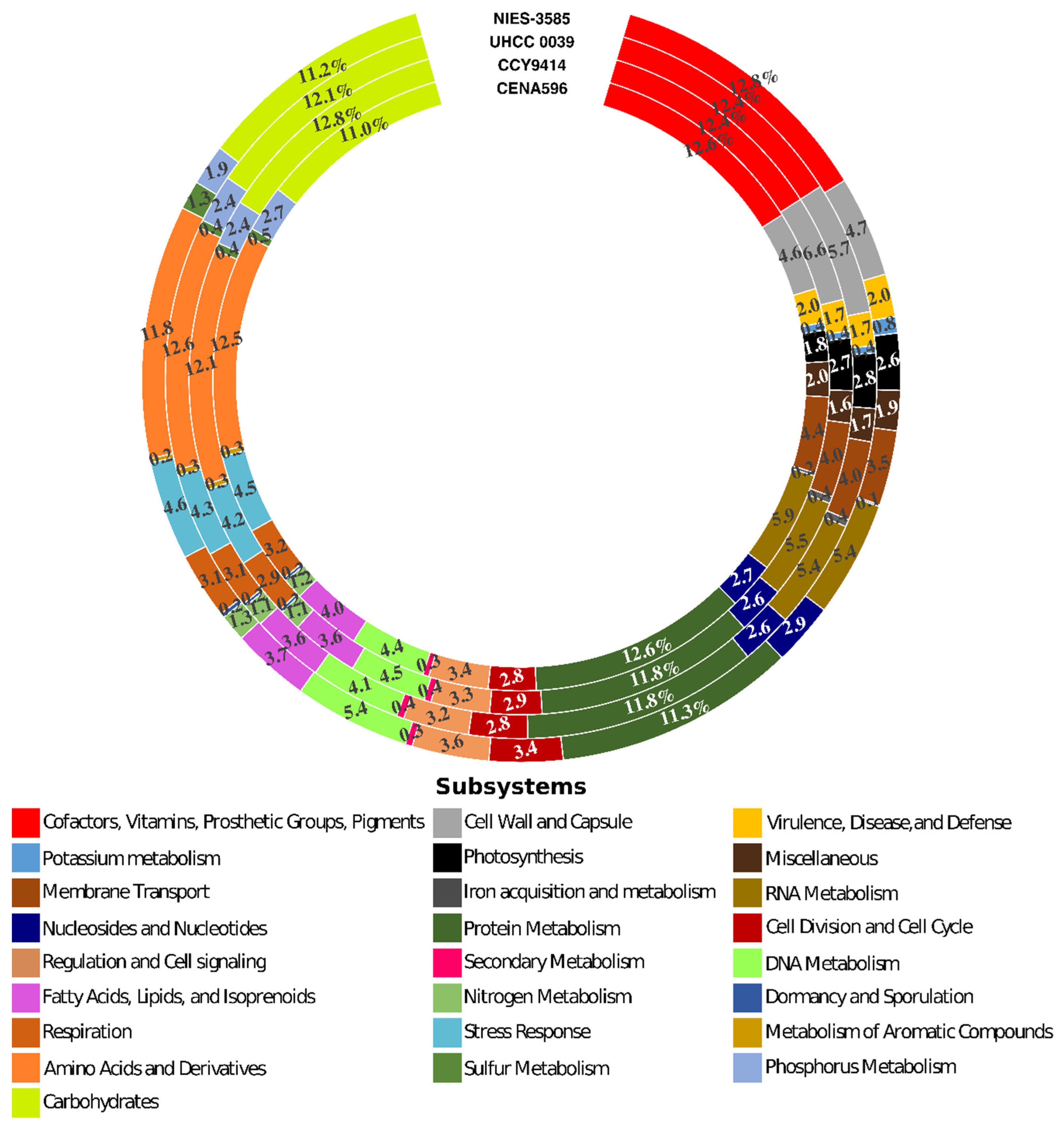
2.3. General Features of Nodularia Genomes

The comparison of CENA596, CCY9414, UHCC 0039, and NIES-3585 genomes assembly statistics revealed resemblances among them (Table 2). Although the CENA596 genome is still in draft status, the total genome size is considerably close to those of CCY9414 and UHCC 0039. Differently, the draft assembly of NIES-3585 showed the largest genome. The GC% content of the strains was similar (≅41.2%), but due to the number of scaffolds, CENA596 showed the lowest N50. The subsystem annotation identified 31–34% of the coding sequences from the four genomes. These sequences were mainly involved in cellular primary metabolism, such as the biosynthesis of vitamins, cofactors, pigments, and the metabolism of proteins, DNA, RNA, amino acids, and carbohydrates, photosynthesis, and respiration (Figure 2). The remaining 66–69% of all the genes were not classified in any subsystems.
Approximately 74% of the protein encoded in the genomes of CENA596, 70% of CCY9414, 71% of UHCC 0039, and 67% of NIES-3585 were orthologs in the four genomes (3122 proteins) (Figure 3A). The percentage of proteins exclusively encoded in each genome (paralogous and singletons) was approximately 8%, 5%, 3%, and 25% for CENA596, CCY9414, UHCC 0039, and NIES-3585, respectively (19.43% of the total). The remaining 18% of the proteins from CENA596, 25% of CCY9414, 26% of UHCC 0039, and 8% of NIES-3585 were orthologs in two or three genomes (10.36% of the total). In terms of the total number of proteins (17,786), 12,488 (or 70.21%) were orthologs in the four genomes, 1842 (or 19.43%) were paralogues and singletons, and 3456 were orthologs in two or three genomes.
The automatic annotation performed with MG-RAST showed that the ortholog genes in the four genomes were involved in a wide range of functions such as primary metabolism (photosynthesis, respiration, metabolism of vitamin, cofactors, etc.) stress responses, secondary metabolism, and other cellular processes (membrane transport, regulation, and cell signaling, and cell division and cycle) (Figure S1A). The specific genes of each strain were classified in equally diverse functions (Figure S1B–E).
Strain CENA596 showed average nucleotide identity (ANI)/average amino acid identity (AAI) of 97.76%/94.45% with CCY9414 and 97.75%/94.32% with UHCC 0039 (Figure 3B,C, respectively). The two Baltic strains were considerably similar (99.64%/97.36%, respectively), whereas the Japanese NIES-3585 had smaller identities (91.15%/87.39%, 91.20%/87.69%, and 91.21%/87.14% to CENA596, CCY9414, and UHCC 0039, respectively).

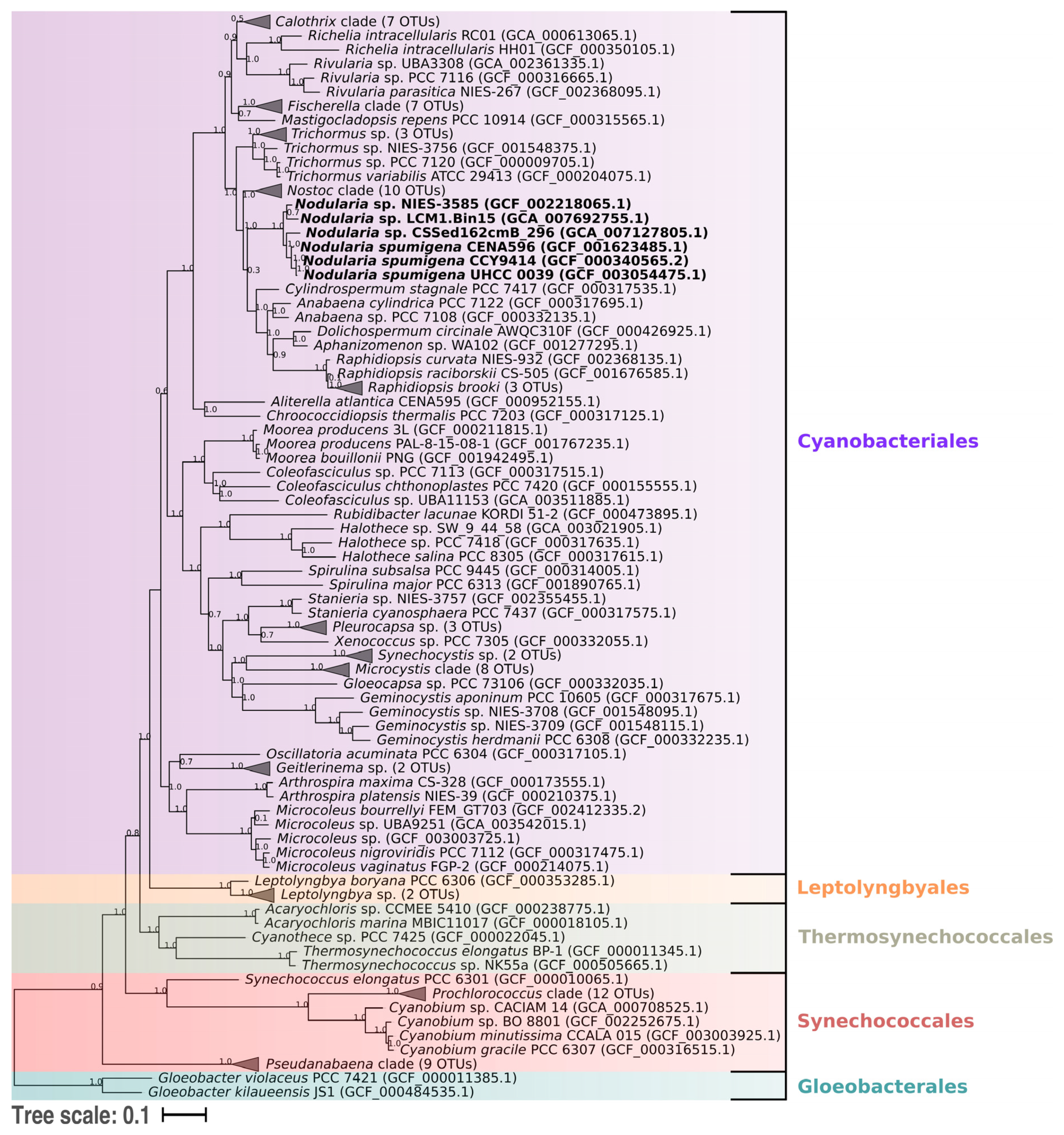
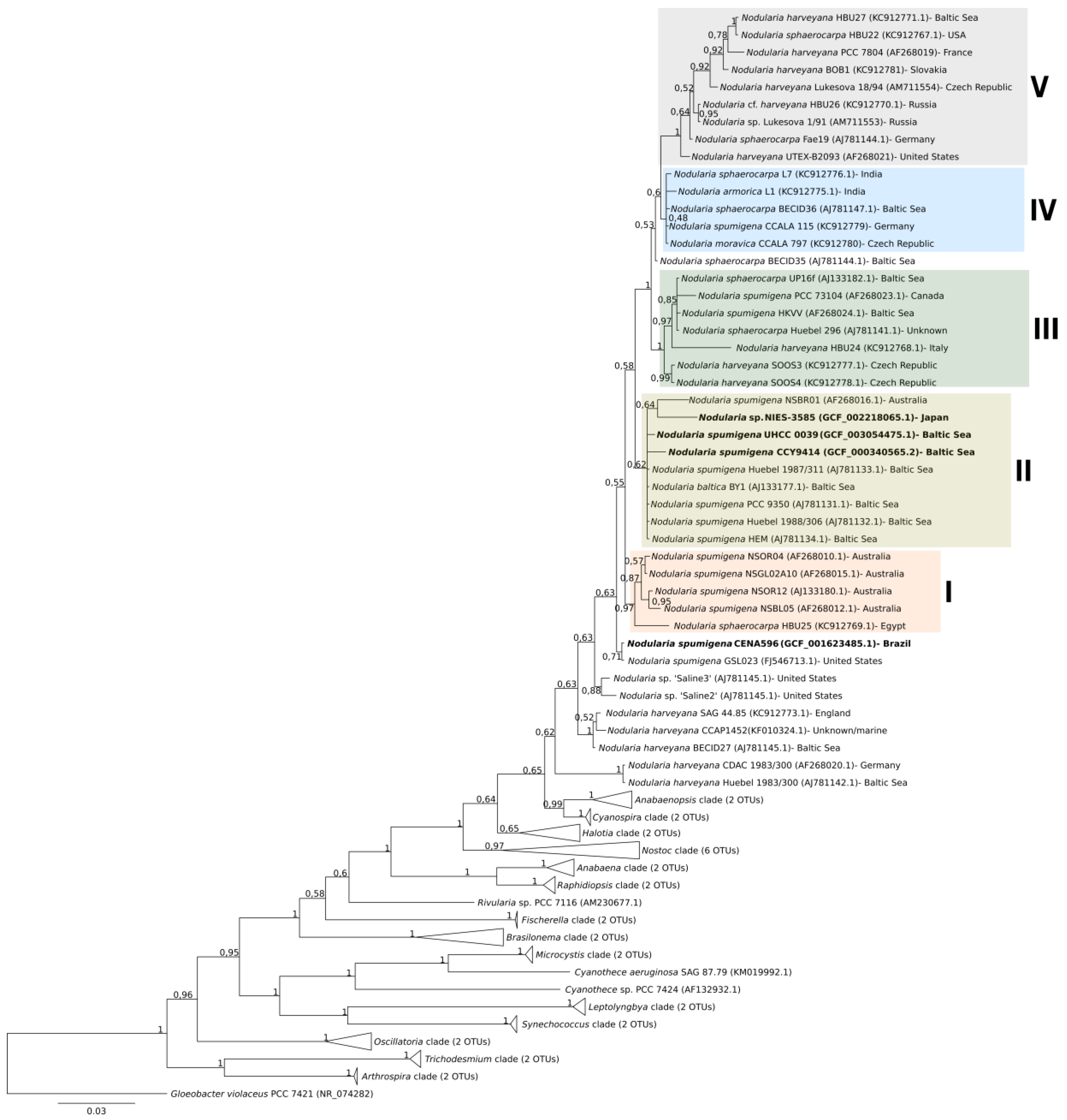
2.4. Phylogenomic Analysis

The phylogenomic tree grouped the strains within a clade containing other cyanobacteria belonging to the order Nostocales (Figure 4). The two evaluated Baltic strains formed a clade that shared a common ancestor with the Brazilian strain. The metagenome-assembled genome (MAG) CSSed162cmB_296 isolated from a Russian soda lake was positioned next to these three Nodularia strains. A sister clade comprised the Japanese strain and the Canadian MAG LCM1.Bin15.
The 16S rRNA gene sequences showed high identities among each other (99%; data not provided), but the strains grouped in distinct clades in the phylogenetic tree (Figure 5). The Brazilian CENA596 was allocated with the strain GSL023 isolated from the Great Salt Lake, UT, USA. This clade was closely related to N. spumigena strains from Australian brackish waters (Subclade I). The sequences of seven N. spumigena strains from the Baltic Sea, including CCY9414 and UHCC 0039, were positioned together in Subclade II. The Japanese NIES-3585 and an Australian N. spumigena strain (NSBR01) were positioned close to these Baltic strains. Nodularia Subclades III, IV, and V included mixed strains from various species and geographical origins.

3. Discussion

Cyanobacteria are known for their abundance and capability of exploring a wide range of environments [53]. Strain CENA596 was isolated from shrimp production ponds in the southern region of Brazil [47]. The crustaceans are cultivated using the Biofloc Technology System, in which fertilizers are used to stimulate microbial action to mineralize and assimilate nutrients from the feeding and excretion of shrimp [54]. In this way, water can be used in several production cycles without needing to exchange it with the surrounding aquatic environment [55]. Although cyanobacteria naturally compete with algae in aquaculture tanks, cyanobacteria often dominate eutrophic tanks due to their higher ability to thrive with low dissolved oxygen, high temperature, and turbidity [56]. Moreover, the water salinities commonly used in shrimp ponds are optimal for N. spumigena growth [49,57,58,59]. Therefore, cyanobacteria may form blooms in these environments and negatively interfere with shrimp production [60]. Strain CCY9414 was isolated from samples collected near Bornholm, Denmark in 1996 [61]. During the summer, N. spumigena blooms overcome phosphorous limitation by degrading phosphonate and are favored in the Baltic Sea due to stable, stratified, and warm brackish water [46,48]. Strain UHCC 0039 (also known as AV1) was isolated from a water sample collected in 1987 from the open Gulf of Finland [48,49].
The application of genome sequencing and mining to analyze and classify BGCs in the N. spumigena CENA596 genome gave new insights on the potential of this strain to synthesize natural products. BGCs are the core organization of cyanobacterial biosynthetic pathways at the genome level and generally code for multidomain enzymes, such as PKSs and NRPSs, transporters, and tailoring enzymes (such as halogenases, oxidases, and cyclases) [62,63]. BGC expression is regulated at the transcriptional level, and regulatory mechanisms are frequently found flanking the BGC [64,65]. A search for gene clusters responsible for the synthesis of natural products in N. spumigena CENA596 revealed a similar genomic construction to that found in the genomes of the Baltic N. spumigena CCY9414 and UHCC 0039 strains. However, LC-HR-QTOF mass spectrometry analyses showed lower variant production of the nonribosomal peptides NOD, SPU, APT, and AER by the Brazilian strain than the Baltic strains [42,44,46,50]. The co-production of toxin (NOD) and odorous (geosmin) metabolites by N. spumigena CENA596 during bloom episodes in shrimp ponds raises concern for human and animal health issues along with economic losses. When consumed by humans and other animals, NOD may lead to hepatic structure modification and consequent tissue damage, organ failure, and hemorrhagic shock [66]. Bioaccumulation of this molecule in the environment is also of great concern [39,40,67,68].
Gene cluster nda was first identified in N. spumigena NSOR12 as being composed of nine genes 47kbp wide in total [69]. This gene cluster was later found in N. spumigena strain AV1 [70], followed by CCY9414 [46] and CENA596 [47]. The reason behind NOD production by cyanobacteria is unknown but probable functions have been proposed such as protection against oxidative and luminous stress, predation, and allelopathic competition [66,71]. We note that nda was absent in the Japanese benthic Nodularia sp. NIES-3585, which corroborates the report that benthic Nodularia strains are not NOD producers [72]. Therefore, the fact that benthic Nodularia does not produce NOD and the high conservation of nda among the three planktonic N. spumigena strains suggests an important adaptive advantage of NOD-producing strains for the exploration of the water surface.
The nonribosomal spu and apt found in the genome of N. spumigena CENA596 showed nearly identical gene organization as in N. spumigena CCY9414 and UHCC 0039 and Sphaerospermopsis torques-reginae ITEP-024. In these strains, spu and apt together are in a genome region separated only by a 12 kbp nucleotide sequence that is believed to encode biosynthetic enzymes of substrates involved in the synthesis of both metabolites [73]. SPU and APT are protease inhibitors [42,43]. Proteases are responsible for catalyzing the breakdown of proteins, and therefore are intimately related to cell cycle progression, cell proliferation, and death; DNA replication; tissue remodeling, homeostasis; wound healing; and the immune response in complex animals: the dysregulation of those enzymes may lead to cardiovascular and inflammatory diseases, cancer, osteoporosis, and neurological disorders [74]. Despite the presence of apt in the genome of CENA596, only namalides were identified by chemical analysis. The production of namalides by the APT peptide synthase pathway has been attributed to a module skipping event and has already been identified in Nostoc sp. CENA543 and S. torques-reginae ITEP-024 [75,76]. Namalides are cyclic tetrapeptides with an exogenous amino acid attached to the macrocycle by a urethane linkage and are structurally related to APT but lacking two amino acid residues [77]. These authors also reported the carboxypeptidase A inhibitory activity of namalide at submicromolar concentrations.
The aer of the Brazilian CENA596 showed an incomplete aerI gene acting as a transferase in the molecule biosynthesis. Events of gene inversion, deletion, rearrangements, fusion, and fission are frequent in bacterial genomes and are known as mechanisms of gene evolution, in which multi-domain proteins evolve [78,79]. The alteration found in gene aerI in CENA596 did not affect its expression, as two AER variants were detected by mass spectrometry analysis. Variability in the aer biosynthetic pathway has been associated with a varying production of AER analogs [80]. Many cyanobacterial genera produce AERs, and ~100 variants have been described [81]. In the genus Nodularia, aer was first described in N. spumigena CCY9414 [46]. AERs constitute a group of linear modified tetrapeptides, and since the first isolation [82], this class of natural products gained attention as protease inhibitors [83] and as potent biotoxins [84].
The ribosomal gene cluster involved in the synthesis of geosmin was identified only in the genome of N. spumigena CENA596, and gas chromatography analysis confirmed the production of this terpenoid. This is the second report of geosmin being produced by N. spumigena [85]. Geosmin is the dominant cause of the muddy and earthy taste and odor in drinking water and causes consumer complaints and economic losses in drinking water supplies and the fishing industry [45]. Although this odorous metabolite is produced by a wide range of organisms, including actinobacteria, proteobacteria, fungi, amoeba, and liverwort, cyanobacteria are considered to be the major source of geosmin in aquatic environments where photosynthetic growth is viable [86]. This terpene seems to be part of oxidative stress responses in fungi and may have the same function in cyanobacteria [87].
Gene clusters mys and scy found in the genomes of the four Nodularia and the three planktonic N. spumigena, respectively, are responsible for the synthesis of MAAs and SCY, which are strong UV-absorbing compounds. Their biosynthesis is a mechanism developed by certain cyanobacteria to avoid harmful biological effects of exposure to solar UV radiation [88]. In addition to their photoprotective function, these pigments are important antioxidants, compatible solutes, and intracellular nitrogen reservoirs that may show anti-inflammatory and antiproliferative activities without chemical toxicity [89,90]. Therefore, these alkaloid compounds have gained research attention due to potential economic importance as candidates for pharmaceutical and cosmetic applications. Mass spectrometry analysis of the Brazilian CENA596 detected the production of two MAAs—shinorine and porphyra-334—as previously identified [89]. SCY was not detected despite the presence of scy. The explanation for this may be that the genes are not being expressed or the production level is below the detection limit, as multiple environmental signals may reportedly act to determine the level of this pigment in various cyanobacteria species [91].
Cyanobacteria from genus Nodularia were divided into three ecological groups: planktonic, benthic, and terrestrial [92]. Phenotypic (presence or absence of aerotopes), genetic (16S rRNA gene sequences), and genomic features (short tandemly repeated repetitive sequence fingerprinting) differentiated aquatic lineages in the planktonic or benthic groups [72]. The genetic organization of the operons responsible for the synthesis of aerotope structure is susceptible to rearrangements that may lead to the loss of these structures [93]. The benthic NIES-3585 did not possess the gvp, and therefore, is expected to not form aerotopes, as commonly found in a strain of this lifestyle. Although planktonic Nodularia has been extensively studied due to the formation of toxic blooms, the existing studies on benthic and soil Nodularia studies are limited to a few areas [26,72,94]. This lack of studies may be related to them not being dominant in these biotopes and generally not producing nodularin, with the exception of the strain N. sphaerocarpa PCC7804, which was isolated from a benthonic mantle of a thermal spring in France and produces NOD and [L-Har2]NOD [95,96].
The subsystem annotation of the genomes revealed a similar pattern of functional annotation. This result point to a noticeable genetic similarity as found in a previous comparative study of the N. spumigena strains [49]. The majority of the orthologs from the four genomes remain unidentified, whereas those that were annotated were involved in key metabolic pathways necessary for the survival of the organisms but also adaptive advantages and the secondary metabolism. This set of orthologous genes originated during speciation [97,98]. Similarly to the orthologs, just a few of the specific genes were assigned to a function. Thus, the metabolic diversity of the strains is possibly linked to these unknown genes.
The 16S rRNA gene phylogenetic analysis indicated that strain CENA596 was closely related to North American and Australian strains, whereas CCY9414 and UHCC 0039 were related to Baltic strains. The Japanese NIES-3585 allocated away from other benthic Nodularia but close to the Baltic N. spumigena strains. The phylogeny of this genus is problematic, as several species described based on their morphological characters did not present stable positions in phylogenetic trees [26,99]. Previous studies reported a clear separation between Baltic and Australian N. spumigena, although a limited number of sequences were analyzed [33,100,101]. Due to the inclusion of representatives from various geographical regions, our present results indicate that this geographical separation is not stable. Moreover, divergent subclades of Nodularia were previously proposed based on a limited number of 16S rRNA sequences [33,99]. The new subclades established here comprise strains from varied species and geographical regions and provide no clear separation based on these aspects. This result corroborates that the analysis of 16S rRNA is not an appropriate approach for studying the species categories and geographical origin of Nodularia [99].
16S rRNA gene-based phylogenies have been widely used for the identification of cyanobacteria [102], although the method has several limitations that may lead to poor resolutions in other bacterial taxa [103,104,105]. Therefore, the use of single-copy vertically inherited proteins has been proposed as a better approach to infer bacterial phylogeny [106,107]. In the specific case of cyanobacteria, analyses of 31 concatenated proteins provided robust results for the taxonomic study of these organisms [108,109,110]. The use of 120 conserved proteins has been recently proposed as a standard method for bacterial taxonomy and we introduced it to investigate the currently available Nodularia genomes and MAGs [111].
The approach used in our present work requires genome sequences of the organisms, only four of which are available for Nodularia. Although the two available MAGs of Nodularia were included, we were unable to conduct a robust analysis of the evolutionary relationship of this genus and therefore our analysis resembled a previous analysis constructed with 31 conserved proteins [49]. So far, no evident geographical difference among American, European, and Asian Nodularia can be pointed out. Both metagenomes are from soda lakes in the Northern Hemisphere. MAG CSSed162cmB_296 was grouped with the planktonic N. spumigena strains, whereas LCM1.Bin15 is positioned close to the benthic one. Future inclusion of new genomic sequences from distinct ecological niches will improve the taxonomy of Nodularia species and may establish subgroups containing various taxa.

4. Conclusions

Comparative analyses showed that the genomes of the Brazilian strain N. spumigena CENA596 and both the Baltic strains N. spumigena CCY9414 and UHCC 0039 are considerably more similar than the Japanese strain Nodularia sp. NIES-3585. As the function of most of the genes encoded in those genomes remains unclear, the biosynthetic diversity of the genus Nodularia was only evaluated based on known pathways, leaving potential BGCs to be further explored. So far, genome mining and chemical analyses of the available N. spumigena strains have already demonstrated the vast potential of this species to produce bioactive molecules, highlighting the need for monitoring harmful blooms in aquatic environments explored by human activities. The phylogeny of genus Nodularia still needs improvements, as the majority of genomic information available is from the species N. spumigena, which lacks representatives for most of the geographic regions. Future studies on the genus should use a phylogenomic approach based on the analysis of a large number of conserved proteins rather than the traditional 16S rRNA gene for a robust taxonomy. Last, generating new genomic information of isolates from other regions may contribute to better understanding of this genus, help prevent damages caused by toxic blooms, and enable the discovery of new natural products with pharmacological and biological activities.

5. Materials and Methods

5.1. Cyanobacterial Genomes

The draft genome of the Brazilian strain N. spumigena CENA596 was published earlier [47] (accession number GCF_001623485.1). This strain was isolated from a N. spumigena bloom sample collected on 5 December 2013 in a shrimp production pond (32°12′19″S, 52°10′42″W) at the Marine Aquaculture Station of the Federal University of Rio Grande. The station is located on Cassino beach in the southwest Atlantic Ocean, Rio Grande municipality, Rio Grande do Sul state, Brazil. The strain N. spumigena CCY9414, with a possibly complete published genome, was isolated from a sample of brackish surface water collected in 1996 from the Baltic Sea near Bornholm [46,61] (accession number GCF_000340565.2). Strain N. spumigena UHCC 0039 (or N. spumigena AV1), with a complete genome, was isolated from a water sample collected in 1987 from the open Gulf of Finland [12,48,49] (accession number GCF_003054475.1). The draft genome of the Japanese strain Nodularia sp. NIES-3585 (accession number GCF_002218065.1) was included in this study because it showed a nucleotide identity of 99% to the N. spumigena CENA596 genome.

5.2. Natural Product Pathways in N. spumigena CENA596

Automatic annotation of secondary metabolite gene clusters in the analyzed genomes was performed using the antiSMASH server v4.0 [112]. Manual annotation and curation were then performed with the Artemis program v17.0.1 [113] and BLASTP (http://blast.ncbi.nlm.nih.gov/Blast.cgi?PAGE=Proteins). For that, protein sequences of known secondary metabolite pathways were compared with the GenBank protein database using the BLASTP tool to detect orthologs (e-value ≤ 1 × 10⁻²⁰, identity ≥ 30%). The specificity of the adenylation domain of nonribosomal peptide synthetases (NRPSs) enzymes was predicted using the server NRPSpreditor2 [114].

5.3. Targeted Metabolomics

N. spumigena CENA596 was cultured in an F/2 liquid medium [115] without silica. Cultures were maintained at 22 ± 1 °C following a light–dark cycle of 14:10 h with fluorescent light (40–50 µmol photons∙m−2∙s−1) for 20 days. Fresh samples (200 mL) were vacuum-filtered on glass fiber (Sartorius GmbH, Germany) for cell concentration. Samples were extracted with 1 mL of MeOH:H2O 60/40 (v/v) at ultrasound probe (Sonic Ruptor 400, Omni, GA, USA) during 1 min on ice. After centrifugation for 10 min at 12,000× g (5804R, Eppendorf, Germany), the supernatant was separated and the extraction repeated. Combined supernatants were filtered (Millex 0.45 μm, Millipore, MA, USA) into vials. Analyses of NOD, SPU, APT, AER, mycosporine-like amino acids (MAA), and scytonemin (SCY) were performed on a Luna C18(2) column (150 × 2.1 mm, 3 μm; Phenomenex, State abbreviation, USA) using (A) 0.1% formic acid and (B) acetonitrile as the mobile phase (250 μL∙min−1 at 35 °C). The separation was obtained in a Shimadzu Prominence equipment (Kyoto, Japan) with a linear gradient from 5% to 90% B in 34 min (a total time of 45 min). High-resolution mass spectrometry data were acquired on a quadrupole/time-of-flight instrument (MicroTOF-QII, Bruker Daltonics, MA, USA) equipped with an electrospray source (ESI) operated in the positive ionization mode. Compound scanning was performed from m/z 100 to 1500 using a sodium acetate solution for mass calibration. Product ion spectra were acquired using argon as a collision gas under variable dissociation energies. Geosmin analysis was performed as described previously [45] using solid-phase microextraction and gas chromatography coupled to mass spectrometry (7890A/5975C, Agilent Technologies, CA, USA). The identification of compounds in CENA596 was based on their exact mass (<5 ppm) and their MS2 profile compared to previous spectra available in the literature (molecular data is shown in File S1). The terpenoid geosmin was identified based on its retention time and mass spectrum profile by GC-MS compared to an analytical standard and the NIST library.

5.4. Comparative Analyses

Genome assembly statistics were obtained using Assemblathon 2 [116]. A genome-wide comparison of the four N. spumigena and Nodularia sp. genomes was conducted using Blast Atlas analysis in GView Server v.3 [117]. Subsystems annotation was performed with the RAST server [51] and SEED tool [52]. The OrthoVenn server was used in the analysis of orthologous clusters [118] while MG-RAST v4.0.3 was used for the automatic annotation of the orthologs and specific proteins [119]. Heatmaps were estimated using the program GET_HOMOLOGUES [120,121] and generated using a seaborn v0.9 library script (https://github.com/mwaskom/seaborn/releases).

5.5. Phylogenomic Analysis

The phylogenomic placement was inferred with GTDB-Tk v0.3.2 (database release 89, https://github.com/Ecogenomics/GTDBTk) based on the Genome Taxonomy Database (GTDB), which is a recently proposed genomic-based taxonomy for Bacteria and Archaea [111]. The pipeline generates the tree through the identification and alignment of 120 bacterial single-copy conserved marker genes, then infers the phylogeny of the concatenated sequences using FastTree [122] with the WAG+GAMMA models and maximum likelihood algorithm. MAG of Nodularia publicly available in the NCBI was included in the analysis to expand the number of representatives of the genus.
The 16S ribosomal RNA (rRNA) gene tree was generated using an alignment performed with Muscle alignment in MEGA v10.0.5 (default parameters) of 81 cyanobacterial sequences [123]. The evolutionary model GTR+I+G best fitted the data set according to jModelTest v2.1.1 [124] and the Bayesian inference was constructed by MrBayes v3.2.6 with 5,000,000 generations [125].

Supplementary Materials

The following are available online at https://0-www-mdpi-com.brum.beds.ac.uk/2072-6651/12/3/141/s1, Figure S1: Orthologs (A) and specific proteins to Nodularia spumigena CENA596 (B), CCY9414 (C), and UHCC 0039 (D), and Nodularia sp. NIES-3585 (E) annotated according to KEGG Orthology (KO) and SEED Subsystems, File S1: Results of the chemical analyses of the strain N. spumigena CENA 596. The name of variants, the method used, and the mass spectrums of nodularins, spumigins, namalides, aeruginosins, and mycosporine-like amino acids; and the results of geosmin analysis using SPME-GC-MS are shown, Table S1: Results of the antiSMASH automatic annotation of Nodularia spumigena CENA596, CCY9414 and UHCC 0039, and Nodularia sp. NIES-3585. The number of synthases and synthetases, and the biosynthetic gene cluster identified in the genomes through manual curation are shown, Table S2: Proposed function of proteins encoded by the biosynthetic gene cluster identified through manual curation in the genome of Nodularia spumigena strains CENA596, CCY9414, and UHCC 0039, and Nodularia sp. NIES-3585, Table S3: Conservation of the adenylation domain-binding pockets in genes ndaA-D, spuA-B, aptA-D, and aerM-G in the genome of Nodularia spumigena strains CENA596, CCY9414, and UHCC 0039; and other cyanobacteria.

Author Contributions

M.F.F. and R.V.P. designed the study. R.V.P., E.D., V.A.C.d.A., J.R., K.S., and M.F.F. were responsible for the generation and analysis of the genomic data. F.A.D. and E.P. performed and interpreted the chemical analyses. All authors participated in discussing the results and writing the manuscript. The authors approved the final article. All authors have read and agreed to the published version of the manuscript.

Funding

This research was supported by a grant from the State of São Paulo Research Foundation (FAPESP) 2013/50425-8 and 2014/50420-9 to MFF and EP, respectively, and the Academy of Finland (1273798) to KS. Also, EP acknowledges the support of the University of São Paulo Foundation (FUSP) (Project #1979), the Brazilian Federal Agency for the Support and Evaluation of Graduate Education (CAPES, Project # 23038.001401/2018-92), and Brazilian National Council for Scientific and Technological Development (CNPq, 311048/2016-1 and 439065/2018-6). RVP received a graduate scholarship from CAPES, Finance code 001, an EDUFI fellowship from the Center from International Mobility (CIMO) and funding from the Doctoral Programme in Microbiology and Biotechnology. ED received scholarship from CAPES, Finance code 001. JR and VACA received a postdoctoral fellowship from the CAPES-PNPD program. MF received a research fellowship (310244/2015-3) from the CNPq.

Conflicts of Interest

The authors declare no conflicts of interest.

References

  1. FAO. The State of World Fisheries and Aquaculture 2018—Meeting the Sustainable Development Goals; FAO: Rome, Italy, 2018; ISBN 978-92-5-130562-1. [Google Scholar]
  2. Ferreira, N.C.; Bonetti, C.; Seiffert, W.Q. Hydrological and Water Quality Indices as management tools in marine shrimp culture. Aquaculture 2011, 318, 425–433. [Google Scholar] [CrossRef]
  3. Ma, Z.; Song, X.; Wan, R.; Gao, L. A modified water quality index for intensive shrimp ponds of Litopenaeus vannamei. Ecol. Indic. 2013, 24, 287–293. [Google Scholar] [CrossRef]
  4. Jescovitch, L.N.; Ullman, C.; Rhodes, M.; Davis, D.A. Effects of different feed management treatments on water quality for Pacific white shrimp Litopenaeus vannamei. Aquac. Res. 2018, 49, 526–531. [Google Scholar] [CrossRef]
  5. Barraza-Guardado, R.H.; Arreola-Lizárraga, J.A.; López-Torres, M.A.; Casillas-Hernández, R.; Miranda-Baeza, A.; Magallón-Barrajas, F.; Ibarra-Gámez, C. Effluents of Shrimp Farms and Its Influence on the Coastal Ecosystems of Bahía de Kino, Mexico. Sci. World J. 2013, 2013, 1–8. [Google Scholar] [CrossRef]
  6. Alonso-Rodríguez, R.; Páez-Osuna, F. Nutrients, phytoplankton and harmful algal blooms in shrimp ponds: A review with special reference to the situation in the Gulf of California. Aquaculture 2003, 219, 317–336. [Google Scholar]
  7. Am, A.; Silvester, R.; Alexander, D.; M, N.; Abdulla, M.H. Characterization of blooming algae and bloom-associated changes in the water quality parameters of traditional pokkali cum prawn fields along the South West coast of India. Environ. Monit. Assess. 2016, 188. [Google Scholar] [CrossRef]
  8. Gao, J.; Zuo, H.; Yang, L.; He, J.-H.; Niu, S.; Weng, S.; He, J.; Xu, X. Long-term influence of cyanobacterial bloom on the immune system of Litopenaeus vannamei. Fish Shellfish Immunol. 2017, 61, 79–85. [Google Scholar] [CrossRef]
  9. Yang, P.; Lai, D.Y.F.; Jin, B.; Bastviken, D.; Tan, L.; Tong, C. Dynamics of dissolved nutrients in the aquaculture shrimp ponds of the Min River estuary, China: Concentrations, fluxes and environmental loads. Sci. Total Environ. 2017, 603–604, 256–267. [Google Scholar] [CrossRef] [Green Version]
  10. Main, D.C.; Berry, P.H.; Peet, R.L.; Robertson, J.P. Sheep mortalities associated with the blue green alga Nodularia spumigena. Aust. Vet. J. 1977, 53, 578–581. [Google Scholar] [CrossRef]
  11. Carmichael, W.W.; Eschedor, J.T.; Patterson, G.M.; Moore, R.E. Toxicity and partial structure of a hepatotoxic peptide produced by the cyanobacterium Nodularia spumigena Mertens emend. L575 from New Zealand. Appl. Environ. Microbiol. 1988, 54, 2257–2263. [Google Scholar] [CrossRef] [Green Version]
  12. Sivonen, K.; Kononen, K.; Carmichael, W.W.; Dahlem, A.M.; Rinehart, K.L.; Kiviranta, J.; Niemela, S.I. Occurrence of the hepatotoxic cyanobacterium Nodularia spumigena in the Baltic Sea and structure of the toxin. Appl. Environ. Microbiol. 1989, 55, 1990–1995. [Google Scholar] [CrossRef] [PubMed] [Green Version]
  13. Seaman, M.T.; Ashton, P.J.; Williams, W.D. Inland salt waters of southern Africa. Hydrobiologia 1991, 210, 75–91. [Google Scholar] [CrossRef]
  14. Nehring, S. Mortality of dogs associated with a mass development of Nodularia spumigena (Cyanophyceae) in a brackish lake at the German North Sea coast. J. Plankton Res. 1993, 15, 867–872. [Google Scholar] [CrossRef]
  15. Jones, G.; Blackburn, S.; Parker, N. A toxic bloom of Nodularia spumigena Mertens in Orielton Lagoon, Tasmania. Mar. Freshw. Res. 1994, 45, 787–800. [Google Scholar] [CrossRef]
  16. Harding, W.R.; Rowe, N.; Wessels, J.C.; Beattie, K.A.; Codd, G.A. Death of a dog attributed to the cyanobacterial (blue-green algal) hepatotoxin nodularin in South Africa. J. S. Afr. Vet. Assoc. 1995, 66, 256–259. [Google Scholar]
  17. Blackburn, S.I.; McCausland, M.A.; Bolch, C.J.S.; Newman, S.J.; Jones, G.J. Effect of salinity on growth and toxin production in cultures of the bloom-forming cyanobacterium Nodularia spumigena from Australian waters. Phycologia 1996, 35, 511–522. [Google Scholar] [CrossRef]
  18. Mazur-Marzec, H.; Meriluoto, J.; Pliński, M.; Szafranek, J. Characterization of nodularin variants in Nodularia spumigena from the Baltic Sea using liquid chromatography/mass spectrometry/mass spectrometry. Rapid Commun. Mass Spectrom. 2006, 20, 2023–2032. [Google Scholar] [CrossRef]
  19. Akcaalan, R.; Mazur-Marzec, H.; Zalewska, A.; Albay, M. Phenotypic and toxicological characterization of toxic Nodularia spumigena from a freshwater lake in Turkey. Harmful Algae 2009, 8, 273–278. [Google Scholar] [CrossRef]
  20. Galat, D.L.; Verdin, J.P.; Sims, L.L. Large-scale patterns of Nodularia spumigena blooms in Pyramid Lake, Nevada, determined from Landsat imagery: 1972–1986. Hydrobiologia 1990, 197, 147–164. [Google Scholar] [CrossRef]
  21. Beutel, M.W.; Horne, A.J.; Roth, J.C.; Barratt, N.J. Limnological effects of anthropogenic desiccation of a large, saline lake, Walker Lake, Nevada. Hydrobiologia 2001, 466, 91–105. [Google Scholar] [CrossRef]
  22. Falcón, L.I.; Escobar-Briones, E.; Romero, D. Nitrogen fixation patterns displayed by cyanobacterial consortia in Alchichica crater-lake, Mexico. Hydrobiologia 2002, 467, 71–78. [Google Scholar] [CrossRef]
  23. Oliva, M.G.; Lugo, J.; Alcocer, L.; Peralta, L.; Oseguera, L. Planktonic bloom-forming Nodularia in the saline lake Alchichica, Mexico. Nat. Resour. Environ. Issues 2009, 15, 121–126. [Google Scholar]
  24. Bazán, G.I.; Alvarez, S.B. Cianoficeas continentals de la provincia de la pampa (Argentina)-I. Rev. Fac. Agron. Luz. 1994, 7, 43–62. [Google Scholar]
  25. Pérez Baliero, M.; Del, C.; Bonilla, S.; De León, L.; Šmarda, J.; Komárek, J. A bloom of Nodularia baltica-spumigena group (Cyanobacteria) in a shallow coastal lagoon of Uruguay, South America. Algol. Stud. 1999, 93, 91–101. [Google Scholar]
  26. Laamanen, M.J.; Gugger, M.F.; Lehtimaki, J.M.; Haukka, K.; Sivonen, K. Diversity of Toxic and Nontoxic Nodularia Isolates (Cyanobacteria) and Filaments from the Baltic Sea. Appl. Environ. Microbiol. 2001, 67, 4638–4647. [Google Scholar] [CrossRef] [Green Version]
  27. Mazur-Marzec, H.; Kaczkowska, M.; Blaszczyk, A.; Akcaalan, R.; Spoof, L.; Meriluoto, J. Diversity of Peptides Produced by Nodularia spumigena from Various Geographical Regions. Mar. Drugs 2012, 11, 1–19. [Google Scholar] [CrossRef] [Green Version]
  28. Francis, G. Poisonous Australian Lake. Nature 1878, 18, 11–12. [Google Scholar] [CrossRef] [Green Version]
  29. Edler, L.; Fernö, S.; Lind, M.G.; Lundberg, R.; Nilsson, P.O. Mortality of dogs associated with a bloom of the cyanobacterium Nodularia spumigena in the Baltic Sea. Ophelia 1985, 24, 103–109. [Google Scholar] [CrossRef]
  30. Van Halderen, A.; Harding, W.R.; Wessels, J.C.; Schneider, D.J.; Heine, E.W.; Van der Merwe, J.; Fourie, J.M. Cyanobacterial (blue-green algae) poisoning of livestock in the western Cape Province of South Africa. J. S. Afr. Vet. Assoc. 1995, 66, 260–264. [Google Scholar]
  31. Simola, O.; Wiberg, M.; Jokela, J.; Wahlsten, M.; Sivonen, K.; Syrja, P. Pathologic Findings and Toxin Identification in Cyanobacterial (Nodularia spumigena) Intoxication in a Dog. Vet. Pathol. 2012, 49, 755–759. [Google Scholar] [CrossRef] [Green Version]
  32. Pacheco, L.; Kunrath, N. Identification of the Toxic Pentapeptide Nodularin in a Cyanobacterial Bloom in a Shrimp Farm in South American Atlantic Coast. Pharm. Anal. Acta 2016, 7, 479. [Google Scholar]
  33. Da Silveira, S.B.; Wasielesky, W.; Andreote, A.P.D.; Fiore, M.F.; Odebrecht, C. Morphology, phylogeny, growth rate and nodularin production of Nodularia spumigena from Brazil. Mar. Biol. Res. 2017, 13, 1095–1107. [Google Scholar] [CrossRef]
  34. Rinehart, K.L.; Harada, K.; Namikoshi, M.; Chen, C.; Harvis, C.A.; Munro, M.H.G.; Blunt, J.W.; Mulligan, P.E.; Beasley, V.R.; Dahlem, A.M.; et al. Nodularin, microcystin, and the configuration of Adda. J. Am. Chem. Soc. 1988, 110, 8557–8558. [Google Scholar] [CrossRef]
  35. Yoshizawa, S.; Matsushima, R.; Watanabe, M.F.; Harada, K.; Ichihara, A.; Carmichael, W.W.; Fujiki, H. Inhibition of protein phosphatases by microcystins and nodularin associated with hepatotoxicity. J. Cancer Res. Clin. Oncol. 1990, 116, 609–614. [Google Scholar] [CrossRef] [PubMed]
  36. Honkanen, R.E.; Dukelow, M.; Zwiller, J.; Moore, R.E.; Khatra, B.S.; Boynton, A.L. Cyanobacterial nodularin is a potent inhibitor of type 1 and type 2A protein phosphatases. Mol. Pharmacol. 1991, 40, 577–583. [Google Scholar] [PubMed]
  37. Ohta, T.; Sueoka, E.; Iida, N.; Komori, A.; Suganuma, M.; Nishiwaki, R.; Tatematsu, M.; Kim, S.J.; Carmichael, W.W.; Fujiki, H. Nodularin, a potent inhibitor of protein phosphatases 1 and 2A, is a new environmental carcinogen in male F344 rat liver. Cancer Res. 1994, 54, 6402–6406. [Google Scholar]
  38. Kankaanpää, H.; Turunen, A.-K.; Karlsson, K.; Bylund, G.; Meriluoto, J.; Sipiä, V. Heterogeneity of nodularin bioaccumulation in northern Baltic Sea flounders in 2002. Chemosphere 2005, 59, 1091–1097. [Google Scholar] [CrossRef]
  39. Sipiä, V.; Kankaanpää, H.; Peltonen, H.; Vinni, M.; Meriluoto, J. Transfer of nodularin to three-spined stickleback (Gasterosteus aculeatus L.), herring (Clupea harengus L.), and salmon (Salmo salar L.) in the northern Baltic Sea. Ecotoxicol. Environ. Saf. 2007, 66, 421–425. [Google Scholar] [CrossRef]
  40. Barda, I.; Kankaanpää, H.; Purina, I.; Balode, M.; Sjövall, O.; Meriluoto, J. Bioaccumulation of hepatotoxins—A considerable risk in the Latvian environment. Environ. Pollut. 2015, 196, 313–320. [Google Scholar] [CrossRef]
  41. Drobac, D.; Tokodi, N.; Lujić, J.; Marinović, Z.; Subakov-Simić, G.; Dulić, T.; Važić, T.; Nybom, S.; Meriluoto, J.; Codd, G.A.; et al. Cyanobacteria and cyanotoxins in fishponds and their effects on fish tissue. Harmful Algae 2016, 55, 66–76. [Google Scholar] [CrossRef]
  42. Fewer, D.P.; Jokela, J.; Rouhiainen, L.; Wahlsten, M.; Koskenniemi, K.; Stal, L.J.; Sivonen, K. The non-ribosomal assembly and frequent occurrence of the protease inhibitors spumigins in the bloom-forming cyanobacterium Nodularia spumigena. Mol. Microbiol. 2009, 73, 924–937. [Google Scholar] [CrossRef] [PubMed]
  43. Rouhiainen, L.; Jokela, J.; Fewer, D.P.; Urmann, M.; Sivonen, K. Two Alternative Starter Modules for the Non-Ribosomal Biosynthesis of Specific Anabaenopeptin Variants in Anabaena (Cyanobacteria). Chem. Biol. 2010, 17, 265–273. [Google Scholar] [CrossRef] [PubMed] [Green Version]
  44. Fewer, D.P.; Jokela, J.; Paukku, E.; Österholm, J.; Wahlsten, M.; Permi, P.; Aitio, O.; Rouhiainen, L.; Gomez-Saez, G.V.; Sivonen, K. New Structural Variants of Aeruginosin Produced by the Toxic Bloom Forming Cyanobacterium Nodularia spumigena. PLoS ONE 2013, 8, e73618. [Google Scholar] [CrossRef] [Green Version]
  45. Suurnäkki, S.; Gomez-Saez, G.V.; Rantala-Ylinen, A.; Jokela, J.; Fewer, D.P.; Sivonen, K. Identification of geosmin and 2-methylisoborneol in cyanobacteria and molecular detection methods for the producers of these compounds. Water Res. 2015, 68, 56–66. [Google Scholar] [CrossRef] [PubMed]
  46. Voß, B.; Bolhuis, H.; Fewer, D.P.; Kopf, M.; Möke, F.; Haas, F.; El-Shehawy, R.; Hayes, P.; Bergman, B.; Sivonen, K.; et al. Insights into the Physiology and Ecology of the Brackish-Water-Adapted Cyanobacterium Nodularia spumigena CCY9414 Based on a Genome-Transcriptome Analysis. PLoS ONE 2013, 8, e60224. [Google Scholar] [CrossRef] [PubMed] [Green Version]
  47. Popin, R.V.; Rigonato, J.; Abreu, V.A.C.; Andreote, A.P.D.; Silveira, S.B.; Odebrecht, C.; Fiore, M.F. Draft Genome Assembly of the Bloom-Forming Cyanobacterium Nodularia spumigena Strain CENA596 in Shrimp Production Ponds. Genome Announc. 2016, 4, e00466-16. [Google Scholar] [CrossRef] [Green Version]
  48. Teikari, J.E.; Fewer, D.P.; Shrestha, R.; Hou, S.; Leikoski, N.; Mäkelä, M.; Simojoki, A.; Hess, W.R.; Sivonen, K. Strains of the toxic and bloom-forming Nodularia spumigena (cyanobacteria) can degrade methylphosphonate and release methane. ISME J. 2018, 12, 1619–1630. [Google Scholar] [CrossRef] [Green Version]
  49. Teikari, J.E.; Hou, S.; Wahlsten, M.; Hess, W.R.; Sivonen, K. Comparative Genomics of the Baltic Sea Toxic Cyanobacteria Nodularia spumigena UHCC 0039 and Its Response to Varying Salinity. Front. Microbiol. 2018, 9. [Google Scholar] [CrossRef] [Green Version]
  50. Mazur-Marzec, H.; Bertos-Fortis, M.; Toruńska-Sitarz, A.; Fidor, A.; Legrand, C. Chemical and Genetic Diversity of Nodularia spumigena from the Baltic Sea. Mar. Drugs 2016, 14, 209. [Google Scholar] [CrossRef] [Green Version]
  51. Aziz, R.K.; Bartels, D.; Best, A.A.; DeJongh, M.; Disz, T.; Edwards, R.A.; Formsma, K.; Gerdes, S.; Glass, E.M.; Kubal, M.; et al. The RAST Server: Rapid Annotations using Subsystems Technology. BMC Genom. 2008, 9, 75. [Google Scholar] [CrossRef] [Green Version]
  52. Overbeek, R. The Subsystems Approach to Genome Annotation and its Use in the Project to Annotate 1000 Genomes. Nucleic Acids Res. 2005, 33, 5691–5702. [Google Scholar] [CrossRef] [PubMed] [Green Version]
  53. Beck, C.; Knoop, H.; Axmann, I.M.; Steuer, R. The diversity of cyanobacterial metabolism: Genome analysis of multiple phototrophic microorganisms. BMC Genom. 2012, 13, 56. [Google Scholar] [CrossRef] [PubMed] [Green Version]
  54. Crab, R.; Defoirdt, T.; Bossier, P.; Verstraete, W. Biofloc technology in aquaculture: Beneficial effects and future challenges. Aquaculture 2012, 356, 351–356. [Google Scholar] [CrossRef]
  55. Krummenauer, D.; Peixoto, S.; Cavalli, R.O.; Poersch, L.H.; Wasielesky, W. Superintensive Culture of White Shrimp, Litopenaeus vannamei, in a Biofloc Technology System in Southern Brazil at Different Stocking Densities. J. World Aquac. Soc. 2011, 42, 726–733. [Google Scholar] [CrossRef]
  56. Sunda, W.G.; Graneli, E.; Gobler, C.J. Positive feedback and the development and persistence of ecosystem disruptive algal blooms. J. Phycol. 2006, 42, 963–974. [Google Scholar] [CrossRef]
  57. Rosas, C.; López, N.; Mercado, P.; Martínez, E. Effect of Salinity Acclimation on Oxygen Consumption of Juveniles of the White Shrimp Litopenaeus vannamei. J. Crustacean Biol. 2001, 21, 912–922. [Google Scholar] [CrossRef] [Green Version]
  58. Roy, L.A.; Davis, D.A.; Saoud, I.P.; Boyd, C.A.; Pine, H.J.; Boyd, C.E. Shrimp culture in inland low salinity waters: Shrimp culture in inland low salinity waters. Rev. Aquac. 2010, 2, 191–208. [Google Scholar] [CrossRef]
  59. Silveira, S.B.; Odebrecht, C. Effects of Salinity and Temperature on the Growth, Toxin Production, and Akinete Germination of the Cyanobacterium Nodularia spumigena. Front. Mar. Sci. 2019, 6, 339. [Google Scholar] [CrossRef]
  60. Yusoff, F.M.; Zubaidah, M.S.; Matias, H.B.; Kwan, T.S. Phytoplankton succession in intensive marine shrimp culture ponds treated with a commercial bacterial product. Aquac. Res. 2002, 33, 269–278. [Google Scholar] [CrossRef]
  61. Hayes, P.K.; Barker, G.L.A. Genetic diversity within Baltic sea populations of Nodularia (cyanobacteria). J. Phycol. 1997, 33, 919–923. [Google Scholar] [CrossRef]
  62. Walsh, C.T.; Chen, H.; Keating, T.A.; Hubbard, B.K.; Losey, H.C.; Luo, L.; Marshall, C.G.; Miller, D.A.; Patel, H.M. Tailoring enzymes that modify nonribosomal peptides during and after chain elongation on NRPS assembly lines. Curr. Opin. Chem. Biol. 2001, 5, 525–534. [Google Scholar] [CrossRef]
  63. Welker, M.; Von Döhren, H. Cyanobacterial peptides—Nature’s own combinatorial biosynthesis. FEMS Microbiol. Rev. 2006, 30, 530–563. [Google Scholar] [CrossRef] [PubMed] [Green Version]
  64. Jones, A.C.; Gerwick, L.; Gonzalez, D.; Dorrestein, P.C.; Gerwick, W.H. Transcriptional analysis of the jamaicamide gene cluster from the marine cyanobacterium Lyngbya majuscula and identification of possible regulatory proteins. BMC Microbiol. 2009, 9, 247. [Google Scholar] [CrossRef] [PubMed] [Green Version]
  65. Pearson, L.A.; Dittmann, E.; Mazmouz, R.; Ongley, S.E.; D’Agostino, P.M.; Neilan, B.A. The genetics, biosynthesis and regulation of toxic specialized metabolites of cyanobacteria. Harmful Algae 2016, 54, 98–111. [Google Scholar] [CrossRef] [PubMed]
  66. Pearson, L.; Mihali, T.; Moffitt, M.; Kellmann, R.; Neilan, B. On the Chemistry, Toxicology and Genetics of the Cyanobacterial Toxins, Microcystin, Nodularin, Saxitoxin and Cylindrospermopsin. Mar. Drugs 2010, 8, 1650–1680. [Google Scholar] [CrossRef] [Green Version]
  67. Mazur-Marzec, H.; Tymińska, A.; Szafranek, J.; Pliński, M. Accumulation of nodularin in sediments, mussels, and fish from the Gulf of Gdańsk, southern Baltic Sea. Environ. Toxicol. 2007, 22, 101–111. [Google Scholar] [CrossRef]
  68. Persson, K.-J.; Legrand, C.; Olsson, T. Detection of nodularin in European flounder (Platichthys flesus) in the west coast of Sweden: Evidence of nodularin mediated oxidative stress. Harmful Algae 2009, 8, 832–838. [Google Scholar] [CrossRef]
  69. Moffitt, M.C.; Neilan, B.A. Characterization of the Nodularin Synthetase Gene Cluster and Proposed Theory of the Evolution of Cyanobacterial Hepatotoxins. Appl. Environ. Microbiol. 2004, 70, 6353–6362. [Google Scholar] [CrossRef] [Green Version]
  70. Jonasson, S.; Vintila, S.; Sivonen, K.; El-Shehawy, R. Expression of the nodularin synthetase genes in the Baltic Sea bloom-former cyanobacterium Nodularia spumigena strain AV1: Expression of nda genes in Nodularia spumigena. FEMS Microbiol. Ecol. 2008, 65, 31–39. [Google Scholar] [CrossRef] [Green Version]
  71. Brutemark, A.; Engström-Öst, J. Does the presence of zooplankton influence growth and toxin production of Nodularia spumigena? Int. Rev. Hydrobiol. 2013, 98, 225–234. [Google Scholar]
  72. Lyra, C.; Laamanen, M.; Lehtimäki, J.M.; Surakka, A.; Sivonen, K. Benthic cyanobacteria of the genus Nodularia are non-toxic, without gas vacuoles, able to glide and genetically more diverse than planktonic Nodularia. Int. J. Syst. Evol. Microbiol. 2005, 55, 555–568. [Google Scholar] [CrossRef] [PubMed] [Green Version]
  73. Lima, S.T.; Alvarenga, D.O.; Etchegaray, A.; Fewer, D.P.; Jokela, J.; Varani, A.M.; Sanz, M.; Dörr, F.A.; Pinto, E.; Sivonen, K.; et al. Genetic Organization of Anabaenopeptin and Spumigin Biosynthetic Gene Clusters in the Cyanobacterium Sphaerospermopsis torques-reginae ITEP-024. ACS Chem. Biol. 2017, 12, 769–778. [Google Scholar] [CrossRef]
  74. Turk, B. Targeting proteases: Successes, failures and future prospects. Nat. Rev. Drug Discov. 2006, 5, 785–799. [Google Scholar] [CrossRef] [PubMed]
  75. Sanz, M.; Salinas, R.K.; Pinto, E. Namalides B and C and Spumigins K–N from the Cultured Freshwater Cyanobacterium Sphaerospermopsis torques-reginae. J. Nat. Prod. 2017, 80, 2492–2501. [Google Scholar] [CrossRef] [PubMed]
  76. Shishido, T.K.; Jokela, J.; Fewer, D.P.; Wahlsten, M.; Fiore, M.F.; Sivonen, K. Simultaneous Production of Anabaenopeptins and Namalides by the Cyanobacterium Nostoc sp. CENA543. ACS Chem. Biol. 2017, 12, 2746–2755. [Google Scholar] [CrossRef] [PubMed]
  77. Cheruku, P.; Plaza, A.; Lauro, G.; Keffer, J.; Lloyd, J.R.; Bifulco, G.; Bewley, C.A. Discovery and Synthesis of Namalide Reveals a New Anabaenopeptin Scaffold and Peptidase Inhibitor. J. Med. Chem. 2012, 55, 735–742. [Google Scholar] [CrossRef] [Green Version]
  78. Tillier, E.R.M.; Collins, R.A. Genome rearrangement by replication-directed translocation. Nat. Genet. 2000, 26, 195–197. [Google Scholar] [CrossRef]
  79. Kummerfeld, S.K.; Teichmann, S.A. Relative rates of gene fusion and fission in multi-domain proteins. Trends Genet. 2005, 21, 25–30. [Google Scholar] [CrossRef]
  80. Ishida, K.; Welker, M.; Christiansen, G.; Cadel-Six, S.; Bouchier, C.; Dittmann, E.; Hertweck, C.; Tandeau de Marsac, N. Plasticity and Evolution of Aeruginosin Biosynthesis in Cyanobacteria. Appl. Environ. Microbiol. 2009, 75, 2017–2026. [Google Scholar] [CrossRef] [Green Version]
  81. Dittmann, E.; Gugger, M.; Sivonen, K.; Fewer, D.P. Natural Product Biosynthetic Diversity and Comparative Genomics of the Cyanobacteria. Trends Microbiol. 2015, 23, 642–652. [Google Scholar] [CrossRef]
  82. Murakami, M.; Okita, Y.; Matsuda, H.; Okino, T.; Yamaguchi, K. Aeruginosin 298-A, a thrombin and trypsin inhibitor from the blue-green alga Microcystis aeruginosa (NIES-298). Tetrahedron Lett. 1994, 35, 3129–3132. [Google Scholar] [CrossRef]
  83. Ersmark, K.; Del Valle, J.R.; Hanessian, S. Chemistry and Biology of the Aeruginosin Family of Serine Protease Inhibitors. Angew. Chem. Int. Ed. 2008, 47, 1202–1223. [Google Scholar] [CrossRef] [PubMed]
  84. Kohler, E.; Grundler, V.; Häussinger, D.; Kurmayer, R.; Gademann, K.; Pernthaler, J.; Blom, J.F. The toxicity and enzyme activity of a chlorine and sulfate containing aeruginosin isolated from a non-microcystin-producing Planktothrix strain. Harmful Algae 2014, 39, 154–160. [Google Scholar] [CrossRef] [PubMed] [Green Version]
  85. Scholz, B.; Liebezeit, G. Biochemical composition, biological activities and toxicological effects of two non-nodularin producing strains of Nodularia spumigena Mertens in Jürgens. J. Appl. Phycol. 2013, 25, 643–660. [Google Scholar] [CrossRef]
  86. Jüttner, F.; Watson, S.B. Biochemical and Ecological Control of Geosmin and 2-Methylisoborneol in Source Waters. Appl. Environ. Microbiol. 2007, 73, 4395–4406. [Google Scholar] [CrossRef] [PubMed] [Green Version]
  87. Behr, M.; Serchi, T.; Cocco, E.; Guignard, C.; Sergeant, K.; Renaut, J.; Evers, D. Description of the mechanisms underlying geosmin production in Penicillium expansum using proteomics. J. Proteom. 2014, 96, 13–28. [Google Scholar] [CrossRef] [PubMed]
  88. Sinha, R.P.; Häder, D.-P. UV-protectants in cyanobacteria. Plant. Sci. 2008, 174, 278–289. [Google Scholar] [CrossRef]
  89. Matsui, K.; Nazifi, E.; Hirai, Y.; Wada, N.; Matsugo, S.; Sakamoto, T. The cyanobacterial UV-absorbing pigment scytonemin displays radical-scavenging activity. J. Gen. Appl. Microbiol. 2012, 58, 137–144. [Google Scholar] [CrossRef] [Green Version]
  90. D’Agostino, P.M.; Javalkote, V.S.; Mazmouz, R.; Pickford, R.; Puranik, P.R.; Neilan, B.A. Comparative Profiling and Discovery of Novel Glycosylated Mycosporine-Like Amino Acids in Two Strains of the Cyanobacterium Scytonema cf. crispum. Appl. Environ. Microbiol. 2016, 82, 5951–5959. [Google Scholar] [CrossRef] [Green Version]
  91. Rastogi, R.P.; Sonani, R.R.; Madamwar, D. Cyanobacterial Sunscreen Scytonemin: Role in Photoprotection and Biomedical Research. Appl. Biochem. Biotechnol. 2015, 176, 1551–1563. [Google Scholar] [CrossRef]
  92. Komárek, J.; Mareš, J. An update to modern taxonomy (2011) of freshwater planktic heterocytous cyanobacteria. Hydrobiologia 2012, 698, 327–351. [Google Scholar] [CrossRef]
  93. Wang, H.; Sivonen, K.; Rouhiainen, L.; Fewer, D.P.; Lyra, C.; Rantala-Ylinen, A.; Vestola, J.; Jokela, J.; Rantasärkkä, K.; Li, Z.; et al. Genome-derived insights into the biology of the hepatotoxic bloom-forming cyanobacterium Anabaena sp. strain 90. BMC Genom. 2012, 13, 613. [Google Scholar] [CrossRef] [PubMed] [Green Version]
  94. Lopes, V.R.; Antunes, A.; Welker, M.; Martins, R.F.; Vasconcelos, V.M. Morphological, toxicological and molecular characterization of a benthic Nodularia isolated from Atlantic estuarine environments. Res. Microbiol. 2010, 161, 9–17. [Google Scholar] [CrossRef] [PubMed]
  95. Beattie, K.A.; Kaya, K.; Codd, G.A. The cyanobacterium Nodularia PCC 7804, of freshwater origin, produces [L-Har2]nodularin. Phytochemistry 2000, 54, 57–61. [Google Scholar] [CrossRef]
  96. Moffitt, M.C.; Neilan, B.A. On the presence of peptide synthetase and polyketide synthase genes in the cyanobacterial genus Nodularia. FEMS Microbiol. Lett. 2001, 196, 207–214. [Google Scholar] [CrossRef]
  97. Tatusov, R.L.; Koonin, E.V.; Lipman, D.J. A genomic perspective on protein families. Science 1997, 278, 631–637. [Google Scholar] [CrossRef] [Green Version]
  98. Gabaldón, T.; Koonin, E.V. Functional and evolutionary implications of gene orthology. Nat. Rev. Genet. 2013, 14, 360–366. [Google Scholar] [CrossRef] [Green Version]
  99. Řeháková, K.; Mareš, J.; Lukešová, A.; Zapomělová, E.; Bernardová, K.; Hrouzek, P. Nodularia (Cyanobacteria, Nostocaceae): A phylogenetically uniform genus with variable phenotypes. Phytotaxa 2014, 172, 235. [Google Scholar] [CrossRef] [Green Version]
  100. Lehtimäki, J.; Lyra, C.; Suomalainen, S.; Sundman, P.; Rouhiainen, L.; Paulin, L.; Salkinoja-Salonen, M.; Sivonen, K. Characterization of Nodularia strains, cyanobacteria from brackish waters, by genotypic and phenotypic methods. Int. J. Syst. Evol. Microbiol. 2000, 50, 1043–1053. [Google Scholar] [CrossRef] [Green Version]
  101. Bolch, C.J.S.; Orr, P.T.; Jones, G.J.; Blackburn, S.I. Genetic, morphological, and toxicological variation among globally distributed strains of Nodularia (cyanobacteria). J. Phycol. 1999, 35, 339–355. [Google Scholar] [CrossRef]
  102. Komárek, J. Recent changes (2008) in cyanobacteria taxonomy based on a combination of molecular background with phenotype and ecological consequences (genus and species concept). Hydrobiologia 2010, 639, 245–259. [Google Scholar] [CrossRef]
  103. Janda, J.M.; Abbott, S.L. 16S rRNA Gene Sequencing for Bacterial Identification in the Diagnostic Laboratory: Pluses, Perils, and Pitfalls. J. Clin. Microbiol. 2007, 45, 2761–2764. [Google Scholar] [CrossRef] [PubMed] [Green Version]
  104. Schulz, F.; Eloe-Fadrosh, E.A.; Bowers, R.M.; Jarett, J.; Nielsen, T.; Ivanova, N.N.; Kyrpides, N.C.; Woyke, T. Towards a balanced view of the bacterial tree of life. Microbiome 2017, 5, 140. [Google Scholar] [CrossRef] [PubMed]
  105. Větrovský, T.; Baldrian, P. The Variability of the 16S rRNA Gene in Bacterial Genomes and Its Consequences for Bacterial Community Analyses. PLoS ONE 2013, 8, e57923. [Google Scholar] [CrossRef] [PubMed] [Green Version]
  106. Lang, J.M.; Darling, A.E.; Eisen, J.A. Phylogeny of Bacterial and Archaeal Genomes Using Conserved Genes: Supertrees and Supermatrices. PLoS ONE 2013, 8, e62510. [Google Scholar] [CrossRef] [Green Version]
  107. Chun, J.; Rainey, F.A. Integrating genomics into the taxonomy and systematics of the Bacteria and Archaea. Int. J. Syst. Evol. Microbiol. 2014, 64, 316–324. [Google Scholar] [CrossRef]
  108. Shih, P.M.; Wu, D.; Latifi, A.; Axen, S.D.; Fewer, D.P.; Talla, E.; Calteau, A.; Cai, F.; Tandeau de Marsac, N.; Rippka, R.; et al. Improving the coverage of the cyanobacterial phylum using diversity-driven genome sequencing. Proc. Natl. Acad. Sci. USA 2013, 110, 1053–1058. [Google Scholar] [CrossRef] [Green Version]
  109. Calteau, A.; Fewer, D.P.; Latifi, A.; Coursin, T.; Laurent, T.; Jokela, J.; Kerfeld, C.A.; Sivonen, K.; Piel, J.; Gugger, M. Phylum-wide comparative genomics unravel the diversity of secondary metabolism in Cyanobacteria. BMC Genom. 2014, 15, 977. [Google Scholar] [CrossRef] [Green Version]
  110. Komárek, J.; Kaštovský, J.; Mareš, J.; Johansen, J. Taxonomic classification of cyanoprokaryotes (cyanobacterial genera) 2014, using a polyphasic approach. Preslia 2014, 86, 295–335. [Google Scholar]
  111. Parks, D.H.; Chuvochina, M.; Waite, D.W.; Rinke, C.; Skarshewski, A.; Chaumeil, P.-A.; Hugenholtz, P. A standardized bacterial taxonomy based on genome phylogeny substantially revises the tree of life. Nat. Biotechnol. 2018, 36, 996–1004. [Google Scholar] [CrossRef]
  112. Weber, T.; Blin, K.; Duddela, S.; Krug, D.; Kim, H.U.; Bruccoleri, R.; Lee, S.Y.; Fischbach, M.A.; Müller, R.; Wohlleben, W.; et al. antiSMASH 3.0—A comprehensive resource for the genome mining of biosynthetic gene clusters. Nucleic Acids Res. 2015, 43, W237–W243. [Google Scholar] [CrossRef] [PubMed] [Green Version]
  113. Rutherford, K.; Parkhill, J.; Crook, J.; Horsnell, T.; Rice, P.; Rajandream, M.A.; Barrell, B. Artemis: Sequence visualization and annotation. Bioinformatics 2000, 16, 944–945. [Google Scholar] [CrossRef] [PubMed] [Green Version]
  114. Rottig, M.; Medema, M.H.; Blin, K.; Weber, T.; Rausch, C.; Kohlbacher, O. NRPSpredictor2--a web server for predicting NRPS adenylation domain specificity. Nucleic Acids Res. 2011, 39, W362–W367. [Google Scholar] [CrossRef] [PubMed] [Green Version]
  115. Guillard, R.R.L. Culture of Phytoplankton for Feeding Marine Invertebrates. In Culture of Marine Invertebrate Animals; Smith, W.L., Chanley, M.H., Eds.; Springer: Boston, MA, USA, 1975; pp. 29–60. ISBN 978-1-4615-8716-3. [Google Scholar]
  116. Bradnam, K.R.; Fass, J.N.; Alexandrov, A.; Baranay, P.; Bechner, M.; Birol, I.; Boisvert, S.; Chapman, J.A.; Chapuis, G.; Chikhi, R.; et al. Assemblathon 2: Evaluating de novo methods of genome assembly in three vertebrate species. GigaScience 2013, 2. [Google Scholar] [CrossRef] [PubMed]
  117. Petkau, A.; Stuart-Edwards, M.; Stothard, P.; Van Domselaar, G. Interactive microbial genome visualization with GView. Bioinformatics 2010, 26, 3125–3126. [Google Scholar] [CrossRef]
  118. Wang, Y.; Coleman-Derr, D.; Chen, G.; Gu, Y.Q. OrthoVenn: A web server for genome wide comparison and annotation of orthologous clusters across multiple species. Nucleic Acids Res. 2015, 43, W78–W84. [Google Scholar] [CrossRef]
  119. Meyer, F.; Paarmann, D.; D’Souza, M.; Olson, R.; Glass, E.; Kubal, M.; Paczian, T.; Rodriguez, A.; Stevens, R.; Wilke, A.; et al. The metagenomics RAST server—A public resource for the automatic phylogenetic and functional analysis of metagenomes. BMC Bioinformatics 2008, 9, 386. [Google Scholar] [CrossRef] [Green Version]
  120. Contreras-Moreira, B.; Vinuesa, P. GET_HOMOLOGUES, a Versatile Software Package for Scalable and Robust Microbial Pangenome Analysis. Appl. Environ. Microbiol. 2013, 79, 7696–7701. [Google Scholar] [CrossRef] [Green Version]
  121. Vinuesa, P.; Contreras-Moreira, B. Robust Identification of Orthologues and Paralogues for Microbial Pan-Genomics Using GET_HOMOLOGUES: A Case Study of pIncA/C Plasmids. In Bacterial Pangenomics; Mengoni, A., Galardini, M., Fondi, M., Eds.; Springer New York: New York, NY, USA, 2015; Volume 1231, pp. 203–232. ISBN 978-1-4939-1719-8. [Google Scholar]
  122. Price, M.N.; Dehal, P.S.; Arkin, A.P. FastTree 2—Approximately Maximum-Likelihood Trees for Large Alignments. PLoS ONE 2010, 5, e9490. [Google Scholar] [CrossRef]
  123. Kearse, M.; Moir, R.; Wilson, A.; Stones-Havas, S.; Cheung, M.; Sturrock, S.; Buxton, S.; Cooper, A.; Markowitz, S.; Duran, C.; et al. Geneious Basic: An integrated and extendable desktop software platform for the organization and analysis of sequence data. Bioinformatics 2012, 28, 1647–1649. [Google Scholar] [CrossRef]
  124. Posada, D. jModelTest: Phylogenetic Model Averaging. Mol. Biol. Evolut. 2008, 25, 1253–1256. [Google Scholar] [CrossRef] [PubMed]
  125. Ronquist, F.; Huelsenbeck, J.P. MrBayes 3: Bayesian phylogenetic inference under mixed models. Bioinformatics 2003, 19, 1572–1574. [Google Scholar] [CrossRef] [PubMed] [Green Version]
Figure 1. Blast atlas analysis of Nodularia spumigena CENA596, CCY9414, and UHCC 0039, and Nodularia sp. NIES-3585. The genome of the strain N. spumigena UHCC 0039 (a complete genome) was used as a reference to indicate the location of the gene clusters responsible for the biosynthesis of nodularin, anabaenopeptin, spumigin, aeruginosin, geosmin, gas vesicles, mycosporine-like amino acids, and scytonemin.
Toxins 12 00141 g001
Figure 2. RAST Subsystem annotation of the Nodularia spumigena strains CENA596, CCY9414, and UHCC 0039, and Nodularia sp. NIES-3585 genomes. The values represent the percentage of classified sequences in the determined subsystem.
Toxins 12 00141 g002
Figure 3. Comparative genome analyses of the Nodularia spumigena CENA596, CCY9414, and UHCC 0039, and Nodularia sp. NIES-3585. (A) Analysis of homologous proteins, (B) average nucleotide identity heatmap, and (C) average amino acid identity heatmap.
Toxins 12 00141 g003
Figure 4. Maximum Likelihood phylogenomic tree based on 120 conserved proteins in cyanobacterial genomes. The strains Nodularia spumigena CENA596, CCY9414, and UHCC 0039, and Nodularia sp. NIES-3585 are shown in bold. Accession numbers of the sequences are presented in parentheses and GTDB taxonomy of the clades are indicated by different colors.
Toxins 12 00141 g004
Figure 5. Bayesian inference tree based on the 16S rRNA genes from 81 cyanobacterial strains constructed using 1000 bootstrap replicates. The origin of Nodularia strains is presented and the accession number of the sequences is shown in parentheses. Strains Nodularia spumigena CENA596, CCY9414, and UHCC 0039, and Nodularia sp. NIES-3585 are highlighted in bold. Subgroups of strains are presented in different colors.
Toxins 12 00141 g005
Table 1. Natural products identified in the Nodularia spumigena CENA596 and comparison to those previously identified in strains CCY9414 and UHCC 0039 (AV1) [42,44,50]. MW represents the molecular weight of the compounds. The subunits are the amino acids that compose the molecules. See also File S1 for further molecular data on the compounds identified in CENA596.
Natural ProductStructural SubunitsStrain
NodularinMW123456CENA596CCY9414UHCC0039
NOD824MeAspArgAddaGluMeDhb*+++
[D-Asp1] NOD810AspArgAddaGluMeDhb*+++
SpumiginMW123456CENA596CCY9414UHCC0039
A612HplaHtymProArgol**-++
B1626HplaHtymProArg**-++
B2626HplaHtymProArg**-++
D598HplaHtyProArgol**+++
E610HplaHtymProArgal**-++
F596HplaHtyProArgal**+++
G596HplaHphmProArgal**-++
H580HplaHphProArgal**-++
582b582HplaHtymProAgm**--+
AnabaenopeptinMW123456CENA596CCY9414UHCC0039
NP933933PheLysValHtyMeHtyMetO-++
NPA929IleLysMetO2HphMeHtyAcSer-++
NP915a915IleLysMetOHphMeHtyMet-++
NPB913IleLysMetOHphMeHtyAcSer-++
NP 839839IleLysMetHphMeHphSer-++
NP 849849IleLysValHphMeHphAcSer--+
NP 855a855IleLysMetHphMeHtySer-++
NP 855b855IleLysMetOHphMeHphSer-+-
NP 881a881IleLysMetHphMeHphAcSer-++
NP 881b881IleLysIleHphMeHtyMet-++
NP 879879IleLysIleHphMeHtyAcSer-++
[Ser6] NPB871IleLysMetOHphMeHtySer-++
NP 867867IleLysValHphMeHtyMet--+
NP 865865IleLysValHphMeHtyAcSer--+
NP 863863IleLysIleHphMeHphSer--+
NP 883a883IleLysMetHphMeHphMet-++
[Met6] NPC899IleLysMetHphMeHtyMet-++
NPC897IleLysMetHphMeHtyAcSer-++
[MeHph5] NPB897IleLysMetOHphMeHphAcSer-++
NamalideMW123456CENA596CCY9414UHCC0039
B575IleLysIle/LeuHty**+
C561IleLysValHty**+
AeruginosinMW123456CENA596CCY9414UHCC0039
NAL1558BuTyrChoiArgal**-++
NAL2586HexTyrChoiArgal**+++
NAL3614OctTyrChoiArgal**+++
NOL1532AcTyrChoiArgol**-++
NOL2560BuTyrChoiArgol**-++
NOL3588HexTyrChoiArgol**+++
NOL4616OctTyrChoiArgol**-++
TerpenesMW123456CENA596CCY9414UHCC0039
Geosmin*******+--
Mycosporine-like amino acidsMW123456CENA596CCY9414UHCC0039
Shinorine332MaaGlySer***+--
Porphyra 334346MaaGlyThr***+--
MeAsp: Methylaspartate; Arg: Arginine; Adda: 3-amino-9-methoxy-2-6,8-trimethyl-10-phenyl-4,6-decadienoic acid; Glu: Glutamate; MeDhb: N-methyl-dehydrobutyryl;D-MeAsp: D-erythro-ß-methyl-aspartic acid; Asp: Aspartate; Hpla: 3-4-hydroxyphenyl lactic acid; Hty: Homotyrosine; mPro: 4-methyproline; Argol: Argininol; Pro: Proline; Argal: Argininal; Hph: Homophenylalanine; Agm: Agmatine; Phe: Phenylalanine; Lys: Lysine; Val: Valine; MeHty: Methyltyrosine; MetO: Methionine; Ile: Isoleucine; MetO2: Methionine sulfone; AcSer: Acetylserine; MetO: Methionine sulfoxide; Met: Methionine; Ser: Serine; MeHph: Methylhomophenylalanine; Bu: Butyric acid; Tyr: Tyrosine; Hex: Hexanoic acid; Choi: 2-carboxy-6-hydroxyoctahydroindole; Oct: Octanoic acid; Ac: Acetic Acid; Maa: Mycosporine; Gly: Glycine; *: Not applicable; +: Detection of the product; -: Absence of the product.
Table 2. Comparison of genome assembly statistics of Nodularia spumigena strains CENA596, CCY9414, and UHCC 0039, and Nodularia sp. NIES-3585. Subsystem statistics were obtained using the RAST server [51] and SEED tool [52].
Genome StatisticsNodularia spumigenaNodularia sp.
CENA596CCY9414UHCC 0039NIES-3585
Number of contigs29176220
Number of scaffolds-1-4
Number of plasmids--1-
Total size (bp)5,189,6795,462,2715,386,6125,773,538
Max scaffolds length (bp)109,8195,462,2715,294,2865,482,519
Min scaffolds length (bp)5265,462,27192,32658,866
Mean scaffold size (bp)17,8345,462,2712,693,3061,443,384
Median scaffold size (bp)12,4985,462,2712,693,306156,947
GC content (%)41.241.1941.241.2
N50 (bp)32,4745,462,2715,294,2861,022,420
Subsystem annotation statistics----
Number of subsystems370378383387
Number of coding sequences4907527752075546
Coding sequences in subsystems1595 (33%)1704 (33%)1727 (34%)1700 (31%)
Coding sequences not in subsystems3312 (67%)3573 (67%)3480 (66%)3846 (69%)

