Review

Medium Cut-Off Dialysis Membrane and Dietary Fiber Effects on Inflammation and Protein-Bound Uremic Toxins: A Systematic Review and Protocol for an Interventional Study

1
Department of Nephrology, University Medical Centre Ljubljana, 1000 Ljubljana, Slovenia
2
Department of Internal Medicine, Faculty of Medicine, University of Ljubljana, 1000 Ljubljana, Slovenia
*
Author to whom correspondence should be addressed.
Both authors contributed equally to this work.
Submission received: 30 November 2020 / Revised: 22 March 2021 / Accepted: 25 March 2021 / Published: 29 March 2021
(This article belongs to the Special Issue New Strategies for the Reduction of Uremic Toxins)

Abstract

:
The aim of this systematic review is to investigate the effects of the use of a medium cut-off membrane (MCO) and dietary fiber on the concentration of protein-bound uremic toxins (PBUTs) and inflammatory markers in hemodialysis (HD) patients. Of 11,397 papers originally found, eight met the criteria of randomized controlled trial design. No study examined the effects of MCO membranes on PBUTs. Three studies examined the reduction in inflammatory markers with MCO membranes compared to high-flux HD membranes and showed no significant differences. Five studies of dietary fiber supplementation showed an inconclusive positive effect on PBUT levels and a significant positive effect on the reduction in inflammatory markers (interleukin-6 reduction: standardized difference in means −1.18; 95% confidence interval −1.45 to −0.9 for dietary fiber supplementation vs. control; p < 0.001). To date, no study has combined the use of an MCO membrane and fiber supplementation to reduce PBUT levels and inflammation with online hemodiafiltration as a comparator. A rationale and protocol for an interventional trial using a combination of MCO membrane dialysis and fiber supplementation to lower inflammatory markers and PBUT concentrations are presented.
Key Contribution: There are two main options for reducing the components of uremic residual syndrome in HD patients—improving purification techniques and reducing the formation of uremic toxins. Our systematic review found no conclusive evidence w the effects of the novel MCO membrane (improved purification technique) and a more conclusive effect of dietary fiber supplementation (an approach to reduce toxin formation) on systemic inflammation and protein-bound uremic toxins. A rationale and protocol for a study using a combination of both approaches is described.

1. Introduction

Uremic syndrome is caused by the progressive retention of numerous uremic toxins (UTs) in patients with end-stage kidney disease (ESKD). UTs are classified into three main groups: free water-soluble low molecular weight solutes, medium and large molecular weight solutes and protein-bound solutes [1]. Most UTs have negative effects on cardiovascular, inflammatory and fibrogenic systems and may importantly contribute to high morbidity and mortality in chronic kidney disease (CKD) [2]. The clearance of some middle–large uremic toxins is insufficient by modern hemodialysis methods, while protein-bound uremic toxins (PBUTs) are even more difficult to eliminate due to their strong binding to serum proteins, with only a small fraction of the unbound solutes being removed by dialysis [3]. It is quite possible that a significant portion of the adverse effects of the residual uremic syndrome observed in today’s dialysis population is the result of the inadequate clearance of these larger and poorly eliminated PBUTs. Important determinants of the concentration of medium–large uremic toxins and PBUTs are not the dose of dialysis (measured by urea Kt/V) but mainly the residual renal function and the composition of the diet [4]. There are two possible strategies for improving the removal of these toxins: first, increasing clearance by improving dialysis technologies and membranes, and second, reducing the formation and/or absorption of various toxins and toxin precursors.
Online hemodiafiltration, currently considered the best dialysis method for chronic renal replacement therapy, uses high convective volumes for the efficient removal of middle molecules and provides better results compared to high-flux dialysis membranes [5,6,7,8]. High-flux dialysis membranes used in high-flux hemodialysis and online hemodiafiltration provide the better clearance of middle molecules (e.g., β2 microglobulin). However, their cut-off values up to about 20 kDa [5,9] do not provide sufficient clearance to significantly reduce the concentrations of larger uremic toxins. They may improve the survival of patients when treated with large filtration volumes [5,6]. Larger middle molecules, sized > 25 kDa, need to be removed by higher convection rates or by using highly permeable membranes. Recently, a new concept called expanded hemodialysis (HD) (HDx) was developed [9]. HDx is defined as a treatment in which diffusion and convection are conveniently combined inside a hollow fiber dialyzer equipped with a medium cut-off membrane (MCO) [10]. This latest generation of dialysis membranes has been developed to achieve filtration close to that of the natural kidney (i.e., to mimic the filtration ability of the glomerular membrane). Compared to high-flux membranes, MCO membranes have slightly larger pores with tight pore distribution, higher hydraulic water permeability and thus good permeability for larger solutes (15,000–45,000 Da), and at the same time, low permeability for albumin [9,10,11], making them suitable for patients undergoing chronic maintenance hemodialysis.
When considering possible strategies to reduce PBUT generation and absorption, it is important to note that many PBUTs are produced by the metabolism of food constituents in the gut microbiota, mainly amino acid metabolites, such as indoles and phenols [3]. The best studied representatives of this group are p-cresyl sulfate (PCS) and indoxyl sulfate (IS). They are produced by the bacterial fermentation of food protein in the colon. PCS is the metabolite of amino acids tyrosine and phenylalanine, and IS of tryptophan. Both are 90% bound to serum albumin, which explains their poor removal by dialysis. They are mainly excreted in the urine; therefore, their serum concentration is significantly increased in ESKD. With increasing concentration, binding saturation occurs, which leads to an increased free fraction of PCS and IS, which can be further increased in ESKD patients by a low albumin concentration [12]. The production of these toxins is increased in uremia due to shifts in the microbiota in favor of proteolytic to saccharolytic microorganisms. As a result of reduced removal and increased production, the circulating levels of PCS and IS in HD patients can be up to 30 times higher than in healthy individuals [13,14]. PCS and IS are associated with cardiovascular damage, cardiovascular events, increased mortality in CKD, kidney damage and CKD progression [15,16,17]. In general, the higher the protein intake or protein to fiber ratio in the diet, the higher the concentration of PBUTs [4,14]. On the other hand, the higher the dietary fiber intake, the lower the concentration of PBUTs [18,19,20]. Increased dietary fiber intake was associated with reduced inflammation and oxidative stress in HD patients [21,22]. The proposed mechanisms through which increased fiber intake modifies PBUT and inflammation are decreased gut transit time, increased bacterial formation of short-chain fatty acids with improved gut mucosal barrier function, reduced bacterial product translocation and reduced PBUT formation by reduced proteolytic bacterial fermentation [23].
In vitro studies [24,25,26,27] and studies in CKD patients [28] have suggested an association of some PBUTs with inflammatory and oxidative stress markers. Rossi et al. [28] demonstrated a significant correlation of free and total IS with interleukin-6 (IL-6), interferon-gamma (IFN-g) and tumor necrosis factor-alpha (TNF-a), as well as a correlation of free and total PCS with IL-6. In vitro studies showed the ability of PCS and IS to stimulate the gene expression of IL-6 [24] and IS to stimulate the expression of TNF-a [25]. PCS and IS induced reactive oxygen species, which activate the nuclear factor-kappaB (NF-kb) signaling pathway, leading to oxidative stress as well as the production of proinflammatory cytokines [26,27]. In addition, the inflammatory effects of PCS and IS are related to free radical production and fibrosis [29]. PCS and IS are both associated with renal tubulointerstitial fibrosis and cardiovascular disease progression [29,30]. IS has been associated with smooth muscle cell proliferation and vascular calcification, and thus with the progression of atherosclerosis with increased aortic stiffness and the progression of peripheral arterial disease. Through the mechanism of cardiac hypertrophy and fibrosis, increased IS levels have been associated with congestive heart failure and arrhythmia in CKD patients [31,32,33]. IS has been associated with thrombosis in vascular accesses in HD patients [31] and has also been associated with disorders in the hemostatic system [34] and renal bone disease in CKD patients [32,35]. Elevated PCS has been associated with insulin resistance and the development of metabolic syndrome and its complications in CKD patients [30]. All in all, these data show a plethora of evidence for the influence of PBUT on various factors leading to increased morbidity in dialysis patients.
Here, we present a systematic review of MCO membranes and dietary fiber usage in the field of chronic renal replacement therapy on PBUT concentrations and inflammation levels in HD patients. Next, we present a rationale and protocol for an interventional study combining both approaches to reduce these endpoints.

2. Results

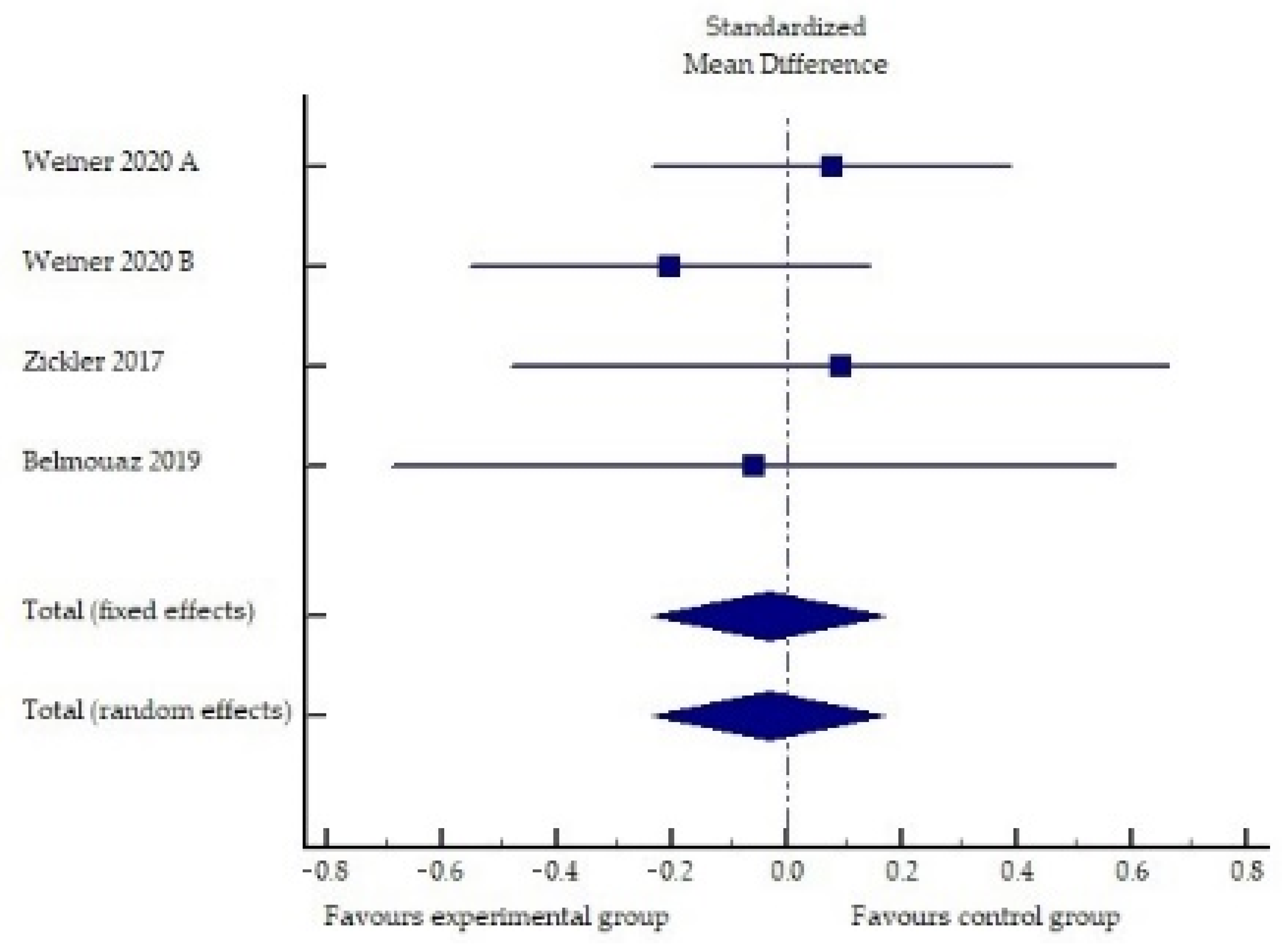
2.1. Performance of Medium Cut-Off Dialyzers in the Elimination of Middle Molecules and Protein-Bound Uremic Toxins Compared to Hemodiafiltration or High-Flux Hemodialysis

Our search strategy generated 4882 potentially relevant articles. Furthermore, 4879 papers were excluded based on the title and abstract, as they did not meet the inclusion criteria due to the type of article, study design or outcome of interest. We reviewed three articles for eligibility, all of which (Table 1) were included in the final review (one randomized controlled trial and two randomized controlled cross-over studies).
The summarized effects of four studies (Figure 1) showed no statistically significant difference between the interventional (MCO membrane) and control treatments (standardized difference of mean values: −0.03; 95% confidence interval: −0.23 to 0.17; p = 0.762).
The analysis of the studies (Table 1, Figure 1) showed predominantly equivocal results, which may show numerically lower values of IL-6 when using the MCO membrane, but the change was not statistically significant compared to high-flux hemodialysis. The reduction in other inflammatory markers (e.g., TNF-α, CRP, IFN-γ and SAA) did not differ significantly with an MCO or a high-flux dialyzer. Only one study reported superior intragroup removal of TNF-α using an MCO membrane, but the difference to controls did not reach significance. Interesting results were reported in a study by Zickler et al. [37], which showed a statistically significant lower TNF-α mRNA and IL-6 mRNA expression in peripheral blood mononuclear cells after MCO membrane dialysis compared to high-flux dialysis. The latter results suggest that the MCO dialyzer might influence the gene expression of immunoregulatory leukocyte subsets, since most of the major inflammatory cytokines were downregulated by lymphocytes and monocytes. Studies conducted to date have several limitations, including a small sample size and a short study duration. A comparison of MCO hemodialysis with hemodiafiltration is needed, since hemodialfiltration would enable the greater removal of pro-inflammatory middle molecules than high-flux hemodialysis. Therefore, further studies are needed to evaluate the ability of the MCO membrane to reduce chronic low-grade inflammation in hemodialysis patients.

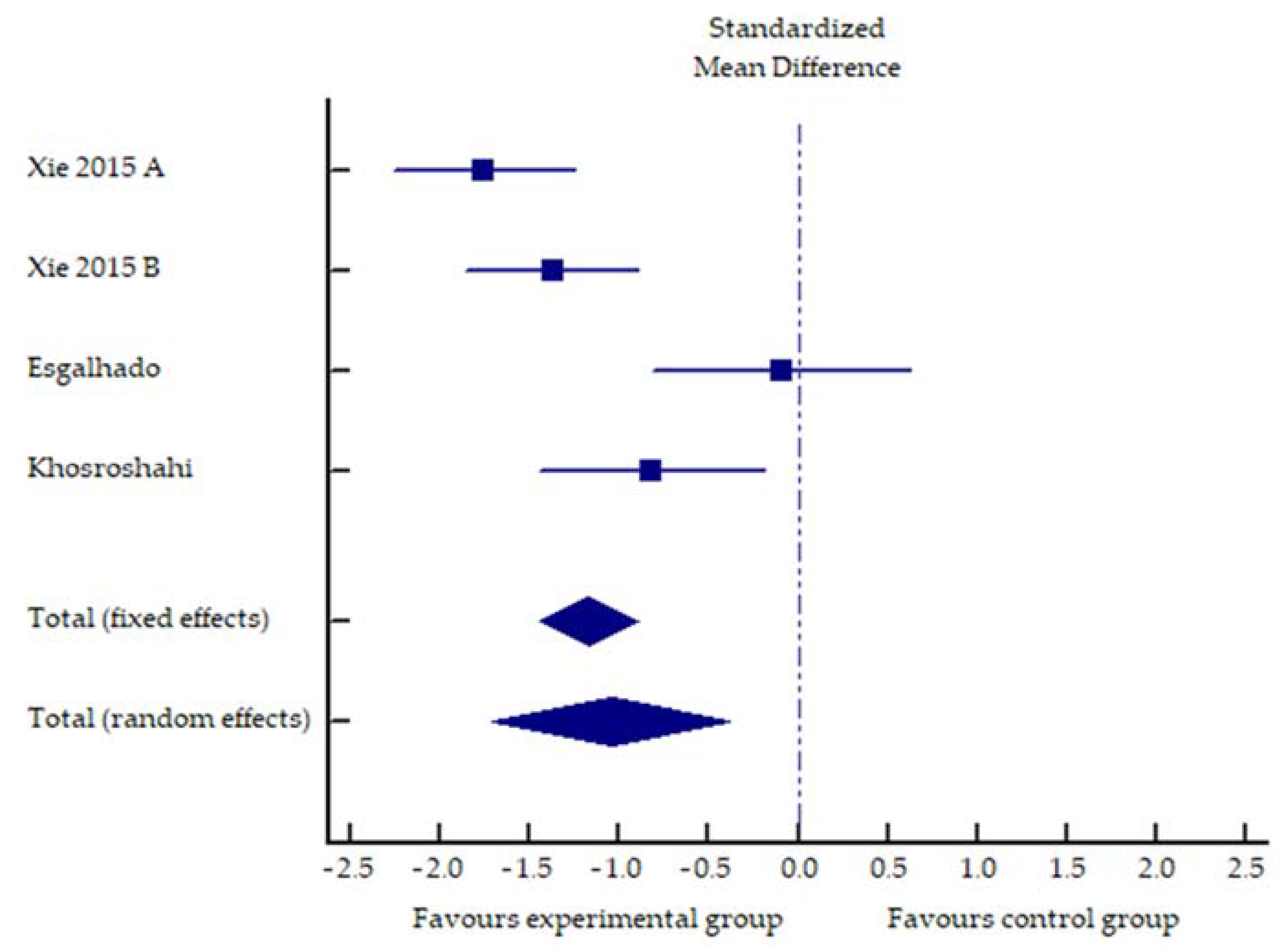
2.2. Impact of Increased Dietary Fiber Supplementation on Serum Concentration of PCS, IS and Inflammatory Markers

Our search strategy yielded 3778 potentially relevant articles. Furthermore, 3769 articles were excluded based on the title and abstract, which clearly indicated that they did not meet the inclusion criteria. Four studies did not satisfy our criteria for inclusion. One study on HD patients was excluded because it was non-randomized. The other three were excluded because the studied population was CKD patients; two of them were also retrospective cross-sectional studies. Finally, only five studies met the inclusion criteria (Table 2).
The summarized effects of four ESs (Figure 2) showed a statistically significant impact of fiber supplementation on IL-6 levels compared to the control treatments (standardized difference of mean values: −1.18; 95% confidence interval: −1.45 to −0.9; p < 0.001).
Table 2 shows that fiber supplementation had a heterogeneous effect on PBUT levels, with studies showing a decrease in either PC or IS with some heterogeneity in terms of the reduction in free or total fraction. On the other hand, the reduction in inflammation was uniform, with all studies showing a decrease at IL-6 and some studies showing a decrease at other markers (TNF-a, hs-CRP). It is possible that the critical variable is perhaps not only the absolute fiber intake but also a protein/fiber ratio in the diet [14], and this may explain some of the discrepancies observed. Nevertheless, these data from a limited number of studies support the assumption of a beneficial effect of dietary fiber supplementation, which should be further investigated.

2.3. Quality Assessment

Two authors (T.V. and T.H.) independently performed the search, selection and evaluation of methodological quality, with a discussion and consensus (J.P.) on all observed differences. PEDro scores ranged from 5 to 9 out of 10, with a median of 7.0 (Table 3). The inter-rater reliability between the two authors was almost perfect (Cohen’s kappa = 0.92). Three studies blinded the assessors, four used intention-to-treat analysis and two concealed allocation.

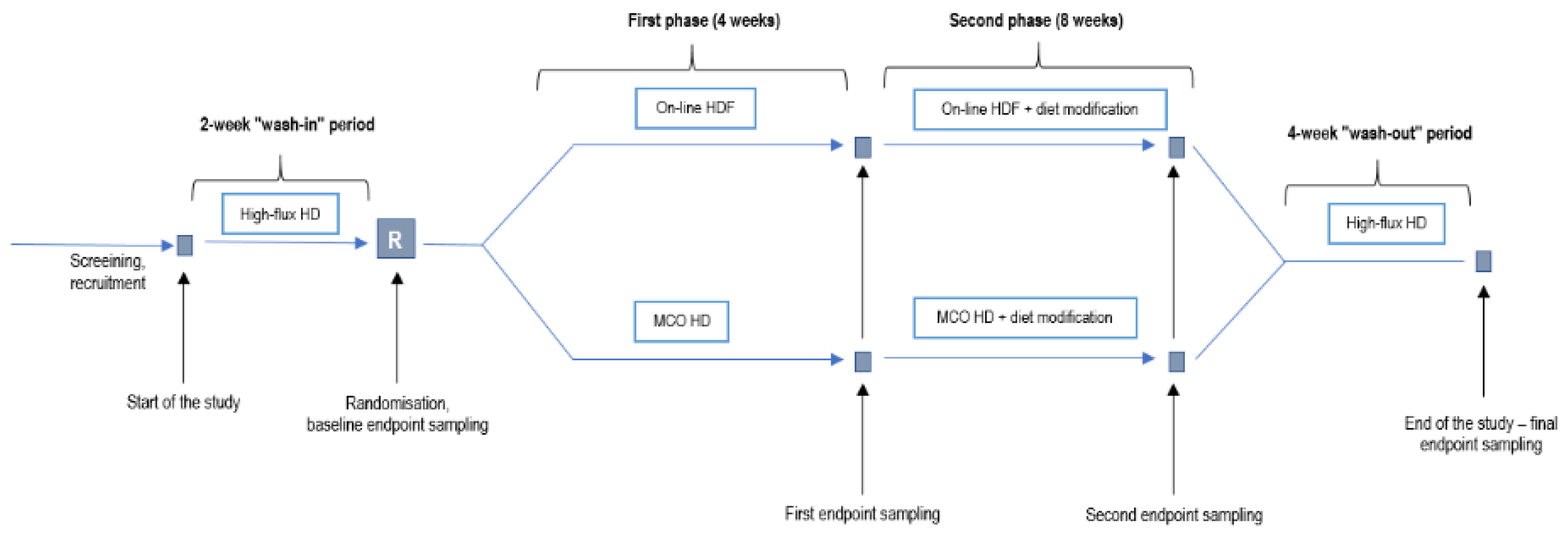
3. Discussion and an Interventional Trial Proposal

The above analysis showed that there is some evidence that fiber supplementation has a potentially positive effect on PBUT levels and inflammation. We could not find any studies investigating the effect of MCO membranes on PBUT values. The effects of MCO membranes on inflammation markers were inconsistent and inconclusive. We found some evidence from recent studies on the use of fiber supplementation to reduce inflammation levels and PBUT concentrations. Furthermore, both interventions are generally safe, and no major adverse effects were reported in the studies reviewed. However, these findings need to be interpreted in light of significant limitations as follows.
First, the number of available and eligible randomized controlled studies is very limited, and the total number of included patients is low. Therefore, this makes our findings susceptible to random effects. Second, as shown in Table 3, in some of the studies, intention-to-treat analyses were not included, outcome assessors were not blinded, there was no concealed allocation and interventions and outcome measures were poorly reported. Since we made no attempt to identify unpublished registered clinical trials, we cannot exclude the possibility of publication bias. In addition, most studies did not calculate the required sample size for accurate effect estimation. Therefore, additional studies are needed to extend the current limited evidence and to address the above limitations.
Against this background, it is a natural step to combine the use of a novel purification therapy (the MCO membrane) and the use of a cheap, easily accessible and adverse effect-free intervention to reduce PBUT generation (dietary fiber supplementation) and to reduce the two important components of residual uremic syndrome—PBUT concentration and inflammation. This proposal could be further supported by a possible synergistic effect of both interventions, as there are known links between PBUTs and inflammation induction (see introductory section above). Additionally, the uremic state per se may induce an increase in PBUT generation due to a modification of the microbiota [39]. Therefore, we speculate whether the use of a medium-cut off dialysis membrane and dietary fiber supplementation could act synergistically and induce a significant and clinically meaningful reduction in inflammation levels and PBUT concentration compared to online hemodiafiltration with a high-flux dialysis membrane.
On this basis, we designed a prospective, randomized, controlled and interventional study in two parallel groups to compare the serum concentrations of selected protein-bound uremic toxins and inflammatory markers obtained by medium cut-off membrane dialysis (Theranova, Baxter AG, USA) and online hemodiafiltration with a standard high-flux dialysis membrane. In the extension phase, patients in both study arms will undergo a dietary change with an increase in daily fiber intake and the addition of a short-chain fatty acid propionate. The two main outcomes of the study will be the serum concentration of p-cresol sulfate and the serum concentration of IL-6. Secondary outcomes will include the serum concentration of indoxyl-sulfate, trimethylamine-N-oxide (TMAO), IL-10, serum amyloid A (SAA), high-sensitivity C-reactive protein (hs-CRP), total leukocyte count and plasma concentration of bacterial 16s rDNA. The serum albumin concentration and lean body weight of patients will represent safety outcomes for this study.
This investigator-initiated research project has been entirely planned and will be conducted by the clinical researchers in a tertiary hospital, University Medical Centre Ljubljana, Department of Nephrology. The study will be conducted in accordance with the ethical principles of the Declaration of Helsinki and was approved by The National Medical Ethics Committee of the Republic of Slovenia (numbers 0120-430/2019/12, date 22 October 2019 and 0120-608/2019/3, date 23 January 2020). All study participants will have to provide informed written consent prior to study enrolment.
The trial will enroll 50 chronic prevalent clinically stable HD patients in the following periods:
First two weeks of the wash-in period with standard bicarbonate hemodialysis and standard high-flux dialysis membrane. Afterwards, the patients will be randomized in a 1:1 ratio to either one of the two study groups:
  • Experimental group: 4 weeks of dialysis with medium cut-off (Theranova) membrane (first phase), and dialysis for 8 weeks with the same membrane and increased fiber and sodium propionate intake (second phase);
  • Control group: 4 weeks of dialysis with a high-flux membrane using online hemodiafiltration (first phase) and 8 weeks of high-flux membrane hemodiafiltration and increased fiber and sodium propionate intake (second phase).
Finally, all patients will undergo a four-week wash-out period with standard bicarbonate hemodialysis using a standard high-flux dialysis membrane identical to the wash-in period.
Measurement of the study endpoints will be carried out at the end of the 2-week wash-in period (baseline), after each interventional period (4-week; 8-week), and at the end of the 4-week wash-out period. The flow diagram of the study is presented in Figure 3.
When determining the duration of wash-in and wash-out periods, we considered the half-life of uremic toxins and inflammation markers, e.g., the half-life for IL-6 is in the range of 2–15 h [40,41,42]; for CRP, 18–19 h [43,44], and it is constant; the reported experimental animal half-life for p-cresol sulfate is up to 12 h [45]. Taking these data into account, we considered that a two-week wash-in period provides an appropriate time margin. A longer wash-out (four weeks) was chosen due to the fact that there may be some microbiota modifying the effect of ingested fiber, which may take a longer time (at least four weeks) to vanish [46]. The duration of the second phase with increased intake of fiber and sodium propionate was set at 8 weeks with the aim of reducing dysbiosis of the microbiota, and demonstrating a synergistic effect of MCO/HDF and fiber supplementation [47].
A clinical nutritionist will monitor the increased daily intake of dietary fiber. The target dietary fiber intake will be 40 g/day, with a minimum intake of 14 g/1000 kcal per day for patients who cannot reach the 40 g/day target, in accordance with the guidelines of European Society of Cardiology for Primary Prevention of Cardiovascular Disease and the American Institute of Medicine [48]. Fiber intake will be achieved by using natural whole grain sources in the habitual meal plan and a supplement of a dietary fiber mixture (psyllium 69% and inulin 30% blend) at a dose of 5 g BID for 8 weeks in the second phase of the study. The fiber supplement will also contain short-chain fatty acid sodium propionate (1 g/day), which will be added to a fiber supplement at the dose of 500 mg BID. It has been reported that short-chain fatty acids have a positive effect on systemic inflammation in patients on maintenance hemodialysis [49,50]. Short-chain fatty acids are a product of indigestible dietary carbohydrates, and we included sodium propionate to enhance the effect of dietary fiber supplementation, especially in patients who cannot achieve the desired goal of fiber intake. We do not expect any significant sodium-, potassium- or acid-based adverse effects from this dietary change [49].
Our study’s inclusion criteria will be the following: patients on chronic hemodialysis or hemodiafiltration for at least 12 weeks, age 18 years or more, a functioning arteriovenous fistula or graft as a permanent dialysis vascular access and being able to give informed consent to participate in the survey. Patients will not be included if planned for kidney transplantation, the transition to peritoneal dialysis or to another dialysis center within 12 weeks of the start of the study, patients with an episode of acute febrile illness 4 weeks prior to study inclusion, active chronic inflammation (e.g., an active autoimmune disease or an open wound), chronic ongoing infection or cancer, new cardiovascular or cerebrovascular event 4 weeks prior to study inclusion, clinically malnourished patients and/or BMI below 19 kg/m2 and/or loss of more than 5% of body mass in the last 3 months, immunosuppressive treatment, expected survival of less than 1 year, pregnancy or breast-feeding, indication for dietary supplements to increase calorie and/or protein intake, patients with specific indication for hemodiafiltration instead of hemodialysis as per attending physician, serum albumin concentration < 32 g/L at screening phase, inability to follow the study diet or test procedures, rapid reduction in residual renal function in the period before entry into the study and being intolerant of online hemodiafiltration (infusion intolerance).
During the dialysis period with the MCO membrane and during high-flux membrane hemodiafiltration, the following basic conditions of the dialysis procedure are planned: blood flow 300 mL/min; dialysate flow 500 mL/min; unchanged duration of dialysis procedures and the unaltered surface area of the dialysis membrane for the total study period; fluid removal by ultrafiltration according to the prescription of the treating physician and the clinical condition; for online hemodiafiltration, the target convection volume will be greater than or equal to 23 L per procedure.
All participants will be under the supervision of dialysis personnel, including the consultant nephrologist and experienced renal dialysis nurses. All adverse events will be prospectively monitored and reported by the study coordinator or principal investigator.

Sample Size

We considered that the expected standard deviation of IL-6 concentration in the HD population was 1.4 pg/mL [49] and estimated that the intraindividual correlation of IL-6 values was at least 0.5. The minimum value of the concentration difference that the study aims to detect is at least half the standard deviation, i.e., 0.7 pg/mL. A sample size of 31 patients is required for the alpha error value of 0.05 and the beta error of 0.2. In the expected dropout of 20% of patients in the 16-week period, a sample size of 39 patients is required, rounded up to 40 patients. When also considering the expected standard deviation of the p-cresol sulfate concentration of 0.13 mg/dL and the expected value of the difference between the two study arms of at least 0.11 mg/dL, at the value of alpha error of 0.05 and beta error of 0.2, a sample size of 44 patients is required. In consideration of the expected dropout, a sample size of 49 patients is required, rounded up to 50 patients, which is our target number for this study.

4. Conclusions

There are two main options for reducing the components of residual uremic syndrome in HD patients—improving purification techniques and reducing the formation and/or absorption of uremic toxins. In recent years, innovation in dialysis membrane technology has been introduced into clinical practice with the development of MCO membranes. In our systematic review, we could not find any studies investigating the effect of dialysis with MCO membrane on PBUT values. The effects of MCO membrane dialysis on inflammation markers were inconsistent and inconclusive. On the other hand, we found compelling evidence from recent studies on the use of dietary fiber supplements to reduce inflammation levels and PBUT concentrations. Therefore, a study that investigates both therapeutic approaches in combination would be justified to answer this question. However, it is premature to conclude the superiority of the combined intervention at this time. Should this combination prove to be effective and safe, it could represent an important step towards improving the important components of residual uremic syndrome in HD patients and possibly contribute to improving their prognosis.

5. Materials and Methods

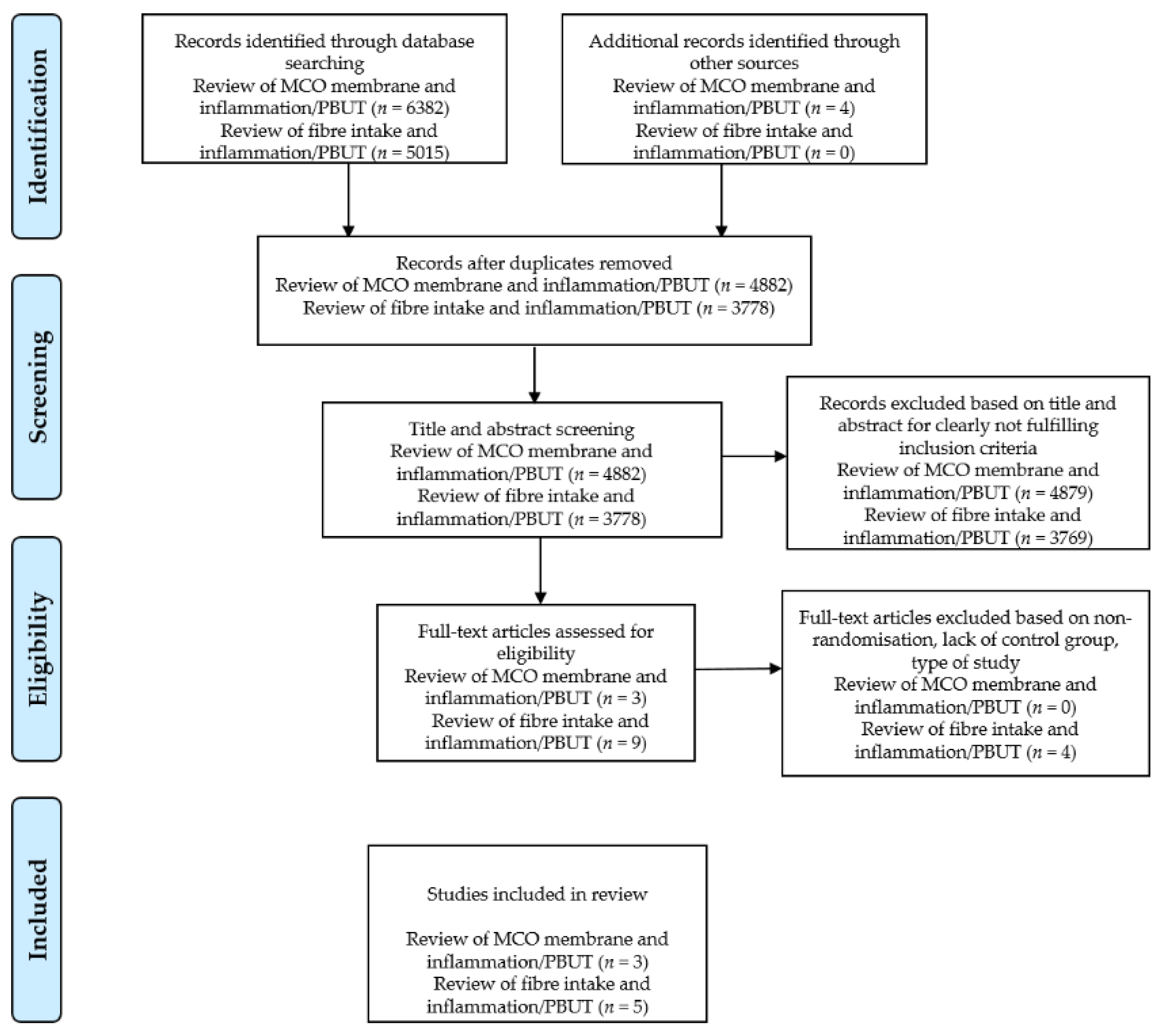
5.1. Literature Search Strategy

For this systematic review, PRISMA guidelines were followed [51]. Our main objective was to find randomized controlled interventional studies that investigated the effects of MCO membranes and/or dietary fiber on PBUT concentration and/or inflammatory markers. The electronic databases investigated were PubMed, Cochrane, Embase and Web of Science from 15 January 2021 to 12 February 2021. The following keywords were used to search for the articles reporting on the performance of medium cut-off dialyzers compared to hemodiafiltration or high-flux HD: “medium cut-off membrane” OR “medium cut-off dialyser” OR “expanded hemodialysis” OR “expanded haemodialysis” OR “high-flux hemodialysis” OR “high-flux haemodialysis” AND “hemodiafiltration” OR “haemodiafiltration” AND “protein-bound uremic toxins” OR “protein-bound uremic solutes” OR “indoxyl sulfate” OR “3-indoxyl sulfate” OR “3-indoxyl sulfuric acid” OR “indol-3-yl sulfate” OR “p-cresol” OR “p-cresyl” OR “4-cresol” OR “4-cresyl” OR “p-cresol sulfate” OR “p-cresyl sulfate” OR “4-cresol sulfate” OR “4-cresyl sulfate” OR “inflammation” OR “inflammation markers” OR “inflammation parameters” OR “inflammatory markers” OR “inflammatory parameters” OR “CRP” OR “hs-CRP” OR “IL-6” OR “serum amyloid A” OR “SAA”.
The keywords in the search for articles on the influence of dietary fiber supplementation on serum concentrations of PCS, IS and inflammation markers were “hemodialysis” OR “haemodialysis” OR “end-stage kidney disease” OR “end-stage renal disease” AND “fiber” OR “fibre” OR “dietary fiber” OR “dietary fibre” OR “nondigestible fiber” OR “nondigestible fibre” OR “prebiotic” OR “starch” OR “resistant starch” AND “indoxyl sulfate” OR “3-Indoxylsulfate” OR “3-Indoxylsulfuric acid” OR “Indol-3-yl sulfate” OR “p-cresol” OR “p-cresyl” OR “4-cresol” OR “4-cresyl” OR “p-cresol sulfate” OR “p-cresyl sulfate” OR “4-cresol sulfate” OR “4-cresyl sulfate” OR “protein-bound uremic toxins” OR “protein-bound uremic solutes” OR “IL-6” OR “CRP” OR “hs-CRP” OR “serum amyloid A” OR “SAA” OR “inflammation” OR “inflammation markers” OR “inflammation parameters” OR “inflammatory markers” OR “inflammatory parameters“.
The literature search was performed independently by two authors (T.H. and T.V.). First, the authors carried out a screening of the titles. Second, the abstracts were selected for further analysis on the basis of previously agreed inclusion and exclusion criteria. Finally, the full text of the included papers was read. In the case of disagreement between the authors, the issue was clarified by a corresponding author (J.P.). If the full text of the paper was not available or some information was missing, we contacted the corresponding and/or the first author by e-mail or via the Research Gate platform. The progress of the screening is shown in Figure 4.

5.2. Inclusion and Exclusion Criteria

The present review included only original articles in English published in peer-reviewed journals. Inclusion criteria were the following: randomized controlled trials in HD patients, studies that investigated the effect of the MCO dialyzer on PBUT and inflammatory marker concentrations or studies that investigated the influence of dietary fiber supplementation on the same endpoints. Due to the small number of eligible studies, we did not include criteria for study duration. Exclusion criteria were non-HD patients, in vitro and animal studies, systematic reviews and meta-analyses, conference abstracts, theses and case reports.

5.3. Quality Assessment

Two authors independently (T.V. and T.H.) assessed the methodological quality and risk of bias of included studies. To assess the methodological quality of the included studies, we used the Physiotherapy Evidence Database (PEDro) scale [52]. The PEDro scale consists of 11 items that assess the methodological quality of a study and can be used in various fields of medicine. Each scale item contributes 1 point to the total PEDro score (0–10 points). The total score is interpreted as follows: ≤3 points (poor methodological quality), 4–5 points (moderate quality) and 6–10 points (high quality). Agreement between the two reviewers was assessed using k-statistics. In the case of disagreement, a corresponding author (J.P.) made the final decision.

Author Contributions

Conceptualization, J.P.; investigation, T.H., T.V., Š.B., and J.P.; resources, J.P.; writing—original draft preparation, T.H., and T.V.; writing—review and editing, J.P. and Š.B. All authors have read and agreed to the published version of the manuscript.

Funding

This research was funded by the public Slovenian Research Agency, grant number L3-1838; University Medical Centre Ljubljana tertiary grant TP20190015; and by the grant from Baxter, d.o.o., Ljubljana, Slovenia. Funding bodies had no part in the study plan or protocol development.

Institutional Review Board Statement

The proposed study protocol is conducted according to the guidelines of the Declaration of Helsinki, and it was approved by the The National Medical Ethics Committee of the Republic of Slovenia (numbers 0120-430/2019/12, date 22 October 2019 and 0120-608/2019/3, date 23 January 2020).

Informed Consent Statement

Informed consent will be obtained from all subjects involved in the study according to the proposed protocol.

Data Availability Statement

Not applicable.

Acknowledgments

The authors would like to thank Armin Paravlić for the help with statistical analysis.

Conflicts of Interest

J.P. has received speaker fees and research grants from Baxter d.o.o. T.H., T.V. and Š.B. declare no conflict of interest.

References

  1. Vanholder, R.; De Smet, R.; Glorieux, G.; Argilés, A.; Baurmeister, U.; Brunet, P.; Clark, W.; Cohen, G.; De Deyn, P.P.; Deppisch, R.; et al. Review on uremic toxins: Classification, concentration, and interindividual variability. Kidney Int. 2003, 63, 1934–1943. [Google Scholar] [CrossRef] [Green Version]
  2. Vanholder, R.; Pletinck, A.; Schepers, E.; Glorieux, G. Biochemical and Clinical Impact of Organic Uremic Retention Solutes: A Comprehensive Update. Toxins 2018, 10, 33. [Google Scholar] [CrossRef] [Green Version]
  3. Graboski, A.L.; Redinbo, M.R. Gut-Derived Protein-Bound Uremic Toxins. Toxins 2020, 12, 590. [Google Scholar] [CrossRef]
  4. Eloot, S.; Van Biesen, W.; Glorieux, G.; Neirynck, N.; Dhondt, A.; Vanholder, R. Does the adequacy parameter Kt/V(urea) reflect uremic toxin concentrations in hemodialysis patients? PLoS ONE 2013, 8, e76838. [Google Scholar] [CrossRef] [Green Version]
  5. Maduell, F.; Moreso, F.; Pons, M.; Ramos, R.; Mora-Macià, J.; Carreras, J.; Soler, J.; Torres, F.; Campistol, J.M.; Martinez-Castelao, A.; et al. High-efficiency postdilution online hemodiafiltration reduces all-cause mortality in hemodialysis patients. J. Am. Soc. Nephrol. 2013, 24, 487–497. [Google Scholar] [CrossRef] [Green Version]
  6. Peters, S.A.E.; Bots, M.L.; Canaud, B.; Davenport, A.; Grooteman, M.P.C.; Kircelli, F.; Locatelli, F.; Maduell, F.; Morena, M.; Nubé, M.J.; et al. Haemodiafiltration and mortality in end-stage kidney disease patients: A pooled individual participant data analysis from four randomized controlled trials. Nephrol. Dial. Transplant. 2016, 31, 978–984. [Google Scholar] [CrossRef]
  7. Mostovaya, I.M.; Blankestijn, P.J.; Bots, M.L.; Covic, A.; Davenport, A.; Grooteman, M.P.C.; Hegbrant, J.; Locatelli, F.; Vanholder, R.; Nubé, M.J. Clinical Evidence on Hemodiafiltration: A Systematic Review and a Meta-analysis. Semin. Dial. 2014, 27, 119–127. [Google Scholar] [CrossRef] [PubMed]
  8. Wang, A.Y.; Ninomiya, T.; Al-Kahwa, A.; Perkovic, V.; Gallagher, M.P.; Hawley, C.; Jardine, M.J. Effect of hemodiafiltration or hemofiltration compared with hemodialysis on mortality and cardiovascular disease in chronic kidney failure: A systematic review and meta-analysis of randomized trials. Am. J. Kidney Dis. 2014, 63, 968–978. [Google Scholar] [CrossRef] [PubMed]
  9. Ronco, C.; La Manna, G. Expanded Hemodialysis: A New Therapy for a New Class of Membranes. Contrib. Nephrol. 2017, 190, 124–133. [Google Scholar] [PubMed]
  10. Ronco, C. The Rise of Expanded Hemodialysis. Blood Purif. 2017, 44, I–VIII. [Google Scholar] [CrossRef] [PubMed]
  11. Zweigart, C.; Boschetti-de-Fierro, A.; Hulko, M.; Nilsson, L.G.; Beck, W.; Storr, M.; Krause, B. Medium cut-off membranes—closer to the natural kidney removal function. Int. J. Artif. Organs 2017, 40, 328–334. [Google Scholar] [CrossRef] [PubMed]
  12. De Smet, R.; David, F.; Sandra, P.; Van Kaer, J.; Lesaffer, G.; Dhondt, A.; Lameire, N.; Vanholder, R. A sensitive HPLC method for the quantification of free and total p-cresol in patients with chronic renal failure. Clin. Chim. Acta 1998, 278, 1–21. [Google Scholar] [CrossRef]
  13. Evenepoel, P.; Glorieux, G.; Meijers, B. P-Cresol Sulfate and Indoxyl Sulfate: Some Clouds Are Gathering in the Uremic Toxin Sky. Kidney Int. 2017, 92, 1323–1324. [Google Scholar] [CrossRef] [PubMed]
  14. Rossi, M.; Johnson, D.W.; Xu, H.; Carrero, J.J.; Pascoe, E.; French, C.; Campbell, K.L. Dietary protein-fiber ratio associates with circulating levels of indoxyl sulfate and p-cresyl sulfate in chronic kidney disease patients. Nutr. Metab. Cardiovasc. Dis. 2015, 25, 860–865. [Google Scholar] [CrossRef] [PubMed]
  15. Rossi, M.; Campbell, K.; Johnson, D.; Stanton, T.; Pascoe, E.; Hawley, C.; Dimeski, G.; McWhinney, B.; Ungerer, J.; Isbel, N. Uraemic toxins and cardiovascular disease across the chronic kidney disease spectrum: An observational study. Nutr. Metab. Cardiovasc. Dis. 2014, 24, 1035–1042. [Google Scholar] [CrossRef]
  16. Wu, I.W.; Hsu, K.H.; Lee, C.C.; Sun, C.Y.; Hsu, H.J.; Tsai, C.J.; Tzen, C.Y.; Wang, Y.C.; Lin, C.Y.; Wu, M.S. P-cresyl sulphate and indoxyl sulphate predict progression of chronic kidney disease. Nephrol. Dial. Transplant. 2011, 26, 938–947. [Google Scholar] [CrossRef] [Green Version]
  17. Vanholder, R.; Massy, Z.; Argiles, A.; Spasovski, G.; Verbeke, F.; Lameire, N.; Beige, J.; Brunet, P.; Cohen, G.; De Deyn, P.P.; et al. Chronic kidney disease as cause of cardiovascular morbidity and mortality. Nephrol. Dial. Transplant. 2005, 20, 1048–1056. [Google Scholar] [CrossRef] [Green Version]
  18. Khosroshahi, H.T.; Abedi, B.; Ghojazadeh, M.; Samadi, A.; Jouyban, A. Effects of fermentable high fiber diet supplementation on gut derived and conventional nitrogenous product in patients on maintenance hemodialysis: A randomized controlled trial. Nutr. Metab. 2019, 16, 1–8. [Google Scholar] [CrossRef] [Green Version]
  19. Esgalhado, M.; Kemp, J.A.; Azevedo, R.; Paiva, B.R.; Stockler-Pinto, M.B.; Dolenga, C.J.; Borges, N.A.; Nakao, L.S.; Mafra, D. Could resistant starch supplementation improve inflammatory and oxidative stress biomarkers and uremic toxins levels in hemodialysis patients? A pilot randomized controlled trial. Food Funct. 2018, 9, 6508–6516. [Google Scholar] [CrossRef]
  20. Sirich, T.L.; Plummer, N.S.; Gardner, C.D.; Hostetter, T.H.; Meyer, T.W. Effect of increasing dietary fiber on plasma levels of colon-derived solutes in hemodialysis patients. Clin. J. Am. Soc. Nephrol. 2014, 9, 1603–1610. [Google Scholar] [CrossRef] [Green Version]
  21. Tayebi Khosroshahi, H.; Vaziri, N.D.; Abedi, B.; Asl, B.H.; Ghojazadeh, M.; Jing, W.; Vatankhah, A.M. Effect of high amylose resistant starch (HAM-RS2) supplementation on biomarkers of inflammation and oxidative stress in hemodialysis patients: A randomized clinical trial. Hemodial. Int. 2018, 22, 492–500. [Google Scholar] [CrossRef] [PubMed]
  22. Xie, L.M.; Ge, Y.Y.; Huang, X.; Zhang, Y.Q.; Li, J.X. Effects of fermentable dietary fiber supplementation on oxidative and inflammatory status in hemodialysis patients. Int. J. Clin. Exp. Med. 2015, 8, 1363–1369. [Google Scholar] [PubMed]
  23. Montemurno, E.; Cosola, C.; Dalfino, G.; Daidone, G.; De Angelis, M.; Gobbetti, M.; Gesualdo, L. What would you like to eat, Mr CKD Microbiota? A Mediterranean Diet, please! Kidney Blood Press. Res. 2014, 39, 114–123. [Google Scholar] [CrossRef] [PubMed]
  24. Sun, C.Y.; Hsu, H.H.; Wu, M.S. P-Cresol sulfate and indoxyl sulfate induce similar cellular inflammatory gene expressions in cultured proximal renal tubular cells. Nephrol. Dial. Transplant. 2013, 28, 70–78. [Google Scholar] [CrossRef] [Green Version]
  25. Lekawanvijit, S.; Adrahtas, A.; Kelly, D.J.; Kompa, A.R.; Wang, B.H.; Krum, H. Does indoxyl sulfate, a uraemic toxin, have direct effects on cardiac fibroblasts and myocytes? Eur. Heart J. 2010, 31, 1771–1779. [Google Scholar] [CrossRef] [Green Version]
  26. Shimizu, H.; Bolati, D.; Adijiang, A.; Muteliefu, G.; Enomoto, A.; Nishijima, F.; Dateki, M.; Niwa, T. NF-κb plays an important role in indoxyl sulfate-induced cellular senescence, fibrotic gene expression, and inhibition of proliferation in proximal tubular cells. Am. J. Physiol. Cell Physiol. 2011, 301, 1201–1213. [Google Scholar] [CrossRef] [PubMed]
  27. Watanabe, H.; Miyamoto, Y.; Honda, D.; Tanaka, H.; Wu, Q.; Endo, M.; Noguchi, T.; Kadowaki, D.; Ishima, Y.; Kotani, S.; et al. P-Cresyl sulfate causes renal tubular cell damage by inducing oxidative stress by activation of NADPH oxidase. Kidney Int. 2013, 83, 582–592. [Google Scholar] [CrossRef] [Green Version]
  28. Rossi, M.; Campbell, K.L.; Johnson, D.W.; Stanton, T.; Vesey, D.A.; Coombes, J.S.; Weston, K.S.; Hawley, C.M.; McWhinney, B.C.; Ungerer, J.P.J.; et al. Protein-bound uremic toxins, inflammation and oxidative stress: A cross-sectional study in stage 3-4 chronic kidney disease. Arch. Med. Res. 2014, 45, 309–317. [Google Scholar] [CrossRef]
  29. Vanholder, R.; Schepers, E.; Pletinck, A.; Nagler, E.V.; Glorieux, G. The uremic toxicity of indoxyl sulfate and p-cresyl sulfate: A systematic review. J. Am. Soc. Nephrol. 2014, 25, 1897–1907. [Google Scholar] [CrossRef]
  30. Nataatmadja, M.; Cho, Y.; Campbell, K.; Johnson, D.W. The Roles of Indoxyl Sulphate and p-Cresyl Sulphate in Patients with Chronic Kidney Disease: A Review of Therapeutic Options. In Chronic Kidney Disease-from Pathophysiology to Clinical Improvements; IntechOpen: London, UK, 2018. [Google Scholar]
  31. Hung, S.C.; Kuo, K.L.; Wu, C.C.; Tarng, D.C. Indoxyl sulfate: A novel cardiovascular risk factor in chronic kidney disease. J. Am. Heart Assoc. 2017, 6, 1–9. [Google Scholar] [CrossRef] [Green Version]
  32. Tan, X.; Cao, X.; Zou, J.; Shen, B.; Zhang, X.; Liu, Z.; Lv, W.; Teng, J.; Ding, X. Indoxyl sulfate, a valuable biomarker in chronic kidney disease and dialysis. Hemodial. Int. 2017, 21, 161–167. [Google Scholar] [CrossRef] [PubMed]
  33. Barreto, F.C.; Barreto, D.V.; Liabeuf, S.; Meert, N.; Glorieux, G.; Temmar, M.; Choukroun, G.; Vanholder, R.; Massy, Z.A.; on behalf of the European Uremic Toxin Work Group (EUTox). Serum indoxyl sulfate is associated with vascular disease and mortality in chronic kidney disease patients. Clin. J. Am. Soc. Nephrol. 2009, 4, 1551–1558. [Google Scholar] [CrossRef] [PubMed] [Green Version]
  34. Kamiński, T.W.; Pawlak, K.; Karbowska, M.; Myśliwiec, M.; Pawlak, D. Indoxyl sulfate—The uremic toxin linking hemostatic system disturbances with the prevalence of cardiovascular disease in patients with chronic kidney disease. BMC Nephrol. 2017, 18, 1–12. [Google Scholar] [CrossRef] [PubMed] [Green Version]
  35. Liu, W.-C.; Tomino, Y.; Lu, K.-C. Impacts of Indoxyl Sulfate and p-Cresol Sulfate on Chronic Kidney Disease and Mitigating Effects of AST-120. Toxins 2018, 10, 367. [Google Scholar] [CrossRef] [PubMed] [Green Version]
  36. Weiner, D.E.; Falzon, L.; Skoufos, L.; Bernardo, A.; Beck, W.; Xiao, M.; Tran, H. Efficacy and safety of expanded hemodialysis with the theranova 400 dialyzer: A randomized controlled trial. Clin. J. Am. Soc. Nephrol. 2020, 15, 1310–1319. [Google Scholar] [CrossRef] [PubMed]
  37. Zickler, D.; Schindler, R.; Willy, K.; Martus, P.; Pawlak, M.; Storr, M.; Hulko, M.; Boehler, T.; Glomb, M.A.; Liehr, K.; et al. Medium Cut-Off (MCO) membranes reduce inflammation in chronic dialysis patients—A randomized controlled clinical trial. PLoS ONE 2017, 12, e0169024. [Google Scholar]
  38. Belmouaz, M.; Bauwens, M.; Hauet, T.; Bossard, V.; Jamet, P.; Joly, F.; Chikhi, E.; Joffrion, S.; Gand, E.; Bridoux, F. Comparison of the removal of uraemic toxins with medium cut-off and high-flux dialysers: A randomized clinical trial. Nephrol. Dial. Transplant. 2020, 35, 328–335. [Google Scholar] [CrossRef] [PubMed]
  39. Sabatino, A.; Regolisti, G.; Brusasco, I.; Cabassi, A.; Morabito, S.; Fiaccadori, E. Alterations of intestinal barrier and microbiota in chronic kidney disease. Nephrol. Dial. Transplant. 2015, 30, 924–933. [Google Scholar] [CrossRef] [Green Version]
  40. Kuribayashi, T. Elimination half-lives of interleukin-6 and cytokine-induced neutrophil chemoattractant-1 synthesized in response to inflammatory stimulation in rats. Lab. Anim. Res. 2018, 34, 80. [Google Scholar] [CrossRef]
  41. Fukagawa, M.; Yumita, S.; Akizawa, T.; Uchida, E.; Tsukamoto, Y.; Iwasaki, M.; Koshikawa, S. Cinacalcet (KRN1493) effectively decreases the serum intact PTH level with favorable control of the serum phosphorus and calcium levels in Japanese dialysis patients. Nephrol. Dial. Transplant. 2008, 23, 328–335. [Google Scholar] [CrossRef] [Green Version]
  42. Wirtz, D.C.; Heller, K.D.; Miltner, O.; Zilkens, K.W.; Wolff, J.M. Interleukin-6: A potential inflammatory marker after total joint replacement. Int. Orthop. 2000, 24, 194–196. [Google Scholar] [CrossRef] [PubMed] [Green Version]
  43. Seo, H.S. The role and clinical significance of high-sensitivity C-reactive protein in cardiovascular disease. Korean Circ. J. 2012, 42, 151–153. [Google Scholar] [CrossRef] [PubMed]
  44. Pepys, M.B.; Hirschfield, G.M. C-reactive protein: A critical update. J. Clin. Investig. 2003, 111, 1805–1812. [Google Scholar] [CrossRef]
  45. Lesaffer, G.; De Smet, R.; D’heuvaert, T.; Belpaire, F.M.; Lameire, N.; Vanholder, R. Comparative kinetics of the uremic toxin p-cresol versus creatinine in rats with and without renal failure. Kidney Int. 2003, 64, 1365–1373. [Google Scholar] [CrossRef] [PubMed] [Green Version]
  46. Meijers, B.K.I.; De Loor, H.; Bammens, B.; Verbeke, K.; Vanrenterghem, Y.; Evenepoel, P. P-Cresyl Sulfate and Indoxyl Sulfate in Hemodialysis Patients. Clin. J. Am. Soc. Nephrol. 2009, 4, 1932–1938. [Google Scholar] [CrossRef] [Green Version]
  47. Rocchetti, M.T.; Cosola, C.; di Bari, I.; Magnani, S.; Galleggiante, V.; Scandiffio, L.; Dalfino, G.; Netti, G.S.; Atti, M.; Corciulo, R.; et al. Efficacy of Divinylbenzenic Resin in Removing Indoxyl Sulfate and P-cresol Sulfate in Hemodialysis Patients: Results from an In Vitro Study and an In Vivo Pilot Trial (xuanro4-Nature 3.2). Toxins 2020, 12, 170. [Google Scholar] [CrossRef] [PubMed] [Green Version]
  48. Perk, J.; De Backer, G.; Gohlke, H.; Graham, I.; Reiner, Ž.; Verschuren, M.; Albus, C.; Benlian, P.; Boysen, G.; Cifkova, R.; et al. European Guidelines on cardiovascular disease prevention in clinical practice (version 2012). Eur. Heart J. 2012, 33, 1635–1701. [Google Scholar]
  49. Marzocco, S.; Fazeli, G.; Di Micco, L.; Autore, G.; Adesso, S.; Dal Piaz, F.; Heidland, A.; Di Iorio, B. Supplementation of Short-Chain Fatty Acid, Sodium Propionate, in Patients on Maintenance Hemodialysis: Beneficial Effects on Inflammatory Parameters and Gut-Derived Uremic Toxins, A Pilot Study (PLAN Study). J. Clin. Med. 2018, 7, 315. [Google Scholar] [CrossRef] [Green Version]
  50. Vinolo, M.A.R.; Rodrigues, H.G.; Nachbar, R.T.; Curi, R. Regulation of inflammation by short chain fatty acids. Nutrients 2011, 3, 858–876. [Google Scholar] [CrossRef] [PubMed] [Green Version]
  51. Moher, D.; Liberati, A.; Tetzlaff, J.; Altman, D.G. Preferred reporting items for systematic reviews and meta-analyses: The PRISMA statement. BMJ 2009, 339, 332–336. [Google Scholar] [CrossRef] [Green Version]
  52. Maher, C.G.; Sherrington, C.; Herbert, R.D.; Moseley, A.M.; Elkins, M. Reliability of the PEDro Scale for Rating Quality of Randomized Controlled Trials. Phys. Ther. 2003, 83, 713–721. [Google Scholar] [CrossRef] [PubMed] [Green Version]
Figure 1. Forest plot of the standardized mean differences of the MCO membrane effect on interleukin 6.
Toxins 13 00244 g001
Figure 2. Forest plot of the standardized mean differences of the fiber intake effect on interleukin 6. Experimental group—fiber supplementation. Control group—various forms of fiberless carbohydrates.
Toxins 13 00244 g002
Figure 3. Study flow diagram. Abbreviations: MCO, medium cut-off membrane (Theranova); HD, hemodialysis; HDF, online hemodiafiltration.
Toxins 13 00244 g003
Figure 4. PRISMA flow diagram. Abbreviations: MCO, medium cut-off membrane (Theranova); PBUT, protein-bound uremic toxins.
Toxins 13 00244 g004
Table 1. Systematic review summary of the impact of hemodialysis (HD) with medium cut-off membrane (MCO) on inflammatory markers.
StudySample SizeExp InterventionCon InterventionDurationEndpointResults
Weiner, 2020 [36]Exp (n = 86)
Con (n = 86)
MCO membraneHigh-flux HD24 weeksIL-6Exp: 15% ↓
Con: 50.6% ↑
p Exp vs. Con n.s.
TNF-αExp: 48.9% ↓
Con: 34.7% ↓
p Exp vs. Con n.s.
Zickler, 2017 [37]Exp (n = 23)
Con (n = 25)
MCO membraneHigh-flux HD4 weeks of each dialysis modality + 8 weeks of extension phaseIL-6Exp: 33.3% ↓ **
Con: 43.9% ↓ *
p Exp vs. Con n.s.
TNF-αExp: 14.5% ↓ **
Con: 5.1% ↓
p Exp vs. Con n.s.
CRPExp: 39.2% ↓
Con: 28.4% ↓
p Exp vs. Con n.s.
Belmouaz, 2020 [38]Exp (n = 20)
Con (n = 20)
MCO membraneHigh-flux HD3 months of each dialysis modalityIL-6Exp: 6.3% ↑
Con: 12.8% ↑
p Exp vs. Con n.s.
TNF-αExp: 19.4% ↓
Con: 16.4% ↓
p Exp vs. Con n.s.
CRPExp: missing data 1
Con: missing data 1
p Exp vs. Con n.s.
Abbreviations: Exp, experimental group; Con, control group; * p < 0.05 within group; ** p < 0.01 within group; RR, reduction ratio; MCO, medium-cut off; HD, hemodialysis; IL-6, interleukin 6; TNF-α, tumor necrosis factor α; CRP, C-reactive protein; 1 only postdialysis values. IL-6 mRNA significantly reduced in Exp group (23.1%) with significant differences between groups (p < 0.001); TNF-α mRNA significantly reduced in both groups (Exp: 18.5%; Con: 14.3%) with significant differences between groups (p < 0.001).
Table 2. Systematic review summary of the impact of dietary fiber on protein-bound uremic toxins and inflammatory markers.
StudySample SizeExp InterventionCon InterventionDurationEndpointResults
Khosroshahi, 2019 [18]Exp (n = 23)
Con (n = 21)
20–25 g of HAM-RS2
20 g week 1–4
25 g week 5–8
20–25 g of waxy corn starch
20 g week 1–4
25 g week 5–8
8 weeks Exp: 31.5% ↓ *
PCCon: 0.7% ↑
p Exp vs. Con 0.992
Exp: 2.96% ↑
ISCon: 11.7% ↑
p Exp vs. Con 0.606
Exp: 110.6% ↑
Con: 25.05% ↑
hs-CRPp Exp vs. Con 0.866
Esgalhado, 2018 [19]Exp (n = 15)
Con (n = 16)
16 g HAM-RS220 g manioc flour4 weeks Exp: 18.15% ↓ **
ISCon: 9.28% ↑
p Exp vs. Con 0.008
Exp: 6.83% ↑
Con: 1.53% ↓
PCSp Exp vs. Con 0.77
Exp: 11.86% ↓ **
Con: 3.28% ↓
IL-6p Exp vs. Con 0.06
Exp: 20.0 ↓
Con: 12.77 ↓
hs-CRPp Exp vs. Con 0.16
Sirich, 2014 [20]Exp (n = 20)
Con (n = 20)
15 g HAM-RS2 week 1,
30 g HAM-RS2 week 2–6
15 g waxy corn starch week 1,
30 g waxy corn starch week 2–6
6 weeks Exp: 30.56% ↓ *
free ISCon: 0%
p Exp vs. Con 0.02
Exp: 17.14% ↓
Con: 3.12% ↓
total ISp Exp vs. Con 0.04
Exp: 22.22% ↓
Con: 4.55% ↑
free PCSp Exp vs. Con 0.05
Exp: 12.12 % ↓
Con: 3.12% ↓
total PCSp Exp vs. Con 0.63
Exp: 10% ↑
Con: 33.33 % ↑
CRPp Exp vs. Con 0.11
Xie, 2015 [22]ExpA (n = 41)
ExpB (n = 39)
Con (n = 44)
ExpA: 10 g water soluble fiber
ExpB: 20 g
water soluble fiber
placebo starch6 weeks ExpA: 32.63% ↓ *
ExpB: 27.22% ↓ *
IL-6Con: 6.19% ↑
p ExpA vs. Con < 0.05
p ExpB vs. Con < 0.05
ExpA: 76.58% ↓ *
ExpB: 62.25% ↓ *
Con: 10.05% ↑
IL-8p ExpA vs. Con < 0.05
p ExpB vs. Con < 0.05
ExpA: 24.06% ↓ *
ExpB: 15.2% ↓ *
Con: 0.77% ↑
TNF-αp ExpA vs. Con < 0.05
p ExpB vs. Con < 0.05
ExpA: 55.14% ↓ *
ExpB: 52.04% ↓ *
Con: 1.06% ↑
p ExpA vs. Con < 0.05
hs-CRPp ExpB vs. Con < 0.05
Khosroshahi, 2018 [21]Exp (n = 22)
Con (n = 22)
20–25 g of HAM-RS2
20 g week 1–4
25 g week 5–8
20–25 g regular wheat-flour
20 g week 1–4
25 g week 5–8
8 weeks Exp: 25.0% ↓ *
IL-6Con: 21.9% ↑
p Exp vs. Con < 0.01
Exp: 6.49% ↓
Con: 20.0% ↑
IL-1βp Exp vs. Con. n.s.
Exp: 18.23% ↓ *
Con: 43.14% ↑ ***
p Exp vs. Con 0.01
TNF-α
Exp: 21.6% ↑
Con: 1.27% ↑
hs-CRPp Exp vs. Con n.s.
Abbreviations: Exp, experimental group; Con, control group; IL-6/8/1β, interleukin 6/8/1β; TNF-α, tumor necrosis factor α; (hs) CRP (high-sensitivity), C-reactive protein; HAM-RS2, high amylose resistant starch (HAM-RS2); PC, p-cresol; PCS, p-cresol sulphate; IS, indoxyl sulfate; * p < 0.05 within group; ** p < 0.01 within group; *** p < 0.001 within group; n.s., non-significant.
Table 3. Physiotherapy Evidence Database (PEDro) scale of included studies.
StudyCriterion 1Criterion 2Criterion 3Criterion 4Criterion 5Criterion 6Criterion 7Criterion 8Criterion 9Criterion 10Criterion 11Score
Weiner 2020110100101116
Zickler 2017111100111118
Belmouaz 2020111100000115
Khosroshahi 2019110111111119
Esgalhado 2018110111100117
Sirich 2014110110000115
Xie 2015110110011117
Khosroshahi 2018110111110118
Criterion 1: eligibility criteria specified; Criterion 2: random allocation; Criterion 3: concealed allocation; Criterion 4: baseline group similarity; Criterion 5: participant blinding; Criterion 6: personnel blinding; Criterion 7: assessor blinding; Criterion 8: more than 85% participants measured for the key endpoint; Criterion 9: intention to treat analysis; Criterion 10: between-group comparison conducted; Criterion 11: point measures /measurement variability. (All criteria except Criterion 1 contribute to the final score.)
Publisher’s Note: MDPI stays neutral with regard to jurisdictional claims in published maps and institutional affiliations.

