Old World Vipers—A Review about Snake Venom Proteomics of Viperinae and Their Variations


1
Department of Chemistry, Technische Universität Berlin, Straße des 17. Juni 135, 10623 Berlin, Germany
2
BIH Center for Regenerative Therapies, Berlin Institute of Health at Charité-Universitätsmedizin Berlin, (BCRT), 10117 Berlin, Germany
*
Author to whom correspondence should be addressed.
Toxins 2021, 13(6), 427; https://0-doi-org.brum.beds.ac.uk/10.3390/toxins13060427
Submission received: 26 May 2021
/
Revised: 11 June 2021
/
Accepted: 16 June 2021
/
Published: 17 June 2021
(This article belongs to the Special Issue Evolution, Genomics and Proteomics of Venom)
Abstract
:Fine-tuned by millions of years of evolution, snake venoms have frightened but also fascinated humanity and nowadays they constitute potential resources for drug development, therapeutics and antivenoms. The continuous progress of mass spectrometry techniques and latest advances in proteomics workflows enabled toxinologists to decipher venoms by modern omics technologies, so-called ‘venomics’. A tremendous upsurge reporting on snake venom proteomes could be observed. Within this review we focus on the highly venomous and widely distributed subfamily of Viperinae (Serpentes: Viperidae). A detailed public literature database search was performed (2003–2020) and we extensively reviewed all compositional venom studies of the so-called Old-World Vipers. In total, 54 studies resulted in 89 venom proteomes. The Viperinae venoms are dominated by four major, four secondary, six minor and several rare toxin families and peptides, respectively. The multitude of different venomics approaches complicates the comparison of venom composition datasets and therefore we differentiated between non-quantitative and three groups of quantitative workflows. The resulting direct comparisons within these groups show remarkable differences on the intra- and interspecies level across genera with a focus on regional differences. In summary, the present compilation is the first comprehensive up-to-date database on Viperinae venom proteomes and differentiating between analytical methods and workflows.
Key Contribution: We present the first comprehensive database for 89 Viperinae venom proteomes of 37 species, from the past two decades. The datasets are categorized into non-quantitative and quantitative workflows and show intra- and interspecies differences across 11 genera.
1. Introduction
Venoms are one of the major traits directly associated with snakes, however, only a small number of the over 3800 different species are highly venomous. Around 10% of all snakes belong to the viper family of Viperidae, which is grouped into three subfamilies with the clade of Azemiopinae and Crotalinae (‘pit vipers’) being sister to the Viperinae subfamily, the so called ‘true vipers’ or ‘pit-less vipers’ [1,2,3]. The evolutionary origin of Viperinae is still elusive, but dated to the middle Eocene and early Miocene 34–42 MA, with the oldest known viperine fossil (Vipera antiqua) found in central Europe [1,3,4,5,6]. From then onwards, they split into several lineages and conquered the ‘Old World’. Apart from a few exceptions, like Madagascar, Ireland, and several Mediterranean islands, these vipers can be found in South Africa, across Europe, the Middle East, up to Asia and even to the far eastern islands of Taiwan and Sakhalin. This vast distribution contributed to the name ‘Old World vipers’.
Each of the 101 true viper species (status: 31 December 2021; reptile-database.reptarium.cz) are venomous and in combination with their wide distribution range led to an increased number of encounters with humans. Especially in warm and densely populated regions of rural communities, interactions are not uncommon, and envenomation is a considerable burden to the public health. About 5.4 million snakebites are estimated to occur per year and while each second bite is a so-called ‘dry-bite’ and no venom is injected, the amount of venom that can be delivered in the other half varies due to several factors [7,8]. The size of the individual snake and thereby the highest possible volume of injectable venom, the kind of species, the time since the last meal, and other aspects are responsible for the outcome of these encounters [7,9,10,11]. Therefore, snakebite envenomation affect over 2.7 million people per year, which gives snakebites a great medical importance and concomitantly more global attention [12,13,14]. Among the most dangerous true vipers, in terms of highest mortality, are the African Bitis arietans (puff adder) and Echis ocellatus (West African saw-scaled viper), as well as two of India’s ‘Big Four’, namely, Echis carinatus (Asian saw-scaled viper) and Daboia russelii (Russell’s viper) [12,15]. Nevertheless, viperid bites are also a critical health issue in the Middle East and even Europe [16,17,18,19].
In recent decades, advances in bioanalytics facilitated deeper molecular insights into the composition of snake venoms, which constitute highly complex mixtures of proteins, peptides, and low molecular components [20]. Ultimately, the full venom composition is responsible for the different medical outcomes of snakebite envenomation [12]. It was shown that these multifaceted venoms differ not only between species but are also highly variable at an intraspecies level. Today, several factors are known to influence the venom composition of snakes [21,22]. One of the most important factors is the available prey and the accompanying diet breadth due to various habitats [23,24,25,26]. Furthermore, age as well as regional separation affect the venom, most likely linked to the available diversity of prey [27,28,29,30,31,32,33]. Sex, long-term captivity effects including stress [34,35,36,37], environmental conditions, like temperature, and the defense against primates are under discussion [38,39]. Venoms and their variations are of great scientific interest and can be seen as a model system for evolutionary biology, reaching from single genes to macroevolutionary contexts [40,41,42].
The investigation of venom diversity is strongly multidisciplinary, in which omics technologies, including genomics, transcriptomics, and proteomics, play an increasingly large role in the field of venom research [43,44]. Nowadays, the bottom-up (BU) and the top-down (TD) approach have become the gold standard in snake venom proteomics and the advantages and disadvantages of both have been extensively discussed [45,46,47,48,49,50]. The integration of high-resolution mass spectrometry (MS)-based workflows, mostly in combination with preceding decomplexation steps, plays a decisive role and has continuously developed over the past decades [51]. Today, de novo and database-dependent annotation methods allow the identification of toxin families, individual toxins, and various proteoforms requiring only minute quantities of venom [52]. In particular, the TD approach is on the rise and allows precise toxin identification directly from crude venoms and in this context the applicability of Fourier transform ion cyclotron resonance (FT-ICR) MS most likely will constitute a decisive step in the coming years [49,53]. While these methods only allow for a relative quantification of venom components, others like inductive coupled plasma (ICP) MS can be used for an absolute quantification, using the statistical abundance of cysteine sulfur in most venom proteins [54,55]. In addition, rather uncommon analytical tools have been used to investigate viper venoms, such as TD in-source decay (ISD) [56], venom on-a-chip [57,58], Fourier transform infrared spectroscopy (FTIR) [59], and the usage of a solid-phase combinatorial hexapeptide ligand library (CPLL) [60].
With constantly evolving technologies, the opportunities to investigate venoms faster and in more detail are surging. As a consequence, the number of new snake venom studies is growing rapidly every year. Some publications list several of those quantified venom proteomes, but Viperinae-related studies in particular constitute only a small part [61,62,63]. Although no up-to-date database summarizes all of these Viperinae studies in a comprehensive manner, there are a certain number of publications reviewing Viperinae venoms, that only focus on a few genera of high medical relevance or from exclusive areas [19,64,65,66,67,68]. However, there are many more studies on true vipers containing fully investigated venoms at the proteomic level. Therefore, we close this gap by providing a comprehensive compilation of recent venom compositions of Old World vipers and related compositional variations.
2. Viperinae Venoms: A Proteomic Database
For a detailed literature search of the Old World viper venom proteomes, we investigated contributions on all genera, species, and subspecies of the Viperinae subfamily up to the end of 2020. We included proteomic studies that analyzed the full venom, either by whole venom analysis or in combination with prior separation steps. In addition, the studies had to confirm the identity of the toxin or toxin family by MS or Edman degradation. For detailed workflows and selection criteria, we refer to the Methods section.
In total, we compiled 89 Viperinae venom proteomes from 54 studies, including 37 different species, belonging to 11 genera. The identified proteomes were analyzed and further classified to their kind of sequence annotation (bottom-up, top-down, Edman degradation), quantification method, and assigned toxin families (Table 1). The detailed database of all analyzed proteomes and values of the venom compositions are given in the Supplementary Materials Table S1. A composition therein corresponds to a single investigated venom or venom pool of a (sub)species within a study.
The number of reported venom proteomes per genus varies enormously and reveals that some genera are given more attention, while others are only analyzed once (Table 1). For example, well-investigated genera include the Oriental vipers Daboia with 24 venom compositions, followed by Palaearctic Vipers Vipera (18 compositions), and saw-scaled vipers Echis (16 compositions). Others like the bush vipers Atheris, with 18 species, only contain three venom proteomes in a single study [69]. Likewise, five out of 18 Bitis species venom compositions are known (Table 1). From the 13 Viperinae genera, three are monotypic: Eristicophis was analyzed only once, while the other two, Montatheris and Proatheris, are the only Old World viper genera not investigated until today [70]. Looking at the species level, the two viperid members of the ‘Big Four’ experienced the most venom proteome analyses: Russel’s viper D. russelii (12 compositions) and Indian saw-scaled viper E. carinatus (11 compositions). They are followed by the Eastern Russel’s Viper D. siamensis (9 compositions), a former subspecies of D. russelii. An additional 10 species were investigated twice or more for their venom composition and 23 venoms were described only once (Table 1).
The first published proteomic Viperinae venom proteome is from 2003 by Nawarak et al. and belongs to D. siamensis (published as V. russelli siamensis and V. russelli formosensis). The study reports on ten different vipers and elapids and employs a multitude of analytical techniques. Since then, each year (except for 2013) one or more new Viperinae venom proteomes was published with a steadily increasing trend (Figure 1A). In the past three years, one year after the reinstated status of snakebites as a neglected tropical disease (NTD) by the WHO, 42 of the 89 venoms were published, which nearly equals the number of all studies in the 15 years prior (Figure 1A) [71]. This reflects the increased awareness in science and public healthcare due to the increasing work of non-profit and governmental organizations, as well as the renewed interest in new antivenom approaches [72,73,74].
2.1. Meta Data of Investigated Snakes
The final venom composition of a study is strongly dependent on the examined specimens. Regarding this aspect, our analysis summarizes different information about the investigated snakes (Figure 1B–F).
The pool size is a parameter that helps to evaluate whether the study represents an average venom of a species, rather a few or only single individuals [22]. The usage of a pooled venom sample reduces the biological variance and increases the statistical power for the investigated population [123]. On the other hand, the analysis of non-pooled venoms from individuals allows for deeper insight into intraspecific variations, including the possibility to detect toxins of lower abundances, when signal intensity would otherwise be suppressed in a large pool size [22,123]. Interestingly, the exact number of pooled individual venoms was only provided for 48 proteomes, while the pool sizes for 41 proteomes were not specified (Figure 1B). The pool sizes range from a single examined specimen to up to 150 individuals, with a median of 8.5 investigated snakes in total, revealing a relatively small pool size. In the studies lacking pool size information, 10 out of 41 venoms reported at least a minimal pool size: >1, >5 or >10. The remaining 31 proteomes were without any information. Understandably, most of the latter venom samples were derived from commercial venom companies or were listed as derived from a serpentarium (Supplementary Material Table S1).
A second important aspect is the origin of the snakes. As mentioned above, the venom can differ in a single species between populations or collection areas. Providing geographical information is highly recommended to allow comparisons between different populations. From all 89 proteomes, we identified 42 which could be directly assigned to a specific region, whereas 32 only mentioned the country of origin and another 15 venoms provided neither a region nor a country (Figure 1C).
Finally, captivity is another factor discussed to influence venom composition. We summarized 40 proteomes that sourced on milked snakes in captivity, 31 from wild captures and 18 without information (Figure 1D). Commercially available venoms and gifts from institutes or antivenom manufactures were counted as ‘captivity’ sources if not stated otherwise. It is worth mentioning that sex as well as age, factors influencing the venom of snakes, are the least given information in proteomic venom studies. For 60 compositions, there was no information about the age and for 62 compositions no information about the sex could be found (Figure 1E,F). Regarding the age, all other proteomes were investigated from adult specimen (21) or different ages (7), and only one study directly compared and distinguished between the venom from juvenile and adult vipers [28].
2.2. Venom Proteome Data Accessibility
The deposition and storage of MS-based proteomics data in publicly accessible databases is of increasing importance and consequently several online platforms are nowadays available [124,125]. Remarkably, to date, the raw data of only 16 venom proteomes from 12 studies between 2016 and 2020 have been uploaded and are freely available. All uploaded proteomes were transmitted to the ProteomeXchange consortium for data repository either by PRIDE or massIVE [125,126,127]. The dataset identifiers, if mentioned in the respective study, are listed in Supplementary Material Table S1. Nevertheless, the comparatively poor number of uploaded proteome data reflects an enormous deficiency in general data accessibility within the snake venomics field.
2.3. Identified Toxin Families
Snake venoms are composed of a broad spectrum of enzymatic and non-enzymatic toxins affecting different biochemical targets. The origins of the components are a multitude of ancestral genes that were functionalized in the venom gland by duplication and neo- or subfunctionalization [20,128,129]. More than 20 different enzymatic and non-enzymatic protein and peptide families were described at the proteomic level from Viperinae venoms and are listed with their common abbreviations in Table 2. Among enzyme functions, this predominantly includes hydrolases (EC 3.-.-.-), but also oxidoreductases (EC 1.-.-.-) and transferases (EC 2.-.-.-).
The identified venom proteins can be classified regarding their average percentual occurrence into four groups: major, secondary, and minor toxin families as well as the kind of rare proteins that were observed only in a few proteomes. Toxins were clustered under their main family nomenclature because many studies did not subdivide respective observations within a single family into subfamilies, like svMP P-I, P-II and P-III or S49 and D49 PLA2. Further detailed information, complementing the identified subfamilies, are listed in Supplementary Material Table S1. Since a detailed examination of each individual toxin (sub)family would go beyond the scope of this review, a variety of reviews and papers are provided in the references section of Table 2.
The four major toxin families (svMP, PLA2, svSP, and CTL) dominate the picture of Old World viper venoms, which explains the hemorrhage and cytotoxic character of most viper envenomation [12]. Neurotoxic clinical profiles mainly caused by PLA2 are less common, but frequently reported for certain species [130,131]. In summary, the major toxin families account for 60–90% of venom compositions, with an average of 75%. The svMP family is the only consistently described toxin among all studies, while CTL is still present in 90% of the compositions. Three of the four major toxin families are enzymes, whereas the secondary toxin families (DI, CRISP, VEGF, and KUN) with the prominent exception of LAAO, mainly display non-enzymatic functions and represent 6–35% (average 17%) of venom compositions. Reported in a higher number of studies, the LAAOs showed a lower percentage compared to DI and are therefore listed behind DI. The minor toxin families (NGF, 5N, PDE, HYAL, PLB, and the Bitis genus unique CYS) were observed in less than half of the proteomes and with a total average share of 13%.
A limited number of toxin families was reported only for a single or few species and grouped as rare toxin families (average < 1%). These included the more abundant QC and AP, but also various other protease classes and even three-finger toxins (3FTx), which will be discussed in more detail below. Additionally, the occurrence of peptides is described in 45 venoms. These polypeptides mostly with masses <8 kDa often represent proteolytic products of C-type natriuretic precursor peptides including tripeptic svMP-i and the blood pressure decreasing BPP. In general, peptides as well as minor and rare toxin families are the least investigated parts of snake venoms, not only in Viperinae [132,133,134].
3. Venom Variations of Old World Vipers
In addition to the identification of specific proteins and their families in different viper species, the differences between toxin abundances are of great interest. Furthermore, knowledge on the venom composition is key to understanding not only the clinical profiles of snakebite envenomation, but also how environmental pressure possibly shaped venom compositions. To correlate these, it is important to consider the performed analysis type. Since the employed quantification approach varies from study to study, and also underlies other variabilities, this will almost certainly impact the general comparability.
3.1. The Bias of Quantification
A quantification approach in the venomics field gives insight into the abundance of toxins and toxin families, either in absolute or relative numbers. In accordance with the aforementioned biological factors, many experimental aspects, such as instrumental implementation, applied protocols, or databases affect the apparent protein composition [46,193,194]. For example, a venom study of the south Indian E. c. carinatus underlines the general importance of a taxonomically specific but not too limited database of protein sequences for annotation. The identified toxin families and toxin abundances in this E. c. carinatus composition were significantly dependent on to the selected database, regarding taxonomic family, genus or species [101].
Although all viper venoms were quantified by relative and label-free (without the usage of isotope or chemical labels) BU approaches and based on a tryptic digest, some workflow details strongly vary (Table 1). Relative protein abundances were either calculated from UV/Vis absorption at a certain wavelength, MS ion counts or by a combination of both. To minimize the effect of different detection and quantification methods, we clustered the quantifying studies into three groups (Figure 2 and Figure 3). A detailed overview of the applied workflows per venom composition can be found in Supplementary Material Table S1.
The most common method applied to true viper venoms is the so called ‘snake venomics’ approach, introduced by Calvete et al. in 2007, which is based on a three-level quantification protocol [46]. Accordingly, the relative abundance of a protein is calculated hierarchically regarding the reversed phase HPLC (RP-HPLC) peak area, 1D SDS-PAGE band intensity and if necessary, the TOP3 relative MS ion intensity.
In addition to the snake venomics approach, several other two-step quantification protocols are in use which are cumulated in a second group. This group consists of a preliminary physicochemical decomplexation step, followed by LC-MS/MS analytics. The separation by gel-filtration (GF), ion-exchange (IE), RP-HPLC or crude venom 1D-SDS PAGE is detected spectroscopically at different wavelengths. In the second step, after an enzymatic digest, the tryptic peptide abundance in a fraction/band is quantified by spectral intensities (SpI), spectral counting (SpC) or the average of both [195]. The combination of various separation methods and a MS-based quantification led to seven different protocols, not considering various normalization factors, like the number of observed or theoretically expected peptides or the molecular mass of the protein. In order to keep this simple, identical wavelength detection methods were summarized (Figure 3A(a–c)). Additionally, the two 2D-SDS PAGE densiometrically quantified and LC-MS/MS identified proteomes were also accounted to this group of two-step quantifications (Figure 3A(d)).
A third group of methods uses whole venom in-solution shotgun, short ‘shotgun’, approaches without additional decomplexation steps prior to LC-MS/MS analytics. These purely MS-based quantifications were calculated by similar SpI or SpC methods, but also the exponentially modified protein abundance index (emPAI) and accurate proteome-wide label-free quantification by delayed normalization and maximal peptide ratio extraction (MaxLFQ) were used [196,197]. The only study comparing two shotgun quantifications is from Kovalchuk et al. who showed that MaxLFQ and SpI-based quantifications render analogous percentual values [121].
All the above-mentioned differences underline the general challenge and complexity of such quantification protocols, which has here a particular focus on the snake subfamily of Viperinae. Considering other non-Viperinae venom studies would reveal even further workflows with major and minor differences. Another bias is the depth and comprehensiveness of the published analyses. While some studies mark non-annotated peaks or peptide-containing venom parts and include them in their calculation of the relative composition, others report only on the identified proteins. Others in turn, use a preliminary mass cut-off filter or a protocol-based detection limit, as e.g., one-step shotgun approaches only consider identified peptides. However, this type of calculation introduces a considerable bias into the true abundances of proteinaceous venom components. Accordingly, interstudy comparisons should always be handled with reservation if different quantification protocols are applied. The usage of more uniform protocols, or at least the online accessibility of raw data for individual reanalysis, as already mentioned, would be an important step to increase the comparability of quantitative data in the venomics field. Nevertheless, the comparison within the quantification groups allows for detailed insight into the venom compositions and trends of single species and genera.
3.2. Snake Venomics
The first group of snake venomics approaches includes 42 proteomes from 24 species. In two studies the identical venom pool sample was used and the more recent composition was included for direct comparison [82,85]. The remaining 41 proteomes are shown as a joint genus-wide comparison and the individual (sub)species level, related to their phylogenetic relationships (Figure 2). To avoid the statistical impact of highly investigated species due to several compositional datasets, a single species proteome was generated by their summed proteomes normalized to the total number of snake venomics studies for this species. These normalized proteomes were then handled equally to calculate the average genus compositions (Figure 2A).
The abundance of single toxin families can strongly differ between genera. However, as already mentioned, svMP and PLA2 in particular dominate the overall picture of the Old World viper venoms (Figure 2A). Other toxin families seem abundant in or even exclusive to some specific genera, like LAAO, CRISP, KUN, and CYS, which will be discussed in the following sections (Supplementary Material Figure S1). The mentioned bias of peptides and not-annotated venom parts are highlighted for the Vipera and Montivipera genera, with a high impact on the relative abundances of the other toxin families.
The dominance of smaller molecular masses and peptides, with often unknown physiological effects, described in only parts of the Old World vipers, shows that peptidomics of the non-classically defined toxins families is still an emerging field [198]. In particular, the combination with TD measurements or intact mass profiling (IMP) can compensate for existing gaps and give a detailed view on the venom content in the low molecular mass range [52,53].
3.3. Clade of African Adders
The venom of the African adders, Bitis genus, is mainly composed of different svMP (28%), like dimeric svMP-PIII, and compared to all other Viperinae they show the highest amount of svSP (22%) and CTL (12%) (Figure 2A). Members of the Bitis genus are the only Old World vipers containing CYS (4%) in their venoms (Supplementary Material Figures S1 and S2). While this protease inhibitor family is more prominent in the evolutionary distinct elapid venoms, CYS was also observed in the venom gland transcriptomes of species of the Viperinae subfamily E. coloratus, V. kaznakovi, and V. anatolica senliki [28,56,78,199]. The venom of B. caudalis is the only Bitis species that lacks on CYS and is dominated by a diversity of PLA2 (60%), forming high aggregate multimers compared to the monomeric ones in the other Bitis venoms (4–20%) [78]. This might be also due to the fact that the small B. caudalis belongs to the dwarf adder Calechidna subgenus, while the others are large-bodied adders of the subgenera Bitis and Macrocerastes [200]. Like B. caudalis, the venom of B. arietans is more divergent compared to all other Bitis species [78]. The major difference between B. arietans and the other Bitis venoms is the occurrence of the long-chain DI Bitistatin (18%) compared to the dimeric DI (2–9%) [75,78]. Studies also mark strong variations in the neutralization effect by polyspecific antivenoms against this genus [201,202,203].
3.4. Clade of Echis and Cerastes
With 15 proteomes from 7 species, the sister-groups Echis and Cerastes have been well studied by the snake venomics approach and allow for a good comparison of inter- as well as intraspecific aspects. Interestingly, the venoms of these vipers, on average, have the highest amounts of svMP and LAAO, while they are exclusively missing VEGF and KUN (Figure 2A).
Saw-scaled vipers, Echis genus, are known for their wide distribution from the western African coast, over the Arabian Peninsula to Sri Lanka and Bangladesh [5]. The intragenus and -species venom variations have been investigated in detail for E. carinatus [23,76,103]. Among all other vipers Echis on average displays the highest svMP content (50%), with strong percentual variations between the different species and regions of origin. E. ocellatus and E. c. sochureki, showed by far the highest svMP content (69–70%) of all Viperinae species. This is contrasted by only 27% svMP for the south Indian E. carinatus and Malian E. leucogaster venom (Figure 2B). This svMP dominance recently made them the target for a new antivenomic approach employing various metal chelators as complexants for cations of the active site from the zinc-dependent svMP [106]. Further, svSP (4%) showed the lowest abundance in the Echis genus and even further proteases were only observed as a very minor component, e.g., aspartic protease (<0.2%, rare protein families) (Supplementary Material Table S1). Overall, the venom profiles of the south Indian E. carinatus venoms with the large share of LAAO and CTL can be separated from the western distributed Echis species.
The North African desert vipers, Cerastes genus, have a simpler venom profile consisting of only seven toxin families (four majors, three secondaries) and several smaller peptides (Table 1). The venoms are based on svMP (46%) and PLA2, the latter with 18 ± 2% are nearly similarly abundant in all six Cerastes proteomes, albeit with remarkable differences in CTL and LAAO contents (Figure 2). With five different proteomic compositions dedicated to C. cerastes, this species is the best investigated by snake venomics approaches. Comparing the three regions of origin (Morocco, Tunisia, and Egypt) essentially covering North Africa, it seems that different venom populations of C. cerastes exist with a contact zone around Tunisia: an eastern lineage with a high amount of CTL (9–24%) and a western lineage with an increased amount of svMP (56–63%) and lower CTL (2–3%) [84]. The appearance of such kind of west and east lineages was already observed at a phylogenetic level of C. cerastes [204,205]. Remarkably, however, the immunorecognition capabilities of two antivenoms were similar for the C. cerastes venoms of all three regions and a low cross-reactivity against C. vipera, with a composition more similar to the western, than the eastern, C. cerastes [83]. The sole appearance of CRISP in the Tunisian venoms of C. cerastes as well as C. vipera, suggests a more complex distribution of venom differences in the Cerates genus. Due to a still comparatively low number of studies, this assumption needs to be further investigated.
3.5. Clade of Eurasian Vipers
Four genera (Daboia, Vipera, Macrovipera, and Montivipera) constitute the group of Eurasian vipers. With several subgenera, species, and subspecies, their taxonomic assignments have been thoroughly discussed [206]. Their venoms are highly diverse, and except for some rare toxins like PLA2 inhibitors or CYS, comprise representatives of each toxin family (Table 1 and Figure 2). The VEGF (1–6%) are restricted to Eurasian vipers, with only a low occurrence in Bitis venoms (0.2%) (Supplementary Material Figure S1). Most secondary toxin families are highly abundant in one genus or another compared to all Old World vipers: Daboia compositions have the highest occurrence of KUN (6%), Vipera of CRISP (6%), and Macrovipera venoms are richest in DI (13%). This finding of clear intergenus differences may be an indicator of evolutionary developments in the compositions (Figure 2A). Furthermore, large variations in the major toxin family ratios between the genera, especially among LAAO, DI, and CRISP are notable (Figure 2B). A problem for the direct comparison of compositions however is the percentage of neglected venom components. Previous discussed genera rarely include non-annotated peaks into their calculations of the venom composition and thus discriminate sizable amounts of peptides, mostly not visible on SDS-gels. In contrast, more recent studies on Vipera and Montivipera venoms, performed by Intact Mass Profiling overcome this bias and reflect a more accurate picture of the compositions.
The Daboia vipers can be phylogenetically divided into a western Mediterranean group and an eastern tropical Asian group, with two species each [206]. The comparison shows that also their venom compositions cluster well according to these lineages (Figure 2B). While the Mediterranean D. mauritanica and D. palaestinae are rich in svMP, increased in their DI and CTL content, and appear more closely related to Macrovipera venoms, the Asian D. russelii and D. siamensis venoms are high in PLA2. In particular, PLA2 are a protein family with various physiological profiles, such as cytotoxic and neurotoxic activities. This is also the reason for similar diverse pathological courses and lethal potential reported for Daboia envenomation, which are often grouped according to their geographic variations [95,98,207]. The physiological effects of PLA2 in the Asian Daboia venom reach from strongly anticoagulant but weakly lethal (e.g., RVV-VD) to neurotoxic and highly lethal (e.g., Drk-a1); hence neurotoxic effects are only clinically significant for D. russelii bites from south India and Sri Lanka [95,208]. Interestingly, the svMP RVV-X and the svSP RVV-V can be found in the venom of both Asian Daboia species, however, they have not been reported for the Mediterranean Daboia venoms. This might be because both venoms of D. mauritanica (former Macrovipera mauritanica) and D. palaestinae (former Vipera xantina palaestinae) were studied in comparison to other snake genera. A reanalysis with a more recent Daboia database would likely yield better insight in terms of comparability. Nevertheless, the venom of D. mauritanica is described with high similarities to the M. lebetina transmediterranea venom [85]. The east–west venom dichotomy within the Daboia genus is supported by the following points: The prominent DI (8–14%) in the Mediterranean Daboia spp. (Viperistatin) were only observed in traces (1%) for the Asian Daboia spp. (Russelstatin). Additionally, the KUN content with 2–18% is more abundant in D. russelii and D. siamensis compared to the other two (>3%) with several unique sequences between the analyzed regions [95].
The common vipers, Vipera genus, comprise 21 different species and a multitude of subspecies. The taxonomic diversity is also reflected by their venom compositions. The genus has the highest CRISP (6%) and lowest DI (>1%) occurrence among all Viperidae, and while some representatives have a high svMP content, others are rich in PLA2, svSP or CRISP (Figure 2B). Apart from this frame, some species show high PLA2-based venom compositions, e.g., V. a. montandoni and V. transcaucasiana (45–52%; e.g., Vaspin, Vipoxin, and Ammodytin variants), with a high level of VEGF (10–11%) which therefore resembles the D. siamensis venom. However, both toxin families are less abundant in the other five proteomes. Interestingly, the amount of svMP increases with a decreasing level of PLA2, and for both V. b. berus proteomes an increasing svSP content is observed. On the other hand, the two V. anatolica venoms are notable with their rich CRISP (10–16%) composition, low in LAAO and the lack of VEGF (Supplementary Material Figure S1).
From an analytical point of view the Vipera genus experienced the most TD investigations, which might be a reason for the high number of annotated peptides (Table 1). This includes a remarkable amount of svMP-i (6%), like the tripeptides pEKW and pENW, known for their occurrence in svMP-rich venoms. Interestingly, despite their low level of svMP, svMP-i were also identified in the proteomes of V. a. montandoni and V. transcaucasiana. For V. a. montandoni the svMP-i content is 11%, almost six times higher than the svMP content (2%) [112,187]. Furthermore, the V. kaznakovi study is the only one looking at the venomic specimen level of the analyzed pool, and thus highlights differences between individual snakes on the level of svSP and CTL as well as LAAO and PLA2, regarding age and sex [28]. This underlines the importance of adequate venom pool sizes of several specimens and is thus highly recommended for future venom proteomics studies in general.
The venom of the large Palearctic vipers, Macrovipera genus, has been quantified for only two M. lebetina subspecies: the northwest African M. l. transmediterranea and the Asian endemic M. l. obstusa. The surprisingly low venom similarity is mainly based on the DI contents, formed by either the dimeric Lebein or short Obustatin [108]. The svMP, as the most abundant toxin family, is for M. l. transmediterranea composed of PIII, while PI leads in M. l. obtusa. Compared to M. l. transmediterranea, both M. l. obtusa compositions show strong differences in the amount of PLA2, svSP, and CTL (Lebecetin) (Supplementary Material Figure S1). The large geographic distance between these subspecies’ populations seems to be a reason for these striking differences. The D. mauritanica (former Macrovipera mauritanica) distributed in northwest Africa shows a higher venom similarity to the co-localized M. l. transmediterranea, with the main content of svMP (64–68%) mostly contributed by svMP-PIII, and the occurrence of VEGF in both venoms which has not been described for M. l. obtusa.
The Middle East mountain vipers, Montivipera genus, consists of eight species in total. However, with only two studied species it is one of the least investigated genera. The genus is rich in PLA2, svMP P-III, and like the Vipera venoms has a high CRISP level (5%). Remarkably, a high peptide content of 20% was reported, which mainly constitutes the directed cleavage products of natriuretic precursor peptides like CNAP in the molecular mass range of 3–4 kDa. The listing of all molecular masses obtained from IMP, like in other Turkish Vipera venom studies, leads to these high peptide numbers. Moreover, the Montivipera compositions should be handled with reservation due to the dominance of non-annotated proteins (>13%) (Figure 2B).
3.6. Other Quantification Workflows
In this section, two other groups of quantifying venomics studies will be discussed: the manifold ‘two-step quantification’ and the ‘shotgun’ approaches (Figure 3). Both approaches, which differ in their workflows and quantification, were less frequently used for the analysis of Viperinae venoms. Even if a direct comparison is not recommended, general trends can be observed within the genera (Figure 2B and Figure 3).
For example, the relative shares of toxin families within a genus are mostly similar across the different quantification groups: KUN are dominant in Daboia, and were only detected in low abundance in Bitis, Echis, and Vipera (Figure 3). Moreover, VEGF are nearly non-existent in Echis and CYS are exclusive to Bitis. The biggest variations in the MS-based quantified compositions are the lower svMP occurrence in Echis, and the remarkably higher content of KUN in Daboia venom, compared to the venom analysis by the snake venomics approach (Figure 2 and Figure 3). However, it cannot be excluded that this might be also an effect caused by the different investigated populations or venom pools.
3.7. Two-Step Quantifications
The second venom analysis group is various and consists of an application mixture by physicochemical separations, followed by a subsequent spectroscopically detection at specific wavelengths, and are combined with different MS quantification methods, like previously discussed, except the 2D SDS PAGE. Hence, due to these different workflows, comparisons within this group had to be considered with some reservation (Figure 3A).
Both venoms of E. c. carinatus show the occurrence of similar toxin families [101,104]. Compared to Echis, the two Vipera venoms (V. berus berus, V. ursinii), quantified by 2D SDS densiometry, are lower in CTL, DI, and KUN, but enriched in CRISP and LAAO [120,122]. Interestingly, these Vipera venoms differ strongly in an intragenus correlation. In detail, the Slovak V. b. berus venom is dominated by PLA2, while the Russian populations analyzed by the snake venomics workflow have a significantly higher svMP and svSP occurrence. The V. ursinii follows this trend of svMP-based Vipera venoms, like the more closely related V. kaznakovi and V. anatolica.
Toxin families within the Daboia genus are dominated by PLA2 and svMP, whereas the CTL content varies in strong dependence on the region of origin [68,89,90,91,92,93]. Interestingly, it seems that the amount of svSP slightly increases from the western to the eastern Daboia distributions and that KUN are less abundant in the most southern Asian populations (D. russelii: south India, Sri Lanka; and D. siamensis: Indonesia) (Supplementary Material Figure S2A). Both tendencies on the content of svSP and KUN have also been observed in other quantification approaches of Daboia (Figure 2B and Figure 3B).
3.8. Whole Venom in-Solution Shotgun
The third group comprises purely MS based in-solution shotgun approaches. Like the others, this group shows clear differences between the single genera and underpins most of the previously mentioned trends (Figure 3B).
The composition of B. arietans obtained by shotgun proteomics is rich in DI (26%) but does not contain any PLA2. This finding, as well as the nearly complete absence of svMP and the high abundance of 3FTx (15%) is uncommon within venoms of this genus [77]. Even 3FTx have been extensively characterized from Elapidae, only few are described for Viperinae, like D. russelii and V. nikolskii, and the occurrence in such high amounts is considered as atypical and a rare toxin family in viper venoms [190,209,210,211,212,213].
The Echis venoms were mostly investigated for E. c. sochureki [102,105]. It shows that the three Iranian populations are highly similar, and that the west Indian E. carinatus venom resembles the Iranian rather than the northwest Indian composition of E. c. sochureki. The northwest Indian derived venom from Rajasthan differs with a high PLA2 content of 62% from all other E. carinatus studies. This implied significant differences in median lethal doses between E. carinatus populations as well as antivenom neutralizing potency with regard to marketed polyvalent antivenoms [102].
The composition of the East Asian D. siamensis venom follows a Daboia pattern in all three quantification groups [99] (Figure 2B and Figure 3). With a commonly low content of CRISP (<1%) in all Daboia studies, D. russelii is the only genus member whose venom displays a higher CRISP level (1–7%) (Supplementary Material Figure S2B).
The venom compositions of all five Vipera are characterized by a mixture of mostly acidic and basic PLA2 (24–65%), while their svMP levels (1–16%) are lower compared to the other studies quantified by group one snake venomics [117,121]. The PLA2-dominated Russian V. nikolskii venom is similar to the previously mentioned Slovak V. b. berus, with variations in the secondary toxin families and a notable low abundance of >1% svMP [120]. This underlines the close relationship often referred to the Nikolsky’s Viper as a subspecies V. berus nikolskii [206]. Within the Russian Vipera the genetically closest related V. orlovi and V. kaznakovi also show the highest similarity. They differ in the PLA2 to svSP ratio, with a larger svSP abundance for V. orlovi (Figure 3B). The Russian V. kaznakovi shows strong differences to the Turkish specimens collected closely to the Turkish-Georgian border. In addition to the different quantification methods applied, the recently described polyphyletic character of Georgian and Russian V. kaznakovi populations within the Pelias group might be another reason for this intraspecific variation [206].
3.9. Non-Quantified Venom Compositions
The last group comprises 21 Viperinae venoms, which was not quantified by any of the aforementioned methods but also includes studies that purely counted 2D-SDS PAGE spots or summarized numbers of identified sequences. Due to the protein intensities, non-quantified images of SDS gels or chromatograms give a rough estimate about the quantitative compositions. Several other compositions belong to rarely described snake venoms in the literature. These four rarely investigated genera (Atheris, Causus, Eristicophis, and Pseudocerastes) contribute eight different species in three studies. Further venomics studies would be interesting for these underinvestigated proteomes.
The venoms of night adders, Causus genus, are of great interest. These (semi-)fossorial snakes are outliers in regard to morphological and ecological aspects of the classical viperid scheme, and their venom compositions are uncommon [81,214]. They consist mainly of LAAO and svMP, with low amounts of svSP and CRISP. The venom of C. lichtensteinii includes only four toxin groups and the C. rhombeatus venom has additionally a few PLA2. Variations at the proteoform level showed remarkable differences in cross reactivity antivenom tests against both species [81]. Nevertheless, night adders have the simplest reported venom composition of all Old World vipers until now, regarding the identified toxin families, and are the only genus lacking single standing DI (Table 1). It is worth mentioning that the peptidome (MW < 10 kDa) is still unknown and might include additional and novel toxins.
The African bush vipers, Atheris genus, have a dominating PLA2 and DI content in their venom with a strong species-specific diversity at high molecular toxins range (30–70 kDa), like svMP and svSP [69]. Size-exclusion chromatograms reveal a higher correlation between A. squamigera and A. nitschei than to A. chlorechis. However, since only three of the 18 Atheris species have been examined at the proteomic level, most of the venom compositions are still unknown.
The three Middle Eastern vipers of Eristicophis and Pseudocerastes show extreme venom variations [70]. The venom of E. macmahoni, from the monophyletic leaf-nosed viper genus, is such an example for a broad diversity of its composition. On a 2D SDS gel it showed >160 proteoform spots, belonging to a mixture of all major and secondary toxin families, except for KUN, and without any minor nor rare toxin families. Kallikrein-like svSP and PLA2 spots are of dominant intensity and pattern. The two false horned vipers, Pseudocerastes genus, are different to Eristicophis in their venom compositions, regarding the lower number of 38–44 2D gel spots, with nearly no proteins in the range of 16–60 kDa [70]. The hemotoxic P. persicus venom is dominated by svMP P-III, PLA2, and CTL, while it is known for P. fieldi that its bite causes strong neurological effects [215,216]. Accordingly, neurotoxic PLA2 are most abundant followed by VGEF, NGF, and KUN, in addition to a few other toxins in traces.
The proteomic analysis of B. arietans, B. gabonica, and the Turkish M. l. obtusa, revealed similar toxins families, with regards to earlier discussions of these genera [60,79,109]. The Vipera genus was further investigated by four compositions of the two European Nose-horned viper subspecies V. a. ammodytes and V. a. meridionalis. They are rich in PLA2 (Ammodytins), followed by svSP, LAAO, CRISP, VEGF like Vammin and two recent studies described KUN in V. a. ammodytes venom as well [113,114,115]. They correspond to V. a. montandoni and V. transcaucasiana, which are discussed as subspecies V. ammodytes transcaucasiana, and underline the leading PLA2 trend in this clade [206].
The non-quantifying Daboia studies underline the division into an Asian and Mediterranean venom group, such as the svMP- and DI-rich Moroccan D. mauritanica venom [86]. In contrast, the venoms of the Indian D. russelii and D. siamensis (Myanmar and Taiwan) are dominated by PLA2, svSP, and KUN [88,97,100]. The earliest study from Nawarak et al. is difficult to classify since the databases in 2003 were much more limited in snake toxin sequences than they are today. Therefore, only PLA2 and a few proteases, including svMP, were described in addition to many unspecific hits [96]. Nevertheless, the two profiles of D. siamensis (formerly described as D. russelii subspecies) were similar and clearly set apart from other viper venoms of the Crotalinae subfamily and Elapidae.
4. Outlook
The in-depth analytical characterization of venom proteomes helps to assess the compositions in relation to geographic locations and trends between genera and lower taxon. This further aids in efforts for the development of more effective antivenom strategies [22,76,217]. Nowadays, after an envenomation, the administration of a mono- or polyclonal antivenom is the only effective clinical treatment. However, this classical antibody approach shows technical and safety issues, like impurities and batch-to-batch differences, particularly with regards to venom variations as a highly limiting factor [21,203,217,218]. Recent considerations in the next-generation antivenomics field are directed to overcome these obstacles by shifting the focus from species-specific, serum-derived to key toxin targeting, recombinant antibodies or the usage of non-antibody-based strategies [21,74,219].
We showed that the majority of Old World viper venoms are composed of a few toxin families (Table 1 and Table 2). Therefore, a general target-based approach would help to treat the major clinical profiles of envenomation. For example, small molecule inhibitors specific against svMP (Batimastat and Marimastat), PLA2 (Varespladib) or svSP (Nafamostat), the three most common toxin families in Viperinae, were already successfully tested against certain viperid venoms [15,220,221,222]. However, for many new potential antivenom therapies, clinical trials for their use against snakebites have not yet been carried out. Even so, several of these drugs have been advanced up to phase III in other indications or already have a validated safety profile in humans [220,223]. The development of such hybrid antivenoms composed of oligoclonal antibodies mixed with a universal toxin-family small molecule inhibitor cocktail, e.g., metal chelators, could be a new kind of broad-spectrum snakebite therapeutics [21,223]. The benefits of a global and not regionally limited ‘universal snake antivenom’ are manifold. The cost reduction due to big batch sizes and the international need would increase the availability in rural regions and tackle snakebites as a NTD.
A considerable number of viper genera were minimally investigated or not investigated at all, and it is estimated that still most of their venom compositions are unknown [102,224]. In the case of Viperinae, this particularly applies to the diverse Atheris, Bitis, and Vipera. Furthermore, in order to obtain more details about the venom compositions and variations, it is not only important to study more species, but also to investigate regional populations. For example, it would be interesting to analyze variations from a species like V. berus, whose populations can be several thousand kilometers apart. This gigantic distribution area also spans different climatic zones and offers a wide range of potential prey, factors which may have an impact on the venom composition.
In the coming years, it is expected that the number of Old World viper venom proteomes will exceed 100. Hence, the systematic compilation in databases and review articles, that summarize and list these data become of ever greater importance in the growing field of venomics. The investigation of further compositions and their variations in detail will help in the development of better snakebite treatments, provide new insights into venoms as an evolutionary model system, and eventually lead to the discovery of medically important treats in human healthcare.
5. Materials and Methods
To identify relevant publications for this review we investigated the genera, species, and subspecies of Old World vipers (Squamata: Serpentes: Viperidae: Viperinae) by an online search using the following search engines/databases and keywords, including a time limitation up to 31 December 2020.
5.1. Online Search and Selection Criteria
The PubMed database (https://pubmed.ncbi.nlm.nih.gov/) of the National Center of Biotechnology Information (NCBI) was used with “<species>/<subspecies>”. Google (https://www.google.com/) as well as Google Scholar (https://0-scholar-google-com.brum.beds.ac.uk/) were used with “snake venom composition (<genus>/ <species>/ <subspecies>)”, “snake venom proteomics (<genus>/<species>/<subspecies>)” and “snake venomics (<species>/<subspecies>)”. Additionally, the online databases of VenomZone (https://venomzone.expasy.org/) and the Snake Venomics Display were consulted [63]. The results were screened manually for proteomic studies. This includes the references of identified studies.
Publications showing only (RP-)HPLC profiles and/or crude venom 1D SDS-PAGE images, often in context of single fraction analysis against e.g., cancer cells or in antivenom studies, were excluded, if no proteomic analysis was performed. This also includes single toxin studies or partial venom characterizations.
5.2. Taxonomic Status and Phylogentic Relationships
The current genus, species, and subspecies status are based on The Reptile Database (http://reptile-database.reptarium.cz, accessed on 31 December 2020) and a list of all searched taxa can be found in Supplementary Material Table S2. For a proteome published under a since revised taxonomic name, it has been changed to reflect The Reptile Database status. The Supplementary Material Table S1 includes the (sub)species name of the original publication as well as the revised name for this review. The phylogenetic relationship between members of the Viperinae subfamily are mainly based on the study of Alencar et al. (2016) and for the Eurasian vipers (Daboia, Vipera, Macrovipera, Montivipera) on the recent work of Freitas et al. (2020) [1,206]. Further references were consulted for intragenus aspects [2,3,200,225,226].
Supplementary Materials
The following are available online at https://0-www-mdpi-com.brum.beds.ac.uk/article/10.3390/toxins13060427/s1, Table S1: Database of 89 Old World viper venom proteomes, Table S2: List of all searched taxa for the detailed literature search, Figure S1: Single toxin families by the snake venomics approach, Figure S2: Single toxin families by various quantification methods.
Author Contributions
Conceptualization, M.D., B.-F.H., R.D.S.; methodology, M.D., B.-F.H.; investigation, M.D.; resources, M.D.; data curation, M.D.; writing—original draft preparation, M.D., B.-F.H., R.D.S.; writing—review and editing, M.D., B.-F.H., R.D.S.; visualization, M.D.; project administration, M.D.; funding acquisition, M.D., R.D.S. All authors have read and agreed to the published version of the manuscript.
Funding
The APC was funded by the German Research Foundation and the Open Access Publication Fund of TU Berlin.
Institutional Review Board Statement
Not applicable.
Informed Consent Statement
Not applicable.
Data Availability Statement
The data presented in this study are available in the Supplementary Materials section.
Conflicts of Interest
The authors declare no conflict of interest.
References
- Alencar, L.R.V.; Quental, T.B.; Grazziotin, F.G.; Alfaro, M.L.; Martins, M.; Venzon, M.; Zaher, H. Diversification in vipers: Phylogenetic relationships, time of divergence and shifts in speciation rates. Mol. Phylogenet. Evol. 2016, 105, 50–62. [Google Scholar] [CrossRef]
- Figueroa, A.; McKelvy, A.D.; Grismer, L.L.; Bell, C.D.; Lailvaux, S.P. A species-level phylogeny of extant snakes with description of a new colubrid subfamily and genus. PLoS ONE 2016, 11, e0161070. [Google Scholar] [CrossRef] [PubMed]
- Šmíd, J.; Tolley, K.A. Calibrating the tree of vipers under the fossilized birth-death model. Sci. Rep. 2019, 9, 5510. [Google Scholar] [CrossRef] [PubMed]
- Wüster, W.; Peppin, L.; Pook, C.E.; Walker, D.E. A nesting of vipers: Phylogeny and historical biogeography of the Viperidae (Squamata: Serpentes). Mol. Phylogenet. Evol. 2008, 49, 445–459. [Google Scholar] [CrossRef] [PubMed]
- Pook, C.E.; Joger, U.; Stümpel, N.; Wüster, W. When continents collide: Phylogeny, historical biogeography and systematics of the medically important viper genus Echis (Squamata: Serpentes: Viperidae). Mol. Phylogenet. Evol. 2009, 53, 792–807. [Google Scholar] [CrossRef]
- Zheng, Y.; Wiens, J.J. Combining phylogenomic and supermatrix approaches, and a time-calibrated phylogeny for squamate reptiles (lizards and snakes) based on 52 genes and 4162 species. Mol. Phylogenet. Evol. 2016, 94, 537–547. [Google Scholar] [CrossRef]
- Mirtschin, P.J.; Dunstan, N.; Hough, B.; Hamilton, E.; Klein, S.; Lucas, J.; Millar, D.; Madaras, F.; Nias, T. Venom yields from Australian and some other species of snakes. Ecotoxicology 2006, 15, 531–538. [Google Scholar] [CrossRef] [PubMed]
- Pucca, M.B.; Knudsen, C.; Oliveira, I.S.; Rimbault, C.; Cerni, F.A.; Wen, F.H.; Sachett, J.; Sartim, M.A.; Laustsen, A.H.; Monteiro, W.M. Current knowledge on snake dry bites. Toxins 2020, 12, 668. [Google Scholar] [CrossRef]
- Allon, N.; Kochva, E. The quantities of venom injected into prey of different size by Vipera palaestinae in a single bite. J. Exp. Zool. 1974, 188, 71–75. [Google Scholar] [CrossRef]
- Hayes, W.K.; Herbert, S.S.; Rehling, G.C.; Gennaro, J.F. Chapter 13. Factors that influence venom expenditure in viperids and other snake species during predatory and defensive contexts. In Biology of the Vipers; Schuett, G.W., Ed.; Eagle Mountain Pub: Eagle Mountain, UT, USA, 2002; pp. 207–333. ISBN 9780972015400. [Google Scholar]
- Morgenstern, D.; King, G.F. The venom optimization hypothesis revisited. Toxicon 2013, 63, 120–128. [Google Scholar] [CrossRef]
- Gutiérrez, J.M.; Calvete, J.J.; Habib, A.G.; Harrison, R.A.; Williams, D.J.; Warrell, D.A. Snakebite envenoming. Nat. Rev. Dis. Primers 2017, 3, 17063. [Google Scholar] [CrossRef]
- Williams, D.J.; Faiz, M.A.; Abela-Ridder, B.; Ainsworth, S.; Bulfone, T.C.; Nickerson, A.D.; Habib, A.G.; Junghanss, T.; Fan, H.W.; Turner, M.; et al. Strategy for a globally coordinated response to a priority neglected tropical disease: Snakebite envenoming. PLoS Negl. Trop. Dis. 2019, 13, e0007059. [Google Scholar] [CrossRef] [Green Version]
- Bolon, I.; Durso, A.M.; Botero Mesa, S.; Ray, N.; Alcoba, G.; Chappuis, F.; Ruiz de Castañeda, R. Identifying the snake: First scoping review on practices of communities and healthcare providers confronted with snakebite across the world. PLoS ONE 2020, 15, e0229989. [Google Scholar] [CrossRef] [Green Version]
- Albulescu, L.-O.; Xie, C.; Ainsworth, S.; Alsolaiss, J.; Crittenden, E.; Dawson, C.A.; Softley, R.; Bartlett, K.E.; Harrison, R.A.; Kool, J.; et al. A therapeutic combination of two small molecule toxin inhibitors provides broad preclinical efficacy against viper snakebite. Nat. Commun. 2020, 11, 6094. [Google Scholar] [CrossRef] [PubMed]
- Wilkins, D.; Burns, D.S.; Wilson, D.; Warrell, D.A.; Lamb, L.E.M. Snakebites in Africa and Europe: A military perspective and update for contemporary operations. J. R. Army Med. Corps 2018, 164, 370–379. [Google Scholar] [CrossRef] [PubMed]
- Paolino, G.; Di Nicola, M.R.; Pontara, A.; Didona, D.; Moliterni, E.; Mercuri, S.R.; Grano, M.; Borgianni, N.; Kumar, R.; Pampena, R. Vipera snakebite in Europe: A systematic review of a neglected disease. J. Eur. Acad. Dermatol. Venereol. 2020, 18, 485. [Google Scholar] [CrossRef]
- Amr, Z.S.; Abu Baker, M.A.; Warrell, D.A. Terrestrial venomous snakes and snakebites in the Arab countries of the Middle East. Toxicon 2020, 177, 1–15. [Google Scholar] [CrossRef] [PubMed]
- Di Nicola, M.R.; Pontara, A.; Kass, G.E.N.; Kramer, N.I.; Avella, I.; Pampena, R.; Mercuri, S.R.; Dorne, J.L.C.M.; Paolino, G. Vipers of major clinical relevance in europe: Taxonomy, venom composition, toxicology and clinical management of human bites. Toxicology 2021, 152724. [Google Scholar] [CrossRef]
- Fry, B.G. (Ed.) Venomous Reptiles and Their Toxins. Evolution, Pathophysiology, and Biodiscovery; Oxford University Press: New York, NY, USA, 2015; ISBN 9780199309399. [Google Scholar]
- Casewell, N.R.; Jackson, T.N.W.; Laustsen, A.H.; Sunagar, K. Causes and Consequences of Snake Venom Variation. Trends Pharmacol. Sci. 2020, 41, 570–581. [Google Scholar] [CrossRef]
- Chippaux, J.-P.; Williams, V.; White, J. Snake venom variability: Methods of study, results and interpretation. Toxicon 1991, 29, 1279–1303. [Google Scholar] [CrossRef]
- Barlow, A.; Pook, C.E.; Harrison, R.A.; Wüster, W. Coevolution of diet and prey-specific venom activity supports the role of selection in snake venom evolution. Proc. Biol. Sci. 2009, 276, 2443–2449. [Google Scholar] [CrossRef] [PubMed] [Green Version]
- Davies, E.-L.; Arbuckle, K. Coevolution of snake venom toxic activities and diet: Evidence that ecological generalism favours toxicological diversity. Toxins 2019, 11, 711. [Google Scholar] [CrossRef] [PubMed] [Green Version]
- Daltry, J.C.; Wüster, W.; Thorpe, R.S. Diet and snake venom evolution. Nature 1996, 379, 537–540. [Google Scholar] [CrossRef] [PubMed]
- Lyons, K.; Dugon, M.M.; Healy, K. Diet breadth mediates the prey specificity of venom potency in snakes. Toxins 2020, 12, 74. [Google Scholar] [CrossRef] [PubMed] [Green Version]
- Gibbs, H.L.; Sanz, L.; Chiucchi, J.E.; Farrell, T.M.; Calvete, J.J. Proteomic analysis of ontogenetic and diet-related changes in venom composition of juvenile and adult Dusky Pigmy rattlesnakes (Sistrurus miliarius barbouri). J. Proteom. 2011, 74, 2169–2179. [Google Scholar] [CrossRef]
- Petras, D.; Hempel, B.-F.; Göçmen, B.; Karis, M.; Whiteley, G.; Wagstaff, S.C.; Heiss, P.; Casewell, N.R.; Nalbantsoy, A.; Süssmuth, R.D. Intact protein mass spectrometry reveals intraspecies variations in venom composition of a local population of Vipera kaznakovi in Northeastern Turkey. J. Proteom. 2019, 199, 31–50. [Google Scholar] [CrossRef]
- Durban, J.; Pérez, A.; Sanz, L.; Gómez, A.; Bonilla, F.; Rodríguez, S.; Chacón, D.; Sasa, M.; Angulo, Y.; Gutiérrez, J.M.; et al. Integrated “omics” profiling indicates that miRNAs are modulators of the ontogenetic venom composition shift in the Central American rattlesnake, Crotalus simus simus. BMC Genom. 2013, 14, 234. [Google Scholar] [CrossRef] [PubMed] [Green Version]
- Zelanis, A.; Tashima, A.K.; Pinto, A.F.M.; Paes Leme, A.F.; Stuginski, D.R.; Furtado, M.F.; Sherman, N.E.; Ho, P.L.; Fox, J.W.; Serrano, S.M.T. Bothrops jararaca venom proteome rearrangement upon neonate to adult transition. Proteomics 2011, 11, 4218–4228. [Google Scholar] [CrossRef]
- Da Silva-Júnior, L.N.; Abreu, L.d.S.; Rodrigues, C.F.B.; Da Galizio, N.C.; Da Aguiar, W.S.; Serino-Silva, C.; Dos Santos, V.S.; Costa, I.A.; Oliveira, L.V.F.; Sant’Anna, S.S.; et al. Geographic variation of individual venom profile of Crotalus durissus snakes. J. Venom. Anim. Toxins Incl. Trop. Dis. 2020, 26, e20200016. [Google Scholar] [CrossRef] [PubMed]
- Lomonte, B.; Rey-Suárez, P.; Fernández, J.; Sasa, M.; Pla, D.; Vargas, N.; Bénard-Valle, M.; Sanz, L.; Corrêa-Netto, C.; Núñez, V.; et al. Venoms of Micrurus coral snakes: Evolutionary trends in compositional patterns emerging from proteomic analyses. Toxicon 2016, 122, 7–25. [Google Scholar] [CrossRef]
- Huang, H.-W.; Liu, B.-S.; Chien, K.-Y.; Chiang, L.-C.; Huang, S.-Y.; Sung, W.-C.; Wu, W.-G. Cobra venom proteome and glycome determined from individual snakes of Naja atra reveal medically important dynamic range and systematic geographic variation. J. Proteom. 2015, 128, 92–104. [Google Scholar] [CrossRef]
- Amorim, F.G.; Costa, T.R.; Baiwir, D.; de Pauw, E.; Quinton, L.; Sampaio, S.V. Proteopeptidomic, Functional and immunoreactivity characterization of bothrops moojeni snake venom: Influence of snake gender on venom composition. Toxins 2018, 10, 177. [Google Scholar] [CrossRef] [Green Version]
- Menezes, M.C.; Furtado, M.F.; Travaglia-Cardoso, S.R.; Camargo, A.C.M.; Serrano, S.M.T. Sex-based individual variation of snake venom proteome among eighteen Bothrops jararaca siblings. Toxicon 2006, 47, 304–312. [Google Scholar] [CrossRef] [PubMed]
- Amazonas, D.R.; Freitas-de-Sousa, L.A.; Orefice, D.P.; de Sousa, L.F.; Martinez, M.G.; Mourão, R.H.V.; Chalkidis, H.M.; Camargo, P.B.; Moura-da-Silva, A.M. Evidence for snake venom plasticity in a long-term study with individual captive Bothrops atrox. Toxins 2019, 11, 294. [Google Scholar] [CrossRef] [PubMed] [Green Version]
- McCleary, R.J.R.; Sridharan, S.; Dunstan, N.L.; Mirtschin, P.J.; Kini, R.M. Proteomic comparisons of venoms of long-term captive and recently wild-caught Eastern brown snakes (Pseudonaja textilis) indicate venom does not change due to captivity. J. Proteom. 2016, 144, 51–62. [Google Scholar] [CrossRef] [PubMed]
- Zancolli, G.; Calvete, J.J.; Cardwell, M.D.; Greene, H.W.; Hayes, W.K.; Hegarty, M.J.; Herrmann, H.-W.; Holycross, A.T.; Lannutti, D.I.; Mulley, J.F.; et al. When one phenotype is not enough: Divergent evolutionary trajectories govern venom variation in a widespread rattlesnake species. Proc. Biol. Sci. 2019, 286, 20182735. [Google Scholar] [CrossRef] [Green Version]
- Kazandjian, T.D.; Petras, D.; Robinson, S.D.; van Thiel, J.; Greene, H.W.; Arbuckle, K.; Barlow, A.; Carter, D.A.; Wouters, R.M.; Whiteley, G.; et al. Convergent evolution of pain-inducing defensive venom components in spitting cobras. Science 2021, 371, 386–390. [Google Scholar] [CrossRef] [PubMed]
- Arbuckle, K. From molecules to macroevolution: Venom as a model system for evolutionary biology across levels of life. Toxicon: X 2020, 6, 100034. [Google Scholar] [CrossRef]
- Barua, A.; Mikheyev, A.S. Many options, few solutions: Over 60 my snakes converged on a few optimal venom formulations. Mol. Biol. Evol. 2019, 36, 1964–1974. [Google Scholar] [CrossRef] [Green Version]
- Zancolli, G.; Casewell, N.R. Venom systems as models for studying the origin and regulation of evolutionary novelties. Mol. Biol. Evol. 2020, 37, 2777–2790. [Google Scholar] [CrossRef]
- Modahl, C.M.; Brahma, R.K.; Koh, C.Y.; Shioi, N.; Kini, R.M. omics technologies for profiling toxin diversity and evolution in snake venom: Impacts on the discovery of therapeutic and diagnostic agents. Annu. Rev. Anim. Biosci. 2020, 8, 91–116. [Google Scholar] [CrossRef] [Green Version]
- Kerkkamp, H.M.I.; Kini, R.M.; Pospelov, A.S.; Vonk, F.J.; Henkel, C.V.; Richardson, M.K. Snake genome sequencing: Results and future prospects. Toxins 2016, 8, 360. [Google Scholar] [CrossRef] [PubMed] [Green Version]
- Fox, J.W.; Serrano, S.M.T. Exploring snake venom proteomes: Multifaceted analyses for complex toxin mixtures. Proteomics 2008, 8, 909–920. [Google Scholar] [CrossRef]
- Calvete, J.J. Snake venomics—From low-resolution toxin-pattern recognition to toxin-resolved venom proteomes with absolute quantification. Expert Rev. Proteom. 2018, 15, 555–568. [Google Scholar] [CrossRef] [PubMed]
- Lomonte, B.; Calvete, J.J. Strategies in ‘snake venomics’ aiming at an integrative view of compositional, functional, and immunological characteristics of venoms. J. Venom. Anim. Toxins Incl. Trop. Dis. 2017, 23, 26. [Google Scholar] [CrossRef] [Green Version]
- Mouchbahani-Constance, S.; Sharif-Naeini, R. Proteomic and transcriptomic techniques to decipher the molecular evolution of venoms. Toxins 2021, 13, 154. [Google Scholar] [CrossRef] [PubMed]
- Ghezellou, P.; Garikapati, V.; Kazemi, S.M.; Strupat, K.; Ghassempour, A.; Spengler, B. A perspective view of top-down proteomics in snake venom research. Rapid Commun. Mass Spectrom. 2019, 33, 20–27. [Google Scholar] [CrossRef] [PubMed] [Green Version]
- Waridel, P.; Frank, A.; Thomas, H.; Surendranath, V.; Sunyaev, S.; Pevzner, P.; Shevchenko, A. Sequence similarity-driven proteomics in organisms with unknown genomes by LC-MS/MS and automated de novo sequencing. Proteomics 2007, 7, 2318–2329. [Google Scholar] [CrossRef]
- Abd El-Aziz, T.M.; Soares, A.G.; Stockand, J.D. Advances in venomics: Modern separation techniques and mass spectrometry. J. Chromatogr. B Analyt. Technol. Biomed. Life Sci. 2020, 1160, 122352. [Google Scholar] [CrossRef]
- Walker, A.A.; Robinson, S.D.; Hamilton, B.F.; Undheim, E.A.B.; King, G.F. Deadly proteomes: A practical guide to proteotranscriptomics of animal venoms. Proteomics 2020, 20, e1900324. [Google Scholar] [CrossRef]
- Melani, R.D.; Nogueira, F.C.S.; Domont, G.B. It is time for top-down venomics. J. Venom. Anim. Toxins Incl. Trop. Dis. 2017, 23, 44. [Google Scholar] [CrossRef] [Green Version]
- Calvete, J.J.; Petras, D.; Calderón-Celis, F.; Lomonte, B.; Encinar, J.R.; Sanz-Medel, A. Protein-species quantitative venomics: Looking through a crystal ball. J. Venom. Anim. Toxins Incl. Trop. Dis. 2017, 23, 27. [Google Scholar] [CrossRef] [Green Version]
- Lemon, D.J.; Horvath, F.P.; Ford, A.A.; May, H.C.; Moffett, S.X.; Olivera, D.S.; Hwang, Y.Y. ICP-MS characterization of seven North American snake venoms. Toxicon 2020, 184, 62–67. [Google Scholar] [CrossRef]
- Hempel, B.-F.; Damm, M.; Mrinalini; Göçmen, B.; Karış, M.; Nalbantsoy, A.; Kini, R.M.; Süssmuth, R.D. Extended snake venomics by top-down in-source decay: Investigating the newly discovered anatolian meadow viper subspecies, vipera anatolica senliki. J. Proteome Res. 2020. [Google Scholar] [CrossRef]
- Zancolli, G.; Sanz, L.; Calvete, J.J.; Wüster, W. Venom on-a-chip: A fast and efficient method for comparative venomics. Toxins 2017, 9, 179. [Google Scholar] [CrossRef] [PubMed] [Green Version]
- Cristina, R.T.; Kocsis, R.; Tulcan, C.; Alexa, E.; Boldura, O.M.; Hulea, C.I.; Dumitrescu, E.; Radulov, I.; Muselin, F. Protein structure of the venom in nine species of snake: From bio-compounds to possible healing agents. Braz. J. Med. Biol. Res. 2020, 53, e9001. [Google Scholar] [CrossRef]
- Igci, N.; OZEL DEMİRALP, F.D. A Fourier transform infrared spectroscopic investigation of macrovipera lebetina lebetina and M. l. obtusa Crude Venoms. Eur. J. Biol. 2020. [Google Scholar] [CrossRef]
- Fasoli, E.; Sanz, L.; Wagstaff, S.; Harrison, R.A.; Righetti, P.G.; Calvete, J.J. Exploring the venom proteome of the African puff adder, Bitis arietans, using a combinatorial peptide ligand library approach at different pHs. J. Proteom. 2010, 73, 932–942. [Google Scholar] [CrossRef] [PubMed] [Green Version]
- Junqueira-de-Azevedo, I.L.M.; Campos, P.F.; Ching, A.T.C.; Mackessy, S.P. Colubrid venom composition: An -omics perspective. Toxins 2016, 8, 230. [Google Scholar] [CrossRef] [PubMed]
- Tasoulis, T.; Isbister, G.K. A Review and Database of Snake Venom Proteomes. Toxins 2017, 9, 290. [Google Scholar] [CrossRef] [PubMed] [Green Version]
- Dam, S.H.; Friis, R.U.W.; Petersen, S.D.; Martos-Esteban, A.; Laustsen, A.H. Snake Venomics Display: An online toolbox for visualization of snake venomics data. Toxicon 2018, 152, 60–64. [Google Scholar] [CrossRef] [PubMed] [Green Version]
- Kalita, B.; Mukherjee, A.K. Recent advances in snake venom proteomics research in India: A new horizon to decipher the geographical variation in venom proteome composition and exploration of candidate drug prototypes. J. Proteins. Proteom. 2019, 10, 149–164. [Google Scholar] [CrossRef]
- Rima, M.; Alavi Naini, S.M.; Karam, M.; Sadek, R.; Sabatier, J.-M.; Fajloun, Z. Vipers of the middle east: A rich source of bioactive molecules. Molecules 2018, 23, 2721. [Google Scholar] [CrossRef] [Green Version]
- Chanda, A.; Mukherjee, A.K. Mass spectrometric analysis to unravel the venom proteome composition of Indian snakes: Opening new avenues in clinical research. Expert Rev. Proteom. 2020, 1–13. [Google Scholar] [CrossRef]
- Siigur, J.; Aaspõllu, A.; Siigur, E. Biochemistry and pharmacology of proteins and peptides purified from the venoms of the snakes Macrovipera lebetina subspecies. Toxicon 2019, 158, 16–32. [Google Scholar] [CrossRef] [PubMed]
- Lingam, T.M.C.; Tan, K.Y.; Tan, C.H. Proteomics and antivenom immunoprofiling of Russell’s viper (Daboia siamensis) venoms from Thailand and Indonesia. J. Venom. Anim. Toxins Incl. Trop. Dis. 2020, 26, e20190048. [Google Scholar] [CrossRef] [PubMed] [Green Version]
- Wang, H.; Chen, X.; König, E.; Zhou, M.; Wang, L.; Chen, T.; Shaw, C. Comparative Profiling of Three Atheris Snake Venoms: A. squamigera, A. nitschei and A. chlorechis. Protein J. 2018, 37, 353–360. [Google Scholar] [CrossRef] [PubMed] [Green Version]
- Ali, S.A.; Jackson, T.N.W.; Casewell, N.R.; Low, D.H.W.; Rossi, S.; Baumann, K.; Fathinia, B.; Visser, J.; Nouwens, A.; Hendrikx, I.; et al. Extreme venom variation in Middle Eastern vipers: A proteomics comparison of Eristicophis macmahonii, Pseudocerastes fieldi and Pseudocerastes persicus. J. Proteom. 2015, 116, 106–113. [Google Scholar] [CrossRef]
- Chippaux, J.-P. Snakebite envenomation turns again into a neglected tropical disease! J. Venom. Anim. Toxins Incl. Trop. Dis. 2017, 23, 38. [Google Scholar] [CrossRef]
- WHO. Snakebite Envenoming—A Strategy for Prevention and Control; WHO: Geneva, Switzerland, 2019; ISBN 978 92 4 151564 1. [Google Scholar]
- Harrison, R.A.; Casewell, N.R.; Ainsworth, S.A.; Lalloo, D.G. The time is now: A call for action to translate recent momentum on tackling tropical snakebite into sustained benefit for victims. Trans. R. Soc. Trop. Med. Hyg. 2019, 113, 835–838. [Google Scholar] [CrossRef] [Green Version]
- Knudsen, C.; Ledsgaard, L.; Dehli, R.I.; Ahmadi, S.; Sørensen, C.V.; Laustsen, A.H. Engineering and design considerations for next-generation snakebite antivenoms. Toxicon 2019, 167, 67–75. [Google Scholar] [CrossRef]
- Juárez, P.; Wagstaff, S.C.; Oliver, J.; Sanz, L.; Harrison, R.A.; Calvete, J.J. Molecular cloning of disintegrin-like transcript BA-5A from a Bitis arietans venom gland cDNA library: A putative intermediate in the evolution of the long-chain disintegrin bitistatin. J. Mol. Evol. 2006, 63, 142–152. [Google Scholar] [CrossRef] [PubMed]
- Casewell, N.R.; Wagstaff, S.C.; Wüster, W.; Cook, D.A.N.; Bolton, F.M.S.; King, S.I.; Pla, D.; Sanz, L.; Calvete, J.J.; Harrison, R.A. Medically important differences in snake venom composition are dictated by distinct postgenomic mechanisms. Proc. Natl. Acad. Sci. USA 2014, 111, 9205–9210. [Google Scholar] [CrossRef] [PubMed] [Green Version]
- Wang, C.R.; Bubner, E.R.; Jovcevski, B.; Mittal, P.; Pukala, T.L. Interrogating the higher order structures of snake venom proteins using an integrated mass spectrometric approach. J. Proteom. 2020, 216, 103680. [Google Scholar] [CrossRef] [PubMed]
- Calvete, J.J.; Escolano, J.; Sanz, L. Snake venomics of Bitis species reveals large intragenus venom toxin composition variation: Application to taxonomy of congeneric taxa. J. Proteome Res. 2007, 6, 2732–2745. [Google Scholar] [CrossRef]
- Francischetti, I.M.B.; My-Pham, V.; Harrison, J.; Garfield, M.K.; Ribeiro, J.M.C. Bitis gabonica (Gaboon viper) snake venom gland: Toward a catalog for the full-length transcripts (cDNA) and proteins. Gene 2004, 337, 55–69. [Google Scholar] [CrossRef] [PubMed] [Green Version]
- Calvete, J.J.; Marcinkiewicz, C.; Sanz, L. Snake venomics of Bitis gabonica gabonica. Protein family composition, subunit organization of venom toxins, and characterization of dimeric disintegrins bitisgabonin-1 and bitisgabonin-2. J. Proteome Res. 2007, 6, 326–336. [Google Scholar] [CrossRef]
- Coimbra, F.C.P.; Dobson, J.; Zdenek, C.N.; Op den Brouw, B.; Hamilton, B.; Debono, J.; Masci, P.; Frank, N.; Ge, L.; Kwok, H.F.; et al. Does size matter? Venom proteomic and functional comparison between night adder species (Viperidae: Causus) with short and long venom glands. Comp. Biochem. Physiol. Part C Toxicol. Pharmacol. 2018, 211, 7–14. [Google Scholar] [CrossRef]
- Bazaa, A.; Marrakchi, N.; El Ayeb, M.; Sanz, L.; Calvete, J.J. Snake venomics: Comparative analysis of the venom proteomes of the Tunisian snakes Cerastes cerastes, Cerastes vipera and Macrovipera lebetina. Proteomics 2005, 5, 4223–4235. [Google Scholar] [CrossRef]
- Fahmi, L.; Makran, B.; Pla, D.; Sanz, L.; Oukkache, N.; Lkhider, M.; Harrison, R.A.; Ghalim, N.; Calvete, J.J. Venomics and antivenomics profiles of North African Cerastes cerastes and C. vipera populations reveals a potentially important therapeutic weakness. J. Proteom. 2012, 75, 2442–2453. [Google Scholar] [CrossRef]
- Ozverel, C.S.; Damm, M.; Hempel, B.-F.; Göçmen, B.; Sroka, R.; Süssmuth, R.D.; Nalbantsoy, A. Investigating the cytotoxic effects of the venom proteome of two species of the Viperidae family (Cerastes cerastes and Cryptelytrops purpureomaculatus) from various habitats. Comp. Biochem. Physiol. Part C Toxicol. Pharmacol. 2019, 220, 20–30. [Google Scholar] [CrossRef]
- Makran, B.; Fahmi, L.; Pla, D.; Sanz, L.; Oukkache, N.; Lkhider, M.; Ghalim, N.; Calvete, J.J. Snake venomics of Macrovipera mauritanica from Morocco, and assessment of the para-specific immunoreactivity of an experimental monospecific and a commercial antivenoms. J. Proteom. 2012, 75, 2431–2441. [Google Scholar] [CrossRef] [PubMed]
- Chakir, S.; Daoudi, K.; Darkaoui, B.; Lafnoune, A.; Hmyene, A.; Oukkache, N. Screening of Active Biomolecules from the Venom of the Moroccan Viper Daboia mauritanica. EC Pharmacol. Toxicol. 2019, 7, 144–149. [Google Scholar]
- Momic, T.; Arlinghaus, F.T.; Arien-Zakay, H.; Katzhendler, J.; Eble, J.A.; Marcinkiewicz, C.; Lazarovici, P. Pharmacological aspects of Vipera xantina palestinae venom. Toxins 2011, 3, 1420–1432. [Google Scholar] [CrossRef]
- Sharma, M.; Das, D.; Iyer, J.K.; Kini, R.M.; Doley, R. Unveiling the complexities of Daboia russelii venom, a medically important snake of India, by tandem mass spectrometry. Toxicon 2015, 107, 266–281. [Google Scholar] [CrossRef] [PubMed]
- Tan, N.H.; Fung, S.Y.; Tan, K.Y.; Yap, M.K.K.; Gnanathasan, C.A.; Tan, C.H. Functional venomics of the Sri Lankan Russell’s viper (Daboia russelii) and its toxinological correlations. J. Proteom. 2015, 128, 403–423. [Google Scholar] [CrossRef]
- Mukherjee, A.K.; Kalita, B.; Mackessy, S.P. A proteomic analysis of Pakistan Daboia russelii russelii venom and assessment of potency of Indian polyvalent and monovalent antivenom. J. Proteom. 2016, 144, 73–86. [Google Scholar] [CrossRef]
- Kalita, B.; Patra, A.; Mukherjee, A.K. Unraveling the proteome composition and Immuno-profiling of western india russell’s viper venom for in-depth understanding of its pharmacological properties, clinical manifestations, and effective antivenom treatment. J. Proteome Res. 2017, 16, 583–598. [Google Scholar] [CrossRef]
- Faisal, T.; Tan, K.Y.; Sim, S.M.; Quraishi, N.; Tan, N.H.; Tan, C.H. Proteomics, functional characterization and antivenom neutralization of the venom of Pakistani Russell’s viper (Daboia russelii) from the wild. J. Proteom. 2018, 183, 1–13. [Google Scholar] [CrossRef]
- Kalita, B.; Patra, A.; Das, A.; Mukherjee, A.K. Proteomic Analysis and Immuno-Profiling of Eastern India Russell’s Viper (Daboia russelii) Venom: Correlation between RVV Composition and Clinical Manifestations Post RV Bite. J. Proteome Res. 2018, 17, 2819–2833. [Google Scholar] [CrossRef]
- Kalita, B.; Singh, S.; Patra, A.; Mukherjee, A.K. Quantitative proteomic analysis and antivenom study revealing that neurotoxic phospholipase A2 enzymes, the major toxin class of Russell’s viper venom from southern India, shows the least immuno-recognition and neutralization by commercial polyvalent antivenom. Int. J. Biol. Macromol. 2018, 118, 375–385. [Google Scholar] [CrossRef]
- Pla, D.; Sanz, L.; Quesada-Bernat, S.; Villalta, M.; Baal, J.; Chowdhury, M.A.W.; León, G.; Gutiérrez, J.M.; Kuch, U.; Calvete, J.J. Phylovenomics of Daboia russelii across the Indian subcontinent. Bioactivities and comparative in vivo neutralization and in vitro third-generation antivenomics of antivenoms against venoms from India, Bangladesh and Sri Lanka. J. Proteom. 2019, 207, 103443. [Google Scholar] [CrossRef] [PubMed]
- Nawarak, J.; Sinchaikul, S.; Wu, C.-Y.; Liau, M.-Y.; Phutrakul, S.; Chen, S.-T. Proteomics of snake venoms from Elapidae and Viperidae families by multidimensional chromatographic methods. Electrophoresis 2003, 24, 2838–2854. [Google Scholar] [CrossRef]
- Risch, M.; Georgieva, D.; von Bergen, M.; Jehmlich, N.; Genov, N.; Arni, R.K.; Betzel, C. Snake venomics of the Siamese Russell’s viper (Daboia russelli siamensis)—Relation to pharmacological activities. J. Proteom. 2009, 72, 256–269. [Google Scholar] [CrossRef]
- Sanz, L.; Quesada-Bernat, S.; Chen, P.Y.; Lee, C.D.; Chiang, J.R.; Calvete, J.J. Translational venomics: Third-generation antivenomics of anti-siamese russell’s viper, daboia siamensis, antivenom manufactured in Taiwan CDC’s vaccine center. Trop. Med. Infect. Dis. 2018, 3, 66. [Google Scholar] [CrossRef] [PubMed] [Green Version]
- Tan, K.Y.; Tan, N.H.; Tan, C.H. Venom proteomics and antivenom neutralization for the Chinese eastern Russell’s viper, Daboia siamensis from Guangxi and Taiwan. Sci. Rep. 2018, 8, 8545. [Google Scholar] [CrossRef]
- Liu, C.-C.; Lin, C.-C.; Hsiao, Y.-C.; Wang, P.-J.; Yu, J.-S. Proteomic characterization of six Taiwanese snake venoms: Identification of species-specific proteins and development of a SISCAPA-MRM assay for cobra venom factors. J. Proteom. 2018, 187, 59–68. [Google Scholar] [CrossRef] [PubMed]
- Patra, A.; Kalita, B.; Chanda, A.; Mukherjee, A.K. Proteomics and antivenomics of Echis carinatus carinatus venom: Correlation with pharmacological properties and pathophysiology of envenomation. Sci. Rep. 2017, 7, 17119. [Google Scholar] [CrossRef] [PubMed] [Green Version]
- Senji Laxme, R.R.; Khochare, S.; de Souza, H.F.; Ahuja, B.; Suranse, V.; Martin, G.; Whitaker, R.; Sunagar, K. Beyond the ‘big four’: Venom profiling of the medically important yet neglected Indian snakes reveals disturbing antivenom deficiencies. PLoS Negl. Trop. Dis. 2019, 13, e0007899. [Google Scholar] [CrossRef]
- Bhatia, S.; Vasudevan, K. Comparative proteomics of geographically distinct saw-scaled viper (Echis carinatus) venoms from India. Toxicon X 2020, 7, 100048. [Google Scholar] [CrossRef] [PubMed]
- Patra, A.; Mukherjee, A.K. Proteomic Analysis of Sri Lanka Echis carinatus Venom: Immunological Cross-Reactivity and Enzyme Neutralization Potency of Indian Polyantivenom. J. Proteome Res. 2020, 19, 3022–3032. [Google Scholar] [CrossRef] [PubMed]
- Ghezellou, P.; Albuquerque, W.; Garikapati, V.; Casewell, N.R.; Kazemi, S.M.; Ghassempour, A.; Spengler, B. Integrating top-down and bottom-up mass spectrometric strategies for proteomic profiling of iranian saw-scaled viper, echis carinatus sochureki, venom. J. Proteome Res. 2020. [Google Scholar] [CrossRef]
- Albulescu, L.-O.; Hale, M.S.; Ainsworth, S.; Alsolaiss, J.; Crittenden, E.; Calvete, J.J.; Evans, C.; Wilkinson, M.C.; Harrison, R.A.; Kool, J.; et al. Preclinical validation of a repurposed metal chelator as an early-intervention therapeutic for hemotoxic snakebite. Sci. Transl. Med. 2020, 12. [Google Scholar] [CrossRef]
- Wagstaff, S.C.; Sanz, L.; Juárez, P.; Harrison, R.A.; Calvete, J.J. Combined snake venomics and venom gland transcriptomic analysis of the ocellated carpet viper, Echis ocellatus. J. Proteom. 2009, 71, 609–623. [Google Scholar] [CrossRef]
- Sanz, L.; Ayvazyan, N.; Calvete, J.J. Snake venomics of the Armenian mountain vipers Macrovipera lebetina obtusa and Vipera raddei. J. Proteom. 2008, 71, 198–209. [Google Scholar] [CrossRef] [PubMed]
- Igci, N.; Demiralp, D.O. A preliminary investigation into the venom proteome of Macrovipera lebetina obtusa (Dwigubsky, 1832) from Southeastern Anatolia by MALDI-TOF mass spectrometry and comparison of venom protein profiles with Macrovipera lebetina lebetina (Linnaeus, 1758) from Cyprus by 2D-PAGE. Arch. Toxicol. 2012, 86, 441–451. [Google Scholar] [CrossRef]
- Pla, D.; Quesada-Bernat, S.; Rodríguez, Y.; Sánchez, A.; Vargas, M.; Villalta, M.; Mesén, S.; Segura, Á.; Mustafin, D.O.; Fomina, Y.A.; et al. Dagestan blunt-nosed viper, Macrovipera lebetina obtusa (Dwigubsky, 1832), venom. Venomics, antivenomics, and neutralization assays of the lethal and toxic venom activities by anti-Macrovipera lebetina turanica and anti-Vipera berus berus antivenoms. Toxicon X 2020, 6, 100035. [Google Scholar] [CrossRef] [PubMed]
- Nalbantsoy, A.; Hempel, B.-F.; Petras, D.; Heiss, P.; Göçmen, B.; Iğci, N.; Yildiz, M.Z.; Süssmuth, R.D. Combined venom profiling and cytotoxicity screening of the Radde’s mountain viper (Montivipera raddei) and Mount Bulgar Viper (Montivipera bulgardaghica) with potent cytotoxicity against human A549 lung carcinoma cells. Toxicon 2017, 135, 71–83. [Google Scholar] [CrossRef]
- Hempel, B.-F.; Damm, M.; Göçmen, B.; Karis, M.; Oguz, M.A.; Nalbantsoy, A.; Süssmuth, R.D. Comparative Venomics of the Vipera ammodytes transcaucasiana and Vipera ammodytes montandoni from Turkey Provides Insights into Kinship. Toxins 2018, 10, 23. [Google Scholar] [CrossRef] [Green Version]
- Georgieva, D.; Risch, M.; Kardas, A.; Buck, F.; von Bergen, M.; Betzel, C. Comparative analysis of the venom proteomes of Vipera ammodytes ammodytes and Vipera ammodytes meridionalis. J. Proteome Res. 2008, 7, 866–886. [Google Scholar] [CrossRef]
- Leonardi, A.; Sajevic, T.; Pungerčar, J.; Križaj, I. comprehensive study of the proteome and transcriptome of the venom of the most venomous european viper: Discovery of a new subclass of ancestral snake venom metalloproteinase precursor-derived proteins. J. Proteome Res. 2019, 18, 2287–2309. [Google Scholar] [CrossRef]
- Gopcevic, K.; Karadzic, I.; Izrael-Zivkovic, L.; Medic, A.; Isakovic, A.; Popović, M.; Kekic, D.; Stanojkovic, T.; Hozic, A.; Cindric, M. Study of the venom proteome of Vipera ammodytes ammodytes (Linnaeus, 1758): A qualitative overview, biochemical and biological profiling. Comp. Biochem. Physiol. Part D Genom. Proteom. 2020, 37, 100776. [Google Scholar] [CrossRef]
- Göçmen, B.; Heiss, P.; Petras, D.; Nalbantsoy, A.; Süssmuth, R.D. Mass spectrometry guided venom profiling and bioactivity screening of the Anatolian Meadow Viper, Vipera anatolica. Toxicon 2015, 107, 163–174. [Google Scholar] [CrossRef]
- Giribaldi, J.; Kazandjian, T.; Amorim, F.G.; Whiteley, G.; Wagstaff, S.C.; Cazals, G.; Enjalbal, C.; Quinton, L.; Casewell, N.R.; Dutertre, S. Venomics of the asp viper Vipera aspis aspis from France. J. Proteom. 2020, 103707. [Google Scholar] [CrossRef]
- Latinović, Z.; Leonardi, A.; Šribar, J.; Sajevic, T.; Žužek, M.C.; Frangež, R.; Halassy, B.; Trampuš-Bakija, A.; Pungerčar, J.; Križaj, I. Venomics of Vipera berus berus to explain differences in pathology elicited by Vipera ammodytes ammodytes envenomation: Therapeutic implications. J. Proteom. 2016, 146, 34–47. [Google Scholar] [CrossRef]
- Al-Shekhadat, R.I.; Lopushanskaya, K.S.; Segura, Á.; Gutiérrez, J.M.; Calvete, J.J.; Pla, D. Vipera berus berus Venom from Russia: Venomics, bioactivities and preclinical assessment of microgen antivenom. Toxins 2019, 11, 90. [Google Scholar] [CrossRef] [PubMed] [Green Version]
- Bocian, A.; Urbanik, M.; Hus, K.; Łyskowski, A.; Petrilla, V.; Andrejčáková, Z.; Petrillová, M.; Legath, J. Proteome and peptidome of vipera berus berus venom. Molecules 2016, 21, 1398. [Google Scholar] [CrossRef] [Green Version]
- Kovalchuk, S.I.; Ziganshin, R.H.; Starkov, V.G.; Tsetlin, V.I.; Utkin, Y.N. Quantitative proteomic analysis of venoms from russian vipers of pelias group: Phospholipases A2 are the main venom components. Toxins 2016, 8, 105. [Google Scholar] [CrossRef] [PubMed] [Green Version]
- Lang Balija, M.; Leonardi, A.; Brgles, M.; Sviben, D.; Kurtović, T.; Halassy, B.; Križaj, I. Biological activities and proteomic profile of the venom of Vipera ursinii ssp., a very rare karst viper from Croatia. Toxins 2020, 12, 187. [Google Scholar] [CrossRef] [PubMed] [Green Version]
- Diz, A.P.; Truebano, M.; Skibinski, D.O.F. The consequences of sample pooling in proteomics: An empirical study. Electrophoresis 2009, 30, 2967–2975. [Google Scholar] [CrossRef]
- Perez-Riverol, Y.; Alpi, E.; Wang, R.; Hermjakob, H.; Vizcaíno, J.A. Making proteomics data accessible and reusable: Current state of proteomics databases and repositories. Proteomics 2015, 15, 930–949. [Google Scholar] [CrossRef] [Green Version]
- Deutsch, E.W.; Csordas, A.; Sun, Z.; Jarnuczak, A.; Perez-Riverol, Y.; Ternent, T.; Campbell, D.S.; Bernal-Llinares, M.; Okuda, S.; Kawano, S.; et al. The ProteomeXchange consortium in 2017: Supporting the cultural change in proteomics public data deposition. Nucleic Acids Res. 2017, 45, D1100–D1106. [Google Scholar] [CrossRef]
- Vizcaíno, J.A.; Deutsch, E.W.; Wang, R.; Csordas, A.; Reisinger, F.; Ríos, D.; Dianes, J.A.; Sun, Z.; Farrah, T.; Bandeira, N.; et al. ProteomeXchange provides globally coordinated proteomics data submission and dissemination. Nat. Biotechnol. 2014, 32, 223–226. [Google Scholar] [CrossRef]
- Perez-Riverol, Y.; Csordas, A.; Bai, J.; Bernal-Llinares, M.; Hewapathirana, S.; Kundu, D.J.; Inuganti, A.; Griss, J.; Mayer, G.; Eisenacher, M.; et al. The PRIDE database and related tools and resources in 2019: Improving support for quantification data. Nucleic Acids Res. 2019, 47, D442–D450. [Google Scholar] [CrossRef]
- Hargreaves, A.D.; Swain, M.T.; Hegarty, M.J.; Logan, D.W.; Mulley, J.F. Restriction and recruitment-gene duplication and the origin and evolution of snake venom toxins. Genome Biol. Evol. 2014, 6, 2088–2095. [Google Scholar] [CrossRef] [PubMed] [Green Version]
- Reyes-Velasco, J.; Card, D.C.; Andrew, A.L.; Shaney, K.J.; Adams, R.H.; Schield, D.R.; Casewell, N.R.; Mackessy, S.P.; Castoe, T.A. Expression of venom gene homologs in diverse python tissues suggests a new model for the evolution of snake venom. Mol. Biol. Evol. 2015, 32, 173–183. [Google Scholar] [CrossRef] [Green Version]
- Silva, A.; Kuruppu, S.; Othman, I.; Goode, R.J.A.; Hodgson, W.C.; Isbister, G.K. Neurotoxicity in Sri Lankan Russell’s Viper (Daboia russelii) envenoming is primarily due to U1-viperitoxin-Dr1a, a pre-synaptic neurotoxin. Neurotox. Res. 2017, 31, 11–19. [Google Scholar] [CrossRef] [PubMed]
- Zanetti, G.; Duregotti, E.; Locatelli, C.A.; Giampreti, A.; Lonati, D.; Rossetto, O.; Pirazzini, M. Variability in venom composition of European viper subspecies limits the cross-effectiveness of antivenoms. Sci. Rep. 2018, 8, 9818. [Google Scholar] [CrossRef] [PubMed]
- Boldrini-França, J.; Cologna, C.T.; Pucca, M.B.; Bordon, K.d.C.F.; Amorim, F.G.; Anjolette, F.A.P.; Cordeiro, F.A.; Wiezel, G.A.; Cerni, F.A.; Pinheiro-Junior, E.L.; et al. Minor snake venom proteins: Structure, function and potential applications. Biochim. Biophys. Acta Gen. Subj. 2017, 1861, 824–838. [Google Scholar] [CrossRef] [PubMed]
- Almeida, J.R.; Resende, L.M.; Watanabe, R.K.; Carregari, V.C.; Huancahuire-Vega, S.; da S. Caldeira, C.A.; Coutinho-Neto, A.; Soares, A.M.; Vale, N.; de C. Gomes, P.A.; et al. Snake venom peptides and low mass proteins: Molecular tools and therapeutic agents. Curr. Med. Chem. 2017, 24, 3254–3282. [Google Scholar] [CrossRef] [PubMed]
- Villar-Briones, A.; Aird, S.D. Organic and peptidyl constituents of snake venoms: The picture is vastly more complex than we imagined. Toxins 2018, 10, 392. [Google Scholar] [CrossRef] [Green Version]
- Olaoba, O.T.; Karina Dos Santos, P.; Selistre-de-Araujo, H.S.; Ferreira de Souza, D.H. Snake Venom Metalloproteinases (SVMPs): A structure-function update. Toxicon X 2020, 7, 100052. [Google Scholar] [CrossRef]
- Fox, J.W.; Serrano, S.M.T. Insights into and speculations about snake venom metalloproteinase (SVMP) synthesis, folding and disulfide bond formation and their contribution to venom complexity. FEBS J. 2008, 275, 3016–3030. [Google Scholar] [CrossRef] [PubMed]
- Casewell, N.R.; Wagstaff, S.C.; Harrison, R.A.; Renjifo, C.; Wüster, W. Domain loss facilitates accelerated evolution and neofunctionalization of duplicate snake venom metalloproteinase toxin genes. Mol. Biol. Evol. 2011, 28, 2637–2649. [Google Scholar] [CrossRef] [PubMed] [Green Version]
- Markland, F.S.; Swenson, S. Snake venom metalloproteinases. Toxicon 2013, 62, 3–18. [Google Scholar] [CrossRef] [PubMed]
- Ferreira, B.A.; Deconte, S.R.; de Moura, F.B.R.; Tomiosso, T.C.; Clissa, P.B.; Andrade, S.P.; Araújo, F.d.A. Inflammation, angiogenesis and fibrogenesis are differentially modulated by distinct domains of the snake venom metalloproteinase jararhagin. Int. J. Biol. Macromol. 2018, 119, 1179–1187. [Google Scholar] [CrossRef] [PubMed]
- Kini, R.M. Excitement ahead: Structure, function and mechanism of snake venom phospholipase A2 enzymes. Toxicon 2003, 42, 827–840. [Google Scholar] [CrossRef] [PubMed]
- Hiu, J.J.; Yap, M.K.K. Cytotoxicity of snake venom enzymatic toxins: Phospholipase A2 and l-amino acid oxidase. Biochem. Soc. Trans. 2020, 48, 719–731. [Google Scholar] [CrossRef] [PubMed] [Green Version]
- Harris, J.B.; Scott-Davey, T. Secreted phospholipases A2 of snake venoms: Effects on the peripheral neuromuscular system with comments on the role of phospholipases A2 in disorders of the CNS and their uses in industry. Toxins 2013, 5, 2533–2571. [Google Scholar] [CrossRef] [Green Version]
- Mora-Obando, D.; Fernández, J.; Montecucco, C.; Gutiérrez, J.M.; Lomonte, B. Synergism between basic Asp49 and Lys49 phospholipase A2 myotoxins of viperid snake venom in vitro and in vivo. PLoS ONE 2014, 9, e109846. [Google Scholar] [CrossRef] [Green Version]
- Jan, V.M.; Guillemin, I.; Robbe-Vincent, A.; Choumet, V. Phospholipase A2 diversity and polymorphism in European viper venoms: Paradoxical molecular evolution in Viperinae. Toxicon 2007, 50, 1140–1161. [Google Scholar] [CrossRef] [PubMed]
- Serrano, S.M.T. The long road of research on snake venom serine proteinases. Toxicon 2013, 62, 19–26. [Google Scholar] [CrossRef] [PubMed]
- Ullah, A.; Masood, R.; Ali, I.; Ullah, K.; Ali, H.; Akbar, H.; Betzel, C. Thrombin-like enzymes from snake venom: Structural characterization and mechanism of action. Int. J. Biol. Macromol. 2018, 114, 788–811. [Google Scholar] [CrossRef] [PubMed]
- Castro, H.C.; Zingali, R.B.; Albuquerque, M.G.; Pujol-Luz, M.; Rodrigues, C.R. Snake venom thrombin-like enzymes: From reptilase to now. Cell. Mol. Life Sci. 2004, 61, 843–856. [Google Scholar] [CrossRef]
- Latinović, Z.; Leonardi, A.; Koh, C.Y.; Kini, R.M.; Trampuš Bakija, A.; Pungerčar, J.; Križaj, I. The procoagulant snake venom serine protease potentially having a dual, blood coagulation factor V and X-Activating activity. Toxins 2020, 12, 358. [Google Scholar] [CrossRef]
- Eble, J.A. Structurally Robust and Functionally Highly Versatile-C-Type Lectin (-Related) Proteins in Snake Venoms. Toxins 2019, 11, 136. [Google Scholar] [CrossRef] [Green Version]
- Morita, T. Structures and functions of snake venom CLPs (C-type lectin-like proteins) with anticoagulant-, procoagulant-, and platelet-modulating activities. Toxicon 2005, 45, 1099–1114. [Google Scholar] [CrossRef]
- Clemetson, K.J. Snaclecs (snake C-type lectins) that inhibit or activate platelets by binding to receptors. Toxicon 2010, 56, 1236–1246. [Google Scholar] [CrossRef]
- Calvete, J.J. Chapter 18: Brief history and molecular determinants of snake venom disintegrin evolution. In Toxins and Hemostasis; Kini, R.M., Clemetson, K.J., Markland, F.S., McLane, M.A., Morita, T., Eds.; Springer: Dordrecht, The Netherlands, 2011; pp. 285–300. ISBN 978-90-481-9294-6. [Google Scholar]
- Rivas-Mercado, E.A.; Garza-Ocañas, L. Disintegrins obtained from snake venom and their pharmacological potential. Med. Univ. 2017, 19, 32–37. [Google Scholar] [CrossRef]
- Calvete, J.J.; Marcinkiewicz, C.; Monleón, D.; Esteve, V.; Celda, B.; Juárez, P.; Sanz, L. Snake venom disintegrins: Evolution of structure and function. Toxicon 2005, 45, 1063–1074. [Google Scholar] [CrossRef]
- Calvete, J.J.; Moreno-Murciano, M.P.; Theakston, R.D.G.; Kisiel, D.G.; Marcinkiewicz, C. Snake venom disintegrins: Novel dimeric disintegrins and structural diversification by disulphide bond engineering. Biochem. J. 2003, 372, 725–734. [Google Scholar] [CrossRef] [PubMed]
- Ullah, A. Structure-function studies and mechanism of action of snake venom L-Amino acid oxidases. Front. Pharmacol. 2020, 11, 110. [Google Scholar] [CrossRef] [PubMed]
- Izidoro, L.F.M.; Sobrinho, J.C.; Mendes, M.M.; Costa, T.R.; Grabner, A.N.; Rodrigues, V.M.; da Silva, S.L.; Zanchi, F.B.; Zuliani, J.P.; Fernandes, C.F.C.; et al. Snake venom L-amino acid oxidases: Trends in pharmacology and biochemistry. Biomed Res. Int. 2014, 2014, 196754. [Google Scholar] [CrossRef] [PubMed] [Green Version]
- Fox, J.W. A brief review of the scientific history of several lesser-known snake venom proteins: L-amino acid oxidases, hyaluronidases and phosphodiesterases. Toxicon 2013, 62, 75–82. [Google Scholar] [CrossRef]
- Tadokoro, T.; Modahl, C.M.; Maenaka, K.; Aoki-Shioi, N. Cysteine-rich secretory proteins (CRISPs) from venomous snakes: An overview of the functional diversity in A large and underappreciated superfamily. Toxins 2020, 12, 175. [Google Scholar] [CrossRef] [Green Version]
- Sunagar, K.; Johnson, W.E.; O’Brien, S.J.; Vasconcelos, V.; Antunes, A. Evolution of CRISPs associated with toxicoferan-reptilian venom and mammalian reproduction. Mol. Biol. Evol. 2012, 29, 1807–1822. [Google Scholar] [CrossRef] [Green Version]
- Ramazanova, A.S.; Starkov, V.G.; Osipov, A.V.; Ziganshin, R.H.; Filkin, S.Y.; Tsetlin, V.I.; Utkin, Y.N. Cysteine-rich venom proteins from the snakes of Viperinae subfamily—Molecular cloning and phylogenetic relationship. Toxicon 2009, 53, 162–168. [Google Scholar] [CrossRef]
- Yamazaki, Y.; Matsunaga, Y.; Tokunaga, Y.; Obayashi, S.; Saito, M.; Morita, T. Snake venom vascular endothelial growth Factors (VEGF-Fs) exclusively vary their structures and functions among species. J. Biol. Chem. 2009, 284, 9885–9891. [Google Scholar] [CrossRef] [PubMed] [Green Version]
- Tokunaga, Y.; Yamazaki, Y.; Morita, T. Specific distribution of VEGF-F in Viperinae snake venoms: Isolation and characterization of a VGEF-F from the venom of Daboia russelli siamensis. Arch. Biochem. Biophys. 2005, 439, 241–247. [Google Scholar] [CrossRef]
- Yamazaki, Y.; Takani, K.; Atoda, H.; Morita, T. Snake venom vascular endothelial growth factors (VEGFs) exhibit potent activity through their specific recognition of KDR (VEGF receptor 2). J. Biol. Chem. 2003, 278, 51985–51988. [Google Scholar] [CrossRef] [Green Version]
- Župunski, V.; Kordiš, D.; Gubenšek, F. Adaptive evolution in the snake venom Kunitz/BPTI protein family. FEBS Lett. 2003, 547, 131–136. [Google Scholar] [CrossRef] [Green Version]
- Mukherjee, A.K.; Dutta, S.; Kalita, B.; Jha, D.K.; Deb, P.; Mackessy, S.P. Structural and functional characterization of complex formation between two Kunitz-type serine protease inhibitors from Russell’s Viper venom. Biochimie 2016, 128–129, 138–147. [Google Scholar] [CrossRef]
- Mourão, C.B.F.; Schwartz, E.F. Protease inhibitors from marine venomous animals and their counterparts in terrestrial venomous animals. Mar. Drugs 2013, 11, 2069–2112. [Google Scholar] [CrossRef] [Green Version]
- Sunagar, K.; Fry, B.G.; Jackson, T.N.W.; Casewell, N.R.; Undheim, E.A.B.; Vidal, N.; Ali, S.A.; King, G.F.; Vasudevan, K.; Vasconcelos, V.; et al. Molecular evolution of vertebrate neurotrophins: Co-option of the highly conserved nerve growth factor gene into the advanced snake venom arsenalf. PLoS ONE 2013, 8, e81827. [Google Scholar] [CrossRef]
- Hogue-Angeletti, R.A.; Bradshaw, R.A. Chapter 8: Nerve growth factors in snake venoms. In Snake Venoms; Lee, C.-Y., Ed.; Springer: Berlin/Heidelberg, Germany, 1979; pp. 276–294. ISBN 978-3-642-66915-6. [Google Scholar]
- Trummal, K.; Tõnismägi, K.; Paalme, V.; Järvekülg, L.; Siigur, J.; Siigur, E. Molecular diversity of snake venom nerve growth factors. Toxicon 2011, 58, 363–368. [Google Scholar] [CrossRef]
- Dhananjaya, B.L.; D’Souza, C.J.M. The pharmacological role of nucleotidases in snake venoms. Cell Biochem. Funct. 2010, 28, 171–177. [Google Scholar] [CrossRef] [PubMed]
- Sales, P.B.V.; Santoro, M.L. Nucleotidase and DNase activities in Brazilian snake venoms. Comp. Biochem. Physiol. Part C Toxicol. Pharmacol. 2008, 147, 85–95. [Google Scholar] [CrossRef]
- Uzair, B.; Khan, B.A.; Sharif, N.; Shabbir, F.; Menaa, F. Phosphodiesterases (PDEs) from Snake Venoms: Therapeutic Applications. Protein Pept. Lett. 2018, 25, 612–618. [Google Scholar] [CrossRef] [PubMed]
- Ullah, A.; Ullah, K.; Ali, H.; Betzel, C.; Ur Rehman, S. The sequence and a three-dimensional structural analysis reveal substrate specificity among snake venom phosphodiesterases. Toxins 2019, 11, 625. [Google Scholar] [CrossRef] [PubMed] [Green Version]
- Girish, K.S.; Jagadeesha, D.K.; Rajeev, K.B.; Kemparaju, K. Snake venom hyaluronidase: An evidence for isoforms and extracellular matrix degradation. Mol. Cell. Biochem. 2002, 240, 105–110. [Google Scholar] [CrossRef] [PubMed]
- Kemparaju, K.; Girish, K.S.; Nagaraju, S. Chapter 11: Hyaluronidases, a neglected class of glycosidases from snake venom. In Handbook of Venoms and Toxins of Reptiles; Mackessy, S.P., Ed.; Taylor & Francis: Boca Raton, FL, USA, 2010; pp. 237–258. ISBN 978-0849391651. [Google Scholar]
- Ullah, A.; Masood, R. The sequence and three-dimensional structure characterization of snake venom phospholipases B. Front. Mol. Biosci. 2020, 7, 175. [Google Scholar] [CrossRef] [PubMed]
- Inagaki, H. Chapter 8: Snake venom protease inhibitors: Enhanced identification, expanding biological function, and promising future. In Snake Venoms; Inagaki, H., Vogel, C.-W., Mukherjee, A.K., Rahmy, T.R., Eds.; Springer: Dordrecht, The Netherlands, 2017; pp. 161–186. ISBN 978-94-007-6409-5. [Google Scholar]
- Mashiko, H.; Takahashi, H. Cysteine proteinase inhibitors in elapid and hydrophiid snake venoms. Toxicon 2002, 40, 1275–1281. [Google Scholar] [CrossRef]
- Evans, H.J.; Barrett, A.J. A cystatin-like cysteine proteinase inhibitor from venom of the African puff adder (Bitis arietans). Biochem. J. 1987, 246, 795–797. [Google Scholar] [CrossRef] [Green Version]
- Pawlak, J.; Manjunatha Kini, R. Snake venom glutaminyl cyclase. Toxicon 2006, 48, 278–286. [Google Scholar] [CrossRef] [PubMed]
- Wang, Y.-M.; Huang, K.-F.; Tsai, I.-H. Snake venom glutaminyl cyclases: Purification, cloning, kinetic study, recombinant expression, and comparison with the human enzyme. Toxicon 2014, 86, 40–50. [Google Scholar] [CrossRef]
- Vaiyapuri, S.; Wagstaff, S.C.; Watson, K.A.; Harrison, R.A.; Gibbins, J.M.; Hutchinson, E.G. Purification and functional characterisation of rhiminopeptidase A, a novel aminopeptidase from the venom of Bitis gabonica rhinoceros. PLoS Negl. Trop. Dis. 2010, 4, e796. [Google Scholar] [CrossRef] [Green Version]
- Faiz, M.A.; Falkous, G.; Harris, J.B.; Mantle, D. Comparison of protease and related enzyme activities in snake venoms. Comp. Biochem. Physiol. Part B Biochem. Mol. Biol. 1996, 113, 199–204. [Google Scholar] [CrossRef]
- Ogawa, Y.; Murayama, N.; Fujita, Y.; Yanoshita, R. Characterization and cDNA cloning of aminopeptidase A from the venom of Gloydius blomhoffi brevicaudus. Toxicon 2007, 49, 1172–1181. [Google Scholar] [CrossRef]
- Huang, K.-F.; Chiou, S.-H.; Ko, T.-P.; Wang, A.H.-J. Determinants of the inhibition of a Taiwan habu venom metalloproteinase by its endogenous inhibitors revealed by X-ray crystallography and synthetic inhibitor analogues. Eur. J. Biochem. 2002, 269, 3047–3056. [Google Scholar] [CrossRef] [PubMed] [Green Version]
- Wagstaff, S.C.; Favreau, P.; Cheneval, O.; Laing, G.D.; Wilkinson, M.C.; Miller, R.L.; Stöcklin, R.; Harrison, R.A. Molecular characterisation of endogenous snake venom metalloproteinase inhibitors. Biochem. Biophys. Res. Commun. 2008, 365, 650–656. [Google Scholar] [CrossRef] [PubMed]
- Sciani, J.M.; Pimenta, D.C. The modular nature of bradykinin-potentiating peptides isolated from snake venoms. J. Venom. Anim. Toxins Incl. Trop. Dis. 2017, 23, 45. [Google Scholar] [CrossRef]
- Péterfi, O.; Boda, F.; Szabó, Z.; Ferencz, E.; Bába, L. Hypotensive snake venom components-a mini-review. Molecules 2019, 24, 2778. [Google Scholar] [CrossRef] [Green Version]
- Babenko, V.V.; Ziganshin, R.H.; Weise, C.; Dyachenko, I.; Shaykhutdinova, E.; Murashev, A.N.; Zhmak, M.; Starkov, V.; Hoang, A.N.; Tsetlin, V.; et al. Novel bradykinin-potentiating peptides and three-finger toxins from viper venom: Combined NGS venom gland transcriptomics and quantitative venom proteomics of the Azemiops feae viper. Biomedicines 2020, 8, 249. [Google Scholar] [CrossRef] [PubMed]
- Vink, S.; Jin, A.H.; Poth, K.J.; Head, G.A.; Alewood, P.F. Natriuretic peptide drug leads from snake venom. Toxicon 2012, 59, 434–445. [Google Scholar] [CrossRef] [PubMed]
- Mosbah, A.; Marrakchi, N.; Mansuelle, P.; Kouidhi, S.; Giralt, E.; El Ayeb, M.; Herbette, G.; Cherif, A.; Gigmes, D.; Darbon, H.; et al. Lebetin peptides, a new class of potent platelet aggregation inhibitors: Chemical synthesis, biological Activity and NMR spectroscopic study. Int. J. Pept. Res. Ther. 2020, 26, 21–31. [Google Scholar] [CrossRef]
- Bantscheff, M.; Schirle, M.; Sweetman, G.; Rick, J.; Kuster, B. Quantitative mass spectrometry in proteomics: A critical review. Anal. Bioanal. Chem. 2007, 389, 1017–1031. [Google Scholar] [CrossRef] [PubMed] [Green Version]
- Knudsen, G.M.; Chalkley, R.J. The effect of using an inappropriate protein database for proteomic data analysis. PLoS ONE 2011, 6, e20873. [Google Scholar] [CrossRef] [Green Version]
- Zhu, W.; Smith, J.W.; Huang, C.-M. Mass spectrometry-based label-free quantitative proteomics. J. Biomed. Biotechnol. 2010, 2010, 840518. [Google Scholar] [CrossRef]
- Ishihama, Y.; Oda, Y.; Tabata, T.; Sato, T.; Nagasu, T.; Rappsilber, J.; Mann, M. Exponentially modified protein abundance index (emPAI) for estimation of absolute protein amount in proteomics by the number of sequenced peptides per protein. Mol. Cell. Proteom. 2005, 4, 1265–1272. [Google Scholar] [CrossRef] [Green Version]
- Cox, J.; Hein, M.Y.; Luber, C.A.; Paron, I.; Nagaraj, N.; Mann, M. Accurate proteome-wide label-free quantification by delayed normalization and maximal peptide ratio extraction, termed MaxLFQ. Mol. Cell. Proteom. 2014, 13, 2513–2526. [Google Scholar] [CrossRef] [Green Version]
- Tashima, A.K.; Zelanis, A. Snake venom peptidomics. In Venom Genomics and Proteomics; Gopalakrishnakone, P., Calvete, J.J., Eds.; Springer: Dordrecht, The Netherlands, 2016; pp. 317–331. ISBN 978-94-007-6415-6. [Google Scholar]
- Hargreaves, A.D.; Swain, M.T.; Logan, D.W.; Mulley, J.F. Testing the Toxicofera: Comparative transcriptomics casts doubt on the single, early evolution of the reptile venom system. Toxicon 2014, 92, 140–156. [Google Scholar] [CrossRef] [PubMed] [Green Version]
- Barlow, A.; Wüster, W.; Kelly, C.M.R.; Branch, W.R.; Phelps, T.; Tolley, K.A. Ancient habitat shifts and organismal diversification are decoupled in the African viper genus Bitis (Serpentes: Viperidae). J. Biogeogr. 2019, 46, 1234–1248. [Google Scholar] [CrossRef] [Green Version]
- Laing, G.; Renjifo, J.; Ruiz, F.; Harrison, R.; Nasidi, A.; Gutierrez, J.-M.; Rowley, P.; Warrell, D.; Theakston, R. A new Pan African polyspecific antivenom developed in response to the antivenom crisis in Africa. Toxicon 2003, 42, 35–41. [Google Scholar] [CrossRef]
- Segura, A.; Villalta, M.; Herrera, M.; León, G.; Harrison, R.; Durfa, N.; Nasidi, A.; Calvete, J.J.; Theakston, R.D.G.; Warrell, D.A.; et al. Preclinical assessment of the efficacy of a new antivenom (EchiTAb-Plus-ICP) for the treatment of viper envenoming in sub-Saharan Africa. Toxicon 2010, 55, 369–374. [Google Scholar] [CrossRef]
- Youngman, N.J.; Debono, J.; Dobson, J.S.; Zdenek, C.N.; Harris, R.J.; Op den Brouw, B.; Coimbra, F.C.P.; Naude, A.; Coster, K.; Sundman, E.; et al. Venomous landmines: Clinical implications of extreme coagulotoxic diversification and differential neutralization by antivenom of venoms within the viperid snake genus bitis. Toxins 2019, 11, 422. [Google Scholar] [CrossRef] [Green Version]
- Martínez-Freiría, F.; Biogeographic History of Cerastes Vipers, CIBIO/InBIO, Research Center in Biodiversity and Genetic Resources of the University of Porto, Vairão, Portugal. Personal communication, 2020.
- Barros, M.I.O. Reconstructing the Evolutionary History of Desert-Adapted Cerastes Vipers in North Africa and the Arabian Peninsula. Master’s Thesis, University of Porto, Potro, Portugal, 2019. [Google Scholar]
- Freitas, I.; Ursenbacher, S.; Mebert, K.; Zinenko, O.; Schweiger, S.; Wüster, W.; Brito, J.C.; Crnobrnja-Isailović, J.; Halpern, B.; Fahd, S.; et al. Evaluating taxonomic inflation: Towards evidence-based species delimitation in Eurasian vipers (Serpentes: Viperinae). Amphib. Reptil. 2020, 1–27. [Google Scholar] [CrossRef]
- Kalita, B.; Mackessy, S.P.; Mukherjee, A.K. Proteomic analysis reveals geographic variation in venom composition of Russell’s Viper in the Indian subcontinent: Implications for clinical manifestations post-envenomation and antivenom treatment. Expert Rev. Proteom. 2018, 15, 837–849. [Google Scholar] [CrossRef]
- Chauhan, V.; Thakur, S. The North-South divide in snake bite envenomation in India. J. Emerg. Trauma Shock 2016, 9, 151–154. [Google Scholar] [CrossRef]
- Makarova, Y.V.; Kryukova, E.V.; Shelukhina, I.V.; Lebedev, D.S.; Andreeva, T.V.; Ryazantsev, D.Y.; Balandin, S.V.; Ovchinnikova, T.V.; Tsetlin, V.I.; Utkin, Y.N. The first recombinant viper three-finger toxins: Inhibition of muscle and neuronal nicotinic acetylcholine receptors. Dokl. Biochem. Biophys. 2018, 479, 127–130. [Google Scholar] [CrossRef]
- Maity, G.; Mandal, S.; Chatterjee, A.; Bhattacharyya, D. Purification and characterization of a low molecular weight multifunctional cytotoxic phospholipase A2 from Russell’s viper venom. J. Chromatogr. B Analyt. Technol. Biomed. Life Sci. 2007, 845, 232–243. [Google Scholar] [CrossRef]
- Utkin, Y.N. Last decade update for three-finger toxins: Newly emerging structures and biological activities. World J. Biol. Chem. 2019, 10, 17–27. [Google Scholar] [CrossRef]
- Doley, R.; Pahari, S.; Mackessy, S.P.; Kini, R.M. Accelerated exchange of exon segments in Viperid three-finger toxin genes (Sistrurus catenatus edwardsii; Desert Massasauga). BMC Evol. Biol. 2008, 8, 196. [Google Scholar] [CrossRef] [Green Version]
- Pawlak, J.; Mackessy, S.P.; Fry, B.G.; Bhatia, M.; Mourier, G.; Fruchart-Gaillard, C.; Servent, D.; Ménez, R.; Stura, E.; Ménez, A.; et al. Denmotoxin, a three-finger toxin from the colubrid snake Boiga dendrophila (Mangrove Catsnake) with bird-specific activity. J. Biol. Chem. 2006, 281, 29030–29041. [Google Scholar] [CrossRef] [Green Version]
- INEICH, I.; BONNET, X.; SHINE, R.; SHINE, T.; BRISCHOUX, F.; LEBRETON, M.; CHIRIO, L. What, if anything, is a ‘typical’ viper? Biological attributes of basal viperid snakes (genus Causus Wagler, 1830). Biol. J. Linn. Soc. 2006, 89, 575–588. [Google Scholar] [CrossRef]
- Tsai, M.C.; Lee, C.Y.; Bdolah, A. Mode of neuromuscular blocking action of a toxic phospholipase A2 from Pseudocerastes fieldi (Field’s horned viper) snake venom. Toxicon 1983, 21, 527–534. [Google Scholar] [CrossRef]
- Bdolah, A. Comparison of venoms from two subspecies of the false horned viper (Pseudocerastes persicus). Toxicon 1986, 24, 726–729. [Google Scholar] [CrossRef]
- Ainsworth, S.; Menzies, S.K.; Casewell, N.R.; Harrison, R.A. An analysis of preclinical efficacy testing of antivenoms for sub-Saharan Africa: Inadequate independent scrutiny and poor-quality reporting are barriers to improving snakebite treatment and management. PLoS Negl. Trop. Dis. 2020, 14, e0008579. [Google Scholar] [CrossRef] [PubMed]
- Alangode, A.; Rajan, K.; Nair, B.G. Snake antivenom: Challenges and alternate approaches. Biochem. Pharmacol. 2020, 181, 114135. [Google Scholar] [CrossRef]
- Jenkins, T.P.; Fryer, T.; Dehli, R.I.; Jürgensen, J.A.; Fuglsang-Madsen, A.; Føns, S.; Laustsen, A.H. Toxin neutralization using alternative binding proteins. Toxins 2019, 11, 53. [Google Scholar] [CrossRef] [Green Version]
- Xie, C.; Albulescu, L.-O.; Bittenbinder, M.A.; Somsen, G.W.; Vonk, F.J.; Casewell, N.R.; Kool, J. Neutralizing effects of small molecule inhibitors and metal chelators on coagulopathic viperinae snake venom toxins. Biomedicines 2020, 8, 297. [Google Scholar] [CrossRef]
- Escalante, T.; Franceschi, A.; Rucavado, A.; Gutiérrez, J.M. Effectiveness of batimastat, a synthetic inhibitor of matrix metalloproteinases, in neutralizing local tissue damage induced by BaP1, a hemorrhagic metalloproteinase from the venom of the snake Bothrops asper. Biochem. Pharmacol. 2000, 60, 269–274. [Google Scholar] [CrossRef]
- Lewin, M.; Samuel, S.; Merkel, J.; Bickler, P. Varespladib (LY315920) appears to be a potent, broad-spectrum, inhibitor of snake venom phospholipase A2 and a possible pre-referral treatment for envenomation. Toxins 2016, 8, 248. [Google Scholar] [CrossRef] [Green Version]
- Kini, R.M.; Sidhu, S.S.; Laustsen, A.H. Biosynthetic oligoclonal antivenom (BOA) for snakebite and next-generation treatments for snakebite victims. Toxins 2018, 10, 534. [Google Scholar] [CrossRef] [Green Version]
- Jackson, T.N.W.; Jouanne, H.; Vidal, N. Snake venom in context: Neglected clades and concepts. Front. Ecol. Evol. 2019, 7, 60. [Google Scholar] [CrossRef] [Green Version]
- Menegon, M.; Loader, S.P.; Marsden, S.J.; Branch, W.R.; Davenport, T.R.B.; Ursenbacher, S. The genus Atheris (Serpentes: Viperidae) in East Africa: Phylogeny and the role of rifting and climate in shaping the current pattern of species diversity. Mol. Phylogenet. Evol. 2014, 79, 12–22. [Google Scholar] [CrossRef] [PubMed]
- Thorpe, R.S.; Pook, C.E.; Malhotra, A. Phylogeography of the Russell’s viper (Daboia russelii) complex in relation to variation in the colour pattern and symptoms of envenoming. Herpetol. J. 2007, 17, 209–218. [Google Scholar]
Figure 1.
Meta data analysis for the 89 venom proteome studies of the Viperinae subfamily. (A) Overview of published true viper venom composition per year and the total number sum in red. (B) Variation of investigated pool sizes. Unknown ones in some cases were noted as a minimal or total unknown number. Given information in the reports of the investigated snakes about (C) the regional origin, (D) the source of the animal, like captured in the wild or kept in captivity, (E) age, and (F) sex. Detailed values and locations for each proteome are available in Supplementary Material Table S1.

Figure 2.
Snake venomics of Old World viper venom proteomes. (A) Overview of the genus venom compositions with the number of investigated species in brackets. Multiple studies per species have been equivalently summarized and used as a single species entry for the node compositions. Nodes were calculated in relation to the associated species numbers per branch. (B) Forty-one comparative proteomics data of 24 different Viperinae species and subspecies are lined up according to taxonomic relation and genera are separated by a dotted line. Origins of investigated specimen are mentioned after the (sub)species. Up and down arrows mark prominent abundance changes of a toxin family in the corresponding color to the previous node. The study references are listed behind the corresponding composition. Schematic cladograms of the phylogenetic relationships are based on phylogenetic studies mentioned in Materials and Methods (Section 5.2).

Figure 3.
Old World viper venom proteomes by various quantification methods. (A) Five Viperinae species investigated by two-step quantifications and (B) 8 Viperinae species by whole venom in-solution shotgun approaches, with 13 proteomic data each. Used quantification methods are mentioned by bold letters: a (GF/EI+MSQ), b (1D SDS PAGE+MSQ), c (RP-HPLC+MSQ), d (2D SDS PAGE), e (emPAI), f (MSQ), g (MaxLFQ), with MSQ indicating MS-based quantification of spectral intensity or spectral count calculations. Species and subspecies are lined up according to taxonomic relation and genera are separated by a dotted line. Origin of investigated specimen are mentioned after the (sub)species name. The study references are listed behind the corresponding composition. Schematic cladograms of the phylogenetic relationships are based on phylogenetic studies mentioned in the Materials and Methods (Section 5.2).


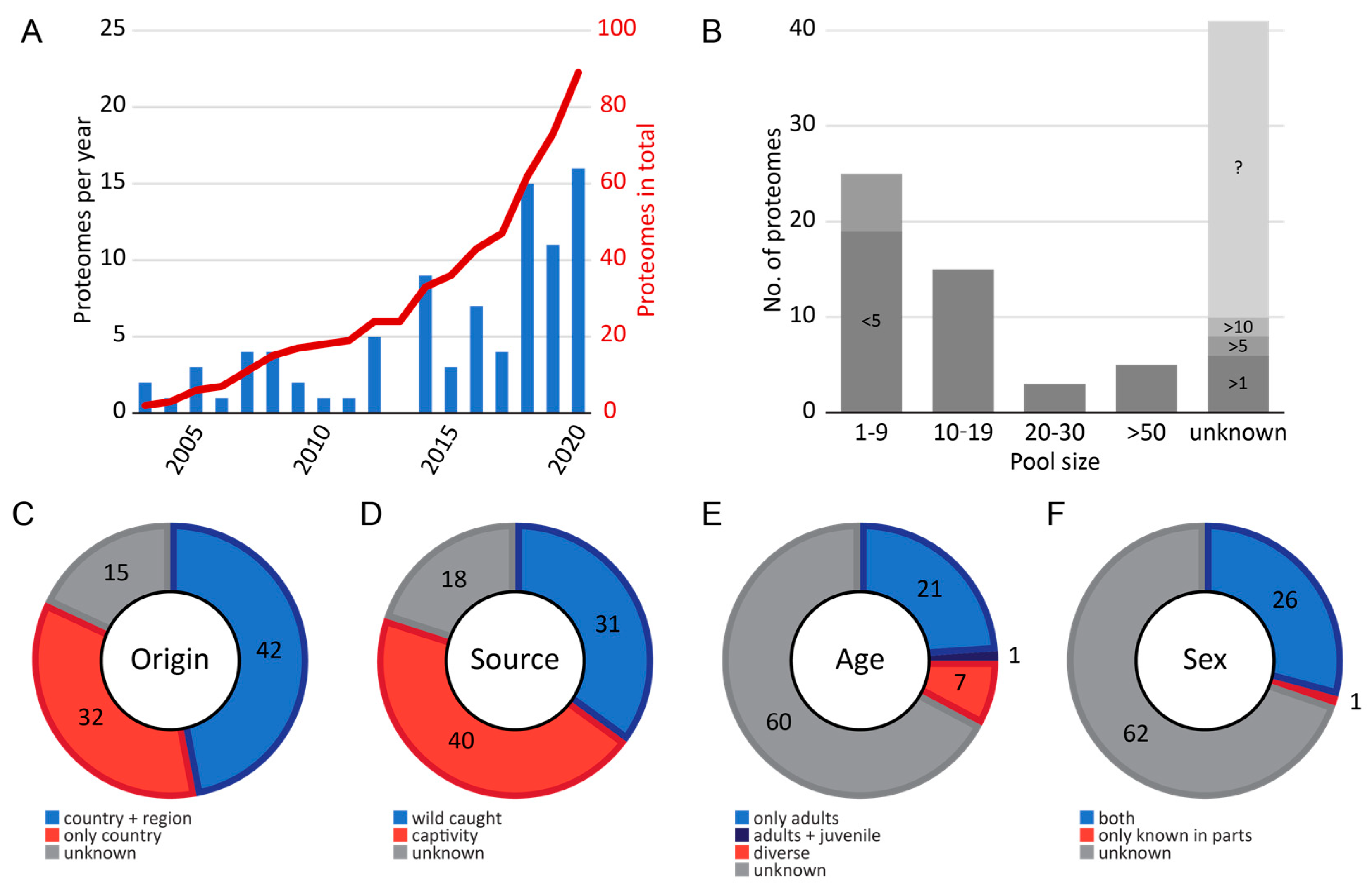
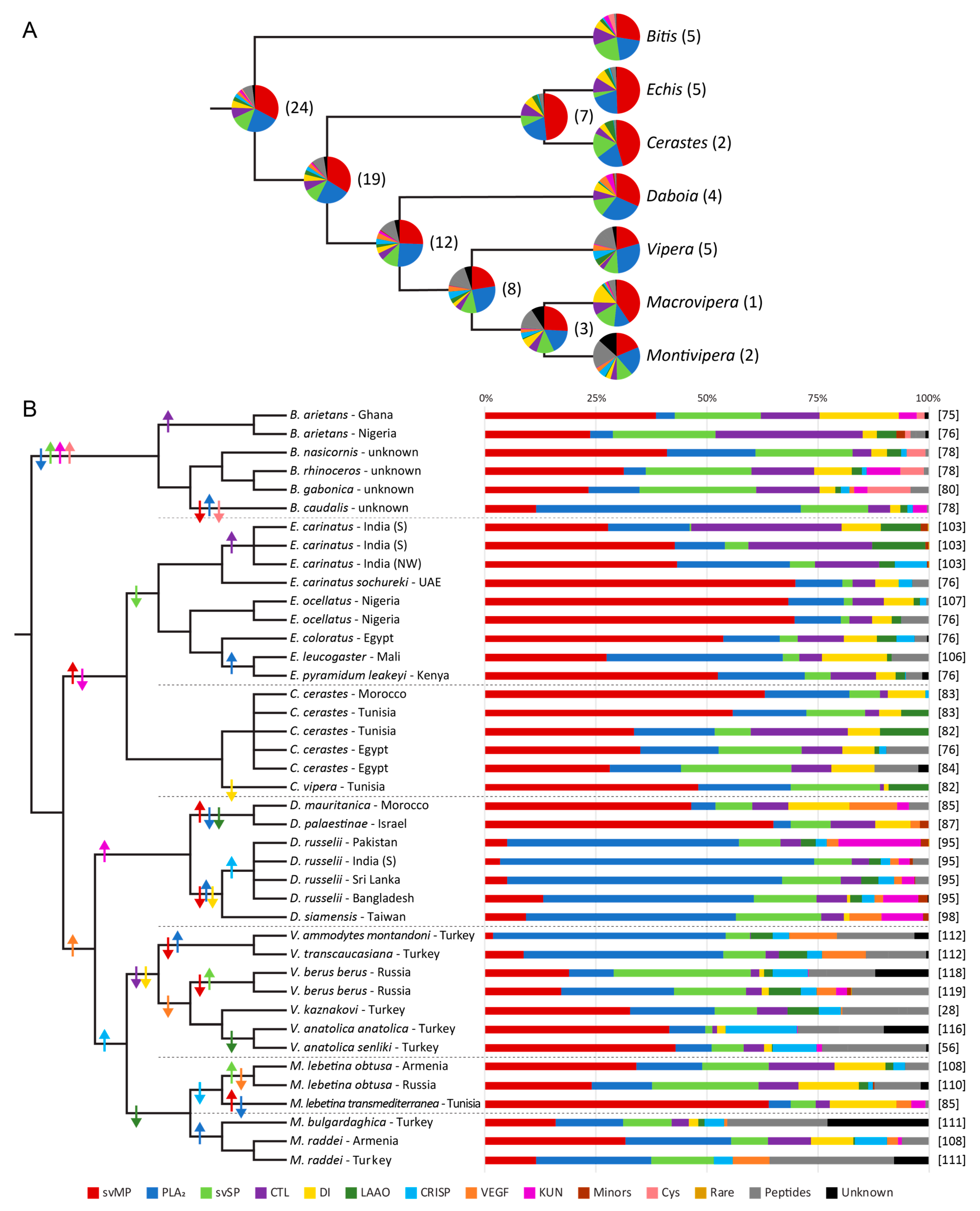
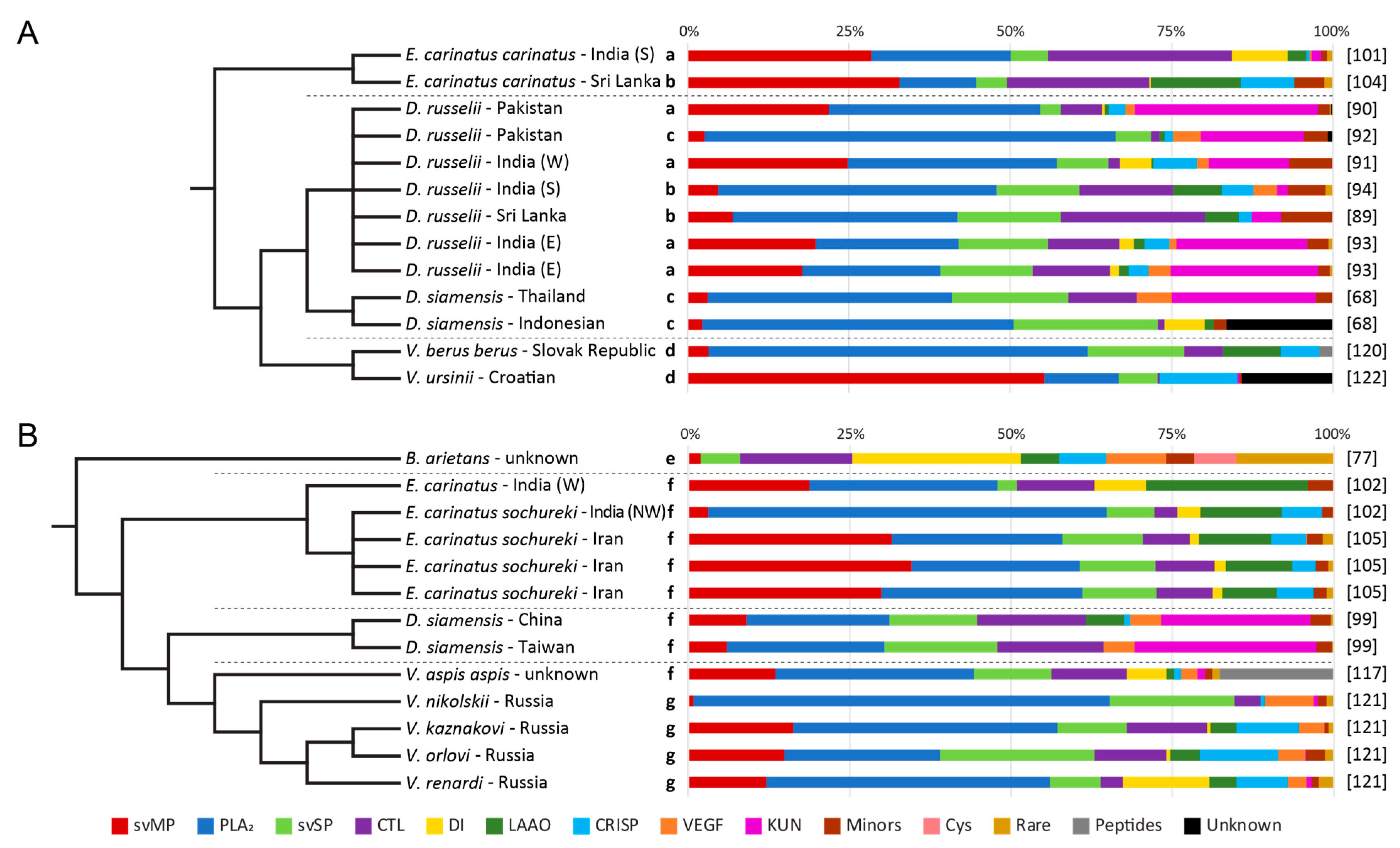
Table 1.
Complete overview of the 89 Viperinae venom proteomes. The number of studied venom compositions per genus (grey), species, and subspecies (bracketed) are mentioned. Types of different sequence annotations (black bars) and quantification (black dot) are marked for each species. The checklist shows identified toxin families, sorted by their general abundance in the venom of all Viperinae into major, secondary and further toxin families (minor, rare families and peptides). Abbreviations: svMP (snake venom metalloproteinase), PLA2 (phospholipase A2), svSP (snake venom serine protease), CTL (C-type lectin-related protein), DI (disintegrins), LAAO (L-amino acid oxidase), CRISP (cysteine-rich secretory protein), VEGF (vascular endothelial growth factors F), KUN (Kunitz-type trypsin inhibitor), CYS (cystatin), svMP-i (svMP inhibitor), NP (natriuretic peptides).
| Majors | Secondaries | ||||||||||||||||||||||||
|---|---|---|---|---|---|---|---|---|---|---|---|---|---|---|---|---|---|---|---|---|---|---|---|---|---|
| Genus Species | No. of Compositions | Examined Subspecies | Bottom-Up | Top-Down | Edman Degradation | Q—Snake Venomics | Q—Two-Step Workflows | Q—in-Solution Shotgun | No Quantification | ■svMP | ■PLA2 | ■svSP | ■CTL | ■DI | ■LAAO | ■CRISP | ■VEGF | ■KUN | ■Minor families | ■CYS | ■Rare Families | ■svMP-i | ■NP | ■Other Peptides | References |
| Atheris | 3 | ||||||||||||||||||||||||
| A. chlorechis | 1 | ■ | ● | 🗸 | 🗸 | - | - | 🗸 | - | - | - | - | - | - | - | - | - | 🗸 | [69] | ||||||
| A. nitschei | 1 | ■ | ● | 🗸 | 🗸 | 🗸 | - | 🗸 | - | - | - | - | - | - | - | - | - | 🗸 | [69] | ||||||
| A. squamigera | 1 | ■ | ● | 🗸 | 🗸 | 🗸 | - | 🗸 | - | - | - | - | - | - | - | - | - | 🗸 | [69] | ||||||
| Bitis | 9 | ||||||||||||||||||||||||
| B. arietans | 4 | ■ | ■ | ● | ● | ● | 🗸 | 🗸 | 🗸 | 🗸 | 🗸 | 🗸 | 🗸 | 🗸 | 🗸 | 🗸 | 🗸 | 🗸 | 🗸 | 🗸 | 🗸 | [60,75,76,77] | |||
| B. caudalis | 1 | ■ | ■ | ● | 🗸 | 🗸 | 🗸 | 🗸 | 🗸 | 🗸 | 🗸 | - | 🗸 | - | - | - | - | - | 🗸 | [78] | |||||
| B. gabonica | 2 | ■ | ■ | ● | ● | 🗸 | 🗸 | 🗸 | 🗸 | 🗸 | 🗸 | 🗸 | 🗸 | 🗸 | - | 🗸 | - | - | 🗸 | 🗸 | [79,80] | ||||
| B. nasicornis | 1 | ■ | ■ | ● | 🗸 | 🗸 | 🗸 | 🗸 | 🗸 | 🗸 | 🗸 | - | - | - | 🗸 | - | - | - | 🗸 | [78] | |||||
| B. rhinoceros | 1 | ■ | ■ | ● | 🗸 | 🗸 | 🗸 | 🗸 | 🗸 | 🗸 | 🗸 | - | 🗸 | - | 🗸 | - | - | 🗸 | 🗸 | [78] | |||||
| Causus | 2 | ||||||||||||||||||||||||
| C. lichtensteinii | 1 | ■ | ● | 🗸 | - | 🗸 | - | - | 🗸 | 🗸 | - | - | - | - | - | - | - | - | [81] | ||||||
| C. rhombeatus | 1 | ■ | ● | 🗸 | 🗸 | 🗸 | - | - | 🗸 | 🗸 | - | - | - | - | - | - | - | - | [81] | ||||||
| Cerastes | 6 | ||||||||||||||||||||||||
| C. cerastes | 5 | ■ | ■ | ■ | ● | 🗸 | 🗸 | 🗸 | 🗸 | 🗸 | 🗸 | 🗸 | - | - | - | - | - | 🗸 | - | 🗸 | [76,82,83,84] | ||||
| C. vipera | 1 | ■ | ■ | ● | 🗸 | 🗸 | 🗸 | 🗸 | 🗸 | 🗸 | - | - | - | - | - | - | 🗸 | - | - | [82] | |||||
| Daboia | 24 | ||||||||||||||||||||||||
| D. mauritanica | 2 | ■ | ■ | ● | ● | 🗸 | 🗸 | 🗸 | 🗸 | 🗸 | - | - | 🗸 | 🗸 | - | - | - | - | 🗸 | - | [85,86] | ||||
| D. palaestinae | 1 | ■ | ■ | ● | 🗸 | 🗸 | 🗸 | 🗸 | 🗸 | - | - | 🗸 | - | 🗸 | - | - | - | - | - | [87] | |||||
| D. russelii | 12 | ■ | ● | ● | 🗸 | 🗸 | 🗸 | 🗸 | 🗸 | 🗸 | 🗸 | 🗸 | 🗸 | 🗸 | - | 🗸 | 🗸 | 🗸 | 🗸 | [88,89,90,91,92,93,94,95] | |||||
| D. siamensis | 9 | ■ | ■ | ● | ● | ● | ● | 🗸 | 🗸 | 🗸 | 🗸 | 🗸 | 🗸 | 🗸 | 🗸 | 🗸 | 🗸 | - | 🗸 | - | - | - | [68,96,97,98,99,100] | ||
| Echis | 16 | ||||||||||||||||||||||||
| E. carinatus | 11 | (2) | ■ | ■ | ■ | ● | ● | ● | 🗸 | 🗸 | 🗸 | 🗸 | 🗸 | 🗸 | 🗸 | 🗸 | 🗸 | 🗸 | - | 🗸 | 🗸 | 🗸 | 🗸 | [76,101,102,103,104,105] | |
| E. coloratus | 1 | ■ | ■ | ● | 🗸 | 🗸 | 🗸 | 🗸 | 🗸 | 🗸 | 🗸 | - | - | - | - | - | 🗸 | - | - | [76] | |||||
| E. leucogaster | 1 | ■ | ● | 🗸 | 🗸 | 🗸 | 🗸 | 🗸 | 🗸 | - | - | - | - | - | - | 🗸 | - | - | [106] | ||||||
| E. ocellatus | 2 | ■ | ■ | ● | ● | 🗸 | 🗸 | 🗸 | 🗸 | 🗸 | 🗸 | 🗸 | - | - | - | - | - | 🗸 | - | 🗸 | [76,107] | ||||
| E. pyramidum | 1 | ■ | ■ | ● | 🗸 | 🗸 | 🗸 | 🗸 | 🗸 | 🗸 | 🗸 | - | - | - | - | - | 🗸 | - | - | [76] | |||||
| Eristicophis | 1 | ||||||||||||||||||||||||
| E. macmahoni | 1 | ■ | ● | 🗸 | 🗸 | 🗸 | 🗸 | 🗸 | 🗸 | 🗸 | 🗸 | - | - | - | - | - | 🗸 | - | [70] | ||||||
| Macrovipera | 5 | ||||||||||||||||||||||||
| M. lebetina | 5 | (2) | ■ | ■ | ● | ● | 🗸 | 🗸 | 🗸 | 🗸 | 🗸 | 🗸 | 🗸 | 🗸 | 🗸 | 🗸 | - | - | 🗸 | 🗸 | - | [82,85,108,109,110] | |||
| Montivipera | 3 | ||||||||||||||||||||||||
| M. bulgardaghica | 1 | ■ | ● | 🗸 | 🗸 | 🗸 | 🗸 | 🗸 | 🗸 | 🗸 | 🗸 | - | - | - | - | 🗸 | - | 🗸 | [111] | ||||||
| M. raddei | 2 | ■ | ■ | ● | 🗸 | 🗸 | 🗸 | 🗸 | 🗸 | 🗸 | 🗸 | 🗸 | - | 🗸 | - | - | 🗸 | 🗸 | 🗸 | [108,111] | |||||
| Pseudocerastes | 2 | ||||||||||||||||||||||||
| P. fieldi | 1 | ■ | ● | 🗸 | 🗸 | 🗸 | 🗸 | - | 🗸 | 🗸 | 🗸 | 🗸 | 🗸 | - | - | - | - | - | [70] | ||||||
| P. persicus | 1 | ■ | ● | 🗸 | 🗸 | 🗸 | 🗸 | 🗸 | 🗸 | 🗸 | 🗸 | 🗸 | 🗸 | - | - | - | - | - | [70] | ||||||
| Vipera | 18 | ||||||||||||||||||||||||
| V. ammodytes | 5 | (3) | ■ | ■ | ■ | ● | ● | 🗸 | 🗸 | 🗸 | 🗸 | 🗸 | 🗸 | 🗸 | 🗸 | 🗸 | 🗸 | - | 🗸 | 🗸 | 🗸 | 🗸 | [112,113,114,115] | ||
| V. anatolica | 2 | (2) | ■ | ■ | ● | 🗸 | 🗸 | 🗸 | 🗸 | 🗸 | 🗸 | 🗸 | - | 🗸 | 🗸 | - | 🗸 | 🗸 | 🗸 | 🗸 | [56,116] | ||||
| V. aspis | 1 | ■ | ● | 🗸 | 🗸 | 🗸 | 🗸 | 🗸 | 🗸 | 🗸 | 🗸 | 🗸 | 🗸 | - | 🗸 | 🗸 | - | 🗸 | [117] | ||||||
| V. berus | 3 | ■ | ■ | ● | ● | 🗸 | 🗸 | 🗸 | 🗸 | 🗸 | 🗸 | 🗸 | 🗸 | 🗸 | 🗸 | - | 🗸 | 🗸 | 🗸 | 🗸 | [118,119,120] | ||||
| V. kaznakovi | 2 | ■ | ■ | ● | ● | 🗸 | 🗸 | 🗸 | 🗸 | 🗸 | 🗸 | 🗸 | 🗸 | - | 🗸 | - | 🗸 | 🗸 | 🗸 | 🗸 | [28,121] | ||||
| V. nikolskii | 1 | ■ | ● | 🗸 | 🗸 | 🗸 | 🗸 | - | 🗸 | 🗸 | 🗸 | 🗸 | 🗸 | - | 🗸 | - | 🗸 | - | [121] | ||||||
| V. orlovi | 1 | ■ | ● | 🗸 | 🗸 | 🗸 | 🗸 | 🗸 | 🗸 | 🗸 | 🗸 | 🗸 | 🗸 | - | 🗸 | - | 🗸 | - | [121] | ||||||
| V. renardi | 1 | ■ | ● | 🗸 | 🗸 | 🗸 | 🗸 | 🗸 | 🗸 | 🗸 | 🗸 | 🗸 | 🗸 | - | 🗸 | - | - | - | [121] | ||||||
| V. transcaucasiana | 1 | ■ | ■ | ● | 🗸 | 🗸 | 🗸 | 🗸 | - | 🗸 | 🗸 | 🗸 | - | 🗸 | - | - | 🗸 | 🗸 | 🗸 | [112] | |||||
| V. ursinii | 1 | ■ | ● | 🗸 | 🗸 | 🗸 | 🗸 | - | - | 🗸 | - | 🗸 | 🗸 | - | 🗸 | - | - | - | [122] | ||||||
Table 2.
Toxin families in the venom proteomes of Viperinae. The proteins and peptides were grouped into families (bold) according to their general abundance in the venom compositions. LAAO and PLB were reported from more studies but show lower average percentages in the venoms than DI and HYAL. Enzymes are mentioned by their Enzyme Commission number (EC). Additional information about the average monomeric mass, number of disulfide bridges (No. of S-S) and their appearance in the proteomic studies is given. Asterisked entries mark that no exact number was derived from the literature. A more detailed list of the rare protein families is available in Supplementary Material Table S1.
| Abbreviation | Snake Venom Toxin Family | Enzyme Class | Monomeric Size in kDa | No. of S-S | Observed in No. of Studies | References |
|---|---|---|---|---|---|---|
| Major toxin families | ||||||
| svMP | snake venom metalloproteinase | EC 3.4.24.- | 20–100 | 4–18 | 89 | [135,136,137,138,139] |
| PLA2 | phospholipase A2 | EC 3.1.1.4 | 13–15 | 6–8 | 87 | [140,141,142,143,144] |
| svSP | snake venom serine protease | EC 3.4.21.- | 22–67 | 6 | 86 | [145,146,147,148] |
| CTL incl. Snaclec | C-type lectin-related protein | - | 13–15 | 3 | 80 | [149,150,151] |
| Secondary toxin families | ||||||
| DI | disintegrin | - | 4–10 | 4–8 | 63 | [152,153,154,155] |
| LAAO | l-amino acid oxidase | EC 1.4.3.2 | 50–70 | 2 | 68 | [156,157,158] |
| CRISP | cysteine-rich secretory protein | - | 20–33 | 8 | 63 | [159,160,161] |
| VEGF | vascular endothelial growth factors F | - | 10–15 | 5 | 48 | [162,163,164] |
| KUN | Kunitz-type inhibitor | - | 6–7 | 3 | 42 | [20,165,166,167] |
| Minor toxin families | ||||||
| NGF | nerve growth factor | - | 12–37 | 3 | 41 | [168,169,170] |
| 5N | 5′-nucleotidase | EC 3.1.3.5 | 73–100 | 4 | 34 | [171,172] |
| PDE | phosphodiesterase | EC 3.1.4.1 | 90–140 | 16 | 33 | [173,174] |
| HYAL | hyaluronidase | EC 3.2.1.35 | 33–110 | 5 | 17 | [158,175,176] |
| PLB | phospholipase B | EC 3.1.1.5 | ~55 | 2 | 21 | [177] |
| CYS | cystatin | - | 12–15 | 2 | 8 | [178,179,180] |
| Rare families (selection) | ||||||
| QC | glutaminyl cyclotransferase | EC 2.3.2.5 | 33–40 | 1 | 17 | [181,182] |
| AP | Aminopeptidase | EC 3.4.11.- | 100–150 | * | 17 | [183,184,185] |
| Peptides | ||||||
| svMP-i | svMP-inhibitor | - | 0.3 | 0 | 22 | [134,186,187] |
| BPP | bradykinin potentiating peptide | - | 0.5–1.5 | 0 | 24 | [133,188,189,190] |
| other peptides | incl. further natriuretic peptides | - | 1–10 | - | 24 | [133,134,189,191,192] |
Publisher’s Note: MDPI stays neutral with regard to jurisdictional claims in published maps and institutional affiliations. |
© 2021 by the authors. Licensee MDPI, Basel, Switzerland. This article is an open access article distributed under the terms and conditions of the Creative Commons Attribution (CC BY) license (https://creativecommons.org/licenses/by/4.0/).
Share and Cite
MDPI and ACS Style
Damm, M.; Hempel, B.-F.; Süssmuth, R.D. Old World Vipers—A Review about Snake Venom Proteomics of Viperinae and Their Variations. Toxins 2021, 13, 427. https://0-doi-org.brum.beds.ac.uk/10.3390/toxins13060427
AMA Style
Damm M, Hempel B-F, Süssmuth RD. Old World Vipers—A Review about Snake Venom Proteomics of Viperinae and Their Variations. Toxins. 2021; 13(6):427. https://0-doi-org.brum.beds.ac.uk/10.3390/toxins13060427
Chicago/Turabian StyleDamm, Maik, Benjamin-Florian Hempel, and Roderich D. Süssmuth. 2021. "Old World Vipers—A Review about Snake Venom Proteomics of Viperinae and Their Variations" Toxins 13, no. 6: 427. https://0-doi-org.brum.beds.ac.uk/10.3390/toxins13060427
Note that from the first issue of 2016, this journal uses article numbers instead of page numbers. See further details here.