Article

A Biochemical and Pharmacological Characterization of Phospholipase A2 and Metalloproteinase Fractions from Eastern Russell’s Viper (Daboia siamensis) Venom: Two Major Components Associated with Acute Kidney Injury

by
Janeyuth Chaisakul
1,*,
Orawan Khow
2,
Kulachet Wiwatwarayos
3,
Muhamad Rusdi Ahmad Rusmili
4,
Watcharamon Prasert
1,
Iekhsan Othman
5,
Syafiq Asnawi Zainal Abidin
5,
Mongkon Charoenpitakchai
6,
Wayne C. Hodgson
7,
Lawan Chanhome
2 and
Narongsak Chaiyabutr
2
1
Department of Pharmacology, Phramongkutklao College of Medicine, Bangkok 10400, Thailand
2
Queen Saovabha Memorial Institute, Thai Red Cross Society, Bangkok 10330, Thailand
3
Institute of Pathology, Ministry of Public Health, Bangkok 10400, Thailand
4
Kulliyyah of Pharmacy, International Islamic University Malaysia, Bandar Indera Mahkota, Kuantan 25200, Malaysia
5
Jeffrey Cheah School of Medicine and Health Sciences, Monash University Malaysia, Bandar Sunway 46150, Malaysia
6
Department of Pathology, Phramongkutklao College of Medicine, Bangkok 10400, Thailand
7
Monash Venom Group, Department of Pharmacology, Biomedical Discovery Institute, Monash University, Clayton, VIC 3800, Australia
*
Author to whom correspondence should be addressed.
Submission received: 30 June 2021 / Revised: 19 July 2021 / Accepted: 21 July 2021 / Published: 26 July 2021
(This article belongs to the Special Issue Venom-Induced Tissue Damage)

Abstract

:
Acute kidney injury (AKI) following Eastern Russell’s viper (Daboia siamensis) envenoming is a significant symptom in systemically envenomed victims. A number of venom components have been identified as causing the nephrotoxicity which leads to AKI. However, the precise mechanism of nephrotoxicity caused by these toxins is still unclear. In the present study, we purified two proteins from D. siamensis venom, namely RvPLA2 and RvMP. Protein identification using LCMS/MS confirmed the identity of RvPLA2 to be snake venom phospholipase A2 (SVPLA2) from Thai D. siamensis venom, whereas RvMP exhibited the presence of a factor X activator with two subunits. In vitro and in vivo pharmacological studies demonstrated myotoxicity and histopathological changes of kidney, heart, and spleen. RvPLA2 (3–10 µg/mL) caused inhibition of direct twitches of the chick biventer cervicis muscle preparation. After administration of RvPLA2 or RvMP (300 µg/kg, i.p.) for 24 h, diffuse glomerular congestion and tubular injury with minor loss of brush border were detected in envenomed mice. RvPLA2 and RvMP (300 µg/kg; i.p.) also induced congestion and tissue inflammation of heart muscle as well as diffuse congestion of mouse spleen. This study showed the significant roles of PLA2 and SVMP in snake bite envenoming caused by Thai D. siamensis and their similarities with observed clinical manifestations in envenomed victims. This study also indicated that there is a need to reevaluate the current treatment strategies for Thai D. siamensis envenoming, given the potential for irreversible nephrotoxicity.
Key Contribution: We demonstrated that PLA2 and SVMP isolated from D. siamensis venom play a significant role in causing acute kidney injury experimentally. Early evaluation and treatment would help to minimize the severity of snakebite-induced nephrotoxicity.

1. Introduction

Daboia spp. (Russell’s vipers) are a widely distributed snake genera in Asia and responsible for a high incidence of morbidity and mortality [1]. In 2016, the World Health Organization (WHO) classified Russell’s vipers as category 1 medically important venomous snakes in India, Nepal, Sri Lanka, Myanmar, Thailand and some Indonesian islands, i.e., Java, Komodo, Flores, and Lomblen [1]. Russell’s vipers are recognised as two distinct species, i.e., Daboia russelii (the Western Russell’s viper) and Daboia siamensis (the Eastern Russell’s viper). D. russelii is found throughout India, Pakistan, Bangladesh, Sri Lanka, and the west Bay of Bengal, whilst D. siamensis has a wide distribution across Southeast Asia (i.e., Myanmar, Thailand, and some Indonesian Islands), and Southern China (i.e., Guangdong and Guangxi), including Taiwan [1,2,3].
Acute kidney injury (AKI) caused by disseminated intravascular coagulopathy is commonly seen following envenoming by Daboia spp. regardless of the geographical range of the envenoming snake [4,5,6,7,8]. In contrast, the presence of neurotoxicity and myotoxicity following Daboia spp. envenoming displays remarkable geographical variation. According to the available clinical data, hematotoxicity and nephrotoxicity are prominently observed following D. siamensis envenoming in Taiwan and Myanmar [9,10]. Whereas, envenoming by the Sri Lankan and South Indian population of D. russelii causes significant skeletal muscle paralysis and myoglobinuria [11,12,13]. These clinical outcomes have been attributed to the presence of neurotoxic and myotoxic snake venom phospholipase A2 (PLA2) in the venom of this Daboia spp. population.
A proteomic investigation demonstrated that more than 90% of D. siamensis venom proteins belong to the Kunitz-type serine protease inhibitors, PLA2, C-type lectin/lectin-like protein, serine protease, and metalloproteinase families [14]. PLA2 and snake venom metalloproteinase (SVMP) from the venoms of Daboia spp. have been shown to be predominately responsible for coagulopathy, nephrotoxicity, neurotoxicity, cytotoxicity, and myotoxicity [15,16,17,18]. Daborhagin-M and daborhagin-K are P-III hemorrhagic metalloproteinases isolated from Russell’s viper venoms from Myanmar and eastern India, respectively. These SVMPs both induce severe hemorrhagic symptoms in experimental animals and also hydrolyze the Aα-chain of fibrinogen, fibronectin, and type IV collagen in in vitro studies [19]. PLA2s from Daboia spp. cause multiple toxic activities. For example, a myotoxic PLA2 [20] and a pre-synaptic PLA2 neurotoxin, called U1-viperitoxin [10], both isolated from Sri Lankan Russell’s viper (D. russelii) have been characterized in the chicken biventer preparation.
Recently, RvPLA2 and RvMP were isolated from D. siamensis venom and showed physiological function and histopathological changes in ex vivo perfused rabbit kidney model after incubation for 9 h [21]. However, potential contributing factors following administration of RvPLA2 and RvMP, for example, myotoxicity involvement, the effects of purified fractions in an in vivo model extending beyond 9 h, and their roles in the mechanism of AKI have not been fully investigated. The aim of the present study was to characterize RvPLA2 and RvMP from Thai D. siamensis venom using in vitro myotoxic and mice in vivo experiments. Protein characterization of RvPLA2 and RvMP was also undertaken to identify the amino acid sequences for both proteins.

2. Results

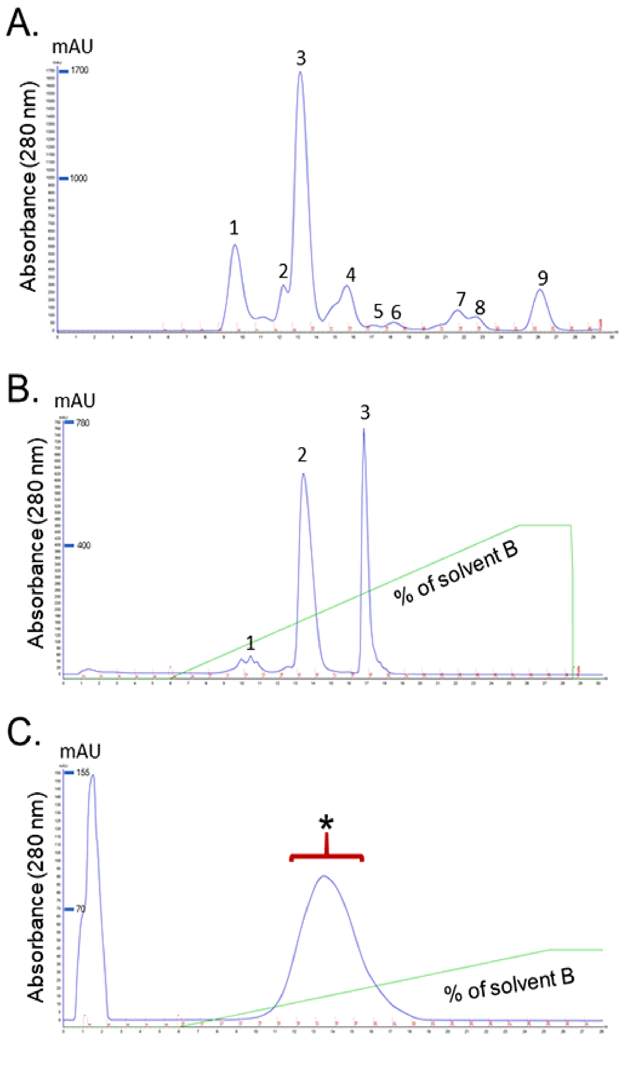
2.1. Identification of Purified Fractions

2.1.1. Isolation and Purification of RvPLA2 and RvMP

Isolation and Purification of RvPLA2 and RvMP were completed as previously described [21]. In brief, for RvPLA2 isolation, D. siamensis venom (DSV) was fractionated using HiTrap CM FF ion-exchange column and the elution chromatogram yielded three major peaks (Figure 1A). Determination of PLA2 activity of the peaks showed that only fraction 2 contained PLA2 activity. This fraction was further purified using a SuperdexTM 75 10/300GL size exclusion column and the highest peak was chosen for further analysis (Figure 1B).
For RvMP fractionation, D. siamensis venom was fractionated using a SuperdexTM 75 10/300GL size exclusion column and the elution chromatogram yielded nine peaks (Figure 2A). Fraction 1 was found to have the highest protease activity. Further purification of fraction 1, using a Mono Q column, yielded three main peaks and the highest protease activity was detected in Peak 2 (Figure 2B). Peak 2 was freeze-dried, and further fractionated using Resource S column. The second peak eluted from this step was chosen for further analysis (Figure 2C).

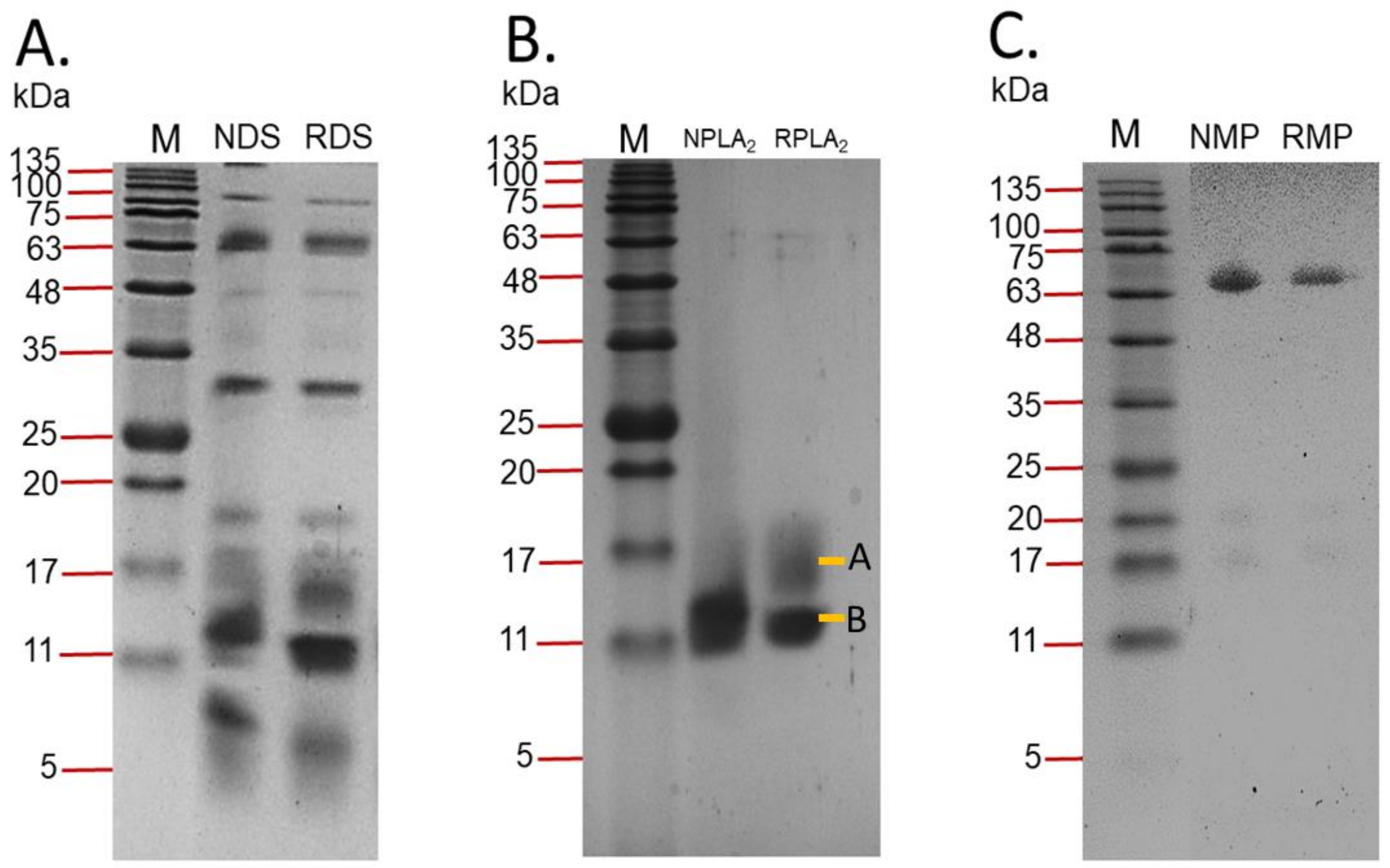
2.1.2. Sodium Dodecyl Sulphate-Polyacrylamide Gel Electrophoresis (SDS-PAGE)

D. siamensis venom, and the fractions of interest, were separated in a gel under non-reducing and reducing conditions (Figure 3). Thick and high intensity bands were observed in the MW range below 20 kDa in reduced and non-reduced venom. Several single protein bands of reduced and non-reduced venoms were also detected above the MW of 25 kDa (Figure 3A). The homogeneity and molecular weight of fractions were also assessed with SDS-PAGE. A thick protein band was detected in the lane loaded with non-reduced RvPLA2 with an estimated MW of 14 kDa. The two different bands, labeled as A and B, were detected in the lane loaded with reduced RVPLA2 and were estimated to have MW’s of 14 kDa and 12 kDa, respectively (Figure 3B). In lanes that were loaded with RvMP, the estimated molecular weights of RvMP under non-reducing and reducing conditions were 68 kDa and 65 kDa, respectively (Figure 3C).

2.1.3. Phospholipase A2 Activity

The PLA2 activity of venom and fractions was determined using a secretory PLA2 colourmetric assay kit (Cayman Chemical, MI, USA). PLA2 activity for D. siamensis venom, RvPLA2, and RvMP were 2000 ± 245, 1620 ± 173, and 18 ± 7 µmol/min/mg (n = 3), respectively. The PLA2 activity of bee venom, used as a positive control, was 540 ± 22 µmol/min/mg (n = 3).

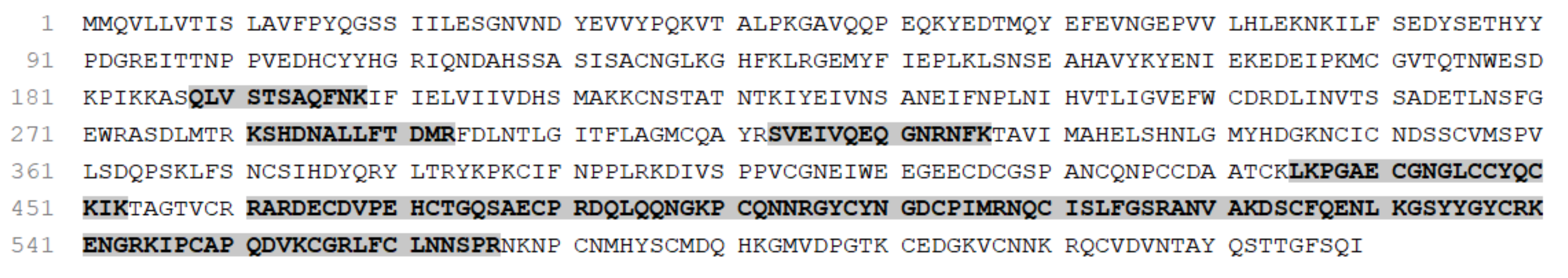
2.1.4. Protein Identification for RvMP

Protein identification and de novo sequencing showed RvMP to be identical to a factor X activator (Supplementary Data S1). Two proteins were detected in the sample: coagulation factor X activating enzyme heavy chain (UNIPROT accession number Q7LZ61, Figure 4) and snaclec coagulation factor X activating enzyme light chain (UNIPROT accession number Q4PRD1, Figure 5). The detected peptides showed 26% sequence similarity for the heavy chain and 36% for the light chain. Significant amino acid mutation was not detected from the de novo sequences.

2.1.5. Protein Identification for RvPLA2

Protein identification by de novo sequencing from LCMS/MS analysis of the digested RvPLA2 peptides showed that they have 88% sequence similarity with acidic phospholipase A2 Daboia siamensis (UniPROT accession number P31100, Figure 6) (Supplementary Data S2). No amino acid mutation was detected.

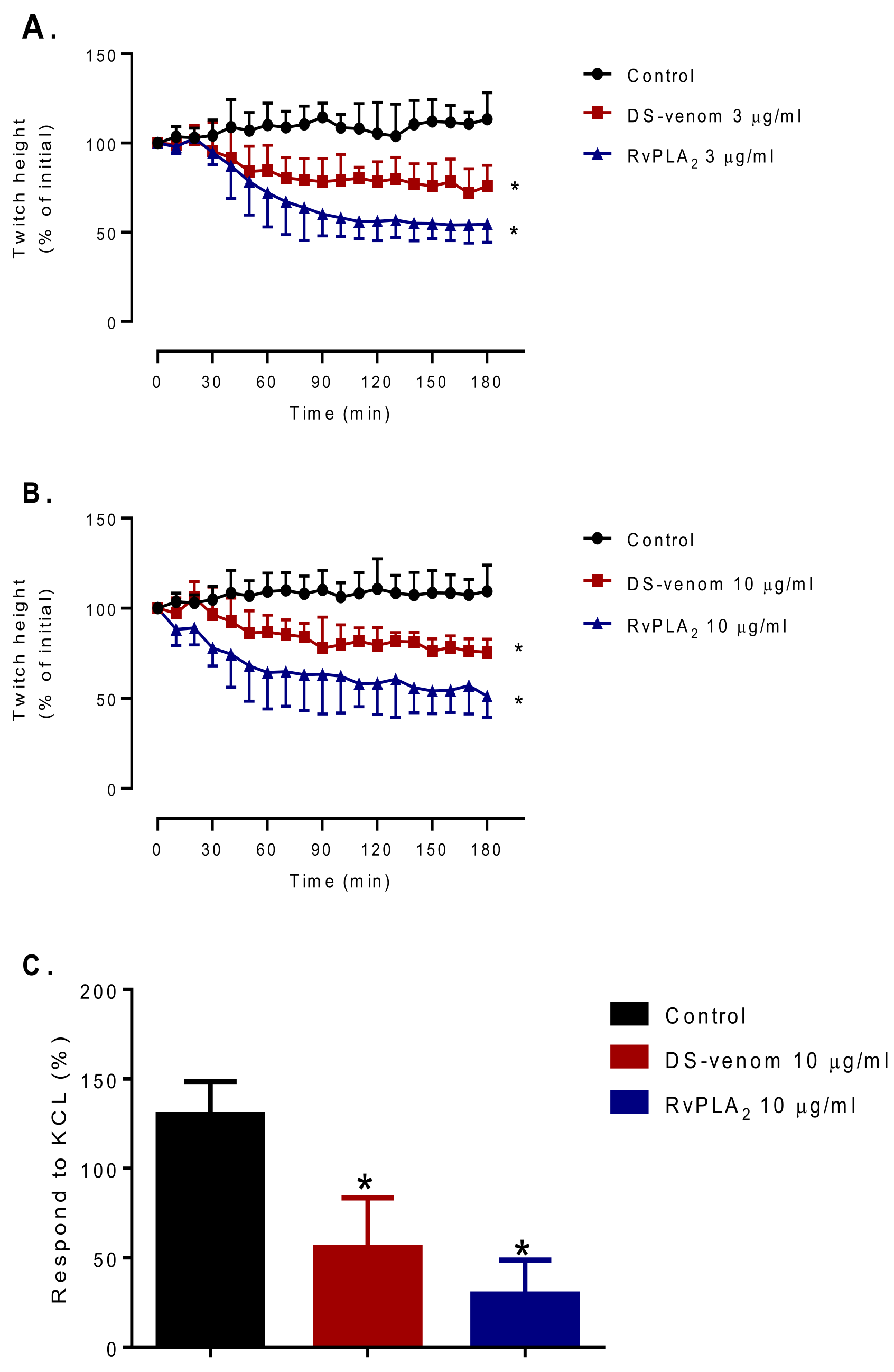
2.2. Myotoxicity Studies

D. siamensis venom (3–10 µg/mL, n = 4, one-way ANOVA, p < 0.05) and RvPLA2 (3–10 µg/mL, n = 4, one-way ANOVA, p < 0.05, Figure 7A,B) significantly reduced direct twitches in the chick biventer preparation as compared with vehicle. Neither venom (3–10 µg/mL, n = 4) nor RvPLA2 (3–10 µg/mL, n = 4, one-way ANOVA, p < 0.05) had a significant effect on baseline tension as compared with vehicle. Venom and RvPLA2 (3–10 µg/mL, n = 4, one-way ANOVA, p < 0.05) both significantly decreased contractile response to KCl as compared with the control (Figure 7C).

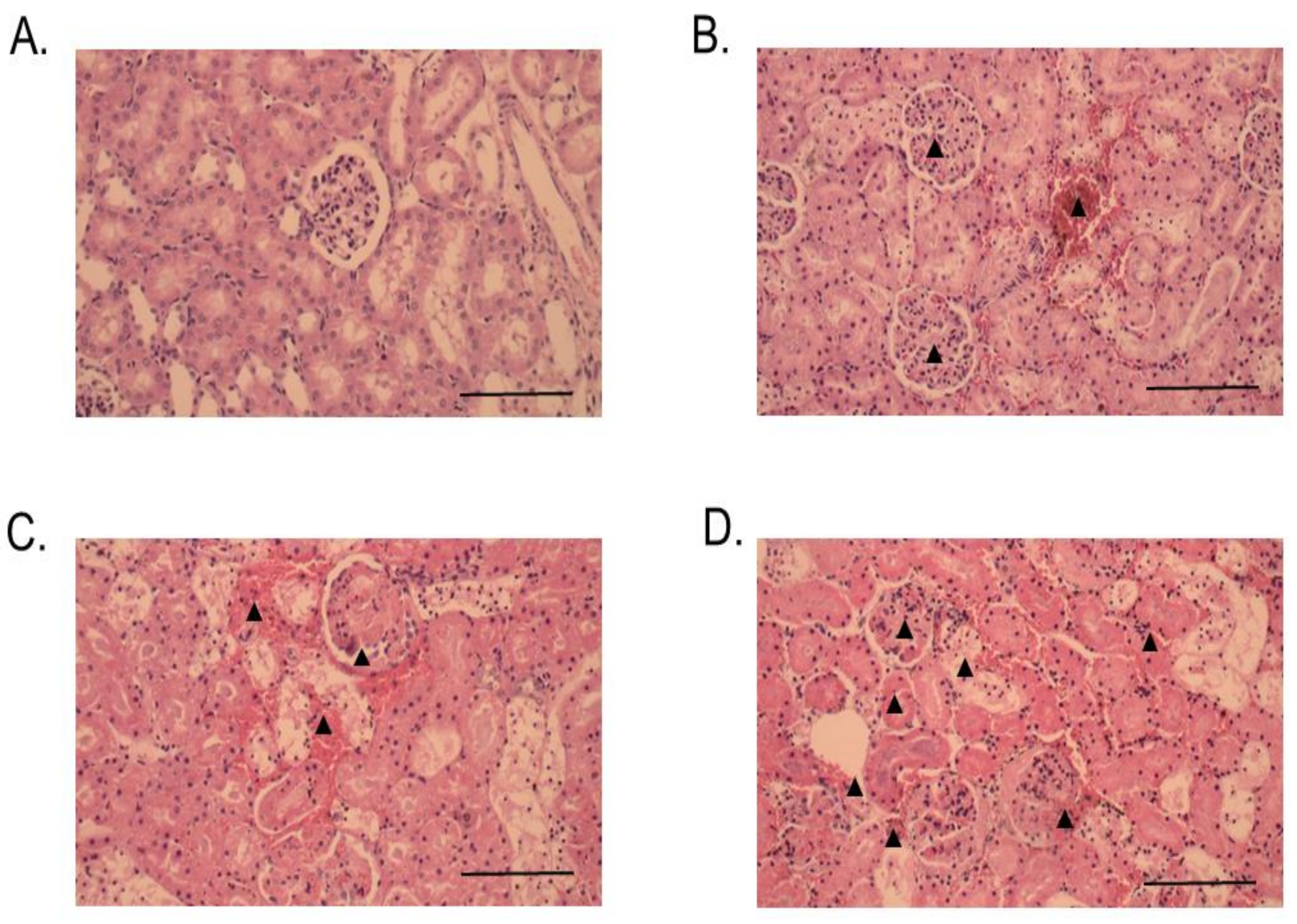
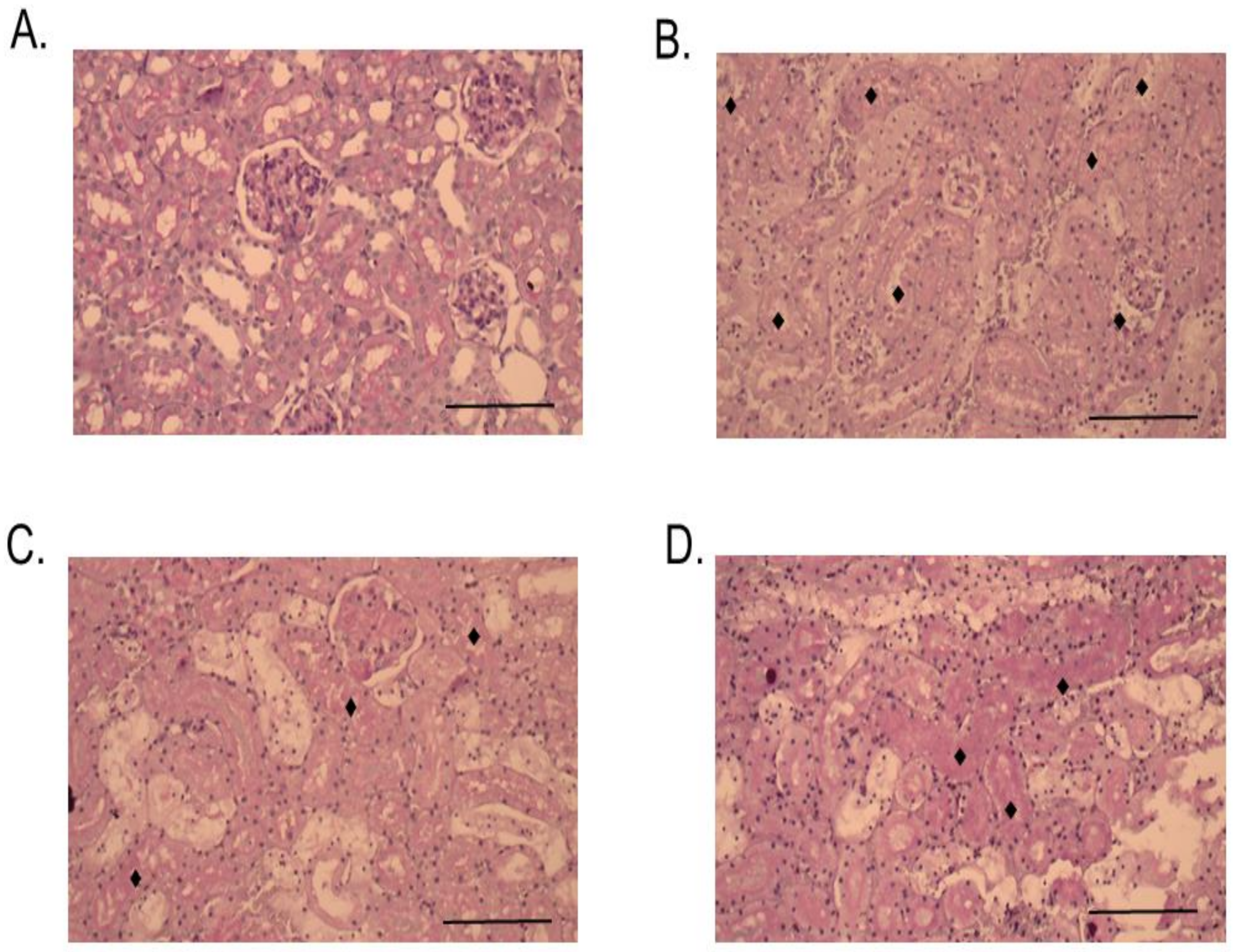
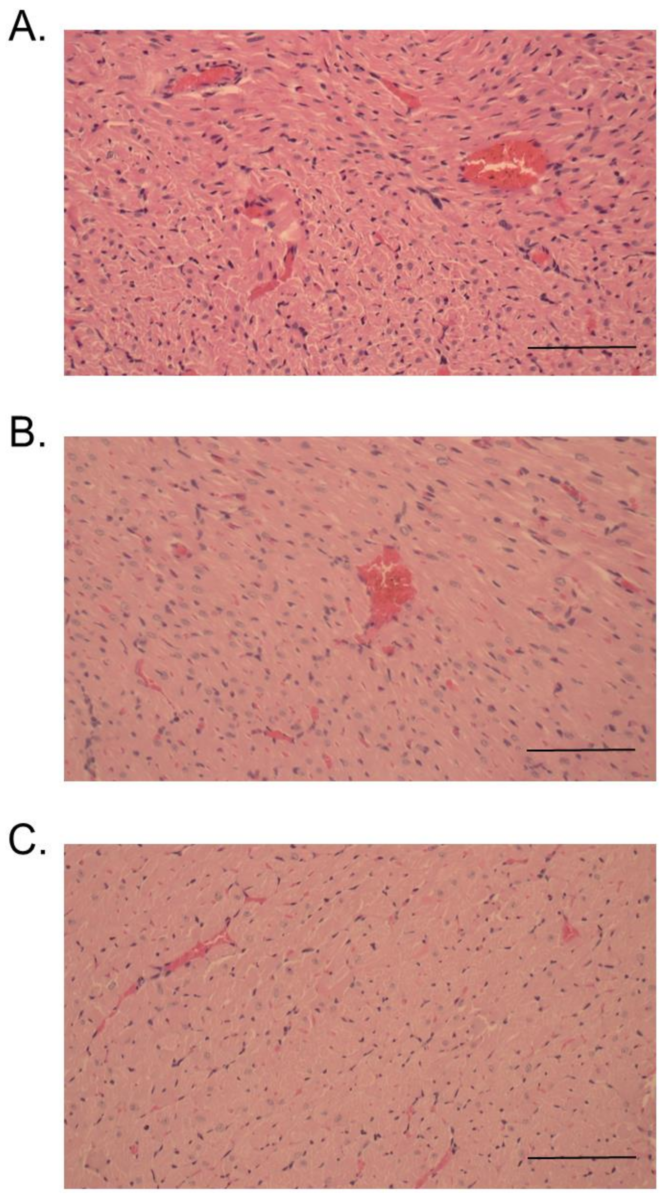
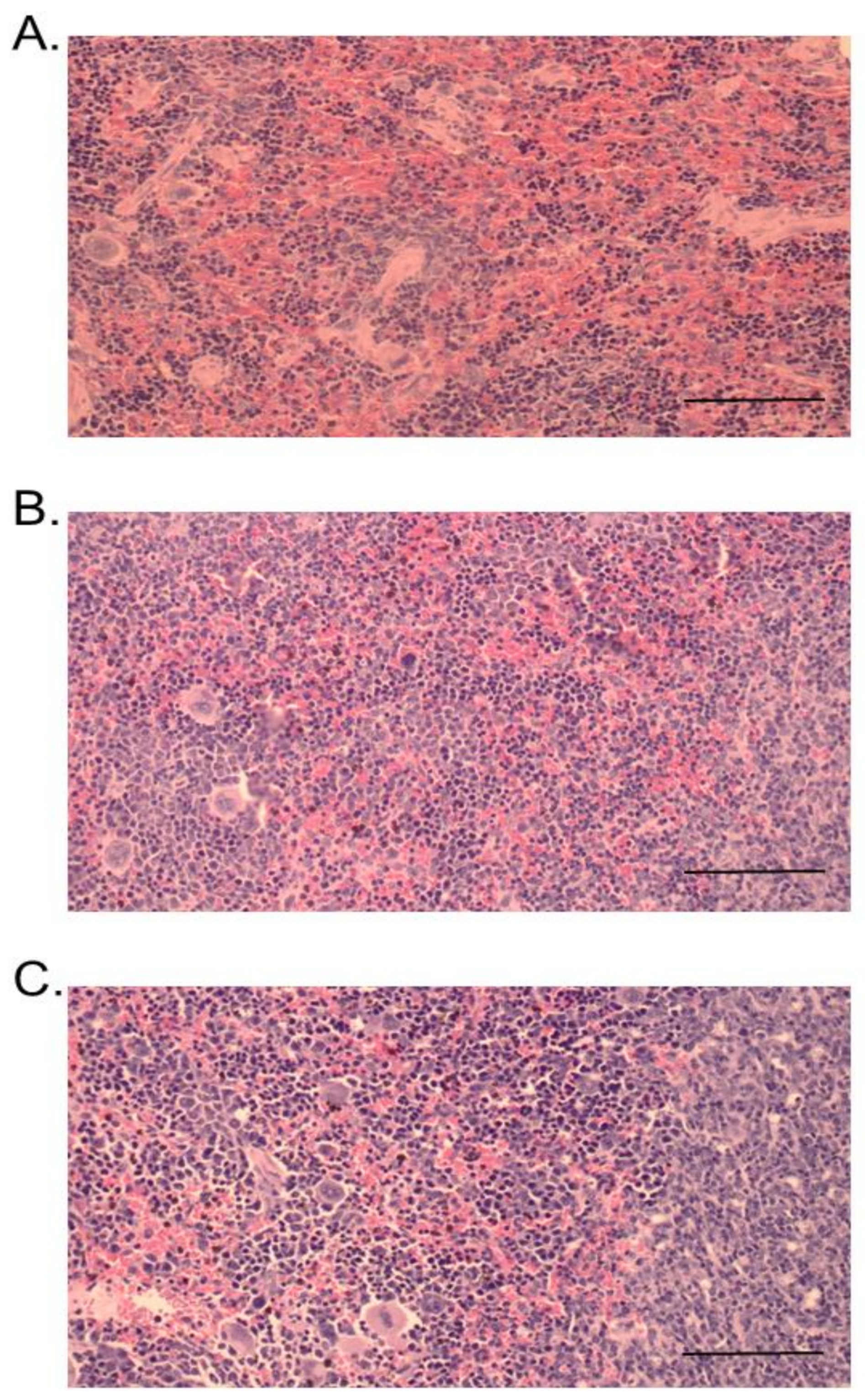
2.3. Histopathological Studies

Histopathological changes in mice caused by intraperitoneal (i.p.) administration of venom, RvPLA2, and RvMP were determined by light microscopic examination of kidney, heart, and spleen tissue samples. No significant histopathological change was observed in heart, kidney, and spleen tissues from animals that were administered with venom, RvPLA2, and RvMP at doses lower than 300 µg/kg (i.p.). Histopathological analysis of mice kidneys which were dissected at the 24 h time point following venom or RvPLA2 administration (300 µg/kg, i.p.) exhibited moderate (+2) morphological changes as compared with the negative control (Figure 8). Changes were defined by the presence of hyaline cast, dilatation of renal capillary, inflammatory foci, and glomeruli and/or congestion of interstitial vessels (Figure 8B,C). Severe congestion (+3) with hemorrhage was observed in glomeruli and interstitial vessels following the injection of RvMP (300 µg/kg, i.p.) after 24 h (Figure 8D). Administration of venom caused moderate tubular injury with partial loss of brush border with cell sloughing off and the presence of hyaline cast after 24 h (Figure 9B), whereas RvPLA2 and RvMP (300 µg/kg, i.p.) caused mild tubular injury (+1) with partial loss of brush border (Figure 9C,D).
At 24 h, administration of venom or RvPLA2 (300 µg/kg, i.p.) caused a moderate degree (+2) of cardiac muscle necrosis and congestion (Figure 10A,B). A mild cardiac muscle necrosis was observed following administration of RvMP (Figure 10C). The spleen tissue samples for mice injected with venom and its fractions displayed hemorrhage and congestion 24 h after administration (Figure 11A–C).

3. Discussion

The present study demonstrated histopathological changes in tissues which are associated with the toxic activities of RvPLA2 and RvMP. We purified RvPLA2 and RvMP from Thai D. siamensis venom using ion-exchange and size-exclusion chromatography. The SDS-PAGE analysis detected two different protein bands of reducing RvPLA2, suggesting there might be some low molecular weight impurities in the sample. No band was observed at 25–35 kDa range in non-reduced RvPLA2 lane which exclude the possibility of the presence of a dimeric complex of PLA2. The identity of RvPLA2 was confirmed using LCMS/MS data which indicated that it was an acidic phospholipase A2 from Daboia siamensis with 88% sequence similarity. Previously, a purified anticancer PLA2 toxin, Drs-PLA2, was demonstrated to share 100% N-terminus sequence homology with a basic PLA2 from D. r. siamensis venom (viperotoxin). However, the pI for Drs-PLA2 was slightly more acidic, suggesting that viperotoxin was not Drs-PLA2 [22]. It was also shown that Drs-PLA2 had in vitro hemolytic, anticoagulant, and cytotoxicity activities. Although many Russell’s viper PLA2 toxins share a high degree of similarity in amino acid sequence, their enzymatic and other biological activities might be different due to slight differences in amino acid sequence. This could be due to the presence of post-translational modification of the proteins that was not able to be detected by the method used in the current study. This also indicates possible intra- and inter-species variation and a structure–function relationship of viperid venom PLA2 [23]. Snake venom metalloproteases (SVMPs) are significant components in most viper venoms, and cause hemostatic and cellular homeostasis disturbances. According to the presence and absence of various non proteinase domains, as observed via mRNA transcripts and proteins in the venom, SVMPs are categorized into PI, PII (a and b), and PIII (a and b) classes [24]. Functional variabilities of SVMPs were reported to indicate distinct mechanisms in the coagulation processes of human, avian, and rodents [25]. SVMPs from viper venoms displayed molecular weights between 20 and 70 kDa [25]. In the present study, the estimated molecular weight of RvMP from SDS-PAGE was 65 kDa when reduced, and 68 kDa when not reduced. RvMP is a PIII snake venom metalloproteinase based on its molecular weight, detected peptides, and matched protein. BnMPIIIc-SVMP, from Bothrops neuwiedi venom, also displayed a molecular weight of 67 kDa [25]. The matched protein, coagulation factor X activating enzyme heavy chain (Q7LZ61), was reported to exist as a heterotrimeric, with one heavy chain and two light chains linked by disulfide bonds [26]. However, based on the SDS-PAGE band profiles of reduced and non-reduced samples, RvMP does not possess other subunits. Although a snaclec coagulation factor X activating enzyme light chain (Q4PRD1) was also detected in the sample, it was not present in sufficient concentration to be detected by SDS-PAGE under non-reducing and reducing conditions but enough for the highly sensitive mass-spectrometry system. This indicates that it could be a co-eluted protein in the RvMP-containing fraction.
A recent study, using an ex vivo perfused rabbit kidney model, showed that RvPLA2 and RvMP (280 µg/mL) significantly increased perfusion pressure, urinary flow rate, glomerular filtration rate, osmolar clearance, and renal vascular resistance [21]. Histopathological determination of perfused rabbit kidney sections revealed crystal deposits in the glomerular capillary lumen, dilatation of proximal and distal convoluted tubules, as well as tubulonephrosis, following incubation with RvPLA2 or RvMP for 90 min. In the current study, we determined histopathological changes of mouse kidneys following intraperitoneal administration of venom or fractions (300 µg/kg) for 24 h. A moderate degree of histopathological lesions was detected as diffuse and/or focal glomeruli, and congestion of interstitial vessels and tubular injury. These are almost the same characteristics of kidney lesion as identified in previous studies using perfused rabbit kidneys [21] and rat in vivo experiments [27]. However, the degree of morphological changes observed in our present work could not be compared with previous work performed by our group due to the differences in species of experimental animal used and time of contact with venom or toxins. The degree of histopathologic change observed following snake envenoming is significantly associated with the time of contact with venom or toxin [28]. The effects of RvPLA2 and RvMP on physiological function and morphological changes were abolished by the prior addition of the platelet activating factor (PAF) inhibitor, WEB 2086 [21]. This suggests a role for PAF to modulate glomerular functions via PAF receptor within the renal glomeruli [29].
Acute kidney injury (AKI) appears to be an important clinical outcome associated with mortality, especially those bitten by Daboia species [10]. The pathogenesis of AKI after Russell’s viper envenoming is incompletely understood, but seems to involve microvascular fibrin deposition, direct nephrotoxicity, and cardiovascular impairments [30]. The mechanisms behind AKI following viper envenoming have been suggested to be associated with DIC or coagulopathy [31]. Here, we demonstrated that administration of D. siamensis venom, either RvPLA2 or RvMP, for 24 h to mice produced diffuse glomerular congestion, hyaline cast, and moderate tubular necrosis, suggesting Russell’s viper envenoming induces acute kidney injury [27]. Our histopathological data correlates with clinical observations in envenomed patients indicating that acute tubular necrosis as well as renal cortical necrosis become the predominant pathologies detected in kidney biopsy [32,33].
Myotoxicity with a significant increase in creatine kinase (CK) is a common clinical observation following envenoming by myotoxic snake species including sea snakes (e.g., Hydrophiinae), some elapids (e.g., Pseudechis australis and Bungarus candidus) [34,35] and some vipers (e.g., Daboia russelii and Bothrops asper) [9,36]. Systemic myotoxicity, which causes rhabdomyolysis, is induced by systemic myotoxins that cause widespread muscle injury resulting in elevation of CK, hyperkalaemia, and AKI with myoglobinuria. In the present study, we demonstrated that RvPLA2 from D. siamensis venom attenuated twitch height of directly stimulated chick biventer cervicis muscle and also decreased contractile response to KCl, both indicative of myotoxicity [37]. Previously, myotoxic PLA2 toxins, U1-viperitoxin-Dr1a and U1-viperitoxin-Dr-b, were purified from Sri Lankan D. russelii venom and exhibited mild myotoxic activity in anaesthetised experimentally envenomed animals and in vitro chick biventer muscle [20]. In fact, marked myotoxicity with skeletal muscle breakdown is likely to be absent following envenoming by D. siamensis, in agreement with a clinical observation in Sri Lanka indicating mild myotoxicity following D. russelii envenoming [20]. Indeed, the delayed onset of myotoxicity seen in envenomed patients, and its correlation with the pharmacokinetic profile of the venom and the rise in CK in envenomed patients, has been shown in a previous study [35].
Clinically, administration of antivenom is the only reliable treatment for snakebite-envenomed patients. The time interval from bite to antivenom administration is the strongest predictor of AKI [10]. Early administration of antivenom was associated with a shorter duration of coagulopathy and nephrotoxicity [10,27]. It has been shown that early administration of monovalent D. siamensis antivenom prevented a rise of BUN and creatinine in anaesthetized animals, and also prevented venom-induced morphological changes of kidneys [27]. Interesting, D. siamensis monovalent antivenom and Hemato polyvalent antivenom from the Thai Red Cross were able to interact with Sri Lankan D. russelii venom in immunobinding assays, suggesting cross neutralizing effect of these antivenoms for venoms from different species [38].
In the present study, in addition to AKI and nephrotoxicity, D. siamensis venom and its toxins (i.e., RvPLA2 and RvMP) also caused congestion of cardiac muscle and spleen. This indicates that systemic envenoming by D. siamensis venom also affects other organs. Clinically, myocardial infarction with ST elevation was reported in Sri Lanka in patients bitten by D. russelii [39] and Hypnale hypnale [40]. However, our knowledge of myocardial infarction and other systemic outcomes observed following viper envenoming is poorly understood. Indeed, the pathophysiology of the cardiovascular system following viper envenoming needs to be explored further.
In conclusion, RvPLA2 and RvMP from D. siamensis venom cause significant nephrotoxic outcomes. The myotoxic effect of RvPLA2 may contribute to the induction of myoglobinuria resulting in AKI. A study to determine the effectiveness of antivenom to prevent nephrotoxicity induced by these two fractions would be beneficial in order to develop treatment strategies for acute kidney injury in envenomed patients.

4. Materials and Methods

4.1. Snake Venoms

Venom was extracted from more than 20 captive specimens (both male and female) of Eastern Russell’s viper (D. siamensis) held at QSMI, The Thai Red Cross Society Bangkok, Thailand. Venoms were pooled, frozen, and then freeze-dried. Dried venoms were weighed and stored at −20 °C prior to use. The venom was reconstituted in phosphate-buffered saline (PBS) when needed, unless stated otherwise.

4.2. Protein Concentration

The protein concentration of venom and fractions was determined, as per manufacturer’s instructions, using a BCA Protein Assay Kit (Pierce Biotechnology, Rockford, IL, USA). In brief, venom (25 µL) was pipetted onto a 96-well plate in triplicate. Then, reagent buffer mix (200 µL) was added to each well. The plate was incubated at 37 °C for 30 min, and then analyzed at 562 nm using an ELISA plate reader spectrophotometer (Enspire® multimode plate reader, Waltham, MA, USA). Protein concentration of the sample was determined from the bovine serum albumin standard curve.

4.3. Fractionation of Venom

Isolation and purification of snake venom phospholipase A2 (RvPLA2) and snake venom metalloproteinase (RvMP) were performed following previously described methods [21].

4.3.1. Purification RvPLA2

D. siamensis venom was dissolved in buffer A (50 mM phosphate pH 6.0) and centrifugated at 10,000 rpm for 5 min. The supernatant was loaded onto a HiTrap CM FF column (GE Healthcare, Uppsala, Sweden). The column was equilibrated with 5 volumes of buffer A and elution was carried out with an increasing linear gradient of 0–1 M NaCl in buffer A at a flow rate of 0.5 mL/min for 15 column volumes. Absorbance of the elution was monitored at 280 nm and the eluted fractions were collected using an AKTA pure Fast Protein Liquid Chromatography system (GE Healthcare, Uppsala, Sweeden). Four peaks were obtained from the elution chromatogram and each peak was tested for PLA2 activity using the Holzer and Mackessy method [21,41]. Peaks with PLA2 activity were pooled and further fractionated using a SuperdexTM 75 10/300GL column (GE Healthcare, Uppsala, Sweden) mounted on a AKTA pure Fast Protein Liquid Chromatography system (GE Healthcare, Uppsala, Sweden). The fraction was eluted using 10 mM PBS, pH 7.4, at room temperature. The flow rate was set at 0.5 mL/min and 1 mL fraction was collected in each tube and elution was run for 50 min. The eluted proteins were detected by absorbance at 280 nm.

4.3.2. Purification of RvMP

The venom supernatant was loaded onto a SuperdexTM 75 10/300GL column equilibrated with 0.1 M sodium acetate buffer pH 6.7. The elution was carried out using 0.1 M sodium acetate buffer pH 6.7 at a flow rate of 0.4 mL/min and fractions of 1 mL per tube were collected using an AKTA Pure FPLC system (GE Healthcare, Sweden) for 50 min. Each fraction was assayed for protease activity, as previously described [21,42]. The fractions with protease activity were collected, desalted, and concentrated by centrifugal ultrafiltration (Macrosep® 10K, Pall Corp., Portsmouth, UK). Then, the pooled fraction was loaded onto a Mono Q column (5/15 GL, GE healthcare) washed with 5 column volumes of buffer A (50 mM Tris-HCl buffer, pH 8.0) and eluted with 0–60% linear gradient buffer B (1 M NaCl) for 20 column volumes. Three peaks were separated, the active peak was further purified by Resource S column which was equilibrated with 10 mM sodium phosphate buffer pH 6.7, and eluted with a linear gradient of 0–0.3 M NaCl. The eluted proteins were detected by absorbance at 280 nm.

4.4. Determination of PLA2 Activity

The PLA2 activity for venom and fraction was determined, according to the manufacturer’s instructions, using a secretory PLA2 colormetric assay kit (Cayman Chemical, Ann Harbor, MI, USA). In brief, the 1,2-dithio analog of diheptanoyl phosphatidylcholine was used as a substrate for venom PLA2 enzymes. Free thiols generated following the hydrolysis of the thio ester bond at the sn-2 position by PLA2 were detected using DTNB (5,5′-dithio-bis-(2-nitrobenzoic acid)). The change of absorbance was monitored at 405 nm and sampled every minute for 10 min period using a plate reader spectrophotometer (EnSpire® Multimode Plate Reader, Perkin Elmer, MA, USA). The PLA2 activity was calculated as micromoles of phosphatidylcholine hydrolyzed per min per mg of enzyme. Determination of PLA2 activity was performed in triplicate for all samples including bee venom (positive control).

4.5. Sodium Dodecyl Sulphate-Polyacrylamide Gel Electrophoresis (SDS-PAGE)

Venom or fractions (10 μg) was mixed with reducing and non-reducing sample buffers before being loaded into 15% separating gel with 5% stacking gel. The gel was electrophoresed at 90 V for 30 min, and then 120–150 V for 1 h, using the method previously described [43]. TriColor Broad Protein Ladder (Biotechrabbit GmbH, Henigsdorf, Germany) was used as a protein molecular weight marker. Protein bands were visualized by staining the gel with X-Press Blue Protein Stain (Himedia, L.B.S. Marg, Mumbai, India), followed by de-staining using distilled water. Then, the gel was scanned using a Chemi Imager, Alliance Mini HD9 Auto (UVITEC, Cambridge, UK).

4.6. In-Solution Digestion of Protein

Protein digestion was performed using the method recommended by the mass spectrometry manufacturer (Agilent Technologies, Santa Clara, CA, USA). In brief, freeze dried samples were treated with ammonium bicarbonate (25 µL, 100 mM), trifluoroethanol (25 µL), and DTT (1 µL; 200 mM). This mixture was briefly vortexed, centrifuged, and then incubated at 60 °C for 1 h. Iodoacetamide (4 µL, 200 mM) was added into the tubes and left for 1 h in the dark. Then, DTT (1 µL) was added into the tubes and left for 1 h at room temperature. Prior to trypsin addition, the pH of the treated samples was adjusted to pH 7–9 using Milli-Q water and ammonium bicarbonate (100 mM). Then, the samples were incubated overnight at 37 °C. Formic acid was used to terminate the trypsin reaction at the end of the incubation. The samples were dried using a vacuum concentrator and stored at −20 °C prior to analysis. The sample was re-dissolved using 0.1% formic acid prior to loading into an ESI-LCMS/MS system.

4.7. Nanoflow Liquid Chromatography-Ionization Coupled with Mass Spectrometry/Mass Spectrometry (ESI-LCMS/MS)

The digested sample was loaded into an Agilent C18 300 Å Large Capacity Chip (Agilent Technologies, Santa Clara, CA, USA) mounted onto an Agilent 1200 HPLC-Chip/MS Interface, coupled with an Agilent 6550 iFunnel Q-ToF LC/MS (Agilent Technologies, Santa Clara, CA, USA). The flow rate was set at 4 µL/min and 0.5 µL/min for the capillary pump and the nano pump, respectively. The chip column was equilibrated with 0.1% formic acid in water and peptides were eluted with an increasing gradient of 90% ACN in 0.1% formic acid using the following gradient: 0–75% from 0 to 30 min and 75% for 4 min. The mass spectrometry was set at positive ion polarity mode, the capillary voltage was set at 2050 V, and the fragmentor voltage was set at 360 V. Gas temperature was 325 °C and drying gas flow was set at 5 L/min.

4.8. Main Venom Protein Identification

Samples were identified using a PEAK Studio software (version 7.0, Bioinformatics Solution, Waterloo, ON, Canada). The homology search was performed by comparing de novo sequence tags generated from the software using mass spectrometry data with UniProt Serpentes database from July 2017. In the software, carbamidomethylation was set as the fixed modification, trypsin as the digestion enzyme, and parent mass error tolerance and fragment mass error tolerance were set at 0.1 Da. The identity of the protein was accepted if they fulfilled the following criteria: the maximum number of missed cleavages and maximum variable pot-translational modification per peptide ≤3, false detection rate (FDR) <0.1%, the minimum value for protein -10logP is 30, and the minimum number of unique peptides is 2. In addition to these parameters, the identity of the protein was determined based on the source of organism of the matched protein, the highest -10logP value and the highest number of unique and matched peptides.

4.9. Animal Care and Ethics

All animal experiments were performed under protocols approved by the Subcommittee for Multidisciplinary Laboratory and Animal Usage of Phramongkutklao College of Medicine and the Institutional Review Board, Royal Thai Army (IRBRTA) Department, Bangkok, Thailand (Ethical Clearance code IRBRTA 456/2560, approval date 16 August 2017 and IRBRTA 222/2562, approval date 21 February 2019) in accordance with the UK Animal (Scientific Procedure) Act, 1986 and the National Institutes of Health guide for the care and use of Laboratory animals (NIH Publications No. 8023, revised 1978).

4.10. Chick Biventer Cervicis Nerve-Muscle Preparation for Myotoxicity Determination

Male chickens (Gallus gallus domesticus), aged between 4–10 days, were obtained from a local poultry hatchery (Bangkok, Thailand) and kept in a well-lit cage with access to food and drinking water ad libitum. Chickens were humanely killed by CO2 asphyxiation. The biventer cervicis nerve-muscles were both removed and mounted in 5 mL organ baths containing physiological salt solution of the following composition: 118.4 mM NaCl, 4.7 mM KCl, 1.2 mM MgSO4, 1.2 mM KH2PO4, 2.5 mM CaCl2, 25 mM NaHCO3, and 11.1 mM glucose. The solution was maintained at 34 °C and bubbled with carbogen (95% O2 and 5% CO2) under 1 g resting tension. Direct stimulation (rate 0.1 Hz and pulse duration 2 ms) was applied to tissues at supramaximal voltage (20–30 V) using a Grass SD9 stimulator with an electrode placed around the middle of the muscle. To ensure selective muscle stimulation, d-tubocurarine (10 µM) was added and left in the organ bath throughout the duration of experiment. Contractile responses to potassium chloride (KCl, 40 mM for 30 s) was obtained in the absence of electrical stimulation. In all experiments, venom or purified fractions (3–10 µg/mL) were left in contact with the preparation for up to 3 h. Twitch responses were measured using Grass force-displacement transducers (FT03) and recorded using a MacLab System. A significant contracture of skeletal muscle (i.e., a rise in baseline) and/or inhibition of direct twitches were considered as signs of myotoxicity.

4.11. Histopathological Effects of RvPLA2 and RvMP on Rodent Tissues

4.11.1. Animal Treatments

Male Jcl:ICR mice, weighing between 20 and 30 g, were purchased from Nomura-Siam International Co. Ltd., Bangkok, Thailand. Animals were housed in stainless steel containers with access to food and drinking water ad libitum. RvPLA2 and RvMP dose 50, 150, and 300 µg/kg were chosen for preliminary test. D. siamensis venom, saline, RvPLA2, or RvMP was intraperitoneally (i.p.) administered to 3 rats per dose. All animals were humanely euthanized by cervical dislocation at 24 h after i.p. administration.

4.11.2. Histopathological Studies

The kidneys, heart, and spleen were removed from euthanized animals and preserved in 10% formaldehyde before being embedded in paraffin. Embedded tissues were cut and stained with hematoxylin and eosine (H&E) and/or periodic acid Schiff (PAS). Tissue determination for morphological change was done under a light microscope (Olympus BH-2, Olympus Optical Co., Tokyo, Japan). Areas in the slide with morphological changes due to typical myotoxicity and nephrotoxicity were photographed using an Olympus C-35AD camera (Olympus Optical Co.).

4.12. Data Analysis and Statistics

Prism 6.0 software (GraphPad Software, La Jolla, CA, USA) was used for statistical analysis. Twitch height and contractile responses to KCl were expressed as a percentage of the corresponding value prior to the administration of venom or purified fractions. Multiple comparisons were done using a one-way analysis of variance (ANOVA) followed by a Bonferroni multiple comparison test. Values of p < 0.05 were accepted as significant. Data were expressed as mean ± SEM.

Supplementary Materials

The following are available online at https://0-www-mdpi-com.brum.beds.ac.uk/article/10.3390/toxins13080521/s1, Data S1: Sequence of RvMP, Data S2: Sequence of RvPLA2.

Author Contributions

Conceptualization, J.C. and O.K.; methodology, J.C., O.K., S.A.Z.A. and W.P.; software, S.A.Z.A. and M.R.A.R.; validation, J.C., M.R.A.R. and O.K.; formal analysis, J.C., M.R.A.R. and O.K.; investigation, J.C., M.R.A.R., K.W., M.C., W.P. and O.K.; resources, N.C., L.C., W.C.H. and I.O.; data curation, J.C., M.R.A.R. and O.K.; writing—original draft preparation, J.C. and M.R.A.R.; writing—review and editing, W.C.H. and M.R.A.R.; visualization, J.C., M.R.A.R. and O.K; supervision, W.C.H., M.C., N.C. and L.C.; project administration, J.C.; funding acquisition, J.C. All authors have read and agreed to the published version of the manuscript.

Funding

This research was funded by Research Grant from Phramongkutklao College of Medicine, Bangkok, Thailand. M.R.A.R. was supported by the Research Incentive Grant Scheme of International Islamic University Malaysia (grant no. RIGS 17-007-0582). W.C.H. was supported by an Australian National Health and Medical Research Council (NHMRC) Centres for Research Excellence Grant (ID:1110343).

Institutional Review Board Statement

All animal experiments were done using protocols approved by the Subcommittee for Multidisciplinary Laboratory and Animal Usage of the Phramongkutklao College of Medicine and the Institutional Review Board, Royal Thai Army Department, Bangkok, Thailand (Documentary Proof of Ethical Clearance numbers IRBRTA 456/2560 and IRBRTA 222/2562) in accordance with the UK Animal (Scientific Procedure) Act, 1986, and the National Institutes of Health Guide for the Care and Use of Laboratory Animals (NIH Publications No. 8023, revised 1978).

Informed Consent Statement

Not applicable.

Data Availability Statement

Not applicable.

Acknowledgments

The authors wish to acknowledge the Office of Research Development, Phramongkutklao College of Medicine & Phramongkutklao Hospital (ORD, PCM & PMK, Bangkok, Thailand).

Conflicts of Interest

The authors declare no conflict of interest.

References

  1. WHO. Venomous snakes of the South-East Asia Region, their venoms and pathophysiology of human envenoming. In Guidelines for the Management of Snake-Bites, 2nd ed.; WHO: Geneva, Switzerland, 2016; Volume 2. [Google Scholar]
  2. Wuster, W. The genus Daboia (Serpentes: Viperidae): Russell’s viper. Hamadryad 1998, 23, 33–40. [Google Scholar]
  3. O’ Shea, M. Venomous Snake of the World; Princeton University Press: Princeton, NJ, USA, 2011. [Google Scholar]
  4. Hung, D.Z.; Yu, Y.J.; Hsu, C.L.; Lin, T.J. Antivenom treatment and renal dysfunction in Russell’s viper snakebite in Taiwan: A case series. Trans. R. Soc. Trop. Med. Hyg. 2006, 100, 489–494. [Google Scholar] [CrossRef] [PubMed]
  5. Myint, L.; Warrell, D.A.; Phillips, R.E.; Tin Nu, S.; Tun, P.; Maung Maung, L. Bites by Russell’s viper (Vipera russelli siamensis) in Burma: Haemostatic, vascular, and renal disturbances and response to treatment. Lancet 1985, 2, 1259–1264. [Google Scholar] [CrossRef]
  6. Sitprija, V. Snakebite nephropathy. Nephrology 2006, 11, 442–448. [Google Scholar] [CrossRef]
  7. Maduwage, K.; Isbister, G.K. Current treatment for venom-induced consumption coagulopathy resulting from snakebite. PLoS Negl. Trop. Dis. 2014, 8, 3220. [Google Scholar] [CrossRef] [Green Version]
  8. Silva, A.; Samarasinghe, R.; Pilapitiya, S.; Dahanayake, N.; Siribaddana, S. Viper bites complicate chronic agrochemical nephropathy in rural Sri Lanka. J. Venom. Anim. Toxins Incl. Trop. Dis. 2014, 20, 33. [Google Scholar] [CrossRef] [PubMed]
  9. Su, H.Y.; Huang, S.W.; Mao, Y.C.; Liu, M.W.; Lee, K.H.; Lai, P.F.; Tsai, M.J. Clinical and laboratory features distinguishing between Deinagkistrodon acutus and Daboia siamensis envenomation. J. Venom. Anim. Toxins Incl. Trop. Dis. 2018, 24, 43. [Google Scholar] [CrossRef] [PubMed]
  10. Alfred, S.; Bates, D.; White, J.; Mahmood, M.A.; Warrell, D.A.; Thwin, K.T.; Thein, M.M.; Sint San, S.S.; Myint, Y.L.; Swe, H.K.; et al. Acute Kidney Injury Following Eastern Russell’s Viper (Daboia siamensis) Snakebite in Myanmar. Kidney Int. Rep. 2019, 4, 1337–1341. [Google Scholar] [CrossRef] [Green Version]
  11. Kularatne, S.A.; Silva, A.; Weerakoon, K.; Maduwage, K.; Walathara, C.; Paranagama, R.; Mendis, S. Revisiting Russell’s viper (Daboia russelii) bite in Sri Lanka: Is abdominal pain an early feature of systemic envenoming? PLoS ONE 2014, 9, 90198. [Google Scholar] [CrossRef] [PubMed] [Green Version]
  12. Silva, A.; Kuruppu, S.; Othman, I.; Goode, R.J.; Hodgson, W.C.; Isbister, G.K. Neurotoxicity in Sri Lankan Russell’s Viper (Daboia russelii) Envenoming is Primarily due to U1-viperitoxin-Dr1a, a Pre-Synaptic Neurotoxin. Neurotox. Res. 2017, 31, 11–19. [Google Scholar] [CrossRef]
  13. Phillips, R.E.; Theakston, R.D.; Warrell, D.A.; Galigedara, Y.; Abeysekera, D.T.; Dissanayaka, P.; Hutton, R.A.; Aloysius, D.J. Paralysis, rhabdomyolysis and haemolysis caused by bites of Russell’s viper (Vipera russelli pulchella) in Sri Lanka: Failure of Indian (Haffkine) antivenom. Q. J. Med. 1988, 68, 691–715. [Google Scholar] [PubMed]
  14. Tan, K.Y.; Tan, N.H.; Tan, C.H. Venom proteomics and antivenom neutralization for the Chinese eastern Russell’s viper, Daboia siamensis from Guangxi and Taiwan. Sci. Rep. 2018, 8, 8545. [Google Scholar] [CrossRef] [PubMed]
  15. Saikia, D.; Majumdar, S.; Mukherjee, A.K. Mechanism of in vivo anticoagulant and haemolytic activity by a neutral phospholipase A(2) purified from Daboia russelii russelii venom: Correlation with clinical manifestations in Russell’s Viper envenomed patients. Toxicon 2013, 76, 291–300. [Google Scholar] [CrossRef] [PubMed]
  16. Chakraborty, A.K.; Hall, R.H.; Ghose, A.C. Purification and characterization of a potent hemolytic toxin with phospholipase A2 activity from the venom of Indian Russell’s viper. Mol. Cell Biochem. 2002, 237, 95–102. [Google Scholar] [CrossRef] [PubMed]
  17. Risch, M.; Georgieva, D.; von Bergen, M.; Jehmlich, N.; Genov, N.; Arni, R.K.; Betzel, C. Snake venomics of the Siamese Russell’s viper (Daboia russelli siamensis)—Relation to pharmacological activities. J. Proteom. 2009, 72, 256–269. [Google Scholar] [CrossRef]
  18. Mitrmoonpitak, C.; Chulasugandha, P.; Khow, O.; Noiprom, J.; Chaiyabutr, N.; Sitprija, V. Effects of phospholipase A2 and metalloprotease fractions of Russell’s viper venom on cytokines and renal hemodynamics in dogs. Toxicon 2013, 61, 47–53. [Google Scholar] [CrossRef]
  19. Chen, H.S.; Tsai, H.Y.; Wang, Y.M.; Tsai, I.H. P-III hemorrhagic metalloproteinases from Russell’s viper venom: Cloning, characterization, phylogenetic and functional site analyses. Biochimie 2008, 90, 1486–1498. [Google Scholar] [CrossRef]
  20. Silva, A.; Johnston, C.; Kuruppu, S.; Kneisz, D.; Maduwage, K.; Kleifeld, O.; Smith, A.I.; Siribaddana, S.; Buckley, N.A.; Hodgson, W.C.; et al. Clinical and Pharmacological Investigation of Myotoxicity in Sri Lankan Russell’s Viper (Daboia russelii) Envenoming. PLoS Negl. Trop. Dis. 2016, 10, 0005172. [Google Scholar] [CrossRef]
  21. Chaiyabutr, N.; Chanhome, L.; Vasaruchapong, T.; Laoungbua, P.; Khow, O.; Rungsipipat, A.; Sitprija, V. The pathophysiological effects of Russell’s viper (Daboia siamensis) venom and its fractions in the isolated perfused rabbit kidney model: A potential role for platelet activating factor. Toxicon X 2020, 7, 100046. [Google Scholar] [CrossRef]
  22. Khunsap, S.; Pakmanee, N.; Khow, O.; Chanhome, L.; Sitprija, V.; Suntravat, M.; Lucena, S.E.; Perez, J.C.; Sanchez, E.E. Purification of a phospholipase A(2) from Daboia russelii siamensis venom with anticancer effects. J. Venom Res. 2011, 2, 42–51. [Google Scholar]
  23. Tsai, I.H.; Tsai, H.Y.; Wang, Y.M.; Tun, P.; Warrell, D.A. Venom phospholipases of Russell’s vipers from Myanmar and eastern India--cloning, characterization and phylogeographic analysis. Biochim. Biophys. 2007, 1774, 1020–1028. [Google Scholar] [CrossRef]
  24. Fox, J.W.; Serrano, S.M. Insights into and speculations about snake venom metalloproteinase (SVMP) synthesis, folding and disulfide bond formation and their contribution to venom complexity. FEBS J. 2008, 275, 3016–3030. [Google Scholar] [CrossRef] [PubMed]
  25. Bernardoni, J.L.; Sousa, L.F.; Wermelinger, L.S.; Lopes, A.S.; Prezoto, B.C.; Serrano, S.M.; Zingali, R.B.; Moura-da-Silva, A.M. Functional variability of snake venom metalloproteinases: Adaptive advantages in targeting different prey and implications for human envenomation. PLoS ONE 2014, 9, 109651. [Google Scholar] [CrossRef] [PubMed] [Green Version]
  26. Gowda, D.C.; Jackson, C.M.; Hensley, P.; Davidson, E.A. Factor X-activating glycoprotein of Russell’s viper venom. Polypeptide composition and characterization of the carbohydrate moieties. J. Biol. Chem. 1994, 269, 10644–10650. [Google Scholar] [CrossRef]
  27. Chaisakul, J.; Alsolaiss, J.; Charoenpitakchai, M.; Wiwatwarayos, K.; Sookprasert, N.; Harrison, R.A.; Chaiyabutr, N.; Chanhome, L.; Tan, C.H.; Casewell, N.R. Evaluation of the geographical utility of Eastern Russell’s viper (Daboia siamensis) antivenom from Thailand and an assessment of its protective effects against venom-induced nephrotoxicity. PLoS Negl. Trop. Dis. 2019, 13, 0007338. [Google Scholar] [CrossRef] [Green Version]
  28. Charoenpitakchai, M.; Wiwatwarayos, K.; Jaisupa, N.; Rusmili, M.R.A.; Mangmool, S.; Hodgson, W.C.; Ruangpratheep, C.; Chanhome, L.; Chaisakul, J. Non-neurotoxic activity of Malayan krait (Bungarus candidus) venom from Thailand. J. Venom. Anim. Toxins Incl. Trop. Dis. 2018, 24, 9. [Google Scholar] [CrossRef] [Green Version]
  29. Honda, Z.; Ishii, S.; Shimizu, T. Platelet-activating factor receptor. J. Biochem. 2002, 131, 773–779. [Google Scholar] [CrossRef]
  30. Gopalakrishnan, N. Snake Envenoming horizontal line An Underreported Cause of Acute Kidney Injury. Kidney Int. Rep. 2019, 4, 643–646. [Google Scholar] [CrossRef] [PubMed] [Green Version]
  31. Sitprija, V.; Sitprija, S. Renal effects and injury induced by animal toxins. Toxicon 2012, 60, 943–953. [Google Scholar] [CrossRef] [PubMed]
  32. Chugh, K.S. Snake-bite-induced acute renal failure in India. Kidney Int. 1989, 35, 891–907. [Google Scholar] [CrossRef] [PubMed] [Green Version]
  33. Sakthirajan, R.; Dhanapriya, J.; Varghese, A.; Saravanakumar, K.; Dineshkumar, T.; Balasubramaniyan, T.; Gopalakrishnan, N.; Abraham Kurien, A. Clinical profile and outcome of pigment-induced nephropathy. Clin. Kidney J. 2018, 11, 348–352. [Google Scholar] [CrossRef] [PubMed]
  34. Trinh, K.X.; Khac, Q.L.; Trinh, L.X.; Warrell, D.A. Hyponatraemia, rhabdomyolysis, alterations in blood pressure and persistent mydriasis in patients envenomed by Malayan kraits (Bungarus candidus) in southern Viet Nam. Toxicon 2010, 56, 1070–1075. [Google Scholar] [CrossRef] [PubMed]
  35. Hart, A.J.; Hodgson, W.C.; O’Leary, M.; Isbister, G.K. Pharmacokinetics and pharmacodynamics of the myotoxic venom of Pseudechis australis (mulga snake) in the anesthetised rat. Clin. Toxicol 2014, 52, 604–610. [Google Scholar] [CrossRef]
  36. Lomonte, B.; Gutierrez, J.M. A new muscle damaging toxin, myotoxin II, from the venom of the snake Bothrops asper (terciopelo). Toxicon 1989, 27, 725–733. [Google Scholar] [CrossRef]
  37. Harvey, A.L.; Barfaraz, A.; Thomson, E.; Faiz, A.; Preston, S.; Harris, J.B. Screening of snake venoms for neurotoxic and myotoxic effects using simple in vitro preparations from rodents and chicks. Toxicon 1994, 32, 257–265. [Google Scholar] [CrossRef]
  38. Chaisakul, J.; Rusmili, M.R.A.; Alsolaiss, J.; Albulescu, L.O.; Harrison, R.A.; Othman, I.; Casewell, N.R. In Vitro Immunological Cross-Reactivity of Thai Polyvalent and Monovalent Antivenoms with Asian Viper Venoms. Toxins 2020, 12, 766. [Google Scholar] [CrossRef]
  39. Silva, A.; Pilapitiya, S.; Siribaddana, S. Acute myocardial infarction following a possible direct intravenous bite of Russell’s viper (Daboia russelli). BMC Res. Notes 2012, 5, 500. [Google Scholar] [CrossRef] [PubMed] [Green Version]
  40. de Silva, N.L.; Gooneratne, L.; Wijewickrama, E. Acute myocardial infarction associated with thrombotic microangiopathy following a hump-nosed viper bite: A case report. J. Med. Case Rep. 2017, 11, 305. [Google Scholar] [CrossRef] [PubMed]
  41. Holzer, M.; Mackessy, S.P. An aqueous endpoint assay of snake venom phospholipase A2. Toxicon 1996, 34, 1149–1155. [Google Scholar] [CrossRef]
  42. Anson, M.L. The Estimation of Pepsin, Trypsin, Papain, and Cathepsin with Hemoglobin. J. Gen. Physiol. 1938, 22, 79–89. [Google Scholar] [CrossRef] [PubMed]
  43. Laemmli, U.K. Cleavage of structural proteins during the assembly of the head of bacteriophage T4. Nature 1970, 227, 680–685. [Google Scholar] [CrossRef] [PubMed]
Figure 1. (A) A chromatogram of Daboia siamensis venom obtained via ion-exchange chromatography. Venom (25 mg) was applied to a Hitrap CMFF (1 mL) column equilibrated with 50 mM phosphate buffer pH 6.0, the elution was performed with a linear concentration gradient of NaCl from 0 to 1 M in the same buffer at a flow rate of 0.5 mL/min; (B) gel filtration of fraction II, the fraction II was subjected to a column of SuperdexTM 75 10/300 GL equilibrated with 10 mM PBS, pH 7.4. The flow rate was 0.5 mL/min.
Toxins 13 00521 g001
Figure 2. (A) Venom was applied on SuperdexTM 75 10/300GL column equilibrated with 0.1 M sodium acetate buffer, pH 6.7; (B) Peak 1 was then run on a Mono Q column (5/15 GL, GE Healthcare) pre-equilibrated with buffer A (50 mM Tris-HCl buffer, pH 8.0) and eluted with 60% linear gradient buffer B (1 M NaCl) as three fractions; (C) the active peak (i.e., 2nd of the three) was further purified by Resource S column which was pre-equilibrated with 10 mM sodium phosphate buffer, pH 6.7 and eluted with a linear gradient of 0–0.3 M NaCl.
Toxins 13 00521 g002
Figure 3. (A) SDS-PAGE of D. siamensis venom on a 15% separating gel with 5% stacking gel, venom was treated in reducing (RDS) or non-reducing buffer (NDS) prior to loading, electrophoresis, and stained with X-Press Blue Protein Stain; SDS-PAGE of (B) RvPLA2 and (C) RvMP on a 15% separating gel with 5% stacking gel (NPLA2/RvPLA2 in non-reducing buffer, RPLA2/RvPLA2 in reducing buffer (with two different bands, (A,B)), NMP/RvMP in non-reducing buffer, RMP/RvMP in reducing buffer, M is molecular weight marker).
Toxins 13 00521 g003
Figure 4. Sequence of coagulation factor X activating enzyme heavy chain (Q7LZ61). Amino acid sequences in grey are detected amino acids from LCMS/MS.
Toxins 13 00521 g004
Figure 5. Sequence of snaclec coagulation factor X activating enzyme light chain (Q4PRD1). Amino acid sequences in grey are detected amino acids from LCMS/MS.
Toxins 13 00521 g005
Figure 6. Amino acid sequence of acidic phospholipase A2 Daboia siamensis (P31100) in RvPLA2. Amino acid highlighted in grey were detected by LCMS/MS.
Toxins 13 00521 g006
Figure 7. Effect of D. siamensis (DS) venom and RvPLA2 at 3 µg/mL (A) and 10 µg/mL (B) on direct twitches of the chick biventer cervicis nerve-muscle preparation including responses to KCL (40 mM for 30 s: (C). * Significantly different from vehicle control (n = 4, one-way ANOVA, p < 0.05).
Toxins 13 00521 g007
Figure 8. Histopathological changes of mice kidneys following 24-h intraperitoneal administration of: (A) Vehicle control; (B) Thai D. siamensis venom; (C) RvPLA2; (D) RvMP. H&E stain, 20× magnification. Scale bar = 50 µm. ▲ Indicates interstitial vessel congestion.
Toxins 13 00521 g008
Figure 9. Histopathological changes of mice kidneys following 24-h intraperitoneal administration of: (A) Vehicle control; (B) Thai D. siamensis venom; (C) RvPLA2; (D) RvMP. PAS stain, 20× magnification. Scale bar = 50 µm. ♦ Indicates tubular injury.
Toxins 13 00521 g009
Figure 10. Histopathological changes of mice hearts following 24-h intraperitoneal administration of: (A) Thai D. siamensis venom; (B) RvPLA2; (C) RvMP. H&E stain, 20× magnification. Scale bar = 50 µm.
Toxins 13 00521 g010
Figure 11. Histopathological changes of mice spleens following 24-h intraperitoneal administration of: (A) Thai D. siamensis venom; (B) RvPLA2; (C) RvMP. H&E stain, 20× magnification. Scale bar = 50 µm.
Toxins 13 00521 g011
Publisher’s Note: MDPI stays neutral with regard to jurisdictional claims in published maps and institutional affiliations.

