Article

Evaluation of the Adsorption Efficacy of Bentonite on Aflatoxin M1 Levels in Contaminated Milk

1
Department of Food Technology, Arid Lands Cultivation Research Institute, City of Scientific Research and Technological Applications, Alexandria 21934, Egypt
2
Department of Food Hygiene and Control, Faculty of Veterinary Medicine, Alexandria University, Alexandria 22758, Egypt
3
Department of Food Science, Faculty of Agriculture (Saba Basha), Alexandria University, Alexandria 21531, Egypt
4
Department of Forensic Medicine and Toxicology, Faculty of Veterinary Medicine, Alexandria University, Alexandria 22758, Egypt
5
Food Hygiene & Control Department, Faculty of Veterinary Medicine, Mansoura University, Mansoura 35516, Egypt
6
Department of Chemistry, University of La Rioja, 26006 Logroño, Spain
7
Department of Botany and Microbiology, Faculty of Science, Kafrelsheikh University, Kafr Elsheikh 33511, Egypt
8
Department of Food Hygiene, Faculty of Veterinary Medicine, Azzaytuna University, Tarhuna 22131, Libya
9
Department of Food Science, Faculty of Agriculture, Zagazig University, Zagazig 44519, Egypt
10
School of Food Science and Engineering, South China University of Technology, Guangzhou 510641, China
11
Food Microbiology and Biotechnology Laboratory, Food and Nutritional Sciences Program, College of Agriculture and Environmental Sciences, North Carolina A & T State University, Greensboro, NC 27411-1064, USA
12
Plant Protection and Biomolecular Diagnosis, Arid Lands Cultivation Research Institute, City of Scientific Research and Technological Applications, Alexandria 21934, Egypt
13
Department of Food Hygiene, Faculty of Veterinary Medicine, Damanhour University, Damanhour 22511, Egypt
*
Authors to whom correspondence should be addressed.
Submission received: 26 December 2022 / Revised: 20 January 2023 / Accepted: 24 January 2023 / Published: 26 January 2023
(This article belongs to the Special Issue Mycotoxins: Decontamination and Adsorption)

Abstract

:
The existence of aflatoxin M1 (AFM1) in raw milk results in economic losses and public health risks. This research aims to examine the capability of bentonite to adsorb and/or eliminate AFM1 from various raw milk types. In addition, the effects of numerous bentonites (HAFR 1, 2, 3 and 4) on the nutritional characteristics of the milk were studied. Our findings revealed that goat milk had the highest value of AFM1 (490.30 ng/L) in comparison to other milks. AFM1 adsorption was influenced by applying bentonite (0.5 and 1 g) in a concentration-dependent manner for different time intervals (from 0 to 12 h). The percentage of AFM1 reached the maximum adsorption level after 12 h to 100, 98.5 and 98% for bentonites HAFR 3, 1 and 2, respectively. HAFR 3 (1 g bentonite) presented higher adsorption efficiency than other bentonites used in the phosphate buffer saline (PBS) and milk. Residual levels of AFM1 reached their lowest values of 0 and 1.5 ng/L while using HAFR 3 in PBS and milk, respectively. With regard to the influence of bentonite on the nutritional characteristics of milk, there was an increase in fat, protein and solid non-fat ratio while using HAFR 3 and 4, yet decreased lactose in comparison with the control. Scanning Electron Microscopy and Fourier Transform-Infrared Spectroscopy both identified bentonites as superior AFM1 binders. The results demonstrated that bentonite, particularly HAFR 3, was the most effective adsorbent and could thus be a promising candidate for the decontamination of AFM1 in milk.
Key Contribution: Adsorption efficiency of various bentonites with aflatoxin M1 in milk was highlighted. In addition, the X-Ray, spectroscopic, and microscopic characteristics of bentonites were discussed. Bentonite’s adsorption properties depend on its chemical and mineralogical composition. In sum, the bentonite-particularly HAFR 3 was the most effective adsorbent and could thus be a promising candidate for the decontamination of AFM1 in milk.

Graphical Abstract

1. Introduction

According to the World Health Organization, one of the most well-known and extensively studied categories of mycotoxins, aflatoxins (AFs), are present as pollutants in food products throughout the world [1,2]. Numerous fungal species, including Aspergillus flavus, Aspergillus parasiticus and Aspergillus nomius, which grow in many food crops under a variety of climatic circumstances, create AFs as poisonous byproducts of the secondary metabolism of these fungi [3,4]. A. flavus and A. parasiticus produce aflatoxins of four various types: G1, G2, B1 and B2. The most potent naturally occurring genotoxic and carcinogenic toxin is called aflatoxin B1 (AFB1). Mycotoxins are commonly found in various cereal crops and other food stuffs, and are processed in the liver [5]. The primary oxidized metabolite of AFB1 is AFM1, which is present in milk (more specifically, in the water and cream portions) and other dairy foods where it may be persuaded by feed carry-over contamination [6]. The most concerning challenge related to aflatoxins is that they cannot be destroyed by traditional food processing methods including fermentation, refrigeration, heating, freezing or pasteurization [7]. Thus, AFM1 is a potential health risk, especially for children, when it is present in raw milk and some dairy products [8]. The most frequent side effects of AFM1 exposure are teratogenicity, hepatotoxicity and immunotoxicity caused by eating contaminated food and feed [9,10]. In order to limit exposure to AFM1, its maximum allowable levels in milk (50 ng/kg) have been determined by the European Union https://www.legislation.gov.uk/eur/2006/1881 (accessed on 14 September 2022) and many other nations [11].
Bentonites, such as montmorillonite, are heterogeneous deposits made of colloidal and elastic clays formed by volcanic tephra [12]. Bentonites possess a high montmorillonite content and work well as adsorbents for a variety of pollutants [13]. These substances are plentiful in nature, have selective adsorption, are inexpensive, and, most importantly, have low toxicity [14].
In order to reduce the amount of AFs in food and/or feed, several approaches such as physical techniques, biological inactivation and fermentation have been developed and implemented [15,16]. However, none of these methods has yet been proven to be 100 percent effective, secure or practical from an economic standpoint. Another approach is to use non-toxic ingredients, such as bentonites, to bind aflatoxins in feed and food and while maintaining the nutritional value, functionality and organoleptic quality of the food [17,18,19].
Mineral clay-based adsorbents, i.e., activated carbon (charcoal), zeolite, saponite-rich bentonite and HSCA offer an effective decontamination approach and are able to bind AFs, consequently decreasing the absorption of AFB1 in the gastrointestinal tract and its carry-over as AFM1 in dairy [20]. Montmorillonite clay can be effectively modified with plant extracts for the decontamination of T-2 toxin [21]. Similarly, clay adsorbents have been repeatedly proposed as adsorbents for treatment purposes, but natural clays are hydrophilic and can be inefficient for catching hydrophobic pharmaceuticals [22].
Moreover, some researchers have suggested using an adsorbent such as soil bentonite to reduce or detoxify aflatoxin from contaminated milk. It has been demonstrated that these elements are effective at lowering AF levels [23]. Most recently, we investigated the adsorption efficacy of calcium and sodium bentonites for ochratoxin A (OTA) in several cheese samples and concluded that calcium and sodium bentonites could be applied as an innovative food-grade adsorbent for OTA. Moreover, novel enriched feta cheese with calcium bentonite displayed the highly superior organoleptic properties [19].
The viability of any aflatoxin decontamination method will depend on how well the method works and how expensive it is to use. However, there are differences in the effectiveness of using physical, biological and chemical technologies for decontamination alone or in combination. Physical methods are less practical than biological ones since the bacteria have fewer degrading processes and are more stable in the gastrointestinal (GI) tract at various pH values [24]. As a result, the best adsorbents for aflatoxin sequestration are chemically inert bentonite clays. These clays have outstanding physical and chemical characteristics and are frequently used in the food industry. These characteristics include surface specificity, enlargement, adsorption, cation exchange, low cost, high safety and rheological and colloidal features [25].
Testing the adsorption affinity of bentonite against AFM1 is known as one of the commonly approaches for lowering AFM1 in raw milk. Therefore, the aim of this research was to test the ability of various bentonite types as an effective binding/adsorbing agent for AFM1 found in contaminated milk.

2. Results and Discussion

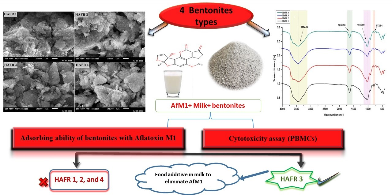
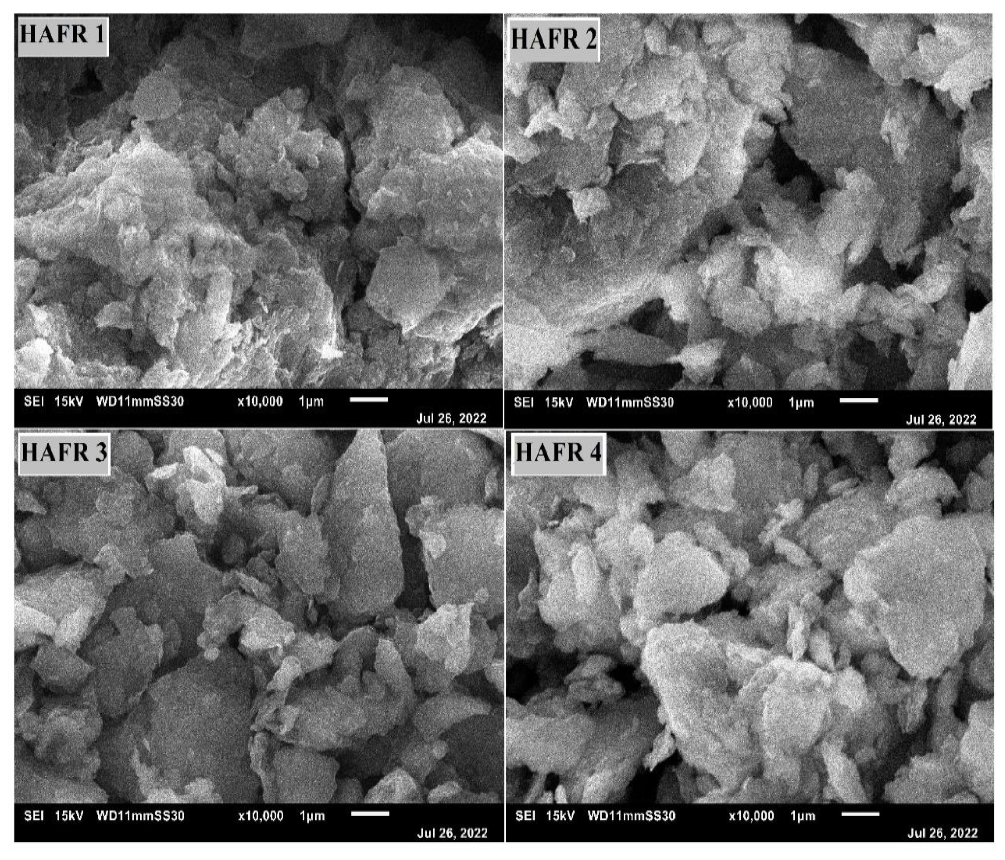
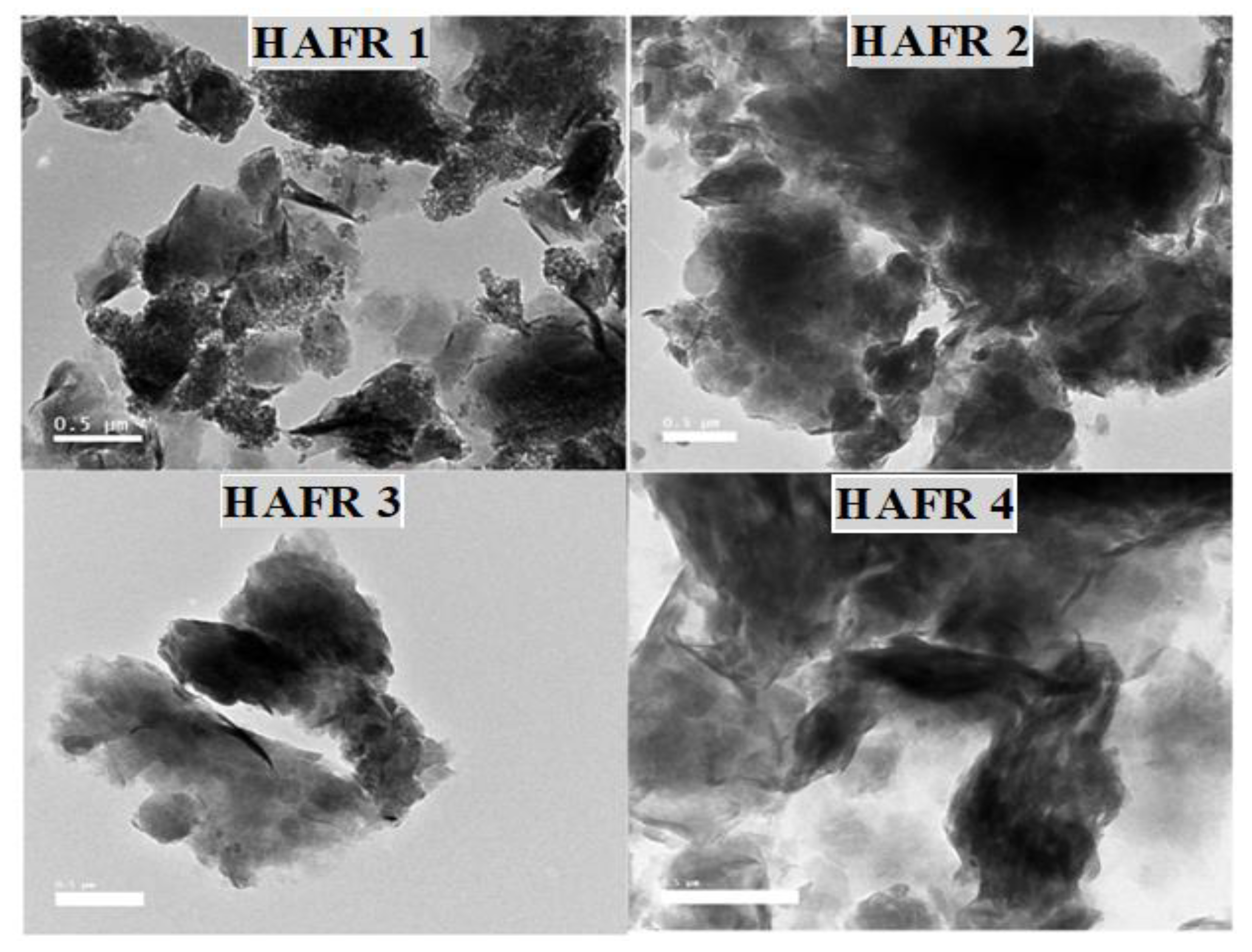
2.1. Spectroscopic and Microscopic Characterization of Bentonites

In the present research, FTIR has been utilized to detect the chemical aspects of resulted peak and settle efficacious enclosure of all the tested components of the four examined bentonites across the spectrum ranging between 972–3442 cm−1. In Figure 1, infrared absorption spectra show the typical peaks of C=C, C=N and C-H in ring structure at 972 cm−1, C-O of carbohydrate at 1035 cm−1 and acetylated amide at 1753 cm−1. Whereas, the typical peaks are stated as C-O stretching of amide I at 1638 cm−1, OH of carbohydrates, proteins and polyphenols at 3442 cm−1. Bentonites showed abroad band at 3442.15 cm−1 because of -OH- stretching band for inter-layer adsorbed water (water current in the mineral bentonites). This implies the opportunity of the hydroxyl linkage among octahedral and tetrahedral layers. From the current findings, we noticed that a very sharp band detected at 1638.98 cm−1 is due to the asymmetric -OH- stretch (deformation mode) of water and a structural part of the mineral, while bands at 1035.09 cm−1 and 972.94 cm−1 were formed by the stretching mode of Si-O (out-of-plane) and Si-O stretching (in-plane) vibration for layered silicate.
Scanning electron microscopy (SEM) and transmission electron microscopy (TEM) were applied to investigate the surface morphology of bentonites. The four bentonites were identified by scanning electron microscopy as being similar in appearance and having sizes, which ranged between 100 and 200 nm. The obtained SEM results presented in (Figure 2) revealed a significantly rougher surface, obstructed with few cracks. These types of surfaces indicate that the fracture toughness of the four examined bentonites could be improved. Images taken with a scanning electron microscope reveal shape irregularities and the dispersion of clay minerals, which may primarily be silica and alumina.
Concerning TEM analysis (Figure 3), it was observed that the diameters of the bentonites range from 100 to 200 nm. The particles’ surface looks like graphite particles with rough borders. The particles are aggregated due to their chemical structure, which harbors many elements, for instance NA, Mg, Al, Si, Cl, K, Ca and Ti, as shown in Table 1. The ratio of the elements Si, Al and Na was high. Meanwhile, Cl was absent in HAFR 1 and 3, and Ti was absent in HAFR 1 (Table 1). This consequence shows the existence of alumina and silica as major ingredients along with traces of Fe, Mg, Na, K, Ti and Ca. The current findings are in line with those reported by various recent studies [26,27,28,29].
The massive changes in the internal surface area between the different types of bentonites (HAFR 3 and 1) are considered the effective parameter that provides abundant sites for attachment. These sites are capable of binding with the AFM1 as a protein and work to diminish it from the treated samples under this investigation. According to Pеtrovi et al. [30], bentonite’s adsorption properties depend mainly on its chemical structures and mineralogical structure, along with its textural and morphological features. Additionally, these minerals have numerous applications in various industrial processes due to their properties, which include surface area, surface charges, the type of exchangeable cations, hydroxyl groups on the edges, charge density, Lewis and Bronsted acidity and silanol groups of crystalline defects or broken surfaces [31]. Thus, these studies confirm our finding that the adsorption ability of bentonites towards AFM1 is dependent mainly on the bentonite’s properties; thus, in the current study, only two bentonites among the four examined showed high activity in aflatoxin removal compared with the other two bentonites. Moreover, the existence of calcium in the four examined bentonites was very low; therefore, it is recommended that these bentonites be used for a different purpose, as Calcium bentonite is less commonly used, as reported by studies on the impact of different bentonite complexes on the removal of aromatic compounds, which is an important indicator that the composition of the used bentonite could be a marker for its removal activity.
Moreover, recent research concluded that bentonites offer great potential to control aflatoxins and improve food and dairy safety and quality [32,33]. Conversely, the AFM1 distribution in milk has non-homogenous aspects, and a significant amount is bound to the milk protein (casein) [34]; this highlight makes the mechanism of adsorption more complex by various bentonite HAFR types. Therefore, the selection of the HAFR type according to its properties provides great potential for adsorbing AFM1 in milk.

2.2. Concentration and Frequency of Detection of Aflatoxins M1 in Raw Milk Samples Using HPLC

Several studies have shown that most raw milk may be contaminated with some types of aflatoxins, especially aflatoxin M1 [11,35]. Many physical, chemical and biological procedures have been applied to get rid of these toxins and their effects, but no methods have succeeded at a satisfactory rate. Therefore, in this study, we tried to use a method to get rid of aflatoxin in milk using four types of Egyptian bentonites. Data presented in Table 2 illustrate that the percentage of AFM1 found in cow, camel, sheep and goat milk samples were 88, 80, 86.7 and 93.3%, with mean values of 54.7, 87.5, 168.4 and 237.1 ng/L, respectively. European Commission No 1881/2006 established that the highest residue amount of AFM1 must not be more than 50 ng/L; though 54, 70, 73.3 and 86.7%, respectively, of the examined cow, camel, sheep and goat milk samples were above the level recommended by European Commission.
The greatest amount of contamination was observed in goat milk (490.3 ng/L), with a range of 5.60–490.30 ng/L and a mean value of 237.08 ng/L. These findings concurred with the findings acquired by de Matos et al. [35], who reported that 100% of examined Brazilian goat milk was contaminated with AFM1, with a mean value of 21.90 ± 10.28 ng/L. In addition, Omar [11] discovered that sheep milk had the greatest levels of AFM1 contamination, and recorded a value of 137.18 ng/kg, with a mean value of 70.25 ± 14.85 ng/kg.
The present study’s results are highly congruent with those reported by Elzupir and Elhussein [36], who observed that 95.45% of cow milk produced in Khartoum city, Sudan was contaminated with AFM1. Similarly, the lower incidence of aflatoxin in camel milk was found by Hussain et al. [37], who investigated AFM1 contamination in milk from five dairy species in Pakistan and discovered that camel milk was free from AFM1. They also discovered that 37.5, 20 and 16.7% of the examined cow, goat and sheep milk samples were contaminated with AFM1.

2.2.1. Adsorption Capability of AFM1 in PBS Solution by Various Bentonite HAFR Types

For aflatoxins M1 adsorption capability via different bentonite concentrations, Table 3 displays AFM1 adsorption by different ratios of bentonite (0.5 and 1 g) in PBS. AFM1 adsorption was influenced by applying bentonite (0.5 and 1 g) in a concentration-dependent manner over different durations of time, i.e., 0, 0.5, 1, 2, 3, 6 and 12 h. Residual levels of AFM1 reached their lowest value of 0 ng/L in HAFR 3 (1 g bentonite), followed by 1.5 ng/L with HAFR 1 (1 g bentonite) and 2 ng/L with HAFR 2 (1 g bentonite). In details and with HAFR 3, they were 100 ng/L at 0 h > 60 ng/L at 0.5 h > 38 ng/L at 1 h >18 ng/L at 2 h > 5 ng/L at 3 h > 1 ng/L at 6 h >0 ng/L at 12 h. The highest adsorption percentage of AFM1 reached the maximum level after 12 h to 100, 98.5 and 98% while using bentonites HAFR 3, 1 and 2, respectively (Table 4).
Adsorbent concentration has an impact on adsorption mechanisms and can alter the dynamic sites available for mycotoxin adsorption. The maximum AFM1 adsorption capacity in this investigation was attained by employing a high concentration of 1 g of bentonite (HAFR 3, then HAFR 1). This indicates that when more bentonite was added to the milk, the amount of AFM1 that was adsorbed increased. These data are in agreement with the findings of Applebaum and Marth [38], who reported that 0.4 g of bentonite per 20 mL of naturally contaminated raw whole milk (at 25 °C for 1 h) was sufficient to accomplish the maximal removal of AFM1 (89%) by bentonite. Moreover, according to Jaynes et al. [39], bentonite can swell up to six times its original size when activated by water, which makes it extremely absorbent and useful for drawing out pollutants.
Time response is a physicochemical mechanism of adsorption that involves moving weights from liquid to solid surfaces [40]. AFM1 was quickly sequestered by bentonite in this investigation, especially at higher doses, with the adsorption percentage peak accumulating after 12 h, according to the adsorption versus time plots. Adsorption behavior revealed that AFM1 was rapidly absorbed in the first 0.5 h and then grew progressively until reaching its peak after 12 h. These findings might support the idea that there are enough vacant adsorption sites open to AFM1 molecules throughout the earliest stages of their reactivity with bentonite up until total adsorbent surface saturation. The subsequent decrease in AFM1 is brought on by the repulsive forces between molecules linked to active regions on the surface of the bentonite [41].
Results presented in Table 5 demonstrate the AFM1 adsorption capability obtained by (1 g bentonite) HAFR 1 and 3 in milk, where no adsorption was observed for untreated milk and all controls and positive control continually measured 100 ng/L AFM1 over different time intervals. After using HAFR 3, the concentration of AFM1 reached the lowest level after 12 h at 1.5 ng/L, followed by HAFR 1, which reached 5 ng/L after 12 h. Collectively, the adsorption percentage for HAFR 3 (98.5%) was relatively higher than that obtained for HAFR 1 (95%), as displayed in Table 6. Our results agreed with Abdel-Wahhab and Kholif [42], who claimed that supplementing early lactating goats with sodium bentonite 1% can significantly reduce the amount of AFM1 in the milk. Furthermore, bentonite, one of the well-known absorbents, can lower the levels of aflatoxin in milk owing to its wide variety and stable way of binding to AFM1 [43]. According to Montaseri et al. [44], milk from cows and goats fed a diet treated with bentonite had a considerably lower AFM1 content.

2.3. Effect of Bentonites on Qualitative Characteristics of the Milk Samples

Regarding the qualitative aspects of the milk samples, Table 7 illustrates the impact of 1 g bentonites (HAFR 1, 2, 3 and 4) on the chemical properties of milk. We can notice that there is an increase in fat, protein and SNF content while using HAFR 3 and 4, but a decreasing trend in lactose was observed compared with negative control. The milk composition of treated milk with HAFR 3 and HAFR 4 was significantly increased in fat and SNF compared with the control, while protein and lactose were reduced according to different bentonite concentrations. The action of bentonite on milk composition may be due to the alterations that occur in milk composition after the treatment with bentonites. Our findings agreed with Awasthi [45], who reported that bentonite may lower milk proteins. Hence, Abdel-Wahhab and Kholif [42] demonstrated that supplementing early lactation goats with 1% bentonite had no effect on the milk’s composition. Another study postulated that bentonite has a low influence on the nutritional characteristics of milk [46].

2.4. Cytotoxicity Evaluation of Bentonites

The estimation of cytotoxicity of bentonite and IC50 (µg/mL) is presented in Table 8. The type of bentonite and its concentration had an impact on the variation in inhibition. At a maximal concentration of 500 µg/mL, bentonite was more cytotoxic, with 100% inhibition and zero viability of the cell in all four examined types. Most cells are still alive at the smallest concentration (0.97 µg/mL) of bentonite, with a very low inhibitory percentage (2 and 6%) for HAFR 1 (98%) and HAFR 3 (94%), respectively. The required level of four types of bentonites for 50% inhibition of cell (IC50) was 57.1, 16.5, 11.35 and 6.92 for HAFR 1, 2, 4 and 3, respectively. This means that HAFR 1 is the most toxic and HAFR 3 is the least cytotoxic. Furthermore, the maximum and minimum toxic constituents are intended to be detected by the cytotoxic evaluation. To select the initial bentonite dosage for cytotoxic activity, the neutral red assay can estimate IC50 [19]. In this study, bentonite was exceedingly hazardous at the highest dosage of 500 µg/mL, while being comparatively non-toxic at the lowest concentration of 0.97 µg/mL. At a dosage of 1.95 µg/mL, there was minimal cytotoxicity. This result agrees with that of Zhang et al. [47], who discovered that the bentonite cytotoxicity on human B lymphocytes increases with an increase in bentonite dose and acquaintance time. The ability of HAFR 1 and 3 to be employed as non-toxic candidates is improved by their low cytotoxicity. The variation of cytotoxicity among the four examined bentonites may be due to the differences in their chemical structures, particle sizes and surface etiologies. The surface characteristics, morphology, and chemical structures of the adsorbent all affect how toxicant induction occurs in cell lines [48]. For studying the chemical, surface and morphological characterization of the four examined bentonites, XRD, FTIR, SEM and TEM will be discussed in the following sections.

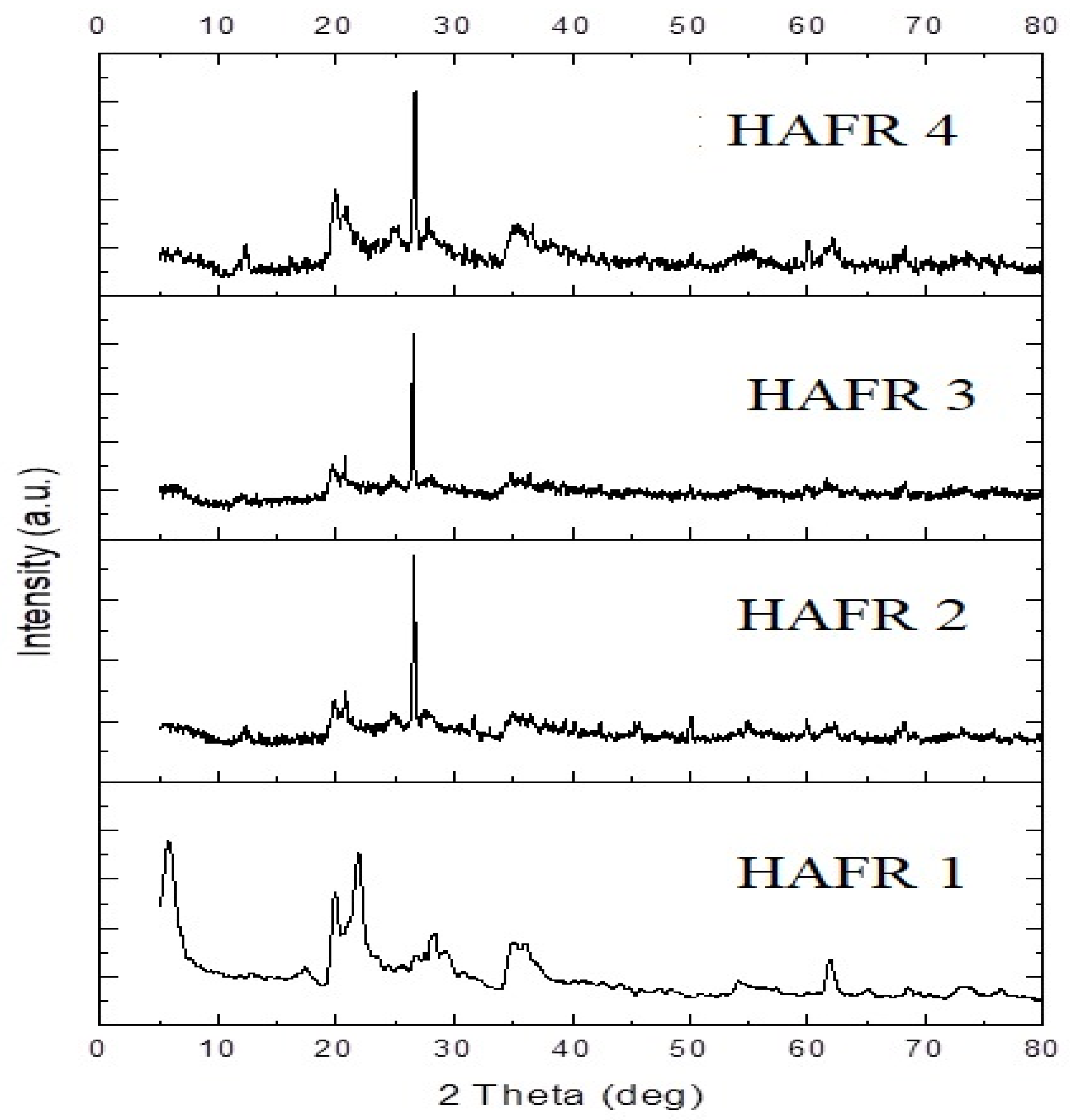
2.5. X-ray Diffraction of Different Types of Bentonites

Figure 4 illustrates the characteristics of crystalline peaks (2 theta) of the four tested bentonites assessed by XRD powder analysis, and their reflections were recorded as follows: 20.2 = (101), 28.6 = (111), 40.23 = (200) and 60.43 = (220). However, the second beak was moved to the left in sample number 1, and was found at 23.2° instead of 28.6°, as shown in the other three bentonites. Samples’ basal reflections at angles (i.e., 20.20°, 28.6°, 40.23° and 60.43°) were observed, which are associated with the presence of impurities, such as aflatoxin, in samples. As shown in Figure 4, bentonite nanoparticles have numerous phases of low quartz and anortithe. This deflection peak has an index miller as follows: 101, 111, 200 and 220. Moreover, it displays that both samples have the phase of low quartz with trigonal structures and anortithe with triklin structures [49]. Therefore, the residual spreading peaks may be because of the crystalline structure and the interaction of the utilized C. colocynthis components [50].

3. Conclusions

The adsorption efficiency of various bentonites with aflatoxin M1 in milk was highlighted. In addition, the X-Ray, spectroscopic and microscopic characteristics of bentonites were assessed. Among the tested milk samples, goat milk was heavily contaminated with AFM1. HAFR 3 (1 g bentonite) had higher adsorption efficiency than other bentonite kinds used in phosphate buffer saline and milk, followed by HAFR 1. With regard to the impact of bentonite on the nutritional characteristics of milk, HAFR 3 and 4 caused moderate changes in fat, protein and SNF. On the other hand, lactose concentration decreased in comparison with the control. Due to their structure and their pH features, HAFR 3 and HAFR 1 demonstrated a high capability to adsorb high amounts of aflatoxin M1. Therefore, the current study confirms that bentonite’s adsorption properties depend on its chemical and mineralogical composition. Based on our results, HAFR 3 and HAFR 1 could be used as promising potential additives to decrease the risks of aflatoxin contamination in raw milk or other fresh food products.

4. Materials and Methods

4.1. Chemicals and Reagents

AFM1 standard solutions (50 µg mL−1 in acetonitrile) were obtained from (Sigma-Aldrich, Dorset, UK). HPLC-grade acetonitrile and methyl alcohol were purchased from (Sigma-Aldrich, Steinheim, Germany). Raw milk samples were acquired from local farms in various regions of Alexandria Governorate, Egypt. The preliminary milk sample was prepared in aliquots of 50 mL and then stored at −20 °C for further analysis.

4.2. Sampling

A total of 100 samples of raw milk from i.e., cow (n = 50), camel (n = 20), sheep (n = 15) and goat (n = 15) were randomly collected within 5 months, obtained from November 2021 to March 2022 from different regions of Alexandria City-Egypt for AFM1 analysis. All samples were transported to the laboratory in an icebox at 2–4 °C. The milk samples were sterilized prior to AFM1 contamination at 140 °C for 2 s. An measurement of 30 mL milk was placed in a dark flask and kept refrigerated until analysis.

Sample Preparation

For milk preparation, the samples were centrifuged (ThermoFisher Scientific Co., Cairo, Egypt) at 3500× g for 10 min at 10 °C. The upper phase was removed by a Pasteur pipette for further testing [51].

4.3. Cleanup/Purification and HPLC Conditions

The extraction of AFM1 from milk was accomplished according to the procedure of Iqbal et al. [52], with slight modifications, and the test kit of immunoaffinity columns was also utilized. The samples were first prepared (see Section 2.2.1). The extracts were then filtered with No. 5 Whatman filter paper, and approximately 50 mL of the sample was passed through an AflaTest immunoaffinity column at a rate ranging between 1–3 mL/min, then washed with 10 mL of deionized water (Milli Q Siemens, Ultra Clear, UV UF TM, Munich, Germany). Further, the bound AFM1 was eluted with acetonitrile reagent (1.5–3.0 mL). Finally, the residue was evaporated (40 °C) with a nitrogen stream. The prepared samples were placed in a dark place for (15 min) at RT. Then, 200 μL of acetonitrile was added to the vials. A 20 μL portion of the solution was subjected to LC analysis. The HPLC system (Agilent 1100 series system, Agilent Technologies, Hewlett-Packard, Waldbronn, Germany) included a model G1311A gradient delivery pump with G1321A fluorescence detector set at ƛʎ λex 360 and λem 430 nm. A Zorbax Eclipse XDB-C8 analytical column (4.6 mm × 150 mm, 5 µm; Agilent Technologies, Santa Clara, CA, USA). The mobile phase was isocratic water: acetonitrile: MeOH (68:24:8, v/v/v), and all separations were conducted at a flow rate of 1.0 mL/min [53]. The calibration curve on HPLC was carried out using several concentrations of AFM1. The detection level limit was 1.4006, hence, the limit of quantification was 1.8116. On the other hand, the AFM1 linear range ranged from 0.1–100 ng/mL.

4.4. Adsorption of AFM1 in Phosphate Buffer Saline (PBS) by Different Types of Bentonite Samples

AFM1 was used as a contaminant solution. The PBS of 100 ng/L AFM1 was exposed to numerous amounts of bentonite (obtained from Egypt’s New Borg El Arab bentonite group) equal to 0.5 and 1 g for several durations i.e., 0, 0.5, 1, 2, 3, 6 and 12 h. Different quantities of bentonite (HAFR 1, HAFR 2, HAFR 3 and HAFR 4) were added to PBS (15 mL) of AFM1 (100 ng/L). The adsorption assessments were performed with constant agitation at RT [19,51]. The adsorbent was separated using a centrifuge, and later, the supernatant was utilized for HPLC analysis, as designated in Section 2.3. The AFM1 removal efficiency (RE) was calculated by the following equation (Equation (1)):
RE (%) = (C0 − C1)/C0 × 100
where RE (%) signifies of AFM1 removal ratio, and C0 and C1 are the concentration of AFM1 before and after adsorption, correspondingly.

4.5. Adsorption of AFM1 in Milk Samples by Bentonite (HAFR 1 and HAFR 3)

The bentonite (HAFR 1 and HAFR 3) was utilized to determine of AFM1 adsorption level in milk samples contaminated with AFM1 (100 ng/L). The amounts of 1 g of bentonite (HAFR 1 and HAFR 3) were added to 30 mL of contaminated milk. The dispersions were shaken vigorously for 12 h. The samples were centrifuged, and the upper part of the milk samples was investigated. The AFM1 quantities were examined by HPLC (HPLC–FLD) as clarified in Section 2.3. [19,54].

4.6. Qualitative Properties of the Tested Milk

Protein, lactose, fat and solid not fat (SNF) ratio in the milk samples was measured using Milk Scan (Broker Optik GMBH, MIRA model, Lenggries, Germany). To contaminate the milk, 100 ng/L of AFM1 was added. Moreover, the adsorbent equal to 1 g was considered to evaluate the adsorption rate for different types of bentonites compared to the positive and negative control [54].

4.7. Cytotoxicity Assessment of Bentonites

Cell viability was examined using peripheral blood mononuclear cells (PBMCs) maintained in the RPMI medium. The PBMCs approach utilizes cells that are isolated from multiple individuals, and could provide a high assessment of the cytotoxicity of drugs in vitro. Moreover, PBMCs cell types provide a principal reflection into how immune cell from several donors respond to the candidate constituents in development. Blank wells (150 µL PBS), control wells (150 µL PBMCs) and tested wells (150 µL PBMCs) were placed on a microtiter plate. Bentonite at several ratios was added to test wells and incubated (New Brunswick™ Galaxy® 170 R CO2 Incubator Series, Eppendorf, Madrid, Spain) for 24 h. Consequently, neutral red (150 µL) was added, and wells were incubated at 37 ℃ for 2 h, then, cells were washed, and plates were shaken with a destaining solution containing (1% acetic acid; 49% deionized water; 50% ethanol; 50 µL/well). Absorbance was determined at 540 nm in a UV-Vis spectrophotometer (PG Instrument Ltd. UK) [55]. The inhibition (%) was assessed by the following Equation (2):
The inhibition (%) = 100 − (O.D Control − O.D Treatment)/(O.D Control)
where: O.D = Optical density; IC50 values were calculated online (www.aatbio.com/tools/ic50-calculator), (Accessed on 25 July 2022).

4.8. Bentonite Characterization

4.8.1. X-ray Diffraction (XRD) Analysis

X-ray powder diffractometry was performed by (Schimadzu 7000 diffractometer, Kyoto, Japan) to determine the structure of the four several types of bentonites (HAFR 1, 2, 3 and 4).

4.8.2. Fourier-Transform Infrared (FTIR)

The FTIR apparatus (Bruker Tensor II FT-IR Spectrometer) was utilized with a resolution (cm−1) with a scan range of 4000–650 cm−1. The resulting spectrum represents molecular absorption, which generates a molecular fingerprint of the four various bentonite types (HAFR 1, 2, 3 and 4). The FTIR approach was conducted by mixing ethanol drops with some particles of bentonites on a glass slide, then placing it on a heater plate at 150 °C for 1 min to obtain a thin layer surface. The ready samples were kept in cold places and dark conditions before the analysis.

4.8.3. Scanning Electron Microscopy (SEM)

The SEM is employed to scan a finely focused electron beam across the surface of a specimen. Specimens were magnified to 300,000×, while maintaining a large depth of focus. The ease of sample scanning by SEM (JEOL JSM 6360LA, Tokyo, Japan) over large distances is quite appealing, in that a large sample viewing area is first surveyed at generally low magnification to seek out particular areas of interest, followed by the high magnification of those specific areas for subsequent detailed investigations.

4.8.4. Transmission Electron Microscope (TEM)

High-resolution images were examined by TEM (a JEOL JEM 2100, Tokyo, Japan) operating at 200 kV. The particle size of the four various types of bentonites (HAFR 1, 2, 3 and 4) was acquired by examining TEM images via Image software (AutoTEM 5 Software) [56].

4.9. Data Analysis

The obtained data were evaluated by the SPSS statistical package, ver. 23 (IBM SPSS Statistics for Windows, IBM Corp., Armonk, NY, USA). The means ± standard deviation (SD) was utilized to express the data. A one-way analysis of variance (ANOVA) was employed applying the Duncan test for data analyses at a significant level (p < 0.01). One-way ANOVA test was used to compare the means of two or more independent groups to determine whether there was statistical evidence that the associated population means were significantly different.

Author Contributions

Conceptualization, G.M.H.; Methodology, G.M.H.; Software, G.M.H., H.S.A.E.-M., M.G.A., O.S.E.O., M.I.E.-T., T.M., Y.E.-H., M.M.F.A., M.S.S., S.A.K., E.E.H., A.A. and E.A.; Validation, G.M.H., T.M., S.A.K. and S.A.I.; Formal analysis, G.M.H., H.S.A.E.-M., M.G.A., O.S.E.O., M.I.E.-T., T.M., Y.E.-H., M.M.F.A., M.S.S., S.A.K., E.E.H., A.A. and E.A.; Investigation, T.M.; Resources, G.M.H. and T.M.; Data curation, T.M., S.A.K. and S.A.I.; Writing–original draft, G.M.H. and T.M.; Writing – review & editing, T.M., S.A.K. and S.A.I.; Visualization, G.M.H., H.S.A.E.-M., M.G.A., O.S.E.O., M.I.E.-T., T.M., Y.E.-H., M.M.F.A., M.S.S., S.A.K., S.A.I., E.E.H., A.A. and E.A.; Supervision, G.M.H.; Project administration, G.M.H., T.M., S.A.K. and S.A.I.; Funding acquisition, T.M., S.A.K. and S.A.I. All authors have read and agreed to the published version of the manuscript.

Funding

This research received no external funding.

Institutional Review Board Statement

Not applicable.

Informed Consent Statement

Not applicable.

Data Availability Statement

The data presented in this study are available on request from the corresponding author.

Conflicts of Interest

The authors declare no conflict of interest.

References

  1. World Health Organization. Aflatoxins. In Food Safety Digest; WHO: Geneva, Switzerland, 2018. [Google Scholar]
  2. Petrova, P.; Arsov, A.; Tsvetanova, F.; Parvanova-Mancheva, T.; Vasileva, E.; Tsigoriyna, L.; Petrov, K. The Complex Role of Lactic Acid Bacteria in Food Detoxification. Nutrients 2022, 14, 2038. [Google Scholar] [CrossRef] [PubMed]
  3. Hamad, G.M.; Mehany, T.; Simal-Gandara, J.; Abou-Alella, S.; Esua, O.J.; Abdel-Wahhab, M.A.; Hafez, E.E. A review of recent innovative strategies for controlling mycotoxins in foods. Food Control 2023, 144, 109350. [Google Scholar] [CrossRef]
  4. Awuchi, C.G.; Ondari, E.N.; Ogbonna, C.U.; Upadhyay, A.K.; Baran, K.; Okpala, C.O.R.; Korzeniowska, M.; Guiné, R.P. Mycotoxins affecting animals, foods, humans, and plants: Types, occurrence, toxicities, action mechanisms, prevention, and detoxification strategies—A revisit. Foods 2021, 10, 1279. [Google Scholar] [CrossRef]
  5. Yang, C.; Song, G.; Lim, W. Effects of mycotoxin-contaminated feed on farm animals. J. Hazard. Mater. 2020, 389, 122087. [Google Scholar] [CrossRef] [PubMed]
  6. Campagnollo, F.B.; Ganev, K.C.; Khaneghah, A.M.; Portela, J.B.; Cruz, A.G.; Granato, D.; Corassin, C.H.; Oliveira, C.A.F.; Sant’Ana, A.S. The occurrence and effect of unit operations for dairy products processing on the fate of aflatoxin M1: A review. Food Control 2016, 68, 310–329. [Google Scholar] [CrossRef]
  7. Durak-Dados, A.; Michalski, M.; Osek, J. Histamine and other biogenic amines in food. J. Vet. Res. 2020, 64, 281–288. [Google Scholar] [CrossRef]
  8. Patyal, A.; Gill, J.P.S.; Bedi, J.S.; Aulakh, R.S. Potential risk factors associated with the occurrence of aflatoxin M1 in raw milk produced under different farm conditions. J. Environ. Sci. Health. Part. B 2020, 55, 827–834. [Google Scholar] [CrossRef] [PubMed]
  9. Nguyen, T.; Flint, S.; Palmer, J. Control of aflatoxin M1 in milk by novel methods: A review. Food Chem. 2020, 311, 125984. [Google Scholar] [CrossRef] [PubMed]
  10. Hamad, G.M.; Amer, A.; El-Nogoumy, B.; Ibrahim, M.; Hassan, S.; Siddiqui, S.A.; EL-Gazzar, A.M.; Khalifa, E.; Omar, S.A.; Abou-Alella, S.A.; et al. Evaluation of the Effectiveness of Charcoal, Lactobacillus rhamnosus, and Saccharomyces cerevisiae as Aflatoxin Adsorbents in Chocolate. Toxins 2023, 15, 21. [Google Scholar] [CrossRef] [PubMed]
  11. Omar, S.S. Aflatoxin M1 levels in raw milk, pasteurised milk and infant formula. Ital. J. Food Saf. 2016, 5, 5788. [Google Scholar] [PubMed] [Green Version]
  12. Huff, W.D. K-bentonites: A review. Am. Mineral. 2016, 101, 43–70. [Google Scholar] [CrossRef]
  13. Nones, J.; Nones, J.; Poli, A.; Trentin, A.G.; Riella, H.G.; Kuhnen, N.C. Organophilic treatments of bentonite increase the adsorption of aflatoxin B1 and protect stem cells against cellular damage. Colloids Surf. B. 2016, 145, 555–561. [Google Scholar] [CrossRef]
  14. Zhou, H. Mixture of palygorskite and montmorillonite (Paly-Mont) and its adsorptive application for mycotoxins. Appl. Clay. Sci. 2016, 131, 140–143. [Google Scholar] [CrossRef]
  15. Rushing, B.R.; Selim, M.I. Aflatoxin B1: A review on metabolism, toxicity, occurrence in food, occupational exposure, and detoxification methods. Food Chem. Toxicol. 2019, 124, 81–100. [Google Scholar] [CrossRef]
  16. Giovati, L.; Magliani, W.; Ciociola, T.; Santinoli, C.; Conti, S.; Polonelli, L. AFM₁ in Milk: Physical, biological, and prophylactic methods to mitigate contamination. Toxins 2015, 7, 4330–4349. [Google Scholar] [CrossRef] [Green Version]
  17. Fleischmann, M.; Pletcher, D. Industrial Applications. In Electrochemistry in Research and Development; Kalvoda, R., Parsons, R., Eds.; Springer: Boston, MA, USA, 1985; pp. 261–281. [Google Scholar]
  18. Di Gregorio, M.C.; Neeff, D.V.D.; Jager, A.V.; Corassin, C.H.; Carão, Á.C.D.P.; Albuquerque, R.D.; Azevedo, A.C.D.; Oliveira, C.A.F. Mineral adsorbents for prevention of mycotoxins in animal feeds. Toxin Rev. 2014, 33, 125–135. [Google Scholar] [CrossRef]
  19. Hamad, G.M.; El-Makarem, H.A.; Abd Elaziz, A.I.; Amer, A.A.; El-Nogoumy, B.A.; Abou-Alella, S.A. Adsorption efficiency of sodium & calcium bentonite for ochratoxin A in some Egyptian cheeses: An innovative fortification model, in vitro and in vivo experiments. World Mycotoxin J. 2022, 15, 285–300. [Google Scholar]
  20. Assaf, J.C.; Nahle, S.; Chokr, A.; Louka, N.; Atoui, A. Assorted methods for decontamination of Aflatoxin M1 in milk using microbial adsorbents. Toxins 2019, 11, 304. [Google Scholar] [CrossRef] [PubMed] [Green Version]
  21. Olopade, B.K.; Oranusi, S.U.; Nwinyi, O.C.; Lawal, I.A.; Gbashi, S.; Njobeh, P.B. Decontamination of T-2 toxin in maize by modified montmorillonite clay. Toxins 2019, 11, 616. [Google Scholar] [CrossRef] [PubMed] [Green Version]
  22. Kryuchkova, M.; Batasheva, S.; Akhatova, F.; Babaev, V.; Buzyurova, D.; Vikulina, A.; Volodkin, D.; Fakhrullin, R.; Rozhina, E. Pharmaceuticals removal by adsorption with montmorillonite nanoclay. Int. J. Mol. Sci. 2021, 22, 9670. [Google Scholar] [CrossRef] [PubMed]
  23. Agag, B.I. Prevention and control of mycotoxins in feeds. Assiut Univ. Bulletin. Environm. Res. 2003, 6, 149–166. [Google Scholar]
  24. Upadhaya, S.D.; Park, M.A.; Ha, J.K. Mycotoxins and their biotransformation in the rumen: A Review. Asian-Australas. J. Anim. Sci. 2010, 23, 1250–1260. [Google Scholar] [CrossRef]
  25. Nones, J.; Solhaug, A.; Eriksen, G.S.; Macuvele, D.L.P.; Poli, A.; Soares, C.; Trentin, A.G.; Riella, H.G.; Nones, J. Bentonite modified with zinc enhances aflatoxin B1 adsorption and increase survival of fibroblasts (3T3) and epithelial colorectal adenocarcinoma cells (Caco-2). J. Hazard. Mater. 2017, 337, 80–89. [Google Scholar] [CrossRef]
  26. Ikhtiyarova, G.A.; Özcan, A.S.; Gök, Ö.; Özcan, A. Characterization of natural- and organo-bentonite by XRD, SEM, FT-IR and thermal analysis techniques and its adsorption behaviour in aqueous solutions. Clay Miner. 2012, 47, 31–44. [Google Scholar] [CrossRef]
  27. Sani, N.; Abdulsalam, A.K.; Abdullahi, U.A. Extraction and quantification of silicon from silica sand obtained from zauma river, zamfara state, Nigeria. Eur. Sci. J. 2013, 99, 160–168. [Google Scholar]
  28. Deepracha, S.; Vibulyaseak, K.G.; Ogawa, M. Complexation of TiO2 with clays and clay minerals for hierarchically designed functional hybrids. Advanced Supramolecular Nanoarchitectonics; William Andrew Publishing: Norwich, NY, USA; Elsevier: Amsterdam, The Netherlands, 2019; pp. 125–150. [Google Scholar] [CrossRef]
  29. Chihi, R.; Blidi, I.; Trabelsi-Ayadi, M.; Ayari, F. Elaboration and characterization of a low-cost porous ceramic support from natural Tunisian bentonite clay. C. R. Chim. 2019, 22, 188–197. [Google Scholar] [CrossRef]
  30. Pеtrović, Z.; Dugić, P.; Аlеksić, V.; Bеgić, S.; Sаdаdinović, Ј.; Мićić, V.; Kljajić, N. Composition, structure and textural characteristics of domestic acid activated bentonite. J. Ergon. Soc. Korea 2014, 1, 133–139. [Google Scholar] [CrossRef]
  31. Djomgoue, P.; Njopwouo, D. FT-IR spectroscopy applied for surface clays characterization. J. Surf. Eng. Mater. Adv. Technol. 2013, 3, 275–282. [Google Scholar] [CrossRef] [Green Version]
  32. Jackson, R.S. Postfermentation treatments and related topics—ScienceDirect. In Wine Science, 5th ed.; Elsevier: Amsterdam, The Netherlands, 2020; pp. 573–723. [Google Scholar]
  33. Lubbers, S.; Charpentier, C.; Feuillat, M. Study of the binding of aroma compounds by bentonites in must, wine and model systems. Vitis 1996, 35, 59–62. [Google Scholar]
  34. Carraro, A.; De Giacomo, A.; Giannossi, M.L.; Medici, L.; Muscarella, M.; Palazzo, L.; Quaranta, V.; Summa, V.; Tateo, F. Clay minerals as adsorbents of aflatoxin M1 from contaminated milk and effects on milk quality. Appl. Clay Sci. 2014, 88–89, 92–99. [Google Scholar] [CrossRef]
  35. De Matos, C.J.; Schabo, D.C.; do Nascimento, Y.M.; Tavares, J.F. Aflatoxin M1 in Brazilian goat milk and health risk assessment. J. Environ. Sci. Health Part B 2021, 56, 415–422. [Google Scholar] [CrossRef]
  36. Elzupir, A.O.; Elhussein, A.M. Determination of aflatoxin M1 in dairy cattle milk in Khartoum State, Sudan. Food Control 2010, 21, 945–946. [Google Scholar] [CrossRef]
  37. Hussain, I.; Anwar, J.; Asi, M.R.; Munawar, M.A.; Kashif, M. Aflatoxin M1 contamination in milk from five dairy species in Pakistan. Food Control 2010, 21, 122–124. [Google Scholar] [CrossRef]
  38. Applebaum, R.S.; Marth, E.H. Use of sulphite or bentonite to eliminate aflatoxin M1 from naturally contaminated raw whole milk. Z. Lebensm. Unters. Forsch. 1982, 174, 303–305. [Google Scholar] [CrossRef]
  39. Jaynes, W.F.; Zartman, R.E.; Hudnall, W.H. Aflatoxin B1 adsorption by clays from water and corn meal. Appl. Clay. Sci. 2007, 36, 197–205. [Google Scholar] [CrossRef]
  40. Subhan, S.; Muhammad, Y.; Sahibzada, M.; Subhan, F.; Tong, Z. Studies on the selection of a catalyst–oxidant system for the energy-efficient desulfurization and denitrogenation of fuel oil at mild operating conditions. Energy Fuels 2019, 33, 8423–8439. [Google Scholar] [CrossRef]
  41. Ma, J.; Yu, F.; Zhou, L.; Jin, L.; Yang, M.; Luan, J.; Tang, Y.; Fan, H.; Yuan, Z.; Chen, J. Enhanced adsorptive removal of methyl orange and methylene blue from aqueous solution by alkali-activated multiwalled carbon nanotubes. ACS Appl. Mater. Interfaces 2012, 4, 5749–5760. [Google Scholar] [CrossRef]
  42. Abdel-Wahhab, M.A.; Kholif, A.E.-K.M. Mycotoxins in animal feeds and prevention strategies: A review. Asian J. Anim. Sci. 2008, 4, 113–131. [Google Scholar] [CrossRef]
  43. Asi, M.R.; Iqbal, S.Z.; Ariño, A.; Hussain, A. Effect of seasonal variations and lactation times on aflatoxin M1 contamination in milk of different species from Punjab, Pakistan. Food Control 2012, 25, 34–38. [Google Scholar] [CrossRef]
  44. Montaseri, H.; Arjmandtalab, S.; Dehghanzadeh, G.; Karami, S.; Oryan, A. Effect of production and storage of probiotic yogurt on aflatoxin M1 residue. J. Food Qual. Hazards Control 2014, 1, 7–14. [Google Scholar]
  45. Awasthi, V.; Bahman, S.; Thakur, L.K.; Singh, S.K.; Dua, A.; Ganguly, S. Contaminants in milk and impact of heating: An assessment study. Indian J. Public Health 2012, 56, 95–99. [Google Scholar]
  46. Naeimipour, F.; Aghajani, J.; Kojuri, S.A.; Ayoubi, S. Useful approaches for reducing aflatoxin M1 content in milk and dairy products. Biomed. Biotechnol. Res. J. 2018, 2, 94–99. [Google Scholar]
  47. Zhang, M.; Li, X.; Lu, Y.; Fang, X.; Chen, Q.; Xing, M.; He, J. Studying the genotoxic effects induced by two kinds of bentonite particles on human B lymphoblast cells in vitro. Mutat. Res. 2011, 720, 62–66. [Google Scholar] [CrossRef]
  48. Michel, C.; Herzog, S.; de Capitani, C.; Burkhardt-Holm, P.; Pietsch, C. Natural mineral particles are cytotoxic to rainbow trout gill epithelial cells in vitro. PLoS ONE 2014, 9, e100856. [Google Scholar] [CrossRef] [PubMed] [Green Version]
  49. Saravanakumar, A.; Peng, M.M.; Ganesh, M.; Jayaprakash, J.; Mohankumar, M.; Jang, H.T. Low-cost and eco-friendly green synthesis of silver nanoparticles using Prunus japonica (Rosaceae) leaf extract and their antibacterial, antioxidant properties. Artif. Cells Nanomed. Biotechnol. 2017, 45, 1–7. [Google Scholar] [CrossRef] [Green Version]
  50. Dahlous, K.A.; Abd-Elkader, O.H.; Fouda, M.M.G.; Al Othman, Z.; El-Faham, A. Eco-friendly method for silver nanoparticles immobilized decorated silica: Synthesis & characterization and preliminary antibacterial activity. J. Taiwan Inst. Chem. Eng. 2019, 95, 324–331. [Google Scholar]
  51. Murshed, S. Evaluation and Assessment of aflatoxin m1 in milk and milk products in yemen using high-performance liquid chromatography. J. Food Qual. 2020, 2020, 8839060. [Google Scholar] [CrossRef]
  52. Iqbal, S.Z.; Asi, M.R.; Jinap, S. Variation of aflatoxin M1 contamination in milk and milk products collected during winter and summer seasons. Food Control 2013, 34, 714–718. [Google Scholar] [CrossRef]
  53. Keskin, Y.; Baskaya, R.; Karsli, S.; Yurdun, T.; Ozyaral, O. Detection of aflatoxin M1 in human breast milk and raw cow’s milk in Istanbul, Turkey. J. Food Prot. 2009, 72, 885. [Google Scholar] [CrossRef]
  54. Jahanmard, E.; Keramat, J.; Nasirpour, A.; Emadi, R. Efficiency of calcined aluminum-magnesium layered double hydroxide for adsorption of aflatoxin m1 from solution and matrix of milk. J. Food Sci. 2021, 86, 5200–5212. [Google Scholar] [CrossRef]
  55. Bouffard, L.; Ryan, R.M.; Deci, E.L. Self-Determination Theory: Basic Psychological Needs in Motivation, Development and Wellness; Guilford Press: New York, NY, USA, 2017. [Google Scholar]
  56. El-Aassar, M.R.; Ibrahim, O.M.; Fouda, M.M.G.; El-Beheri, N.G.; Agwa, M.M. Wound healing of nanofiber comprising Polygalacturonic/Hyaluronic acid embedded silver nanoparticles: In-vitro and in-vivo studies. Carbohydr. Polym. 2020, 238, 116175. [Google Scholar] [CrossRef] [PubMed]
Figure 1. Fourier Transform Infrared (FTIR) of different types of bentonites.
Toxins 15 00107 g001
Figure 2. Scanning Electron Microscopy (SEM) of different bentonites (HAFR 1, HAFR 2, HAFR 3 and HAFR 4).
Toxins 15 00107 g002
Figure 3. Transmission Electron Microscope (TEM) of different bentonites (HAFR 1, HAFR 2, HAFR 3 and HAFR 4).
Toxins 15 00107 g003
Figure 4. X-Ray diffraction of different types of bentonites (HAFR 1, HAFR 2, HAFR 3 and HAFR 4).
Toxins 15 00107 g004
Table 1. Chemical composition of initial raw bentonites by transmission electron microscopy-energy dispersive X-ray spectroscopy.
Element (%)
NaMgAlSiClKCaTi
HAFR (1)0.301.928.7045.1300.770.910
HAFR (2)3.810.9313.8635.170.450.430.370.28
HAFR (3)0.631.6712.1826.9900.650.500.37
HAFR (4)0.080.100.450.950.050.050.050.07
Table 2. Concentration (ng/L) and frequency of detection of aflatoxins M1 in raw milk samples using HPLC.
All Raw SamplesContaminated Samples
SpeciesNMin–MaxMean (±SD)Median (Q1–Q3)N (%)N (%) > MRLMin–MaxMean (±SD)Median (Q1–Q3)
Cow50nd–122.8048.13 ± 34.5851.05 (14.60–76.95)44 (88)26 (54)3.70–122.8054.69 ± 31.5554.90 (25.47–79.80)
Camel20nd–142.4070.0 ± 50.3576.4 (9.82–115.37)16 (80)14 (70)6.50–142.4087.50 ± 39.7382.59 (66.67–127.55)
Sheep15nd–320.3145.93 ± 111.84140.60 (26.40–230.70)13 (86.66)11 (73.33)9.6–320.3168.38 ± 102.46160.10 (81.40–260.55)
Goat15nd–490.30221.28 ± 153.11234.50 (98.40–360.20)14 (93.33)13 (86.66)5.60–490.30237.08 ± 145.64242.90 (118.05–367.67)
P1 = 0.001, P2 = 0.002
N, Numbers of samples; Min-Max, minimum-maximum; ±SD, Standard deviation; Q1, 25th percentile; Q3, 75th percentile; nd, not detected; MRL, Maximum residue level as per the European Commission (EC) Regulation No 1881/2006 (EU); AFM1, 50 ng/L; nd, non-detectable. P1 = Probability values result from the nonparametric comparison among species (Kruskal–Wallis test) involving all samples. P2 = Probability values result from the comparison among frequencies (%) of detection (Fisher’s Exact test).
Table 3. Adsorption of AFM1 (100 ng/L) in PBS solution by various bentonite HAFR types (1, 2, 3 and 4).
Samples0 h0.5 h1 h2 h3 h6 h12 h
PBS (−Ve)0000000
PBS +(100 ng/L) AFM1 (+ve)100100100100100100100
PBS + (1 g) bentonite (HAFR 1)0000000
PBS + (1 g) bentonite (HAFR 2)0000000
PBS +(1 g) bentonite (HAFR 3)0000000
PBS + (1 g) bentonite (HAFR 4)0000000
PBS + (100 ng/L) AFM1+ (0.5 g) bentonite (HAFR 1)100837263555250
PBS +(100 ng/L) AFM1+ (1 g) bentonite (HAFR 1)1006544331041.5
PBS + (100 ng/L) AFM1+ (0.5 g) bentonite (HAFR 2)100827162555351
PBS + (100 ng/L) AFM1+ (1 g) bentonite (HAFR 2)100634121932
PBS + (100 ng/L) AFM1+ (0.5 g) bentonite (HAFR 3)100806859525048
PBS + (100 ng/L) AFM1+ (1 g) bentonite (HAFR 3)100603818510
PBS + (100 ng/L) AFM1+ (0.5 g) bentonite (HAFR 4)100887662555352
PBS + (100 ng/L) AFM1+ (1 g) bentonite (HAFR 4)1006851241164
(−Ve), negative; (+Ve), positive; PBS, phosphate buffer saline.
Table 4. Adsorption percentage (%) of AFM1 (100 ng/L) in PBS solution by different bentonite HAFR types (1, 2, 3 and 4) at various time intervals.
Samples0 h0.5 h1 h2 h3 h6 h12 h
Adsorption (%)
PBS (−Ve)0000000
PBS +(100 ng/L) AFM1 (+ve)0000000
PBS + (1 g) bentonite (HAFR 1)0000000
PBS + (1 g) bentonite (HAFR 2)0000000
PBS +(1 g) bentonite (HAFR 3)0000000
PBS + (1 g) bentonite (HAFR 4)0000000
PBS + (100 ng/L) AFM1+ (0.5 g) bentonite (HAFR 1)0172837454850
PBS +(100 ng/L) AFM1+ (1 g) bentonite (HAFR 1)0355677909698.5
PBS + (100 ng/L) AFM1+ (0.5 g) bentonite (HAFR 2)0182938454749
PBS + (100 ng/L) AFM1+ (1 g) bentonite (HAFR 2)0375979919798
PBS + (100 ng/L) AFM1+ (0.5 g) bentonite (HAFR 3)0203241485052
PBS+ (100 ng/L) AFM1+ (1 g) bentonite (HAFR 3)04062829599100
PBS + (100 ng/L) AFM1+ (0.5 g) bentonite (HAFR 4)0122438454748
PBS + (100 ng/L) AFM1+ (1 g) bentonite (HAFR 4)0324976899496
(−Ve), negative; (+Ve), positive; PBS, phosphate buffer saline.
Table 5. Adsorption of AFM1 (100 ng/L) in milk by bentonite HAFR (1 and 3) at various time intervals.
Samples0 h0.5 h1 h2 h3 h6 h12 h
Milk (−Ve)0000000
Milk +(100 ng/L) AFM1 (+ve)100100100100100100100
Milk + (1 g) bentonite (HAFR 1)0000000
Milk + (1 g) bentonite (HAFR 3)0000000
Milk + (100 ng/L) AFM1+ (1 g) bentonite (HAFR 1)1006746351375
Milk + (100 ng/L) AFM1+ (1 g) bentonite (HAFR 3)100634020751.5
(−Ve), negative; (+Ve), positive.
Table 6. Adsorption (%) of AFM1 (100 ng/L) in milk by bentonite HAFR (1 and 3).
Samples0 h0.5 h1 h2 h3 h6 h12 h
Adsorption (%)
Milk (−Ve)0000000
Milk +(100 ng/L) AFM1 (+ve)0000000
Milk + (1 g) bentonite (HAFR 1)0000000
Milk + (1 g) bentonite (HAFR 3)0000000
Milk + (100 ng/L) AFM1+ (1 g) bentonite (HAFR 1)0335465879395
Milk + (100 ng/L) AFM1 + (1 g) bentonite (HAFR 3)0376080939598.5
(−Ve), negative; (+Ve), positive.
Table 7. Qualitative properties of the milk sample (g /100 mL).
SamplesMean (SD)
FatProteinLactoseSNF
Milk (−Ve)0.55 b ± 0.0053.57 b ± 0.0014.91 a ± 0.0158.49 c ± 0.004
Milk +(100 ng/L) AFM1 (+ve)0.56 b ± 0.0113.56 b ± 0.0044.81 b ± 0.0118.51 b ± 0.00
Milk + (1 g) bentonite (HAFR 1)0.56 b ± 0.0043.58 b ± 0.0034.72 c ± 0.0128.56 a ± 0.003
Milk + (1 g) bentonite (HAFR 2)0.55 b ± 0.0123.54 c ± 0.0023.84 f ± 0.0058.48 c ± 0.001
Milk + (1 g) bentonite (HAFR 3)0.58 a ± 0.0053.59 a ± 0.0034.75 c ± 0.0158.52 b ± 0.002
Milk + (1 g) bentonite (HAFR 4)0.59 a ± 0.0013.61 a ± 0.0054.86 ab ± 0.0048.59 a ± 0.011
Milk + (100 ng/L) AFM1+ (1 g) bentonite (HAFR 1)0.54 c ± 0.0113.53 c ± 0.0044.83 b ± 0.0138.54 a ± 0.012
Milk + (100 ng/L) AFM1+ (1 g) bentonite (HAFR 2)0.53 c ± 0.0023.56 b ± 0.0124.67 d ± 0.028.56 a ± 0.003
Milk + (100 ng/L) AFM1+ (1 g) bentonite (HAFR 3)0.56 b ± 0.0013.54 c ± 0.0114.72 c ± 0.0038.55 a ± 0.004
Milk + (100 ng/L) AFM1+ (1 g) bentonite (HAFR 4)0.58 a ± 0.0163.58 b ± 0.0114.51 e ± 0.0048.56 a ± 0.006
(−Ve), negative; (+Ve), positive; ±SD, standard deviation; SNF, solid non-fat; Values means carrying a different superscript small letter on the same column are significantly different (p < 0.01).
Table 8. Estimation of cytotoxicity of bentonite and IC50 (µg/mL).
Concentration (µg/mL)HAFR (1)HAFR (2)HAFR (3)HAFR (4)
Inhibition %Viability %Inhibition %Viability %Inhibition %Viability %Inhibition %Viability %
5001000100010001000
2508515982991982
1257228928973919
62.5623888129468713
31.254456732786147723
15.62971564479217129
7.81684415966345842
3.9892366451495248
1.95694217942584357
0.9729810906943466
IC5057.116.56.9211.35
Disclaimer/Publisher’s Note: The statements, opinions and data contained in all publications are solely those of the individual author(s) and contributor(s) and not of MDPI and/or the editor(s). MDPI and/or the editor(s) disclaim responsibility for any injury to people or property resulting from any ideas, methods, instructions or products referred to in the content.

