Review

Methylation Markers in Cutaneous Melanoma: Unravelling the Potential Utility of Their Tracking by Liquid Biopsy

by
Valentina Aleotti
1,†,
Cristina Catoni
1,†,
Cristina Poggiana
1,*,
Antonio Rosato
1,2,*,
Antonella Facchinetti
1,2,‡ and
Maria Chiara Scaini
1,‡
1
Immunology and Molecular Oncology Unit, Veneto Institute of Oncology IOV-IRCCS, 35128 Padua, Italy
2
Department of Surgery, Oncology and Gastroenterology, Oncology and Immunology Section, University of Padua, 35128 Padua, Italy
*
Authors to whom correspondence should be addressed.
Equal contribution.
Joint senior authors.
Submission received: 15 November 2021 / Revised: 2 December 2021 / Accepted: 8 December 2021 / Published: 10 December 2021
(This article belongs to the Special Issue DNA Methylation Markers in Liquid Biopsies)

Abstract

:

Simple Summary

Malignant melanoma is the most lethal form of skin cancer. While new therapeutic approaches have improved survival in patients with metastatic melanoma, responses are rarely sustained due to the high degree of heterogeneity at the inter- and intra-metastatic levels. The development of reliable biomarkers to monitor therapeutic response and disease progression is critical. While attention has been focused on dissecting the molecular basis responsible for treatment resistance, it is clear that epigenetic changes warrant further in-depth investigation. Indeed, many aberrantly methylated genes play a role in cell cycle control, apoptosis, and cell invasion, as well as in melanoma progression. Longitudinal monitoring of DNA methylation via liquid biopsy can provide real-time information on the behavior and stage of melanoma.

Abstract

Malignant melanoma is the most serious, life-threatening form of all dermatologic diseases, with a poor prognosis in the presence of metastases and advanced disease. Despite recent advances in targeted therapy and immunotherapy, there is still a critical need for a better understanding of the fundamental mechanisms behind melanoma progression and resistance onset. Recent advances in genome-wide methylation methods have revealed that aberrant changes in the pattern of DNA methylation play an important role in many aspects of cancer progression, including cell proliferation and migration, evasion of cell death, invasion, and metastasization. The purpose of the current review was to gather evidence regarding the usefulness of DNA methylation tracking in liquid biopsy as a potential biomarker in melanoma. We investigated the key genes and signal transduction pathways that have been found to be altered epigenetically in melanoma. We then highlighted the circulating tumor components present in blood, including circulating melanoma cells (CMC), circulating tumor DNA (ctDNA), and tumor-derived extracellular vesicles (EVs), as a valuable source for identifying relevant aberrations in DNA methylation. Finally, we focused on DNA methylation signatures as a marker for tracking response to therapy and resistance, thus facilitating personalized medicine and decision-making in the treatment of melanoma patients.

1. Introduction

Malignant melanoma is the most lethal form of skin cancer, accounting for more than 59,000 deaths worldwide [1]. Indeed, although cutaneous melanoma is usually curable through surgical resection in patients with localized disease, the prognosis is less favorable in patients with regional metastases and advanced disease. Survival has historically been poor due to the infrequent response to conventional chemotherapy. However, in recent years, the systemic treatment of cutaneous melanoma has shifted dramatically as a result of the growing interest and focus on its genetic landscape. A combined approach, consisting of BRAF plus MEK inhibitors, has led to a significant improvement in overall survival (OS) for patients harboring a BRAF V600E/K mutation (35–50% of melanomas) [2,3,4]. Consequently, the definition of the tumor genetic landscape and the identification of molecular predictive factors have become critical for patients with unresectable disease and/or distant metastases [5]. Nonetheless, complete response to MEK/BRAF targeted therapy in approximately 50% of cases is only transitory, and great efforts have been made to characterize the mechanisms underlying the rise of resistance [6]. The primary impediment to achieving a durable response is the high degree of heterogeneity at the inter- and intra- metastatic levels. Therefore, it is now vital to develop reliable biomarkers to monitor therapy response and disease progression [7]. Apart from the effect given by their combined mechanisms of action, there is some evidence that BRAFi/MEKi treatment may promote a favorable immune microenvironment, thus enhancing the efficacy of immune checkpoint inhibition by anti-PD-1 drugs [8,9,10]. Nonetheless, even when used alone, immunotherapy with CTLA-4 and PD-1/PDL-1 blocking antibodies results in a durable response (approximately 2 years), but only in a subset of patients [11,12]. Although new therapeutic approaches have dramatically improved the survival of metastatic melanoma patients, the response is rarely sustained. Many efforts are nowadays concentrated on the dissection of the molecular basis putatively responsible for resistance to treatment, but the whole melanoma molecular landscape remains puzzling and warrants further investigation. Indeed, the extreme hypermutability and heterogeneity of the disease push toward a more complete and exhaustive description of the genomic landscape, able to define the dynamics of the systemic disease. Many studies using next-generation sequencing (NGS) have identified several genetic aberrations [13,14,15,16] that provide insights into the heterogeneity of melanoma, with putative implications for prognosis and therapy. NGS, indeed, can provide a comprehensive mutation profile, and thanks to the development and validation of numerous pipelines, it can successfully identify multiple target alterations with a reduced clinical reporting time [17,18]. Interestingly, the first comprehensive catalog of somatic mutations from an individual cancer, at the whole-genome level, concerned a melanoma cell line [19,20]. The catalog provided remarkable insights into the mutational signature, indicating the presence of a great number of mutations per Mb suggestive of DNA damage due to ultraviolet light exposure. Subsequent Whole Exome Sequencing (WES) studies, performed on clinical samples, demonstrated that NF1, ARID2, PPP6C, RAC1, SNX31, TACC1, and STK19 were significantly mutated in melanoma [13,14]. Moreover, in the era of precision medicine and targeted therapies, molecular subtyping of melanoma has become more and more important by going complementary to the traditional clinicopathological classification [21]. Great support in this direction has been given by the exome sequencing studies of the Cancer Genome Atlas Skin Cutaneous Melanoma (SKCM-TCGA) project that classified cutaneous melanoma into four distinct molecular subtypes: BRAF-mutant, NRAS-mutant, NF1-mutant, and triple-wild-type group [15]. Moreover, through exome sequencing, previously reported melanoma onco- and tumor suppressor- genes have been confirmed (BRAF, NRAS, CDKN2A, TP53, and PTEN), and several additional mutated melanoma genes have been identified (MAP2K1, IDH1, RB1, and DDX3X). In the triple wild-type group, low-frequency mutations were also detected in KIT, CTNNB1, GNA11, and GNAQ [15,20]. A large, high-coverage Whole Genome Sequencing (WGS) study of cutaneous, acral, and mucosal melanoma was set a few years later by Hayward and colleagues [16]. This work supported the involvement of the non-coding genome in melanoma pathogenesis and disclosed different tumorigenic processes among the different melanoma subtypes [16,20]. Interestingly, the number of genes affected by recurrent non-coding mutations (predicted to have a functional impact) was equivalent to that of genes affected by recurrent coding mutations. Interestingly, eight genes, in addition to TERT, showed potential driver promoter mutations (BLCAP, KBTBD8, NSUN6, RALY, RNF185, RPL29, RPS27, and ZNF778). Nonetheless, beyond the genetic landscape depicted above, extensive research into epigenetic modifications is also required to find additional targets and treatment strategies [22,23]. Aberrant promoter methylation, for example, can affect gene expression; its hyper- and hypo- methylation is associated with gene silencing or overexpression, respectively. As already documented, many aberrantly methylated genes play a role in cell cycle control, apoptosis, cell invasion, and, ultimately, melanoma progression [24,25]. One of its strengths is that methylation is a promising biomarker due to the stability of methylated CpG islands (CGIs) in comparison to the hypervariability of melanoma mutational profiles [26].

2. DNA Methylation and Melanoma

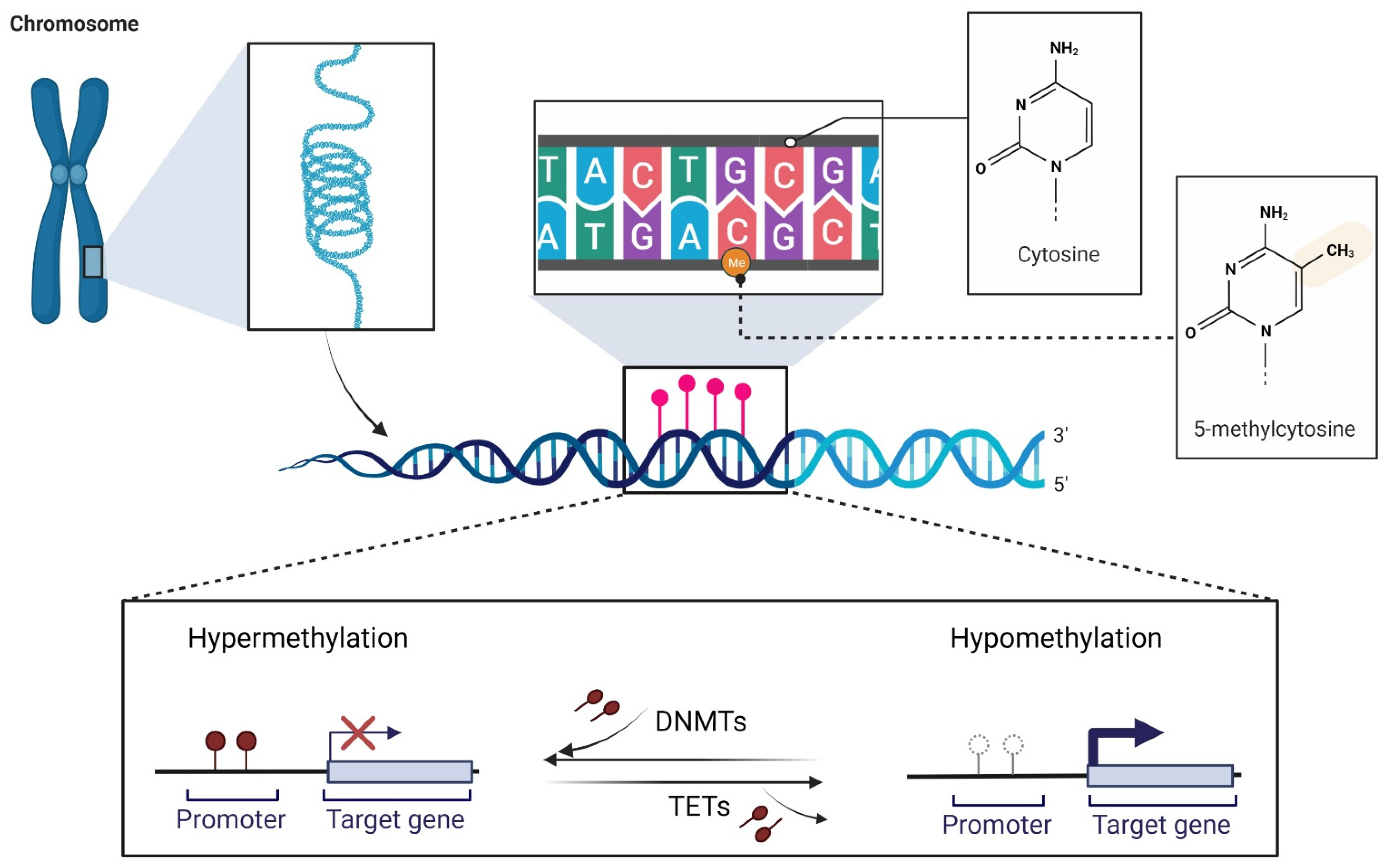
2.1. DNA Methylation at a Glance

DNA methylation, along with histone modifications and non-coding RNAs (ncRNAs), is classified as an epigenetic modification. Epigenetic modifications are defined as heritable alterations in gene expression that occur without modifying the DNA sequence [27,28,29]. As is the case with several other physiological mechanisms, when improperly activated or silenced, it can affect the expression of oncogenes or tumor suppressor genes (TSGs), resulting in tumor initiation, development, and progression [30,31]. DNA methylation consists of the covalent addition of a methyl group (-CH3) to the fifth position of the pyrimidine ring of the cytosine nucleotide (5-methylcytosine, 5-mC) [28,29]. DNA methylation can putatively occur at any cytosine nucleotide in the genome, but its distribution is not random and is mostly restricted to the so-called CpG dinucleotides. CpGs are more abundant at gene promoters, where they tend to congregate and form CGIs [28,29,30]. Two main enzyme families are involved in a balanced and regulated manner in DNA methylation and demethylation processes [28]. DNA methyltransferases (DNMTs), which establish and maintain methylation patterns [30], and ten-eleven translocation (TET) methylcytosine dioxygenases, which are involved in the demethylation pathway (Figure 1) [32,33]. Under normal conditions, DNA methylation encompasses the entire genome, with the exception of short unmethylated regions within the CGIs [34]. The deregulation of DNA methylation mechanisms, characterized mainly by hypermethylation of CGIs in TSG promoters or global loss of DNA methylation, is common in cancer. CGI hypermethylation in promoter regions has been shown to affect genes involved in the regulatory circuits that control cell proliferation and homeostasis, enabling malignant cells to sustain their abnormal growth [29]. Cancer-associated promoter hypermethylation may affect between 5–10% of promoters containing CGIs [35]. Global loss of DNA methylation leads to molecular consequences that are advantageous to tumor development, including the generation of chromosomal instability [36,37], the loss of genomic imprinting [38], and the reactivation of transposable elements such as LINE-1 [29,39,40].

2.2. DNA Methylation in Melanoma Development

Confined hypermethylation at CpG islands and global hypomethylation are epigenetic hallmarks of melanoma, both of which influence tumor behavior [41,42,43]. Melanoma initiation and progression have been associated with loss of tumor suppressors and oncogene activation [43]. Indeed, many TSGs are known to be aberrantly regulated via inactivation caused by specific methylation in the promoter region [44]. These genes appear to be involved in various signaling pathways, which are frequently altered during melanoma development and evolution. These pathways encompass the protein kinase activated by mitogen (MAPK), the phosphoinositide 3-kinase (PI3K), the tumor suppressor retinoblastoma (pRb), and the p53 protein pathways [41,43,45,46] (Figure 2). Since they can act synergistically or independently to control growth and apoptosis, their prolonged and uncontrolled activation is linked to proliferation, invasion, and metastasis. A gradual gain in DNA hypermethylation has been observed to increase in parallel with tumor aggressiveness and is known as the CpG island methylator phenotype (CIMP). It has been suggested that the genes involved in this increasing hypermethylation pattern form the melanoma CIMP [47]. Although less studied, DNA hypomethylation is equally important in the initiation and progression of melanoma. As previously stated, hypomethylation promotes tumor progression by causing genome instability via the demethylation of transposons and pericentromeric repeats, as well as the activation of specific oncogenes [43,48].
A detailed description of all the above aspects connected to melanoma driver epigenetic alterations is included in the next section.

2.2.1. Hypermethylated Genes

As already stated, promoter hypermethylation of tumor-related genes is one of the major mechanisms for gene function loss. Moreover, there is a continuously updating number of tumor-related genes, including TSGs, that have been identified as being silenced as a result of the aberrant hypermethylation of CpG-rich promoter regions [49,50,51,52]. This type of TSG silencing mechanism is frequently found in multiple tumors and, together with the mutation-induced mechanism, is a potent means of disrupting TSG function. A large number of genes have been identified as hypermethylated in melanoma [41,53,54,55], including O6-methylguanine-DNA methyltransferase (MGMT) [52,56], cyclin-dependent kinase inhibitor 2A (CDKN2A) [52,57], and Ras-association domain family 1 isoform A (RASSF1A) [49,58] (Table 1). Interestingly, some of them have been found to contribute to melanoma progression and have been associated with aggressive clinicopathological features and poorer survival [53,55]. Some of the most frequent and best characterized hypermethylated genes are described below. The pathways in which they act are highlighted in Figure 2.

RASSF1A 

The Ras-association domain family 1 (RASSF1) gene, located in the 3p21.3 region, contains eight exons and gives rise to eight different transcripts, from RASSF1A to RASSF1H, by means of alternative splicing and two different promoters [98]. The RASSF1A tumor suppressor is a scaffold protein involved in cell signaling. This protein is situated at the intersection of a complex signaling network that includes key regulators of cellular homeostasis such as RAS, MST2/Hippo, p53, and death receptor pathways [56]. Notably, RASSF1A is involved in the inhibition of cell proliferation and, because its signaling is regulated by pro-apoptotic insults, it activates intrinsic and extrinsic apoptotic cascades [99,100]. The hypermethylation of its promoter and subsequent gene downregulation has been observed in melanoma cell lines, tissues, and serum of stage III/IV cutaneous melanoma patients [47,49,55,65,76,78,93,95,96], and a significant proportion of uveal melanoma [77,97]. Several methods have been used to assess the methylation status of RASSF1A promoter, including methylation-specific polymerase chain reaction (MSP) [47,49,65,76,78,93], real-time quantitative MSP [76], methylation-specific multiplex ligation-dependent probe amplification (MS-MLPA) [55], real-time qPCR [95], and semi-nested PCR [96]. Inactivation of RASSF1A may play an important role in the selective advantage of melanoma cells. In particular, the high frequency with which RASSF1A is inactivated suggests that this could be an important alternate pathway for disrupting Ras signaling [49].

DAPK 

The death-associated protein kinase (DAPK), located on 9q34.1, encodes a serine/threonine kinase belonging to the calmodulin-regulated kinase (CAMK) superfamily, which also includes DRP-1 and ZIP-kinase (ZIPK) [101]. It plays a key role in tumorigenesis by acting as a regulator of caspase-dependent and caspase-independent cell death. DAPK is necessary for apoptosis induced by multiple death signals [102,103] and has also been associated with autophagy activation [103,104,105]. It functions as a tumor suppressor, and its loss occurs as a result of promoter methylation in many cancers [106,107]. DAPK hypermethylation has been detected through MSP investigation in 19% of metastatic tumor tissues from stage III/IV melanoma patients [76] and in only 5% of uveal melanoma tissues (real-time quantitative MSP analysis) [77]. The epigenetic silencing of DAPK, along with RARβ2, RASSF1A, and MGMT, has been observed as a common mechanism of tumor formation in cutaneous melanoma [76]. This may be due to DAPK’s inability to suppress cellular transformation during the early stages of tumor development [108] and to inhibit metastasis [101,109,110].

PTEN 

The phosphatase and tensin homolog gene (PTEN) is located on 10q23.31 and encodes a phosphatase that acts as a negative regulator of the PI3K/Akt pathway by dephosphorylating phosphatidylinositol (3,4,5)-triphosphate (PIP3), an important intracellular second messenger necessary for the activation of the Akt protein, a serine/threonine kinase involved in cell growth and survival [89,111]. The loss of functional PTEN leads to the downregulation of apoptosis and/or increased proliferation [111]. In melanoma, PTEN hypermethylation and constitutive activation of the Akt pathway promotes tumor progression. Indeed, PTEN has been shown to be frequently hypermethylated in malignant melanoma, and a subsequent reduction in its expression in both tissue and serum samples has been noted [55,90,91]. Although the biological impact of the hypermethylated PTEN promoter in melanoma is still not fully elucidated, several studies have examined its prognostic significance [92], and the association of PTEN methylation with advanced stage has been demonstrated. Indeed, an increase in its methylation, detected by MS-MLPA, has been observed in stage III/IV patients compared to early-stage primary tumors, resulting in reduced OS and disease-free survival (DFS) [55].

CDKN2A 

The cyclin-dependent kinase inhibitor 2A (CDKN2A) gene, located on 9p21.3, encodes two tumor suppressor proteins that are frequently mutated in human cancer [52], including melanoma [66]. These two proteins, p16INK4A and p14ARF, are important regulators of the retinoblastoma (Rb) and p53 pathways, respectively, and their gene promoters have been found to be hypermethylated and, consequently, silenced in both primary and metastatic melanoma [65,67].
At the G1-to-S transition in the cell cycle, p16INK4A specifically inhibits the cyclin D-dependent kinase 4/6 (CDK4 and CDK6)-mediated phosphorylation of the retinoblastoma protein (pRB), thus sequestering E2F transcription factors and consequently blocking cell cycle progression [68,69,70,71,112]. Whereas the expression of the p16INK4A protein leads to cell cycle arrest, the p14ARF protein mediates the degradation of MDM-2, which increases p53 protein levels, thus inducing cell cycle arrest to allow for either DNA repair and cell survival or apoptosis to discard the damaged cell [68,69,112]. The p16INK4A promoter is found to be hypermethylated and, consequently, silenced in several cancer types [113]. While the hypermethylated p16INK4A promoter, identified by MSP, was found in 19% of primary cutaneous melanoma cases, the frequency tends to be higher in metastases (33%), indicating that hypermethylation may also occur in later stages [65]. It is associated with increased tumor cell proliferation and reduced patient survival. It is worth noting that p16INK4A promoter methylation, analyzed by MSP, has been predominantly observed in 52% of NRAS-mutated metastatic melanomas but in only 7% of BRAF-mutated metastases [66]. p14ARF has been shown to be hypermethylated in approximately 57% of metastatic melanoma tumors, independently of the p16INK4A promoter [67].

MGMT 

The O6-methylguanine DNA methyltransferase (MGMT) gene is located on 10q26.3 and encodes an enzyme involved in repairing damaged guanine nucleosides [114]. MGMT plays a central role in preventing normal cells from transforming into tumor cells and also protects tumor cells from the cytotoxic effects of chemotherapy with alkylating agents, such as temozolomide (TMZ) and dacarbazine (DTIC), widely used in treating melanoma and glioblastoma [82,115]. This protein catalyzes the transfer of methyl groups from the O6 site of guanine in DNA to its cysteine residue (the protein active site) [116]. MGMT is silenced during the oncogenesis of several human cancer types, leading to inefficient repair of DNA alkylation, and resulting in increased sensitivity to alkylating agents and decreased tumor survival [117]. The hypermethylated state of MGMT has been detected in the cell lines, tissues, and serum of melanoma patients using MSP [65,72,76,78,83], real-time quantitative MSP [76], high-resolution melting point analyses (HRMA), pyrosequencing [83], and MS-MLPA [84]. It is worth noting that the epigenetic silencing of MGMT has been associated with a significantly improved response to DTIC/TMZ single-agent therapy, a longer progression-free survival (PFS) in stage IV melanoma patients [83], and prolonged survival in stage III melanoma patients locoregionally treated with melphalan [84]. All this evidence suggests that the MGMT methylation status may be used as a predictive biomarker of favorable survival in patients receiving chemotherapy [83,84].

RARβ2 

The Retinoic Acid Receptor Beta 2 (RARβ2), located on 3p24.2, encodes a retinoid-inducible tumor suppressor protein. It is a member of the nuclear receptor superfamily, which also includes RARα and RARγ, both of which are expressed differentially during development and adulthood. Together with retinoid X receptors (RXRs), RARs are bound and activated by the biologically active forms of vitamin A (all-trans retinoic, ATRA; 9-cis-retinoic acid, CRA). The activated heterodimer of RAR and RXR binds to the retinoic acid receptor responsive element (RARE) and regulates the transcription of the target genes involved in cell differentiation, proliferation, and apoptosis [118]. Several approaches, including MS-MLPA [55], MSP [76,78,93], real-time quantitative MSP [76,94], and pyrosequencing [85], have been used to disclose that the promoter of this gene is frequently hypermethylated in the cell lines, tissues, and serum of melanoma patients [55,76,78,85,93,94].
In addition to the other TSGs found to be frequently hypermethylated in melanoma, Hoon and colleagues reported that the hypermethylation of RARβ2 was significantly correlated with Breslow thickness, which is an important prognostic factor in patients with early-stage melanoma [76]. The authors concluded that the silencing of this gene might be a key epigenetic factor in melanocyte transformation and primary lesion progression. These results were also confirmed in a more recent study, where a significant reduction in DFS and OS was found in the presence of RARβ2 hypermethylation [55].

CpG Methylator Phenotype (CIMP) in Melanoma

The CpG methylator phenotype (CIMP) was first observed in colorectal cancer [119] and consists of a gradual increase in the level of CGI methylation along with tumor aggressiveness. This phenomenon has also been observed by Tanemura and colleagues in melanoma patients [47]. In this work, they investigated the significance of the CGI methylation status in the progression of malignant melanoma by means of MSP and absolute quantitative assessment of methylated alleles (AQAMA). Methylation of several tumor-related genes, including MINT17, MINT31, TFPI2, WIF1, RASSF1A, and SOCS1, comprising the first melanoma CIMP, increased significantly with advanced clinical stages, suggesting that their inactivation is associated with tumor progression. In recent years, a DNA methylome profiling study, performed by the Illumina Infinium HumanMethylation450 BeadChip, detected additional hypermethylated genes involved in the invasiveness of primary and metastatic melanomas, including ARHGAP22 and NAV2 [53]. In addition, it was recently observed that melanoma CIMP associated with an NRAS-mutant phenotype is more aggressive than melanoma CIMP associated with BRAF-mutant melanoma [120]. However, the strong association between BRAF V600E and CIMP that emerges in colorectal cancer is not observed in melanoma [121]. Although the underlying mechanism of CIMP is still unclear, these data suggest a tissue-specific biological mechanism rather than a universal one for all cancers [121]. All these findings could be essential for developing novel tools to identify early metastasis-promoting epigenetic events.

2.2.2. Hypomethylated Genes

It has been observed that malignant tumor tissues are characterized by global DNA hypomethylation when compared to normal tissues or benign tumors [122,123]. To quantify the global methylation level of an entire genome, it is common practice to analyze the methylation level of the Long Interspersed Nuclear Element-1 (LINE-1) sequences (approximately 20% of the human genome [124]), representing a surrogate marker for global methylation status [125,126]. Global demethylation has been identified as a putative hallmark of the metastatic capacity of primary melanomas [127]. Based on previous studies demonstrating a correlation between LINE-1 hypomethylation and CGI hypermethylation of TSGs [128,129], Hoshimoto and colleagues evaluated the methylation status of LINE-1 and the TSG AIM1 in melanoma patients in order to develop a combination of biomarkers with prognostic utility. Tissue analysis by AQAMA revealed that LINE-1 hypomethylation was higher in stage IV melanoma than in other stages and that AIM1 hypermethylation (MSP detection) was more frequent in metastatic melanoma than in primary melanoma (65% vs. 38%). The combination of LINE-1 hypomethylation and AIM1 hypermethylation was a significant predictor of DFS and OS in stage I/II patients. In addition, they observed that patients with LINE-1 hypomethylation or AIM1 hypermethylation in serum had a worse prognosis [130]. Apart from the global hypomethylated pattern observed in melanoma, specific hypomethylated genes have been identified and correlated with patient outcome (Table 2).

TBC1D16 

It has been observed that the hypomethylation of the TBC1D16 gene, which encodes, among others, the TCB1D16-47KD isoform, generates metastases in melanoma [138]. More in detail, Vizoso and colleagues exploited the use of the Illumina Infinium HumanMethylation450 BeadChip for screening genes with differential DNA methylation in both primary tumor-derived and metastatic melanoma cell lines. This approach led to the identification of the TCB1D16-47KD isoform, whose gene promoter was hypermethylated and downregulated in primary tumor–derived cell lines but was not unmethylated and upregulated in metastatic cell lines. Finally, it was observed that TBC1D16-47KD hypomethylation in metastatic patients was associated with a shorter PFS and OS. Worth noting is that this unmethylated state was observed in 33% of patients carrying the BRAF V600E mutation and has been associated with increased sensitivity to BRAF inhibitors [138].

PDGFD, ZEB1, and THRB

There is currently no targeted therapeutic approach for NRAS-mutant melanomas. Starting from this consideration, Jiang and colleagues performed an integrative analysis encompassing DNA methylation, gene expression, and microRNA expression to identify downstream pathways affected by the most common NRAS driver mutations (Q61K/L/R). DNA methylation profiles of 61 primary melanomas, processed through the Illumina Infinium HumanMethylation450 BeadChip, and obtained from TCGA, were examined [136]. First, global hypomethylation induced by or associated with the NRASQ61 driver mutation was identified as a common feature in melanoma (similar to what Hou and colleagues demonstrated for BRAF V600E signaling), potentially playing an important role in the disease pathogenesis [24,136]. NRASQ61 mutations were then linked to the hypomethylation of PDGFD, ZEB1, and THRB genes, resulting in their increased expression and the dysregulation of the involved downstream pathways. Finally, the expression of PDGFD, ZEB1, and THRB was also associated with patient survival time, thus suggesting that they may have therapeutic potential. PDGFD is located upstream of the MAPK and PI3K pathways and plays a role in the regulation of cell proliferation, transformation, invasion, and angiogenesis [140]. As a transcription factor involved in the epithelial to mesenchymal transition, ZEB1 is an oncogene that can promote neoplastic transformation [141]. Finally, THRB is one of the thyroid hormone receptors [142], and defects in its gene are known to confer generalized thyroid hormone resistance, thus opening up new perspectives for the putative use of thyroid hormone therapy to treat NRAS-mutant melanomas [136].

TKTL1 

The reprogramming of energy metabolism to fuel uncontrolled cell growth and division is a hallmark of cancer. Certain tumor cells are part of a subpopulation of glucose-dependent cells (“Warburg effect”’) that secrete lactate, which is imported and preferentially used as fuel by other tumor cells, resulting in perfect cooperation [143]. Hypomethylation of Transketolase-like 1 (TKTL1), evaluated by quantitative methylation-specific PCR (MS-qPCR), appears to be involved in enhancing the Warburg effect in melanoma by accelerating glucose utilization and lactate production, thereby increasing the likelihood of the successful invasion of melanoma cells [139].

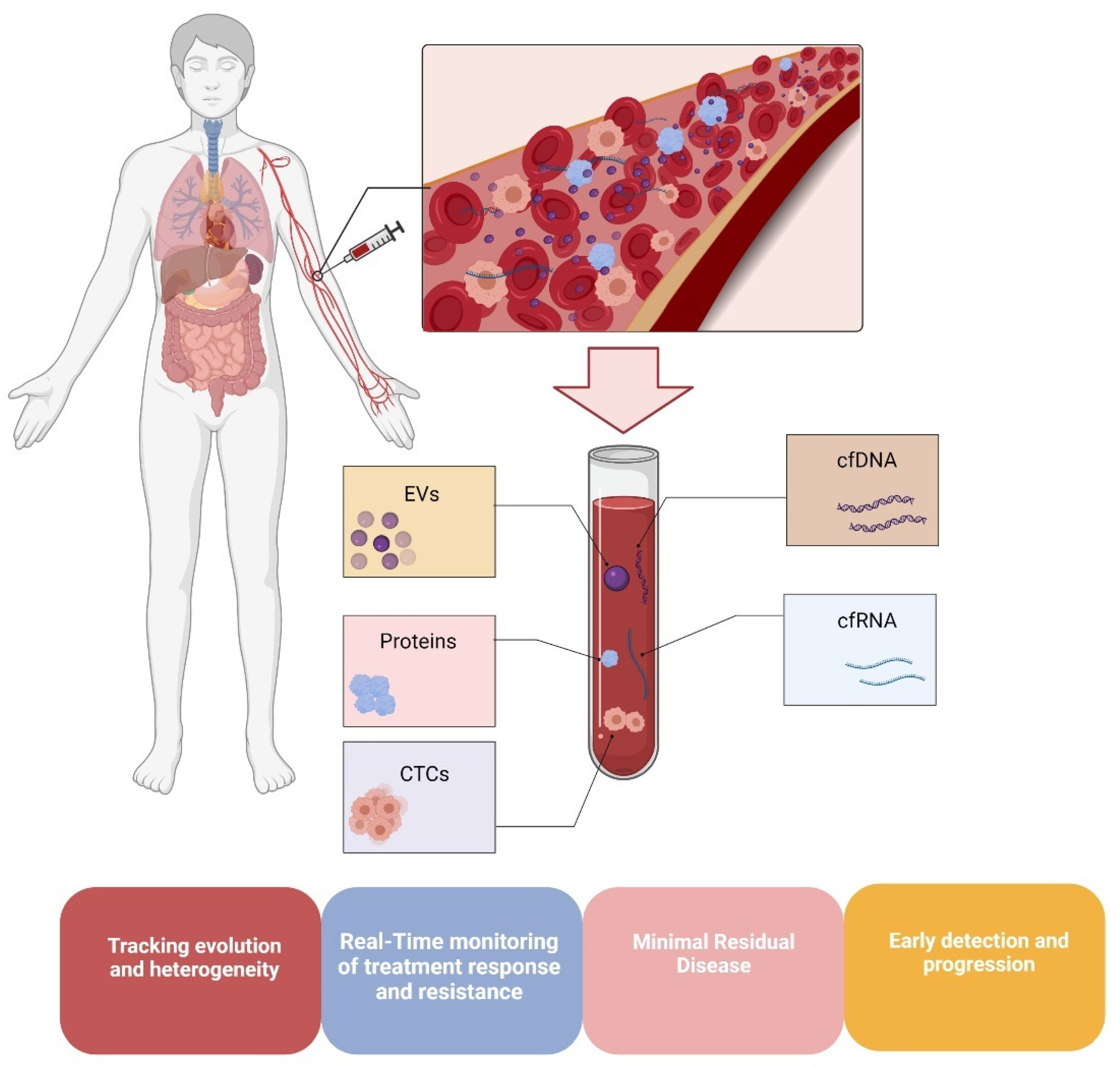
3. DNA Methylation in Melanoma Liquid Biopsies

In this era of “personalized medicine,” much attention is being paid to different approaches that provide repeatable and safer insights into tumor evolution and heterogeneity. Since cutaneous melanoma is characterized by extreme heterogeneity and high tumor mutation burden (TMB) [144], the early detection of tumor-related changes such as chromosomal rearrangements, copy number variation, or mutations in oncogenes and tumor suppressor genes, is mandatory for the choice and adjustment of targeted therapy, treatment monitoring, and detection resistance [145]. In the last decade, the so-called “liquid biopsy” [146], which refers to a test of body fluids (i.e., blood, urine, and saliva), has emerged as a novel biomarker with significant application in translational research due to its ability to provide comparable (or more detailed) information than the conventional tissue biopsy [145]. Indeed, the analysis of circulating tumor cells (CTCs), cell-free circulating nucleic acids (cfDNA and cfRNA), and extracellular vesicles (EVs) via a minimally invasive blood draw has opened new avenues for cancer diagnostics, enhancing risk assessment, real-time monitoring of therapeutic efficacy, early detection of relapse, and monitoring of tumor evolution (Figure 3) [145,147,148,149]. Like all circulating tumor cells, Circulating Melanoma Cells (CMCs) are released into the bloodstream by the primary tumor and/or metastases [145,150]. Due to their high heterogeneity and rarity in the bloodstreams of metastatic melanoma patients [151,152], the continuous improvement of even more sensitive methods to detect and characterize them is of paramount importance. On the other hand, cell-free circulating DNA is produced from physiological functions such as apoptosis, necrosis, or secretion. Moreover, cancer patients have higher amounts of cfDNA than healthy controls [149,153]. Thus, the fraction of cfDNA that originates from tumor cells, called circulating tumor DNA (ctDNA), has been extensively studied as a putative disease marker, both in terms of quantity and composition. Indeed, the identification and monitoring of ctDNA using mutation-detection techniques (i.e., droplet digital PCR (ddPCR) and targeted next-generation sequencing (NGS) panels) has been well documented, and its potential as a promising biomarker for diagnosis, evaluation of treatment effectiveness, and as a tracker of tumor evolution has been well assessed in many cancers, including melanoma [149,154,155,156,157,158,159]. Finally, EVs actively participate in intercellular communication by taking part in the transfer of lipids, proteins, and RNA, thus suggesting a putative active role in cancer development [160,161]. Moreover, they have enormous potential as biomarkers, given that their tumor-derived cargo may be used for different applications, i.e., tumor burden estimation and survival prediction [145,162]. All things considered, the search for reliable biomarkers capable of tracking the disease evolution and heterogeneity in real-time may be successful in liquid biopsy, which is the sum of systemic disease. Since the use of targeted therapies and immunotherapy has significantly altered the natural history of melanoma, it is critical to closely monitor its genetic landscape to ensure its success. As previously stated, aberrant methylation of gene promoters can be a characteristic of cancer [163], and, as a result, the analysis of methylated DNA in liquid biopsy is an emerging field of interest [81,164,165]. It has already been demonstrated that hypermethylation of the promoters of certain selected genes can be used to distinguish melanoma patients from healthy individuals [81], demonstrating its utility as a diagnostic marker. Notably, DNA methylation patterns can change during melanoma progression; thus, longitudinal monitoring of DNA methylation in a non-invasive manner via liquid biopsy can provide real-time information about the behavior and stage of melanoma [80].

3.1. DNA Methylation in Circulating Melanoma Cells

To date, the CTC count as a prognostic/predictive marker is well established for several malignancies for which the cut-off has already been validated [166,167,168]. For others, melanoma included, the process is still ongoing. In any case, this biomarker has garnered considerable attention due to the type of information that its genetic characterization could offer, and more recently, the DNA methylation profile has attracted significantly more attention. However, the study of CTC methylome remains largely unexplored due to the lack of adequate investigation techniques. One of the first studies examined the simultaneous detection of CMCs, inferred by the identification of specific mRNAs in the blood and specific methylated genes. More in detail, Koyanagi and colleagues identified a correlation between the number of melanoma markers (MART-1, GalNAc-T, and MAGE-A3 mRNAs), detected in the blood through quantitative real-time reverse transcription-PCR assay, the presence of circulating methylated RASSF1A and RAR-β2 genes (MSP detection), and patient outcome. Patients with both CMCs and methylated genes showed a significantly poorer response to biochemotherapy, as well as a shorter PFS and OS [169]. Salvianti and colleagues conducted a similar study, focusing on methylated cfDNA and CMCs as two complementary liquid biopsy biomarkers that can be combined to enhance the possibility of disease monitoring [95]. Indeed, they compared RASSF1A promoter methylation tracked in cfDNA by real-time qPCR with the presence of CMCs in both healthy controls and patients at different melanoma stages (in situ, invasive, and metastatic). They found that the percentage of cases with methylated RASSF1A promoter was higher in melanoma patients than in healthy subjects (46% vs. 10%), thus indicating RASSF1A promoter methylation as a good predictor of disease (AUC of 0.905). However, when they checked the presence of CMCs in the three different patient categories, they found no significant association with methylated RASSF1A tracked in cfDNA [95].
Currently, no studies have been conducted to determine the methylation status of CMCs. However, the few studies that have investigated CTC methylation in other cancers are very interesting. Recent publications have focused on finding the optimal technique for studying CTC methylome in lung [170], breast [171,172], and colon cancer [171]. More in detail, Zhao and colleagues developed an approach called LCM-µWGBS that combines laser capture microdissection (LCM) CTC capture and whole-genome bisulfite sequencing (µWGBS), enabling the analysis of a small number of CTCs to obtain information on their DNA methylation landscape [170]. They defined the DNA methylome of CTCs from lung cancer patients and compared it to the global DNA methylation of normal tissues. Interestingly, they found a progressive decrease in global DNA methylation from normal tissue to primary tumors and CTCs, suggesting a gradual loss of DNA methylation during tumorigenesis. Moreover, they observed a tendency toward the increased methylation of several TSG promoters in CTC-DNA compared to the primary tumor.
In a very recent study, Chen and colleagues, by means of single-cell bisulfite sequencing (scBS-seq), provided an important analysis of the single-cell DNA methylome in CTCs, characterizing tumor heterogeneity and the evolution of the tumor cell methylome during cancer progression [171]. Seventeen cancer patients covering six different types of cancers (lung adenocarcinoma, small-cell lung cancer, breast, colon, gastric, and prostate cancer) were tested to assess the methylation level in single CTCs [171]. Firstly, they observed that CTCs, similar to primary tumors, exhibit lower methylation levels than those in normal cells. They also observed that these cells show inter- and intra-patient heterogeneity in terms of promoter methylation. Interestingly, they also investigated the dynamic methylome changes, which occur during cancer metastasis, in the promoter regions of 20 known tumor-associated genes of primary, metastatic tissues, CTCs, and white blood cells of gastric cancer patients accompanied by abdominal ovarian metastasis. For instance, they observed that the methylation level of the LTF gene, often reported down-regulated in tumors, increased with cancer progression. The methylation level of several genes expressed in the ovaries, such as 1orf35, DENND6A, and ZNF285, decreased transiently. Furthermore, the methylation level of genes involved in oncogenic transformation- and cell adhesion-associated pathways (FBP2, HIVEP3, PTPN21, and CEACAM5 genes) decreased, suggesting a role for these pathways in tumor progression.
Since CTCs can be found in patient blood as single CTCs or CTC clusters, Gkountela and colleagues found that CTC clusters are distinguishable from single CTCs based on their methylation status, assessed by single-cell whole-genome bisulfite sequencing. They observed that in CTC clusters, hypomethylated regions are associated with key regulators of stemness and metastasis (OCT4, NANOG, SOX2, and SIN3A), while hypomethylated regions in single CTCs are independent of the pluripotency network [172].

3.2. Methylation in Circulating Melanoma DNA

Several recent studies have focused on ctDNA methylation screening and its potential clinical application, in addition to genomic and/or expression analyses [173,174]. Furthermore, most recent studies have focused on the analysis of ctDNA rather than FFPE samples in an attempt to overcome the limitations associated with the poor quality of the FFPE genetic material. In one of the first studies, based on real-time MSP detection, Hoon and colleagues examined the presence of hypermethylated TSG promoters in the serum of melanoma patients [76]. They observed that the incidence of TSG hypermethylation increased during tumor progression and that MGMT, RASSF1A, and DAPK hypermethylation were significantly lower in primary melanomas compared to metastatic ones. On the other hand, the frequency of hypermethylated RAR-ẞ2 was similar in both primary and metastatic melanomas [76].
A subsequent study by Mori and colleagues highlighted the utility of detecting circulating methylated tumor-related genes in serum (MSP analysis) as a predictive marker of response to biochemotherapy and OS. Among the most frequently hypermethylated genes in melanoma, they discovered a significant correlation between the hypermethylation of RASSF1A and RAR-ẞ2, the response to biochemotherapy, and OS [78]. Interestingly, this correlation with treatment response was not observed when analyzing the MGMT methylation status [78].
A recent study by Liu and colleagues focused on assessing the feasibility of using cfDNA methylation profiles in advanced cancer patients for the detection of metastatic disease [173]. They developed an NGS targeted methylation sequencing assay to measure the methylation status of more than 9000 CpG sites, selected according to TCGA data, and parallel classify the presence of advanced cancer, being also able to predict tumor origin (i.e., melanoma, colorectal cancer, NSCLC, and breast cancer). The authors demonstrated that plasma cfDNA methylation scores detected the presence of cancer in 83.8% of cancer patients with 100% specificity and predicted cancer type in 78.9% of cases. Focusing on melanoma, Diefenbach and colleagues developed an efficient ctDNA methylation analysis workflow using an amplicon-based NGS panel performed on bisulfite-treated DNA. They confirmed the hypermethylation of seven genes (GJB2, HOXA9, MEOX2, OLIG3, PON3, RASSF1, and TFAP2B), known to be hypermethylated in metastatic melanoma patients but not in healthy individuals [81]. Moreover, in contrast to previous studies on cfDNA methylation, they were able to examine the methylation of symmetrical CpG sites in both DNA strands to accurately quantify the level of gene methylation [81].
Some studies have explored the potential of methylation pattern tracking in early-stage tumors. In this regard, the clinical validity of a targeted methylation-based Multi-Cancer Early Detection (MCED) test using cfDNA sequencing has been investigated in several cancer types, including melanoma [174]. This new approach aims to provide the tools to detect tumors at an earlier stage, thus reducing cancer mortality by identifying the cancer signal origin (CSO) [174,175]. The MCED test demonstrated a specificity of 99.5% (false-positive rate of 0.5%) and an overall sensitivity of 51.5% for cancer signal detection. The authors suggested to use this test either in combination with other single-cancer screening tests (for the detection of breast, colorectal, cervical, lung, and prostate cancers) or as a screening for those cancers for which tests are not yet available in the United States. However, the high costs of this type of analysis could represent an obstacle for its application in the clinical setting [174].
In summary, the role of DNA methylation in melanoma deserves deeper investigation, as its role in tumorigenesis is only partially understood. Its usefulness as a biomarker could be helpful in defining a precision medicine workflow. In particular, a suitable and optimal analysis technique should be sought, which is currently lacking. In addition, a cost-benefit analysis should be conducted. While whole-genome bisulfite sequencing can provide a complete methylation profile at a very high cost, a PCR-based approach is cheaper but assesses only a small number of CpG sites [173]. The use of NGS panels, which allows simultaneous analysis of the methylation status of several tens to hundreds of genes, could be an alternative and, if customized and restricted to a limited number of regions of interest, might be an excellent compromise to limit costs.

3.3. Methylation in Melanoma Extracellular Vesicle-Derived DNA (evDNA)

In recent years, there has been growing interest in understanding the role of extracellular vesicles (EVs) in melanoma progression since they are considered to be an alternative means of intercellular communication. EVs are composed of a heterogeneous group of membrane-delimited nanoparticles, usually classified into exosomes (30–120 nm), microvesicles (100–1000 nm), and apoptotic bodies (500–4000 nm) based on their size [176]. They play a key role in the delivery of active cargoes, including DNA fragments, coding and non-coding RNA, proteins, and lipids, from donor to distal cells, and they are released by normal and cancerous cells into the external microenvironment [177]. Tumor-derived EVs have been shown to affect the pathophysiology of recipient cells, modulating a variety of processes involved in cancer progression (increased invasiveness, proliferation rate, and chemoresistance) [178].
The study of EV content is emerging as an innovative and helpful strategy to understand tumor processes due to the fact that the bioactive molecules are packaged and protected within the phospholipid bilayer of EVs. Although research on EV-derived RNA and proteins has been largely explored, only a few studies have been conducted on EV-associated DNA (evDNA). Recent evidence suggests that most of the DNA associated with tumor-derived EVs is double-stranded (dsDNA), representing the entire genome and informing on the mutational status of parental tumor cells [179]. However, the mechanism of DNA loading onto EVs remains unclear. The main hypothesis is the encapsulation of cytosolic DNA during EV biogenesis [160]. In recent years, the methylation analysis of evDNA has attracted attention as a biomarker for the detection of various cancers. The benefit of using evDNA is its stability and protection from digestive enzymes due to encapsulation in the lipid bilayer of EVs. However, major steps forward are needed to overcome issues related to the source of evDNA, sample collection, and DNA extraction methods that appear to affect methylation detection. For example, it is necessary to develop a better strategy for isolating evDNA without cfDNA contamination because it has been observed that DNA tends to stick to any surface, including the lipid envelope of EVs, resulting in the possible co-isolation of cfDNA with the EV during the purification protocol [180]. In this regard, the digestion of the EV pellet by DNAses may be an optimal strategy to overcome the co-isolation of nucleic acids with EVs. The DNA extraction efficiency largely depends on which method of EV isolation and evDNA extraction is used. Moreover, the presence of DNA in exosomes continues to be controversial. In 2019, while Coffey and colleagues concluded that exosomes do not contain DNA, Yokoi and colleagues observed genomic DNA and nucleoprotein in them. Perhaps it may depend on an over-strict exosome isolation strategy that can lead to the loss of evDNA, the levels of which are too low to be detected. Because the most effective approach for EV isolation has not yet been well established, and different methods of EV isolation and DNA extraction are currently used, it is mandatory to develop an optimal common strategy for downstream applications. For example, García-Romero and colleagues compared the most common EV-isolation methods and found that polyethylene glycol precipitation (PEG) seems to be the most feasible and affordable EV isolation technique [181]. Moreover, in a study by Kamyabi and colleagues, a microfluidic platform was described as a method for the rapid isolation of EVs from the plasma of pancreatic cancer patients [182].
Although data on melanoma are still limited, and most of the data are restricted to in vitro experiments, overall findings suggest that the methylation profile of evDNA shows similarities in methylation profile with that of genomic DNA (gDNA) in murine melanoma cells [179]. This evidence has also been confirmed in other cancer types, such as metastatic castration-resistant prostate cancer [183], diffuse large B-cell lymphoma [184], and glioblastoma [185]. A new and interesting approach was recently developed using an electrochemical detection method based on the differential absorption capacities of different methylation levels of DNA on a gold surface [177]. Using this highly sensitive microdevice, Sina and colleagues performed exploratory research to develop a method to isolate evDNA from ctDNA based on methylation-dependent physicochemical properties. They found that evDNA has surface-based properties similar to cellular gDNA, but not to cfDNA, probably due to the longer size of gDNA. Moreover, they demonstrated that this method was able to discriminate cancer and normal evDNA, suggesting a potential use of this device for clinical applications (the adsorption level on the gold surface of patient evDNAs was 20–40%, whereas that of the normal evDNAs was below 20%) [177].
In addition, the promoter regions of specific genes in metastatic castration-resistant prostate cancer (GSTP1, RASSF1A, and SLFN11) and in diffuse large B-cell lymphoma (CDKN2A and CDKN2B) are found to be methylated in both evDNA and primary tumor tissue or CTCs, respectively [183,184]. These findings not only highlight the potential use of evDNA methylation analysis as a biomarker for the detection of cancer but also point toward a better understanding of the evDNA methylation profile in melanoma cancer, which is poorly understood.

4. Circulating Methylated DNA Biomarkers for Tracking Response to Therapy and Resistance Onset

The development of treatment resistance is the main limitation to many anti-cancer therapeutic approaches. DNA methylation has the potential to be used as a biomarker in many clinical situations, including for the prediction of response to therapies and the monitoring of recurrence [41]. As previously discussed, positive signals for a putative prognostic role for methylation status, tracked through liquid biopsy, were found in the early 2000s by Koyanagi and colleagues, who hypothesized the clinical utility of two different molecular variables (CMC-mRNA and serum methylated DNA) in a cohort of patients undergoing biochemotherapy [169]. Similarly, Mori and colleagues assessed the prognostic and predictive significance of detecting specific methylated genes in the serum of melanoma patients undergoing biochemotherapy [78]: they found that hypermethylation of RASSF1A was the best predictor of response and OS, thus corroborating previous evidence found in other malignancies of resistance to cisplatin and tamoxifen associated with RASSF1A hypermethylation [186,187]. Moreover, the methylated status of RAR-β2 was significantly related to survival. As RAR-β2 is involved in the control of cell growth and apoptosis, its expression in tumor cells susceptible to its mediated apoptosis could be an important biomarker of response to biochemotherapy [78]. A recent study by de Vos and colleagues involved a large patient cohort with different malignancies from the Cancer Genome Atlas (TCGA) Research Network to test the value of two hypermethylated genes, SHOX2, and SEPT9, as pan-cancer biomarkers [188]. The quantitative methylation analysis of these genes has shown a correlation with treatment response: when compared to conventional monitoring, cfDNA longitudinal screening revealed an association between an increase in the methylation score (CMS, cumulative cfDNA methylation score) and non-responsiveness to treatment with an 80-day advantage.
Nowadays, chemotherapy with two alkylating agents, DTIC or TMZ, is still an alternative in the event of the development of resistance to immune checkpoint inhibitors (ICI) and/or targeted therapy, or in the case of mutation-negative melanomas [43,189,190]. Therefore, biomarkers capable of identifying the minority of patients who may benefit from this type of approach would be critical, as they are currently lacking. The effects of methylating agents have been putatively related to the expression of MGMT. Its overexpression protects against cell death induced by alkylation. On the other hand, low expression is correlated with a higher possibility of responding to methylating agents [191,192]. As melanomas are likely to express low levels of MGMT [193,194], this could explain why they respond to methylating drugs, including DTIC and TMZ, but not to other anti-cancer drugs. The MGMT methylation status can thus determine the outcome in melanoma patients treated with methylating drugs. In fact, MGMT promoter methylation has been associated with response to single-agent DTIC/TMZ and longer PFS in disseminated cutaneous melanoma [83], even if conflicting data have been reported [190,195]. Melanoma cells appear to be intrinsically resistant to drugs and/or acquire resistance through different strategies involving multiple other players [195]. Ultimately, the simultaneous tracking of the MGMT methylation status and other players could be of great interest for determining the success of TMZ therapy, and liquid biopsy, as a sum of systemic disease, may be the optimal source for performing this type of longitudinal screening.

5. Methylation Markers from Bench to Bedside

Liquid biopsy evaluation of epigenetic biomarkers is an emerging field in oncology that might have a putative impact in improving diagnostic and screening procedures [196]. Several benefits may address the use of DNA methylation as a biomarker, such as its high stability in several biofluids and its dynamism during disease evolution. Speaking about the clinical utility of DNA methylation in cutaneous melanoma, some attempts have already been made in this direction: i.e., the RASSF1A promoter hypermethylation tracked in the cfDNA has been found to be a good diagnostic and prognostic biomarker [78,95]. However, several hindrances make the incorporation of this putative biomarker in the clinical routine still uncertain [197]: first, much efforts are currently being made by the scientific community to standardize the preclinical factors governing the handling of the liquid biopsy samples [198]. The choice of blood collection tubes, for example, is a critical pre-analytical variable [198] as in the clinical setting the fast processing is not always possible. Thus, the levels of the contamination of cfDNA derived from nucleated blood cells caused by an inappropriate collection of blood samples has to be carefully avoided [199]. Second, low yields of ctDNA recovered after bisulfite conversion could impair the downstream workflow. More in detail, even if bisulfite treatment is the gold standard method for mapping methylated cytosines, it presents some hindrances connected with the wet lab procedure that may cause further loss of ctDNA. Finally, in the perspective of translating the analysis of cfDNA methylation into clinical practice, it is necessary to be aware that changes in cfDNA methylation have been observed not only in cancer but also in other situations, including lupus erythematosus [200], liver fibrosis [201], and diabetes [202]. This could be a challenge, especially in the context of cancer screening.
Moreover, from an economical point of view, the new high-throughput technologies (i.e., microarrays and NGS) applied to the DNA methylation analyses present some limits connected with their relatively elevated costs for use in the clinical routine. Nevertheless, it has to be taken into consideration that the accuracy of the information obtained from these approaches helps in identifying the appropriate (target) treatment, thereby limiting the high costs incurred for ineffective therapies. Finally, the training of laboratory professionals able to interpret the large amount of data derived from these new platforms will be mandatory to provide a rapid flow of reliable information from bench to bedside, capable of driving clinical decisions in real-time.
Overall, the still increasing knowledge and continuous technological development, when accompanied by careful preparation of personnel in the field of DNA methylation, will lead to further achievements, hopefully, spendable in the clinic in the near future.

6. Conclusions

The evaluation of epigenetic biomarkers through a liquid biopsy approach is an emerging field that may hold great potential not only for the screening, diagnosis, and identification of tumor types but also for the prediction of response to therapy and/or progression [196,203]. The benefits of this approach include the ability to evaluate tumor markers using non-invasive methods and to gather information on the status and evolution of the systemic, heterogeneous disease. Nowadays, the evaluation of actionable mutations in liquid biopsy has demonstrated clinical value. Nevertheless, the use of epigenetic alterations as biomarkers is still under-exploited, despite their promising potential. DNA methylation tracking is a powerful method for identifying clinically relevant circulating epigenetic biomarkers, facilitating early cancer detection, and resulting in a substantial reduction in cancer—related mortality [203]. From this perspective, all the different components of the circulating compartment deserve to be thoroughly investigated due to the complementary information they may provide. Even though significant efforts have already been made to develop novel biomarkers, much more progress is required. Additional research is required to determine which methylated markers are most accurate to address the questions that need answering. Large-scale targeted methylation sequencing of cfDNA has demonstrated a high potential both for early cancer diagnosis [199] and, most interestingly, for the correlation of methylation scores with treatment outcomes [203]. Since extracellular vesicles carry a molecular “fingerprint” of the cell of origin, they could deliver precious information concerning the cancer status, being prospective biomarkers for melanoma diagnosis or prognosis [145,204]. Finally, circulating melanoma cells, which remain largely unexplored from an epigenetic point of view, are still promising in light of the excellent results obtained in other pathologies.
In conclusion, a better understanding of melanoma epigenetics will enable the identification of more specific diagnostic, prognostic, and predictive biomarkers, which will be advantageous in the era of “precision medicine.” Additionally, it is hoped that more potent epigenetic inhibitors can be developed and tested for the treatment of specific melanoma subtypes.

Author Contributions

Literature revision and draft preparation, V.A., C.C., C.P., A.F. and M.C.S.; image conceptualization, V.A. and C.C.; writing—original draft preparation, V.A., C.C., C.P. and M.C.S.; writing—review and editing, A.F., C.P., A.R. and M.C.S.; supervision, A.R., M.C.S. and A.F.; funding acquisition, M.C.S. All authors have read and agreed to the published version of the manuscript.

Funding

This research was partially funded by the Italian Ministry of Health, Proposal code GR-2016-02361019, Liquid Biopsy in melanoma: investigating the prognostic/predictive potential of circulating melanoma cells (CMCs) and tumor DNA (ctDNA)—P.I. MC Scaini, and Ricerca Corrente, P23-Liquid biopsy in melanoma—P.I. MC Scaini. The work of V.A. and C.C. is supported by 5 × 1000 CANCER PLAT-2.

Acknowledgments

This manuscript is dedicated to the memory of Rita Zamarchi.

Conflicts of Interest

The authors have no conflict of interest to declare.

References

  1. Karimkhani, C.; Green, A.; Nijsten, T.; Weinstock, M.; Dellavalle, R.; Naghavi, M.; Fitzmaurice, C. The global burden of melanoma: Results from the Global Burden of Disease Study 2015. Br. J. Dermatol. 2017, 177, 134–140. [Google Scholar] [CrossRef] [Green Version]
  2. Long, G.V.; Hauschild, A.; Santinami, M.; Atkinson, V.; Mandalà, M.; Sileni, V.C.; Larkin, J.; Nyakas, M.; Dutriaux, C.; Haydon, A.; et al. Adjuvant Dabrafenib plus Trametinib in Stage IIIBRAF-Mutated Melanoma. N. Engl. J. Med. 2017, 377, 1813–1823. [Google Scholar] [CrossRef] [Green Version]
  3. Robert, C.; Grob, J.J.; Stroyakovskiy, D.; Karaszewska, B.; Hauschild, A.; Levchenko, E.; Sileni, V.C.; Schachter, J.; Garbe, C.; Bondarenko, I.; et al. Five-Year Outcomes with Dabrafenib plus Trametinib in Metastatic Melanoma. N. Engl. J. Med. 2019, 381, 626–636. [Google Scholar] [CrossRef] [PubMed]
  4. Eroglu, Z.; Ribas, A. Combination therapy with BRAF and MEK inhibitors for melanoma: Latest evidence and place in therapy. Ther. Adv. Med Oncol. 2016, 8, 48–56. [Google Scholar] [CrossRef] [Green Version]
  5. Long, G.V.; Stroyakovskiy, D.; Gogas, H.; Levchenko, E.; De Braud, F.; Larkin, J.; Garbe, C.; Jouary, T.; Hauschild, A.; Grob, J.J.; et al. Combined BRAF and MEK Inhibition versus BRAF Inhibition Alone in Melanoma. N. Engl. J. Med. 2014, 371, 1877–1888. [Google Scholar] [CrossRef] [PubMed] [Green Version]
  6. Kozar, I.; Margue, C.; Rothengatter, S.; Haan, C.; Kreis, S. Many ways to resistance: How melanoma cells evade targeted therapies. Biochim. Biophys. Acta BBA Rev. 2019, 1871, 313–322. [Google Scholar] [CrossRef]
  7. Gorges, K.; Wiltfang, L.; Gorges, T.M.; Sartori, A.; Hildebrandt, L.; Keller, L.; Volkmer, B.; Peine, S.; Babayan, A.; Moll, I.; et al. Intra-Patient Heterogeneity of Circulating Tumor Cells and Circulating Tumor DNA in Blood of Melanoma Patients. Cancers 2019, 11, 1685. [Google Scholar] [CrossRef] [Green Version]
  8. Frederick, D.T.; Piris, A.; Cogdill, A.; Cooper, Z.; Lezcano, C.; Ferrone, C.R.; Mitra, D.; Boni, A.; Newton, L.P.; Liu, C.; et al. BRAF Inhibition Is Associated with Enhanced Melanoma Antigen Expression and a More Favorable Tumor Microenvironment in Patients with Metastatic Melanoma. Clin. Cancer Res. Off. J. Am. Assoc. Cancer Res. 2013, 19, 1225–1231. [Google Scholar] [CrossRef] [Green Version]
  9. Hodi, F.S.; Sileni, V.C.; Gonzalez, R.; Grob, J.-J.; Rutkowski, P.; Cowey, C.L.; Lao, C.D.; Schadendorf, D.; Wagstaff, J.; Dummer, R.; et al. Nivolumab plus ipilimumab or nivolumab alone versus ipilimumab alone in advanced melanoma (CheckMate 067): 4-year outcomes of a multicentre, randomised, phase 3 trial. Lancet Oncol. 2018, 19, 1480–1492. [Google Scholar] [CrossRef]
  10. Wilmott, J.S.; Long, G.V.; Howle, J.R.; Haydu, L.E.; Sharma, R.N.; Thompson, J.F.; Kefford, R.F.; Hersey, P.; Scolyer, R.A. Selective BRAF Inhibitors Induce Marked T-cell Infiltration into Human Metastatic Melanoma. Clin. Cancer Res. 2011, 18, 1386–1394. [Google Scholar] [CrossRef] [Green Version]
  11. Schumacher, T.N.; Schreiber, R.D. Neoantigens in cancer immunotherapy. Science 2015, 348, 69–74. [Google Scholar] [CrossRef] [PubMed] [Green Version]
  12. Snyder, A.; Makarov, V.; Merghoub, T.; Yuan, J.; Zaretsky, J.M.; Desrichard, A.; Walsh, L.A.; Postow, M.A.; Wong, P.; Ho, T.S.; et al. Genetic Basis for Clinical Response to CTLA-4 Blockade in Melanoma. N. Engl. J. Med. 2014, 371, 2189–2199. [Google Scholar] [CrossRef] [Green Version]
  13. Hodis, E.; Watson, I.R.; Kryukov, G.V.; Arold, S.T.; Imielinski, M.; Theurillat, J.-P.; Nickerson, E.; Auclair, D.; Li, L.; Place, C.; et al. A Landscape of Driver Mutations in Melanoma. Cell 2012, 150, 251–263. [Google Scholar] [CrossRef] [PubMed] [Green Version]
  14. Krauthammer, M.; Kong, Y.; Ha, B.H.; Evans, P.; Bacchiocchi, A.; McCusker, J.; Cheng, E.; Davis, M.J.; Goh, G.; Choi, M.; et al. Exome sequencing identifies recurrent somatic RAC1 mutations in melanoma. Nat. Genet. 2012, 44, 1006–1014. [Google Scholar] [CrossRef] [PubMed] [Green Version]
  15. Akbani, R.; Akdemir, K.C.; Aksoy, B.A.; Albert, M.; Ally, A.; Amin, S.; Arachchi, H.; Arora, A.; Auman, J.T.; Ayala, B.; et al. Genomic Classification of Cutaneous Melanoma. Cell 2015, 161, 1681–1696. [Google Scholar] [CrossRef] [PubMed] [Green Version]
  16. Hayward, N.K.; Wilmott, J.S.; Waddell, N.; Johansson, P.A.; Field, M.A.; Nones, K.; Patch, A.-M.; Kakavand, H.; Alexandrov, L.B.; Burke, H.; et al. Whole-genome landscapes of major melanoma subtypes. Nature 2017, 545, 175–180. [Google Scholar] [CrossRef] [PubMed]
  17. Serratì, S.; De Summa, S.; Pilato, B.; Petriella, D.; Lacalamita, R.; Tommasi, S.; Pinto, R. Next-generation sequencing: Advances and applications in cancer diagnosis. OncoTargets Ther. 2016, 9, 7355–7365. [Google Scholar] [CrossRef] [PubMed] [Green Version]
  18. Park, C.; Kim, M.; Kim, M.J.; Kim, H.; Ock, C.-Y.; Keam, B.; Kim, T.M.; Kim, D.-W.; Kim, J.-I.; Heo, D.S. Clinical application of next-generation sequencing-based panel to BRAF wild-type advanced melanoma identifies key oncogenic alterations and therapeutic strategies. Mol. Cancer Ther. 2019, 19, 937–944. [Google Scholar] [CrossRef] [PubMed]
  19. Pleasance, E.D.; Cheetham, R.K.; Stephens, P.J.; McBride, D.J.; Humphray, S.J.; Greenman, C.D.; Varela, I.; Lin, M.-L.; Ordóñez, G.R.; Bignell, G.R.; et al. A comprehensive catalogue of somatic mutations from a human cancer genome. Nature 2009, 463, 191–196. [Google Scholar] [CrossRef] [PubMed]
  20. Papadodima, O.; Kontogianni, G.; Piroti, G.; Maglogiannis, I.; Chatziioannou, A. Contralateral axillary metastasis: Is surgical treatment the best option? J. Transl. Genet. Genom. 2019, 3, 7. [Google Scholar] [CrossRef] [Green Version]
  21. Scatena, C.; Murtas, D.; Tomei, S. Cutaneous Melanoma Classification: The Importance of High-Throughput Genomic Technologies. Front. Oncol. 2021, 11, 1313. [Google Scholar] [CrossRef]
  22. Jones, P.A.; Baylin, S.B. The Epigenomics of Cancer. Cell 2007, 128, 683–692. [Google Scholar] [CrossRef] [PubMed] [Green Version]
  23. McCabe, M.T.; Brandes, J.; Vertino, P.M. Cancer DNA Methylation: Molecular Mechanisms and Clinical Implications. Clin. Cancer Res. Off. J. Am. Assoc. Cancer Res. 2009, 15, 3927–3937. [Google Scholar] [CrossRef] [PubMed] [Green Version]
  24. Hou, P.; Liu, D.; Dong, J.; Xing, M. The BRAFV600E Causes Widespread Alterations in Gene Methylation in the Genome of Melanoma Cells. Cell Cycle Georget. Tex 2012, 11, 286–295. [Google Scholar] [CrossRef] [PubMed] [Green Version]
  25. Rothhammer, T.; Bosserhoff, A.-K. Epigenetic events in malignant melanoma. Pigment. Cell Res. 2007, 20, 92–111. [Google Scholar] [CrossRef] [PubMed]
  26. Ewarton, K.; Esamimi, G. Methylation of cell-free circulating DNA in the diagnosis of cancer. Front. Mol. Biosci. 2015, 2, 13. [Google Scholar] [CrossRef] [PubMed]
  27. Waterland, R.A. Epigenetic mechanisms and gastrointestinal development. J. Pediatr. 2006, 149, S137–S142. [Google Scholar] [CrossRef]
  28. Yang, Y.; Wang, Y. Role of Epigenetic Regulation in Plasticity of Tumor Immune Microenvironment. Front. Immunol. 2021, 12, 640369. [Google Scholar] [CrossRef]
  29. Ortiz-Barahona, V.; Joshi, R.S.; Esteller, M. Use of DNA methylation profiling in translational oncology. Semin. Cancer Biol. 2020. [Google Scholar] [CrossRef] [PubMed]
  30. Kulis, M.; Esteller, M. 2-DNA Methylation and Cancer. In Advances in Genetics; Herceg, Z., Ushijima, T., Eds.; Epigenetics and Cancer, Part A; Academic Press: Cambridge, MA, USA, 2010; Volume 70, pp. 27–56. [Google Scholar]
  31. Witte, T.; Plass, C.; Gerhauser, C. Pan-cancer patterns of DNA methylation. Genome Med. 2014, 6, 66. [Google Scholar] [CrossRef] [PubMed] [Green Version]
  32. Ito, S.; Shen, L.; Dai, Q.; Wu, S.C.; Collins, L.B.; Swenberg, J.A.; He, C.; Zhang, Y. Tet Proteins Can Convert 5-Methylcytosine to 5-Formylcytosine and 5-Carboxylcytosine. Science 2011, 333, 1300–1303. [Google Scholar] [CrossRef] [Green Version]
  33. Tahiliani, M.; Koh, K.P.; Shen, Y.; Pastor, W.A.; Bandukwala, H.; Brudno, Y.; Agarwal, S.; Iyer, L.M.; Liu, D.R.; Aravind, L.; et al. Conversion of 5-Methylcytosine to 5-Hydroxymethylcytosine in Mammalian DNA by MLL Partner TET1. Science 2009, 324, 930–935. [Google Scholar] [CrossRef] [PubMed] [Green Version]
  34. Suzuki, M.M.; Bird, A. DNA methylation landscapes: Provocative insights from epigenomics. Nat. Rev. Genet. 2008, 9, 465–476. [Google Scholar] [CrossRef] [PubMed]
  35. Baylin, S.B.; Jones, P.A. Epigenetic Determinants of Cancer. Cold Spring Harb. Perspect. Biol. 2016, 8, a019505. [Google Scholar] [CrossRef] [Green Version]
  36. Karpf, A.R.; Matsui, S.-I. Genetic Disruption of Cytosine DNA Methyltransferase Enzymes Induces Chromosomal Instability in Human Cancer Cells. Cancer Res. 2005, 65, 8635–8639. [Google Scholar] [CrossRef] [PubMed] [Green Version]
  37. Sheaffer, K.L.; Elliott, E.N.; Kaestner, K.H. DNA Hypomethylation Contributes to Genomic Instability and Intestinal Cancer Initiation. Cancer Prev. Res. 2016, 9, 534–546. [Google Scholar] [CrossRef] [Green Version]
  38. Holm, T.M.; Jackson-Grusby, L.; Brambrink, T.; Yamada, Y.; Rideout, W.M.; Jaenisch, R. Global loss of imprinting leads to widespread tumorigenesis in adult mice. Cancer Cell 2005, 8, 275–285. [Google Scholar] [CrossRef] [Green Version]
  39. Lee, E.; Iskow, R.; Yang, L.; Gokcumen, O.; Haseley, P.; Luquette, L.J.; Lohr, J.G.; Harris, C.C.; Ding, L.; Wilson, R.K.; et al. Landscape of Somatic Retrotransposition in Human Cancers. Science 2012, 337, 967–971. [Google Scholar] [CrossRef] [Green Version]
  40. Tubio, J.M.; Li, Y.; Ju, Y.S.; Martincorena, I.; Cooke, S.L.; Tojo, M.; Gundem, G.; Pipinikas, C.P.; Zamora, J.; Raine, K.; et al. Extensive transduction of nonrepetitive DNA mediated by L1 retrotransposition in cancer genomes. Science 2014, 345, 1251343. [Google Scholar] [CrossRef] [Green Version]
  41. Micevic, G.; Theodosakis, N.; Bosenberg, M. Aberrant DNA methylation in melanoma: Biomarker and therapeutic opportunities. Clin. Epigenet. 2017, 9, 1–15. [Google Scholar] [CrossRef] [PubMed] [Green Version]
  42. Schinke, C.; Mo, Y.; Yu, Y.; Amiri, K.; Sosman, J.; Greally, J.; Verma, A. Aberrant DNA methylation in malignant melanoma. Melanoma Res. 2010, 20, 253–265. [Google Scholar] [CrossRef]
  43. Dobre, E.-G.; Constantin, C.; Costache, M.; Neagu, M. Interrogating Epigenome toward Personalized Approach in Cutaneous Melanoma. J. Pers. Med. 2021, 11, 901. [Google Scholar] [CrossRef] [PubMed]
  44. Howell, J.P.M.; Liu, S.; Ren, S.; Behlen, C.; Fodstad, O.; Riker, A.I. Epigenetics in Human Melanoma. Cancer Control 2009, 16, 200–218. [Google Scholar] [CrossRef] [Green Version]
  45. Gutiérrez-Castañeda, L.D.; Nova, J.A.; Tovar-Parra, J.D. Frequency of mutations in BRAF, NRAS, and KIT in different populations and histological subtypes of melanoma: A systemic review. Melanoma Res. 2020, 30, 62–70. [Google Scholar] [CrossRef] [PubMed] [Green Version]
  46. Xia, J.; Jia, P.; Hutchinson, K.E.; Dahlman, K.B.; Johnson, D.; Sosman, J.; Pao, W.; Zhao, Z. A Meta-analysis of Somatic Mutations from Next Generation Sequencing of 241 Melanomas: A Road Map for the Study of Genes with Potential Clinical Relevance. Mol. Cancer Ther. 2014, 13, 1918–1928. [Google Scholar] [CrossRef] [PubMed] [Green Version]
  47. Tanemura, A.; Terando, A.M.; Sim, M.-S.; Van Hoesel, A.Q.; De Maat, M.F.; Morton, D.L.; Hoon, D.S. CpG Island Methylator Phenotype Predicts Progression of Malignant Melanoma. Clin. Cancer Res. 2009, 15, 1801–1807. [Google Scholar] [CrossRef] [PubMed] [Green Version]
  48. Ehrlich, M. DNA hypomethylation in cancer cells. Epigenomics 2009, 1, 239–259. [Google Scholar] [CrossRef] [Green Version]
  49. Spugnardi, M.; Tommasi, S.; Dammann, R.; Pfeifer, G.P.; Hoon, D.S.B. Epigenetic inactivation of RAS association domain family protein 1 (RASSF1A) in malignant cutaneous melanoma. Cancer Res. 2003, 63, 1639–1643. [Google Scholar] [PubMed]
  50. Jones, P.A.; Baylin, S.B. The fundamental role of epigenetic events in cancer. Nat. Rev. Genet. 2002, 3, 415–428. [Google Scholar] [CrossRef]
  51. Baylin, S.B.; Herman, J.G. DNA hypermethylation in tumorigenesis: Epigenetics joins genetics. Trends Genet. 2000, 16, 168–174. [Google Scholar] [CrossRef]
  52. Esteller, M.; Corn, P.G.; Baylin, S.B.; Herman, J.G. A gene hypermethylation profile of human cancer. Cancer Res. 2001, 61, 3225–3229. [Google Scholar] [PubMed]
  53. Koroknai, V.; Szász, I.; Hernandez-Vargas, H.; Fernandez-Jimenez, N.; Cuenin, C.; Herceg, Z.; Vízkeleti, L.; Ádány, R.; Ecsedi, S.; Balázs, M. DNA hypermethylation is associated with invasive phenotype of malignant melanoma. Exp. Dermatol. 2020, 29, 39–50. [Google Scholar] [CrossRef] [Green Version]
  54. Fu, S.; Wu, H.; Zhang, H.; Lian, C.G.; Lu, Q. DNA methylation/hydroxymethylation in melanoma. Oncotarget 2017, 8, 78163–78173. [Google Scholar] [CrossRef] [Green Version]
  55. Bustos, B.D.U.; Estal, R.M.; Simó, G.P.; Farinos, J.S.; Marco, C.P.; Mira, M.N.; de Miquel, V.A.; Sánchez, R.B.; Marco, V.S.; Ros, M.L.; et al. Aberrant DNA methylation is associated with aggressive clinicopathological features and poor survival in cutaneous melanoma. Br. J. Dermatol. 2017, 179, 394–404. [Google Scholar] [CrossRef]
  56. Christmann, M.; Pick, M.; Lage, H.; Schadendorf, D.; Kaina, B. Acquired resistance of melanoma cells to the antineoplastic agent fotemustine is caused by reactivation of the DNA repair gene MGMT. Int. J. Cancer 2001, 92, 123–129. [Google Scholar] [CrossRef]
  57. Fujimoto, A.; Morita, R.; Hatta, N.; Takehara, K.; Takata, M. p16INK4a inactivation is not frequent in uncultured sporadic primary cutaneous melanoma. Oncogene 1999, 18, 2527–2532. [Google Scholar] [CrossRef] [Green Version]
  58. Dammann, R.H.; Richter, A.M.; Jiménez, A.P.; Woods, M.; Küster, M.; Witharana, C. Impact of Natural Compounds on DNA Methylation Levels of the Tumor Suppressor Gene RASSF1A in Cancer. Int. J. Mol. Sci. 2017, 18, 2160. [Google Scholar] [CrossRef] [Green Version]
  59. Marzese, D.M.; Scolyer, R.A.; Roqué, M.; Vargas-Roig, L.M.; Huynh, J.L.; Wilmott, J.S.; Murali, R.; Buckland, M.E.; Barkhoudarian, G.; Thompson, J.F.; et al. DNA methylation and gene deletion analysis of brain metastases in melanoma patients identifies mutually exclusive molecular alterations. Neuro-Oncology 2014, 16, 1499–1509. [Google Scholar] [CrossRef] [Green Version]
  60. Worm, J.; Christensen, C.; Grønbæk, K.; Tulchinsky, E.; Guldberg, P. Genetic and epigenetic alterations of the APC gene in malignant melanoma. Oncogene 2004, 23, 5215–5226. [Google Scholar] [CrossRef] [Green Version]
  61. Carmona, F.J.; Villanueva, A.; Vidal, A.; Muñoz, C.; Puertas, S.; Penin, R.M.; Gomà, M.; Lujambio, A.; Piulats, J.M.; Mesía, R.; et al. Epigenetic disruption of cadherin-11 in human cancer metastasis. J. Pathol. 2012, 228, 230–240. [Google Scholar] [CrossRef] [Green Version]
  62. Kaehler, K.C.; Politz, O.; Henderson, D.; Ulbrich, H.-F.; Hauschild, A.; Mund, C.; Egberts, F. Novel DNA methylation markers with potential prognostic relevance in advanced malignant melanoma identified using COBRA assays. Melanoma Res. 2015, 25, 225–231. [Google Scholar] [CrossRef] [PubMed]
  63. Gao, L.; Hurk, K.V.D.; Moerkerk, P.T.; Goeman, J.; Beck, S.; Gruis, N.; Oord, J.J.V.D.; Winnepenninckx, V.J.; van Engeland, M.; van Doorn, R. Promoter CpG Island Hypermethylation in Dysplastic Nevus and Melanoma: CLDN11 as an Epigenetic Biomarker for Malignancy. J. Investig. Dermatol. 2014, 134, 2957–2966. [Google Scholar] [CrossRef] [PubMed] [Green Version]
  64. Kuphal, S.; Martyn, A.C.; Pedley, J.; Crowther, L.M.; Bonazzi, V.F.; Parsons, P.G.; Bosserhoff, A.K.; Hayward, N.K.; Boyle, G.M. H-Cadherin expression reduces invasion of malignant melanoma. Pigment Cell Melanoma Res. 2009, 22, 296–306. [Google Scholar] [CrossRef]
  65. Frequent intra-tumoural heterogeneity of promoter hypermethylation in malignant melanoma. Histol. Histopathol 2007, 22, 1005–1015.
  66. Jonsson, A.; Tuominen, R.; Grafström, E.; Hansson, J.; Egyhazi, S. High Frequency of p16INK4A Promoter Methylation in NRAS-Mutated Cutaneous Melanoma. J. Investig. Dermatol. 2010, 130, 2809–2817. [Google Scholar] [CrossRef] [PubMed] [Green Version]
  67. Freedberg, D.E.; Rigas, S.H.; Russak, J.; Gai, W.; Kaplow, M.; Osman, I.; Turner, F.; Randerson-Moor, J.A.; Houghton, A.; Busam, K.; et al. Frequent p16-Independent Inactivation of p14ARF in Human Melanoma. J. Natl. Cancer Inst. 2008, 100, 784–795. [Google Scholar] [CrossRef] [Green Version]
  68. Sherr, C.J. The Pezcoller lecture: Cancer cell cycles revisited. Cancer Res. 2000, 60, 3689–3695. [Google Scholar]
  69. Pomerantz, J.; Schreiber-Agus, N.; Liégeois, N.J.; Silverman, A.; Alland, L.; Chin, L.; Potes, J.; Chen, K.; Orlow, I.; Lee, H.-W.; et al. The Ink4a Tumor Suppressor Gene Product, p19Arf, Interacts with MDM2 and Neutralizes MDM2’s Inhibition of p53. Cell 1998, 92, 713–723. [Google Scholar] [CrossRef] [Green Version]
  70. Serrano, M.; Lee, H.-W.; Chin, L.; Cordon-Cardo, C.; Beach, D.; DePinho, R. Role of the INK4a Locus in Tumor Suppression and Cell Mortality. Cell 1996, 85, 27–37. [Google Scholar] [CrossRef] [Green Version]
  71. Shapiro, G.I.; Edwards, C.D.; Ewen, M.E.; Rollins, B.J. p16 INK4A Participates in a G 1 Arrest Checkpoint in Response to DNA Damage. Mol. Cell. Biol. 1998, 18, 378–387. [Google Scholar] [CrossRef] [Green Version]
  72. Marini, A.; Mirmohammadsadegh, A.; Nambiar, S.; Gustrau, A.; Ruzicka, T.; Hengge, U.R. Epigenetic Inactivation of Tumor Suppressor Genes in Serum of Patients with Cutaneous Melanoma. J. Investig. Dermatol. 2006, 126, 422–431. [Google Scholar] [CrossRef]
  73. Venza, M.; Visalli, M.; Biondo, C.; Lentini, M.; Catalano, T.; Teti, D.; Venza, I. Epigenetic regulation of p14ARF and p16INK4A expression in cutaneous and uveal melanoma. Biochim. Biophys. Acta BBA Gene Regul. Mech. 2015, 1849, 247–256. [Google Scholar] [CrossRef]
  74. Walesch, S.K.; Richter, A.M.; Helmbold, P.; Dammann, R.H. Claudin11 Promoter Hypermethylation Is Frequent in Malignant Melanoma of the Skin, but Uncommon in Nevus Cell Nevi. Cancers 2015, 7, 1233–1243. [Google Scholar] [CrossRef] [PubMed]
  75. Gao, L.; Smit, M.A.; Oord, J.J.V.D.; Goeman, J.J.; Verdegaal, E.M.E.; Van Der Burg, S.H.; Stas, M.; Beck, S.; Gruis, N.A.; Tensen, C.; et al. Genome-wide promoter methylation analysis identifies epigenetic silencing ofMAPK13in primary cutaneous melanoma. Pigment Cell Melanoma Res. 2013, 26, 542–554. [Google Scholar] [CrossRef]
  76. Hoon, D.S.B.; Spugnardi, M.; Kuo, C.; Huang, S.K.; Morton, D.L.; Taback, B. Profiling epigenetic inactivation of tumor suppressor genes in tumors and plasma from cutaneous melanoma patients. Oncogene 2004, 23, 4014–4022. [Google Scholar] [CrossRef] [Green Version]
  77. Merhavi, E.; Cohen, Y.; Avraham, B.C.R.; Frenkel, S.; Chowers, I.; Pe’Er, J.; Goldenberg-Cohen, N. Promoter Methylation Status of Multiple Genes in Uveal Melanoma. Investig. Opthalmol. Vis. Sci. 2007, 48, 4403–4406. [Google Scholar] [CrossRef] [Green Version]
  78. Mori, T.; O’Day, S.J.; Umetani, N.; Martinez, S.R.; Kitago, M.; Koyanagi, K.; Kuo, C.; Takeshima, T.-L.; Milford, R.; Wang, H.-J.; et al. Predictive Utility of Circulating Methylated DNA in Serum of Melanoma Patients Receiving Biochemotherapy. J. Clin. Oncol. Off. J. Am. Soc. Clin. Oncol. 2005, 23, 9351–9358. [Google Scholar] [CrossRef] [PubMed]
  79. Olvedy, M.; Tisserand, J.C.; Luciani, F.; Boeckx, B.; Wouters, J.; Lopez, S.; Rambow, F.; Aibar, S.; Thienpont, B.; Barra, J.; et al. Comparative Oncogenomics Identifies Tyrosine Kinase FES as a Tumor Suppressor in Melanoma. Available online: https://www.jci.org/articles/view/91291/pdf (accessed on 19 August 2021).
  80. Wouters, J.; Vizoso, M.; Martinez-Cardus, A.; Carmona, F.J.; Govaere, O.; Laguna, T.; Joseph, J.; Dynoodt, P.; Aura, C.; Foth, M.; et al. Comprehensive DNA methylation study identifies novel progression-related and prognostic markers for cutaneous melanoma. BMC Med. 2017, 15, 101. [Google Scholar] [CrossRef]
  81. Diefenbach, R.J.; Lee, J.H.; Chandler, D.; Wang, Y.; Pflueger, C.; Long, G.V.; Scolyer, R.A.; Carlino, M.S.; Menzies, A.M.; Kefford, R.F.; et al. Hypermethylation of Circulating Free DNA in Cutaneous Melanoma. Appl. Sci. 2019, 9, 5074. [Google Scholar] [CrossRef] [Green Version]
  82. Hassel, J.C.; Sucker, A.; Edler, L.; Kurzen, H.; Moll, I.; Stresemann, C.; Spieth, K.; Mauch, C.; Rass, K. MGMT gene promoter methylation correlates with tolerance of temozolomide treatment in melanoma but not with clinical outcome. Br. J. Cancer 2010, 103, 820–826. [Google Scholar] [CrossRef] [PubMed] [Green Version]
  83. Tuominen, R.; Jewell, R.; Oord, J.J.V.D.; Wolter, P.; Stierner, U.; Lindholm, C.; Johansson, C.H.; Lindén, D.; Johansson, H.; Stolt, M.F.; et al. MGMT promoter methylation is associated with temozolomide response and prolonged progression-free survival in disseminated cutaneous melanoma. Int. J. Cancer 2014, 136, 2844–2853. [Google Scholar] [CrossRef] [Green Version]
  84. Guadagni, S.; Fiorentini, G.; Clementi, M.; Palumbo, G.; Masedu, F.; Deraco, M.; De Manzoni, G.; Chiominto, A.; Valenti, M.; Pellegrini, C. MGMT methylation correlates with melphalan pelvic perfusion survival in stage III melanoma patients: A pilot study. Melanoma Res. 2017, 27, 439–447. [Google Scholar] [CrossRef] [PubMed] [Green Version]
  85. Tellez, C.S.; Shen, L.; Estecio, M.; Jelinek, J.; Gershenwald, J.E.; Issa, J.-P. CpG island methylation profiling in human melanoma cell lines. Melanoma Res. 2009, 19, 146–155. [Google Scholar] [CrossRef] [PubMed]
  86. Hartman, M.L.; Czyz, M. MITF in melanoma: Mechanisms behind its expression and activity. Cell. Mol. Life Sci. 2015, 72, 1249–1260. [Google Scholar] [CrossRef] [PubMed] [Green Version]
  87. De Araújo, É.S.S.; Pramio, D.T.; Kashiwabara, A.Y.; Pennacchi, P.C.; Maria-Engler, S.S.; Achatz, M.I.; Campos, A.H.J.F.M.; Duprat, J.P.; Rosenberg, C.; Carraro, D.M.; et al. DNA Methylation Levels of Melanoma Risk Genes Are Associated with Clinical Characteristics of Melanoma Patients. BioMed Res. Int. 2015, 2015, 1–8. [Google Scholar] [CrossRef] [PubMed]
  88. Micevic, G.; Thakral, D.; McGeary, M.; Bosenberg, M.W. PD-L1 methylation regulates PD-L1 expression and is associated with melanoma survival. Pigment Cell Melanoma Res. 2018, 32, 435–440. [Google Scholar] [CrossRef]
  89. Stahl, J.M.; Cheung, M.; Sharma, A.; Trivedi, N.R.; Shanmugam, S.; Robertson, G.P. Loss of PTEN promotes tumor development in malignant melanoma. Cancer Res. 2003, 63, 2881–2890. [Google Scholar]
  90. Mirmohammadsadegh, A.; Marini, A.; Nambiar, S.; Hassan, M.; Tannapfel, A.; Ruzicka, T.; Hengge, U.R. Epigenetic Silencing of the PTEN Gene in Melanoma. Cancer Res. 2006, 66, 6546–6552. [Google Scholar] [CrossRef] [Green Version]
  91. Lahtz, C.; Stranzenbach, R.; Fiedler, E.; Helmbold, P.; Dammann, R.H. Methylation of PTEN as a Prognostic Factor in Malignant Melanoma of the Skin. J. Investig. Dermatol. 2010, 130, 620–622. [Google Scholar] [CrossRef] [Green Version]
  92. Roh, M.R.; Gupta, S.; Park, K.-H.; Chung, K.Y.; Lauss, M.; Flaherty, K.T.; Jönsson, G.; Rha, S.Y.; Tsao, H. Promoter Methylation of PTEN Is a Significant Prognostic Factor in Melanoma Survival. J. Investig. Dermatol. 2016, 136, 1002–1011. [Google Scholar] [CrossRef]
  93. Furuta, J.; Umebayashi, Y.; Miyamoto, K.; Kikuchi, K.; Otsuka, F.; Sugimura, T.; Ushijima, T. Promoter methylation profiling of 30 genes in human malignant melanoma. Cancer Sci. 2004, 95, 962–968. [Google Scholar] [CrossRef] [PubMed]
  94. Liu, S.; Ren, S.; Howell, P.; Fodstad, O.; Riker, A.I. Identification of novel epigenetically modified genes in human melanoma via promoter methylation gene profiling. Pigment. Cell Melanoma Res. 2008, 21, 545–558. [Google Scholar] [CrossRef] [PubMed]
  95. Salvianti, F.; Orlando, C.; Massi, D.; de Giorgi, V.; Grazzini, M.; Pazzagli, M.; Pinzani, P. Tumor-Related Methylated Cell-Free DNA and Circulating Tumor Cells in Melanoma. Front Mol. Biosci. 2016, 2, 76. [Google Scholar] [CrossRef] [Green Version]
  96. Reifenberger, J.; Knobbe, C.B.; Sterzinger, A.A.; Blaschke, B.; Schulte, K.W.; Ruzicka, T.; Reifenberger, G. Frequent alterations of Ras signaling pathway genes in sporadic malignant melanomas. Int. J. Cancer 2004, 109, 377–384. [Google Scholar] [CrossRef]
  97. Maat, W.; Van Der Velden, P.A.; Out-Luiting, C.; Plug, M.; Dirks-Mulder, A.; Jager, M.J.; Gruis, N.A. Epigenetic Inactivation of RASSF1a in Uveal Melanoma. Investig. Ophthalmol. Vis. Sci. 2007, 48, 486–490. [Google Scholar] [CrossRef] [Green Version]
  98. Donninger, H.; Vos, M.D.; Clark, G.J. The RASSF1A tumor suppressor. J. Cell Sci. 2007, 120, 3163–3172. [Google Scholar] [CrossRef] [PubMed] [Green Version]
  99. García-Gutiérrez, L.; McKenna, S.; Kolch, W.; Matallanas, D. RASSF1A Tumour Suppressor: Target the Network for Effective Cancer Therapy. Cancers 2020, 12, 229. [Google Scholar] [CrossRef] [Green Version]
  100. Yi, M.; Yang, J.; Chen, X.; Li, J.; Li, X.; Wang, L.; Tan, Y.; Xiong, W.; Zhou, M.; McCarthy, J.B.; et al. RASSF1A suppresses melanoma development by modulating apoptosis and cell-cycle progression. J. Cell. Physiol. 2011, 226, 2360–2369. [Google Scholar] [CrossRef] [Green Version]
  101. Shiloh, R.; Bialik, S.; Kimchi, A. The DAPK family: A structure—function analysis. Apoptosis 2014, 19, 286–297. [Google Scholar] [CrossRef] [PubMed]
  102. Bialik, S.; Kimchi, A. The Death-Associated Protein Kinases: Structure, Function, and Beyond. Annu. Rev. Biochem. 2006, 75, 189–210. [Google Scholar] [CrossRef] [Green Version]
  103. Singh, P.; Ravanan, P.; Talwar, P. Death Associated Protein Kinase 1 (DAPK1): A Regulator of Apoptosis and Autophagy. Front. Mol. Neurosci. 2016, 9, 46. [Google Scholar] [CrossRef] [Green Version]
  104. Inbal, B.; Bialik, S.; Sabanay, I.; Shani, G.; Kimchi, A. DAP kinase and DRP-1 mediate membrane blebbing and the formation of autophagic vesicles during programmed cell death. J. Cell Biol. 2002, 157, 455–468. [Google Scholar] [CrossRef] [PubMed]
  105. Zalckvar, E.; Berissi, H.; Eisenstein, M.; Kimchi, A. Phosphorylation of Beclin 1 by DAP-kinase promotes autophagy by weakening its interactions with Bcl-2 and Bcl-XL. Autophagy 2009, 5, 720–722. [Google Scholar] [CrossRef] [PubMed] [Green Version]
  106. Gozuacik, D.; Kimchi, A. DAPk protein family and cancer. Autophagy 2006, 2, 74–79. [Google Scholar] [CrossRef] [Green Version]
  107. Kissil, J.L.; Feinstein, E.; Cohen, O.; Jones, P.A.; Tsai, Y.C.; Knowles, M.A.; Eydmann, M.E.; Kimchi, A. DAP-kinase loss of expression in various carcinoma and B-cell lymphoma cell lines: Possible implications for role as tumor suppressor gene. Oncogene 1997, 15, 403–407. [Google Scholar] [CrossRef] [Green Version]
  108. Raveh, T.; Droguett, G.; Horwitz, M.S.; DePinho, R.; Kimchi, A. DAP kinase activates a p19ARF/p53-mediated apoptotic checkpoint to suppress oncogenic transformation. Nat. Cell Biol. 2000, 3, 1–7. [Google Scholar] [CrossRef]
  109. Inbal, B.; Cohen, O.; Polak-Charcon, S.; Kopolovic, J.; Vadai, E.; Eisenbach, L.R.; Kimchi, A. DAP kinase links the control of apoptosis to metastasis. Nature 1997, 390, 180–184. [Google Scholar] [CrossRef]
  110. Kuo, J.-C.; Wang, W.-J.; Yao, C.-C.; Wu, P.-R.; Chen, R.-H. The tumor suppressor DAPK inhibits cell motility by blocking the integrin-mediated polarity pathway. J. Cell Biol. 2006, 172, 619–631. [Google Scholar] [CrossRef] [Green Version]
  111. Simpson, L.; Parsons, R. PTEN: Life as a Tumor Suppressor. Exp. Cell Res. 2001, 264, 29–41. [Google Scholar] [CrossRef] [PubMed]
  112. Zhang, Y.; Xiong, Y.; Yarbrough, W.G. ARF Promotes MDM2 Degradation and Stabilizes p53: ARF-INK4a Locus Deletion Impairs Both the Rb and p53 Tumor Suppression Pathways. Cell 1998, 92, 725–734. [Google Scholar] [CrossRef] [Green Version]
  113. Herman, J.G. Hypermethylation of tumor suppressor genes in cancer. Semin. Cancer Biol. 1999, 9, 359–367. [Google Scholar] [CrossRef]
  114. Tano, K.; Shiota, S.; Collier, J.; Foote, R.S.; Mitra, S. Isolation and structural characterization of a cDNA clone encoding the human DNA repair protein for O6-alkylguanine. Proc. Natl. Acad. Sci. USA 1990, 87, 686–690. [Google Scholar] [CrossRef] [Green Version]
  115. Hegi, M.E.; Hamou, M.-F.; De Tribolet, N.; Kros, J.M.; Mariani, L.; Mirimanoff, R.O.; Stupp, R. MGMT Gene Silencing and Benefit from Temozolomide in Glioblastoma. N. Engl. J. Med. 2005, 352, 997–1003. [Google Scholar] [CrossRef] [Green Version]
  116. Olsson, M.; Lindahl, T. Repair of alkylated DNA in Escherichia coli. Methyl group transfer from O6-methylguanine to a protein cysteine residue. J. Biol. Chem. 1980, 255, 10569–10571. [Google Scholar] [CrossRef]
  117. Costello, J.F.; Futscher, B.W.; Tano, K.; Graunke, D.M.; Pieper, R.O. Graded methylation in the promoter and body of the O6-methylguanine DNA methyltransferase (MGMT) gene correlates with MGMT expression in human glioma cells. J. Biol. Chem. 1994, 269, 17228–17237. [Google Scholar] [CrossRef]
  118. Kato, Y. The Role and Epigenetic Modification of the Retinoic Acid Receptor. In Handbook of Nutrition, Diet, and Epigenetics; Patel, V., Preedy, V., Eds.; Springer International Publishing: Cham, Switzerland, 2018; pp. 1–10. [Google Scholar]
  119. Toyota, M.; Ahuja, N.; Ohe-Toyota, M.; Herman, J.G.; Baylin, S.B.; Issa, J.-P. CpG island methylator phenotype in colorectal cancer. Proc. Natl. Acad. Sci. USA 1999, 96, 8681–8686. [Google Scholar] [CrossRef] [PubMed] [Green Version]
  120. Garcia-Alvarez, A.; Ortiz, C.; Muñoz-Couselo, E. Current Perspectives and Novel Strategies of NRAS-Mutant Melanoma. OncoTargets Ther. 2021, 14, 3709–3719. [Google Scholar] [CrossRef] [PubMed]
  121. Jin, S.-G.; Xiong, W.; Wu, X.; Yang, L.; Pfeifer, G.P. The DNA methylation landscape of human melanoma. Genomics 2015, 106, 322–330. [Google Scholar] [CrossRef] [PubMed]
  122. Gama-Sosa, M.A.; Slagel, V.A.; Trewyn, R.W.; Oxenhandler, R.; Kuo, K.C.; Gehrke, C.W.; Ehrlich, M. The 5-methylcytosine content of DNA from human tumors. Nucleic Acids Res. 1983, 11, 6883–6894. [Google Scholar] [CrossRef]
  123. Feinberg, A.P.; Vogelstein, B. Hypomethylation distinguishes genes of some human cancers from their normal counterparts. Nature 1983, 301, 89–92. [Google Scholar] [CrossRef]
  124. Lander, E.S.; Linton, L.M.; Birren, B.; Nusbaum, C.; Zody, M.C.; Baldwin, J.; Devon, K.; Dewar, K.; Doyle, M.; FitzHugh, W.; et al. Initial sequencing and analysis of the human genome. Nature 2001, 409, 860–921. [Google Scholar] [CrossRef] [Green Version]
  125. Woo, H.D.; Kim, J. Global DNA Hypomethylation in Peripheral Blood Leukocytes as a Biomarker for Cancer Risk: A Meta-Analysis. PLoS ONE 2012, 7, e34615. [Google Scholar] [CrossRef] [PubMed]
  126. Brennan, K.; Flanagan, J. Is There a Link Between Genome-Wide Hypomethylation in Blood and Cancer Risk? Cancer Prev. Res. 2012, 5, 1345–1357. [Google Scholar] [CrossRef] [Green Version]
  127. Ecsedi, S.I.; Hernandez-Vargas, H.; Lima, S.C.; Herceg, Z.; Adany, R.; Balazs, M. Transposable hypomethylation is associated with metastatic capacity of primary melanomas. Int. J. Clin. Exp. Pathol. 2013, 6, 2943–2948. [Google Scholar]
  128. Estécio, M.R.; Gallegos, J.; Vallot, C.; Castoro, R.J.; Chung, W.; Maegawa, S.; Oki, Y.; Kondo, Y.; Jelinek, J.; Shen, L.; et al. Genome architecture marked by retrotransposons modulates predisposition to DNA methylation in cancer. Genome Res. 2010, 20, 1369–1382. [Google Scholar] [CrossRef] [PubMed] [Green Version]
  129. Poage, G.M.; Houseman, E.A.; Christensen, B.C.; Butler, R.A.; Avissar-Whiting, M.; McClean, M.; Waterboer, T.; Pawlita, M.; Marsit, C.; Kelsey, K.T. Global Hypomethylation Identifies Loci Targeted for Hypermethylation in Head and Neck Cancer. Clin. Cancer Res. 2011, 17, 3579–3589. [Google Scholar] [CrossRef] [PubMed] [Green Version]
  130. Hoshimoto, S.; Kuo, C.T.; Chong, K.K.; Takeshima, T.-L.; Takei, Y.; Li, M.W.; Huang, S.K.; Sim, M.-S.; Morton, D.L.; Hoon, D.S. AIM1 and LINE-1 Epigenetic Aberrations in Tumor and Serum Relate to Melanoma Progression and Disease Outcome. J. Investig. Dermatol. 2012, 132, 1689–1697. [Google Scholar] [CrossRef] [Green Version]
  131. Venza, M.; Visalli, M.; Catalano, T.; Beninati, C.; Teti, D.; Venza, I. DSS1 promoter hypomethylation and overexpression predict poor prognosis in melanoma and squamous cell carcinoma patients. Hum. Pathol. 2017, 60, 137–146. [Google Scholar] [CrossRef] [PubMed]
  132. Pramio, D.T.; Pennacchi, P.C.; Maria-Engler, S.S.; Campos, A.H.J.F.M.; Duprat, J.P.; Carraro, D.M.; Krepischi, A.C.V. LINE-1 hypomethylation and mutational status in cutaneous melanomas. J. Investig. Med. 2016, 64, 899–904. [Google Scholar] [CrossRef]
  133. De Smet, C.; De Backer, O.; Faraoni, I.; Lurquin, C.; Brasseur, F.; Boon, T. The activation of human gene MAGE-1 in tumor cells is correlated with genome-wide demethylation. Proc. Natl. Acad. Sci. USA 1996, 93, 7149–7153. [Google Scholar] [CrossRef] [Green Version]
  134. Denk, A.E.; Bettstetter, M.; Wild, P.J.; Hoek, K.; Bataille, F.; Dietmaier, W.; Bosserhoff, A. Loss of maspin expression contributes to a more invasive potential in malignant melanoma. Pigment Cell Res. 2007, 20, 112–119. [Google Scholar] [CrossRef]
  135. Wada, K.; Maesawa, C.; Akasaka, T.; Masuda, T. Aberrant Expression of the Maspin Gene Associated with Epigenetic Modification in Melanoma Cells. J. Investig. Dermatol. 2004, 122, 805–811. [Google Scholar] [CrossRef] [Green Version]
  136. Jiang, W.; Jia, P.; Hutchinson, K.E.; Johnson, D.B.; Sosman, J.A.; Zhao, Z. Clinically relevant genes and regulatory pathways associated with NRASQ61 mutations in melanoma through an integrative genomics approach. Oncotarget 2014, 6, 2496–2508. [Google Scholar] [CrossRef] [PubMed]
  137. Hoffmann, F.; Zarbl, R.; Niebel, D.; Sirokay, J.; Fröhlich, A.; Posch, C.; Holderried, T.A.W.; Brossart, P.; Saavedra, G.; Kuster, P.; et al. Prognostic and predictive value of PD-L2 DNA methylation and mRNA expression in melanoma. Clin. Epigenet. 2020, 12, 1–12. [Google Scholar] [CrossRef]
  138. Vizoso, M.; Ferreira, H.; Lopez-Serra, P.; Carmona, F.J.; Cardus, A.M.; Girotti, M.R.; Villanueva, A.; Guil, S.; Moutinho, C.; Liz, J.; et al. Epigenetic activation of a cryptic TBC1D16 transcript enhances melanoma progression by targeting EGFR. Nat. Med. 2015, 21, 741–750. [Google Scholar] [CrossRef] [PubMed] [Green Version]
  139. Jayachandran, A.; Lo, P.-H.; Chueh, A.C.; Prithviraj, P.; Molania, R.; Davalos-Salas, M.; Anaka, M.; Walkiewicz, M.; Cebon, J.; Behren, A. Transketolase-like 1 ectopic expression is associated with DNA hypomethylation and induces the Warburg effect in melanoma cells. BMC Cancer 2016, 16, 134. [Google Scholar] [CrossRef] [Green Version]
  140. Wang, Z.; Kong, D.; Li, Y.; Sarkar, F.H. PDGF-D Signaling: A Novel Target in Cancer Therapy. Curr. Drug Targets 2009, 10, 38–41. [Google Scholar] [CrossRef]
  141. Wels, C.; Joshi, S.; Koefinger, P.; Bergler, H.; Schaider, H. Transcriptional Activation of ZEB1 by Slug Leads to Cooperative Regulation of the Epithelial–Mesenchymal Transition-Like Phenotype in Melanoma. J. Investig. Dermatol. 2011, 131, 1877–1885. [Google Scholar] [CrossRef] [PubMed] [Green Version]
  142. Kamiya, Y.; Puzianowska-Kuznicka, M.; McPHIE, P.; Nauman, J.; Cheng, S.-Y.; Nauman, A. Expression of mutant thyroid hormone nuclear receptors is associated with human renal clear cell carcinoma. Carcinogenesis 2002, 23, 25–33. [Google Scholar] [CrossRef] [Green Version]
  143. Hanahan, D.; Weinberg, R.A. Hallmarks of Cancer: The Next Generation. Cell 2011, 144, 646–674. [Google Scholar] [CrossRef] [PubMed] [Green Version]
  144. Alexandrov, L.B.; Nik-Zainal, S.; Wedge, D.C.; Aparicio, S.A.J.R.; Behjati, S.; Biankin, A.V.; Bignell, G.R.; Bolli, N.; Borg, A.; Børresen-Dale, A.-L.; et al. Signatures of mutational processes in human cancer. Nature 2013, 500, 415–421. [Google Scholar] [CrossRef] [PubMed] [Green Version]
  145. Kamińska, P.; Buszka, K.; Zabel, M.; Nowicki, M.; Alix-Panabières, C.; Budna-Tukan, J. Liquid Biopsy in Melanoma: Significance in Diagnostics, Prediction and Treatment Monitoring. Int. J. Mol. Sci. 2021, 22, 9714. [Google Scholar] [CrossRef] [PubMed]
  146. Pantel, K.; Alix-Panabières, C. Circulating tumour cells in cancer patients: Challenges and perspectives. Trends Mol. Med. 2010, 16, 398–406. [Google Scholar] [CrossRef] [PubMed]
  147. Alix-Panabières, C.; Pantel, K. Liquid Biopsy: From Discovery to Clinical Application. Cancer Discov. 2021, 11, 858–873. [Google Scholar] [CrossRef]
  148. Cabel, L.; Proudhon, C.; Gortais, H.; Loirat, D.; Coussy, F.; Pierga, J.-Y.; Bidard, F.-C. Circulating tumor cells: Clinical validity and utility. Int. J. Clin. Oncol. 2017, 22, 421–430. [Google Scholar] [CrossRef]
  149. Alix-Panabières, C.; Schwarzenbach, H.; Pantel, K. Circulating Tumor Cells and Circulating Tumor DNA. Annu. Rev. Med. 2012, 63, 199–215. [Google Scholar] [CrossRef]
  150. Siravegna, G.; Marsoni, S.; Siena, S.; Bardelli, A. Integrating liquid biopsies into the management of cancer. Nat. Rev. Clin. Oncol. 2017, 14, 531–548. [Google Scholar] [CrossRef] [PubMed]
  151. Marsavela, G.; Aya-Bonilla, C.A.; Warkiani, M.E.; Gray, E.S.; Ziman, M. Melanoma circulating tumor cells: Benefits and challenges required for clinical application. Cancer Lett. 2018, 424, 1–8. [Google Scholar] [CrossRef]
  152. Rapanotti, M.C.; Campione, E.; Spallone, G.; Orlandi, A.; Bernardini, S.; Bianchi, L. Minimal residual disease in melanoma: Circulating melanoma cells and predictive role of MCAM/MUC18/MelCAM/CD146. Cell Death Discov. 2017, 3, 17005. [Google Scholar] [CrossRef] [Green Version]
  153. Phallen, J.; Sausen, M.; Adleff, V.; Leal, A.; Hruban, C.; White, J.; Anagnostou, V.; Fiksel, J.; Cristiano, S.; Papp, E.; et al. Direct detection of early-stage cancers using circulating tumor DNA. Sci. Transl. Med. 2017, 9, 2415. [Google Scholar] [CrossRef] [Green Version]
  154. Gray, E.S.; Witkowski, T.; Pereira, M.; Calapre, L.; Herron, K.; Irwin, D.; Chapman, B.; Khattak, M.A.; Raleigh, J.; Hatzimihalis, A.; et al. Genomic Analysis of Circulating Tumor DNA Using a Melanoma-Specific UltraSEEK Oncogene Panel. J. Mol. Diagn. JMD 2019, 21, 418–426. [Google Scholar] [CrossRef]
  155. Cheng, F.; Su, L.; Qian, C. Circulating tumor DNA: A promising biomarker in the liquid biopsy of cancer. Oncotarget 2016, 7, 48832–48841. [Google Scholar] [CrossRef] [Green Version]
  156. Cree, I.A.; Uttley, L.; Woods, H.B.; Kikuchi, H.; Reiman, A.; Harnan, S.; Whiteman, B.L.; Philips, S.T.; Messenger, M. The Evidence Base for Circulating Tumour DNA Blood-Based Biomarkers for the Early Detection of Cancer: A Systematic Mapping Review. BMC Cancer 2017, 17, 697. [Google Scholar] [CrossRef] [PubMed]
  157. Gray, E.S.; Rizos, H.; Reid, A.L.; Boyd, S.C.; Pereira, M.R.; Lo, J.; Tembe, V.; Freeman, J.; Lee, J.H.; Scolyer, R.A.; et al. Circulating tumor DNA to monitor treatment response and detect acquired resistance in patients with metastatic melanoma. Oncotarget 2015, 6, 42008–42018. [Google Scholar] [CrossRef] [Green Version]
  158. Santiago-Walker, A.; Gagnon, R.; Mazumdar, J.; Casey, M.; Long, G.; Schadendorf, D.; Flaherty, K.T.; Kefford, R.; Hauschild, A.; Hwu, P.; et al. Correlation of BRAF Mutation Status in Circulating-Free DNA and Tumor and Association with Clinical Outcome across Four BRAFi and MEKi Clinical Trials. Clin. Cancer Res. 2016, 22, 567–574. [Google Scholar] [CrossRef] [Green Version]
  159. Bai, Y.; Wang, Z.; Liu, Z.; Liang, G.; Gu, W.; Ge, Q. Technical progress in circulating tumor DNA analysis using next generation sequencing. Mol. Cell. Probes 2020, 49, 101480. [Google Scholar] [CrossRef] [PubMed]
  160. Kalluri, R. The biology and function of exosomes in cancer. J. Clin. Investig. 2016, 126, 1208–1215. [Google Scholar] [CrossRef]
  161. Lin, J.; Li, J.; Huang, B.; Liu, J.; Chen, X.; Chen, X.-M.; Xu, Y.-M.; Huang, L.-F.; Wang, X.-Z. Exosomes: Novel Biomarkers for Clinical Diagnosis. Sci. World J. 2015, 2015, 1–8. [Google Scholar] [CrossRef]
  162. Alegre, E.; Zubiri, L.; Perez-Gracia, J.L.; González-Cao, M.; Soria, L.; Martín-Algarra, S.; González, A. Circulating melanoma exosomes as diagnostic and prognosis biomarkers. Clin. Chim. Acta Int. J. Clin. Chem. 2016, 454, 28–32. [Google Scholar] [CrossRef] [PubMed]
  163. Moran, B.; Silva, R.; Perry, A.; Gallagher, W.M. Epigenetics of malignant melanoma. Semin. Cancer Biol. 2018, 51, 80–88. [Google Scholar] [CrossRef]
  164. Diefenbach, R.J.; Lee, J.H.; Rizos, H. Methylated circulating tumor DNA as a biomarker in cutaneous melanoma. Melanoma Manag. 2020, 7, MMT46. [Google Scholar] [CrossRef] [PubMed]
  165. Constâncio, V.; Nunes, S.P.; Henrique, R.; Jerónimo, C. DNA Methylation-Based Testing in Liquid Biopsies as Detection and Prognostic Biomarkers for the Four Major Cancer Types. Cells 2020, 9, 624. [Google Scholar] [CrossRef] [PubMed] [Green Version]
  166. Cristofanilli, M.; Budd, G.T.; Ellis, M.J.; Stopeck, A.; Matera, J.; Miller, M.C.; Reuben, J.M.; Doyle, G.V.; Allard, W.J.; Terstappen, L.W.; et al. Circulating Tumor Cells, Disease Progression, and Survival in Metastatic Breast Cancer. N. Engl. J. Med. 2004, 351, 781–791. [Google Scholar] [CrossRef] [Green Version]
  167. De Bono, J.S.; Scher, H.I.; Montgomery, R.B.; Parker, C.; Miller, M.C.; Tissing, H.; Doyle, G.V.; Terstappen, L.; Pienta, K.; Raghavan, D. Circulating Tumor Cells Predict Survival Benefit from Treatment in Metastatic Castration-Resistant Prostate Cancer. Clin. Cancer Res. Off. J. Am. Assoc. Cancer Res. 2008, 14, 6302–6309. [Google Scholar] [CrossRef] [PubMed] [Green Version]
  168. Cohen, S.J.; Punt, C.J.A.; Iannotti, N.; Saidman, B.H.; Sabbath, K.D.; Gabrail, N.Y.; Picus, J.; Morse, M.; Mitchell, E.; Miller, M.C.; et al. Relationship of Circulating Tumor Cells to Tumor Response, Progression-Free Survival, and Overall Survival in Patients with Metastatic Colorectal Cancer. J. Clin. Oncol. Off. J. Am. Soc. Clin. Oncol. 2008, 26, 3213–3221. [Google Scholar] [CrossRef] [PubMed]
  169. Koyanagi, K.; Mori, T.; O’Day, S.J.; Martinez, S.R.; Wang, H.-J.; Hoon, D.S. Association of Circulating Tumor Cells with Serum Tumor-Related Methylated DNA in Peripheral Blood of Melanoma Patients. Cancer Res. 2006, 66, 6111–6117. [Google Scholar] [CrossRef] [Green Version]
  170. Zhao, L.; Wu, X.; Zheng, J.; Dong, D. DNA methylome profiling of circulating tumor cells in lung cancer at single base-pair resolution. Oncogene 2021, 40, 1884–1895. [Google Scholar] [CrossRef]
  171. Chen, H.; Su, Z.; Li, R.; Zhang, N.; Guo, H.; Bai, F. Single-cell DNA methylome analysis of circulating tumor cells. Chin. J. Cancer Res. 2021, 33, 391–404. [Google Scholar] [CrossRef]
  172. Gkountela, S.; Castro-Giner, F.; Szczerba, B.M.; Vetter, M.; Landin, J.; Scherrer, R.; Krol, I.; Scheidmann, M.C.; Beisel, C.; Stirnimann, C.; et al. Circulating Tumor Cell Clustering Shapes DNA Methylation to Enable Metastasis Seeding. Cell 2019, 176, 98–112.e14. [Google Scholar] [CrossRef] [Green Version]
  173. Liu, L.; Toung, J.; Jassowicz, A.; Vijayaraghavan, R.; Kang, H.; Zhang, R.; Kruglyak, K.; Huang, H.; Hinoue, T.; Shen, H.; et al. Targeted methylation sequencing of plasma cell-free DNA for cancer detection and classification. Ann. Oncol. Off. J. Eur. Soc. Med. Oncol. 2018, 29, 1445–1453. [Google Scholar] [CrossRef]
  174. Klein, E.; Richards, D.; Cohn, A.; Tummala, M.; Lapham, R.; Cosgrove, D.; Chung, G.; Clement, J.; Gao, J.; Hunkapiller, N.; et al. Clinical validation of a targeted methylation-based multi-cancer early detection test using an independent validation set. Ann. Oncol. 2021, 32, 1167–1177. [Google Scholar] [CrossRef] [PubMed]
  175. Liu, M.C.; Oxnard, G.R.; Klein, E.A.; Swanton, C.; Seiden, M.V. Sensitive and specific multi-cancer detection and localization using methylation signatures in cell-free DNA. Ann. Oncol. 2020, 31, 745–759. [Google Scholar] [CrossRef]
  176. Catoni, C.; Di Paolo, V.; Rossi, E.; Quintieri, L.; Zamarchi, R. Cell-Secreted Vesicles: Novel Opportunities in Cancer Diagnosis, Monitoring and Treatment. Diagnostics 2021, 11, 1118. [Google Scholar] [CrossRef]
  177. Ibn Sina, A.A.; Lin, T.-Y.; Vaidyanathan, R.; Wang, Z.; Dey, S.; Wang, J.; Behren, A.; Wuethrich, A.; Carrascosa, L.G.; Trau, M. Methylation dependent gold adsorption behaviour identifies cancer derived extracellular vesicular DNA. Nanoscale Horiz. 2020, 5, 1317–1323. [Google Scholar] [CrossRef]
  178. Sadovska, L.; Santos, C.B.; Kalniņa, Z.; Linē, A. Biodistribution, Uptake and Effects Caused by Cancer-Derived Extracellular Vesicles. J. Circ. Biomark. 2015, 4, 4. [Google Scholar] [CrossRef]
  179. Thakur, B.K.; Zhang, H.; Becker, A.; Matei, I.; Huang, Y.; Costa-Silva, B.; Zheng, Y.; Hoshino, A.; Brazier, H.; Xiang, J.; et al. Double-stranded DNA in exosomes: A novel biomarker in cancer detection. Cell Res. 2014, 24, 766–769. [Google Scholar] [CrossRef] [PubMed] [Green Version]
  180. Miranda, K.C.; Bond, D.T.; McKee, M.; Skog, J.; Paunescu, T.; Da Silva, N.; Brown, D.; Russo, L.M. Nucleic acids within urinary exosomes/microvesicles are potential biomarkers for renal disease. Kidney Int. 2010, 78, 191–199. [Google Scholar] [CrossRef] [PubMed] [Green Version]
  181. Garcia-Romero, N.; Madurga, R.; Rackov, G.; Aliana, I.P.; Núñez-Torres, R.; Asensi-Puig, A.; Carrión-Navarro, J.; Esteban-Rubio, S.; Peinado, H.; Gonzalez-Neira, A.; et al. Polyethylene glycol improves current methods for circulating extracellular vesicle-derived DNA isolation. J. Transl. Med. 2019, 17, 75. [Google Scholar] [CrossRef] [PubMed]
  182. Kamyabi, N.; Abbasgholizadeh, R.; Maitra, A.; Ardekani, A.; Biswal, S.L.; Grande-Allen, K.J. Isolation and mutational assessment of pancreatic cancer extracellular vesicles using a microfluidic platform. Biomed. Microdevices 2020, 22, 1–11. [Google Scholar] [CrossRef] [PubMed]
  183. Zavridou, M.; Strati, A.; Bournakis, E.; Smilkou, S.; Tserpeli, V.; Lianidou, E. Prognostic Significance of Gene Expression and DNA Methylation Markers in Circulating Tumor Cells and Paired Plasma Derived Exosomes in Metastatic Castration Resistant Prostate Cancer. Cancers 2021, 13, 780. [Google Scholar] [CrossRef]
  184. Barıs, I.C.; Hacıoglu, S.; Turk, N.S.; Cetın, G.O.; Zencır, S.; Bagcı, G.; Caner, V. Expression and DNA methylation profiles of EZH2-target genes in plasma exosomes and matched primary tumor tissues of the patients with diffuse large B-cell lymphoma. Clin. Transl. Oncol. 2021, 23, 1152–1166. [Google Scholar] [CrossRef]
  185. Maire, C.L.; Fuh, M.M.; Kaulich, K.; Fita, K.D.; Stevic, I.; Heiland, D.H.; A Welsh, J.; Jones, J.C.; Görgens, A.; Ricklefs, T.; et al. Genome-wide methylation profiling of glioblastoma cell-derived extracellular vesicle DNA allows tumor classification. Neuro-Oncology 2021, 23, 1087–1099. [Google Scholar] [CrossRef] [PubMed]
  186. Koul, S.; McKiernan, J.M.; Narayan, G.; Houldsworth, J.; Bacik, J.; Dobrzynski, D.L.; Assaad, A.M.; Mansukhani, M.; Reuter, V.E.; Bosl, G.J.; et al. Role of promoter hypermethylation in Cisplatin treatment response of male germ cell tumors. Mol. Cancer 2004, 3, 16. [Google Scholar] [CrossRef] [Green Version]
  187. Fiegl, H.; Millinger, S.; Mueller-Holzner, E.; Marth, C.; Ensinger, C.; Berger, A.; Klocker, H.; Goebel, G.; Widschwendter, M. Circulating Tumor-Specific DNA: A Marker for Monitoring Efficacy of Adjuvant Therapy in Cancer Patients. Cancer Res. 2005, 65, 1141–1145. [Google Scholar] [CrossRef] [PubMed] [Green Version]
  188. De Vos, L.; Jung, M.; Koerber, R.-M.; Bawden, E.G.; Holderried, T.A.; Dietrich, J.; Bootz, F.; Brossart, P.; Kristiansen, G.; Dietrich, D. Treatment Response Monitoring in Patients with Advanced Malignancies Using Cell-Free SHOX2 and SEPT9 DNA Methylation in Blood. J. Mol. Diagn. JMD 2020, 22, 920–933. [Google Scholar] [CrossRef]
  189. Bouchereau, S.; Chaplain, L.; Fort, M.; Beauchet, A.; Sidibé, T.; Chapalain, M.; Gonzalez-Lara, L.; Longvert, C.; Blom, A.; Saiag, P.; et al. Impact of prior treatment with immune checkpoint inhibitors on dacarbazine efficacy in metastatic melanoma. Br. J. Cancer 2021, 125, 948–954. [Google Scholar] [CrossRef] [PubMed]
  190. Makita, K.; Hara, H.; Sano, E.; Okamoto, Y.; Ochiai, Y.; Harada, T.; Ueda, T.; Nakayama, T.; Aizawa, S.; Yoshino, A. Interferon-β sensitizes human malignant melanoma cells to temozolomide-induced apoptosis and autophagy. Int. J. Oncol. 2019, 54, 1864–1874. [Google Scholar] [CrossRef] [PubMed]
  191. Zuhur, S.S.; Tanik, C.; Karaman, Ö.; Velet, S.; Çil, E.; Öztürk, F.Y.; Özkayalar, H.; Müslüman, A.M.; Altuntaş, Y. MGMT immunoexpression in growth hormone-secreting pituitary adenomas and its correlation with Ki-67 labeling index and cytokeratin distribution pattern. Endocrine 2011, 40, 222–227. [Google Scholar] [CrossRef]
  192. Kaina, B.; Fritz, G.; Mitra, S.; Coquerelle, T.; Bernd, K. Transfection and expression of human O6-methylguanine-DNA methyltransferase (MGMT) cDNA in Chinese hamster cells: The role of MGMT in protection against the genotoxic effects of alkylating agents. Carcinogenesis 1991, 12, 1857–1867. [Google Scholar] [CrossRef]
  193. Kaina, B.; Ziouta, A.; Ochs, K.; Coquerelle, T. Chromosomal instability, reproductive cell death and apoptosis induced by O6-methylguanine in Mex−, Mex+ and methylation-tolerant mismatch repair compromised cells: Facts and models. Mutat. Res. Mol. Mech. Mutagen. 1997, 381, 227–241. [Google Scholar] [CrossRef]
  194. Jian-Min, C.; Yang-Pei, Z.; Wang, C.; Sun, Y.; Fujimoto, J.; Ikenaga, M. O6-Methylguanine-DNA methyltransferase activity in human tumors. Carcinogenesis 1992, 13, 1503–1507. [Google Scholar] [CrossRef]
  195. Naumann, S.C.; Roos, W.; Jöst, E.; Belohlavek, C.; Lennerz, V.; Schmidt, C.; Christmann, M.; Kaina, B. Temozolomide- and fotemustine-induced apoptosis in human malignant melanoma cells: Response related to MGMT, MMR, DSBs, and P53. Br. J. Cancer 2009, 100, 322–333. [Google Scholar] [CrossRef] [PubMed] [Green Version]
  196. Palanca-Ballester, C.; Rodriguez-Casanova, A.; Torres, S.; Calabuig-Fariñas, S.; Exposito, F.; Serrano, D.; Redin, E.; Valencia, K.; Jantus-Lewintre, E.; Diaz-Lagares, A.; et al. Cancer Epigenetic Biomarkers in Liquid Biopsy for High Incidence Malignancies. Cancers 2021, 13, 3016. [Google Scholar] [CrossRef] [PubMed]
  197. Collinson, P. Evidence and Cost Effectiveness Requirements for Recommending New Biomarkers. EJIFCC 2015, 26, 183–189. [Google Scholar]
  198. Schneegans, S.; Lück, L.; Besler, K.; Bluhm, L.; Stadler, J.-C.; Staub, J.; Greinert, R.; Volkmer, B.; Kubista, M.; Gebhardt, C.; et al. Pre-analytical factors affecting the establishment of a single tube assay for multiparameter liquid biopsy detection in melanoma patients. Mol. Oncol. 2020, 14, 1001–1015. [Google Scholar] [CrossRef]
  199. Moss, J.; Magenheim, J.; Neiman, D.; Zemmour, H.; Loyfer, N.; Korach, A.; Samet, Y.; Maoz, M.; Druid, H.; Arner, P.; et al. Comprehensive human cell-type methylation atlas reveals origins of circulating cell-free DNA in health and disease. Nat. Commun. 2018, 9, 5068. [Google Scholar] [CrossRef] [PubMed] [Green Version]
  200. Chan, R.W.Y.; Jiang, P.; Peng, X.; Tam, L.-S.; Liao, G.J.W.; Li, E.K.M.; Wong, P.C.H.; Sun, H.; Chan, K.C.A.; Chiu, R.W.K.; et al. Plasma DNA aberrations in systemic lupus erythematosus revealed by genomic and methylomic sequencing. Proc. Natl. Acad. Sci. USA 2014, 111, E5302–E5311. [Google Scholar] [CrossRef] [PubMed] [Green Version]
  201. Hardy, T.; Zeybel, M.; Day, C.P.; Dipper, C.; Masson, S.; McPherson, S.; Henderson, E.; Tiniakos, D.; White, S.; French, J.; et al. Plasma DNA methylation: A potential biomarker for stratification of liver fibrosis in non-alcoholic fatty liver disease. Gut 2017, 66, 1321–1328. [Google Scholar] [CrossRef] [PubMed]
  202. Syed, F.; Tersey, S.A.; Turatsinze, J.-V.; Felton, J.L.; Kang, N.J.; Nelson, J.B.; Sims, E.K.; Defrance, M.; Bizet, M.; Fuks, F.; et al. Circulating unmethylated CHTOP and INS DNA fragments provide evidence of possible islet cell death in youth with obesity and diabetes. Clin. Epigenet. 2020, 12, 116. [Google Scholar] [CrossRef]
  203. Lianidou, E. Detection and relevance of epigenetic markers on ctDNA: Recent advances and future outlook. Mol. Oncol. 2021, 15, 1683–1700. [Google Scholar] [CrossRef]
  204. Eisenstein, A.; Gonzalez, E.C.; Raghunathan, R.; Xu, X.; Wu, M.; McLean, E.O.; McGee, J.; Ryu, B.; Alani, R.M. Emerging Biomarkers in Cutaneous Melanoma. Mol. Diagn. Ther. 2018, 22, 203–218. [Google Scholar] [CrossRef] [PubMed]
Figure 1. Schematic mechanism of DNA methylation and demethylation. In the DNA sequence, unmethylated cytosines are converted to 5’-methylcytosines (addition of the -CH3 group) by DNMTs. This event encompasses the CpG islands enriched in gene promoters and is generally associated with gene silencing. This reaction is potentially reversible due to the activity of TET enzymes, resulting in the loss of DNA methylation (hypomethylation). The white circles indicate unmethylated CpG sites, and the red circles denote methylated CpG sites. The crossed arrow on the left indicates the absence of transcription after DNA promoter methylation. The thick arrow on the right indicates the start of gene transcription as a consequence of promoter demethylation. Abbreviations: DNMTs, DNA methyltransferases; TETs, ten-eleven translocation methylcytosine dioxygenases. The above diagram was created using BioRender (https://biorender.com/ (accession date 8 October 2021)).
Cancers 13 06217 g001
Figure 2. Pathways and genes involved in melanoma development. Aberrantly methylated genes and signal transduction pathways frequently altered during melanoma development and/or progression. (a) Shows the MAPK and PI3K-Akt pathways; while (b) depicts the p53, Rb, retinoic acid signaling, and DNA repair pathways. Hypermethylated TSGs are marked with a red dot; the alteration induced in the downstream pathway by TSG hypermethylation is represented by a red cross. The diagrams were created by means of https://biorender.com/ (accession date 8 October 2021).
Cancers 13 06217 g002
Figure 3. Liquid biopsy in cancer patients. Circulating tumor cells (CTCs), circulating cell-free tumor DNA (ctDNA), circulating cell-free RNA (cfRNA), and extracellular vesicles (EVs) can be isolated simultaneously from the same blood sample. Their analysis provides real-time information on tumor progression, minimal residual disease, treatment response, and resistance. Abbreviations: CTCs, circulating tumor cells; ctDNA, circulating cell-free tumor DNA; cfRNA, cell-free circulating RNA; EVs, extracellular vesicles. Diagram created by means of Biorender (https://biorender.com/ (accession date 8 October 2021)).
Cancers 13 06217 g003
Table 1. Hypermethylated genes in melanoma.
Gene SymbolGene NameRelevance to Melanoma Ref.
APCAdenomatous Polyposis ColiDecreased expression increases the proliferation potential of melanoma cells. Found in brain metastases in melanoma.[59,60]
CDH11Cadherin 11The hypermethylated CDH11 facilitates the scattering of tumor cells by loosening contacts between them. Increased proliferation following its inactivation may encourage further establishment at secondary sites, contributing to melanoma progression.[61,62,63]
CDH13Cadherin 13The loss of CDH13 is involved in the development of malignant melanoma. Found in brain metastases in melanoma.[55,59,64]
CDKN2ACyclin-Dependent Kinase Inhibitor 2AThe hypermethylated p16INK4A promoter has been predominantly observed in NRAS-mutated metastatic melanomas.[41,55,65,66,67,68,69,70,71,72,73]
CLDN11Claudin 11Its methylation level is a potential tool to help discriminate between malignant melanoma and nevus cell nevi.[74,75]
DAPKDeath-Associated
Protein Kinase
Detected in patients with both cutaneous and uveal melanoma: its epigenetic silencing is a common mechanism for tumor formation.[65,76,77]
ESR1Estrogen Receptor 1 The detection of methylated ESR1 in tissues or sera correlates with tumor progression and is, therefore, of prognostic importance in melanoma patients. In addition, it may identify a population of patients with poor response to systemic therapy, for whom alternative treatment management should be considered.[55,59,78]
FESFES
Proto-Oncogene, Tyrosine Kinase
Its downregulation correlates with poor OS. FES loss drives tumor progression of BRAF V600E-induced murine melanoma.[79]
MAPK13Mitogen-Activated Protein Kinase 13Its epigenetic silencing contributes to melanoma progression: restoration of its expression in melanoma cells with MAPK13 promoter methylation reduces these cells’ proliferative capacity.[63,75]
MEOX2Mesenchyme Homeobox 2This gene’s degree of DNA methylation can predict the prognosis of melanoma patients. Its methylation is associated with melanoma progression and/or poor survival.[80,81]
MGMTO6-Methylguanine-DNA MethyltransferaseIts epigenetic silencing was associated with a better response to DTIC/TMZ therapy and longer PFS in patients with stage IV melanoma and patients with stage III melanoma treated with melphalan locoregional chemotherapy.[65,72,76,78,82,83,84,85]
MITFMelanocyte Inducing Transcription FactorThe MITF gene body was found to be hypermethylated in primary tumors compared to metastases. [86,87]
OLIG3Oligodendrocyte Transcription Factor 3This gene’s degree of DNA methylation can predict the prognosis of melanoma patients. Its methylation is associated with melanoma progression and/or poor survival. [80,81]
OVOL1Ovo Like Transcriptional Repressor 1Patients with high OVOL1 expression in the primary tumor had a significantly better prognosis than those with low expression.[80]
PD-L1Programmed Cell Death 1 Ligand 1Decreased PD-L1 expression correlates with a shorter patient OS.[88]
PON3Paraoxonase 3This gene’s degree of DNA methylation can predict the prognosis of melanoma patients. Its hypermethylation is significantly elevated in patients with metastatic melanoma. [80,81]
PTENPhosphatase And Tensin HomologReduced OS and DFS in stage III/IV patients. Found in brain metastases in melanoma.[55,59,89,90,91,92]
RARβ2Retinoic Acid Receptor Beta 2Correlated with Breslow thickness of the primary tumor: its silencing may be a key epigenetic factor in melanocyte transformation and progression of the primary lesion. Found in brain metastases in melanoma.[55,59,76,78,85,93,94]
RASSF1ARas-Association Domain Family Member 1Detected in patients with cutaneous and uveal melanoma. It can predict the response of patients with stage IV melanoma to biochemotherapy. Found in brain metastases in melanoma.[47,49,55,59,65,76,77,78,93,95,96,97]
SOCS1/2Suppressor of Cytokine Signaling 1/2 Frequently found hypermethylated in the serum of melanoma patients or melanoma cell lines.[72,94]
Table 2. Hypomethylated genes and sequences in melanoma.
Gene/Sequence SymbolGene/Sequence NameRelevance to Melanoma Ref.
DSS1Deleted in Split-Hand/Split-Foot 1Its increased expression has been associated with the presence of metastases, ulceration, and reduced OS and DFS, so that it may be used as a biomarker of poor prognosis in melanoma patients.[131]
LINE-1Long Interspersed Nuclear Element-1 Hypomethylation increase is correlated with advanced stages and a worse prognosis.[85,130,132]
MAGE-1/2/3/4Melanoma-Associated Antigen 1/2/3/4 Frequently hypomethylated in melanoma cell lines.[93,133]
MaspinMaspinIt behaves like a TSG in breast and prostate cancer, but its role in melanoma is controversial.[85,134,135]
PDGFD, THRB, ZEB1Platelet-Derived Growth Factor D, Thyroid Hormone Receptor Beta, Zinc Finger E-Box Binding Homeobox 1Its higher expression in NRASQ61-mutated melanomas has been associated with patients’ survival time. It could be a potential candidate for drug development for NRAS-mutant melanomas.[136]
PD-L2Programmed Cell Death 1 Ligand 2Predictor of longer PFS in patients referred for anti-PD-1 immunotherapy.[137]
TBC1D16TBC1 Domain Family Member 16Associated with increased clinical response to BRAF inhibitors in patients harboring the BRAF V600E missense mutation.[80,138]
TKTL1Transketolase Like 1It increases the metastatic potential of melanoma cells by contributing to the enhancement of the ’Warburg effect.”[139]
Publisher’s Note: MDPI stays neutral with regard to jurisdictional claims in published maps and institutional affiliations.

