Next Article in Journal
Correction: Kwok et al. Targeting the p53 Pathway in CLL: State of the Art and Future Perspectives. Cancers 2021, 13, 4681
Next Article in Special Issue
Folinic Acid Potentiates the Liver Regeneration Process after Selective Portal Vein Ligation in Rats
Previous Article in Journal
Special Issue “Gynaecological Cancers Risk: Breast Cancer, Ovarian Cancer and Endometrial Cancer”
Previous Article in Special Issue
The Incidence of Bacteremia and Risk Factors of Post-Radiofrequency Ablation Fever for Patients with Hepato-Cellular Carcinoma
 
 
cancers-logo
Article Menu
Article

Analysis of Survival and Response to Lenvatinib in Unresectable Hepatocellular Carcinoma

by
Kei Amioka
1,
Tomokazu Kawaoka
1,
Masanari Kosaka
1,
Yusuke Johira
1,
Yuki Shirane
1,
Ryoichi Miura
1,
Serami Murakami
1,
Shigeki Yano
1,
Kensuke Naruto
1,
Yuwa Ando
1,
Yumi Kosaka
1,
Yasutoshi Fujii
1,
Kenichiro Kodama
1,
Shinsuke Uchikawa
1,
Hatsue Fujino
1,
Atsushi Ono
1,
Takashi Nakahara
1,
Eisuke Murakami
1,
Wataru Okamoto
2,
Masami Yamauchi
1,2,
Michio Imamura
1,
Nami Mori
3,
Shintaro Takaki
3,
Keiji Tsuji
3,
Keiichi Masaki
4,
Yoji Honda
4,
Hirotaka Kouno
5,
Hiroshi Kohno
5,
Takashi Moriya
6,
Noriaki Naeshiro
7,
Michihiro Nonaka
8,
Hideyuki Hyogo
8,
Yasuyuki Aisaka
8,
Takahiro Azakami
9,
Akira Hiramatsu
9 and
Hiroshi Aikata
1,*
add Show full author list remove Hide full author list
1
Department of Gastroenterology and Metabolism, Graduate School of Biomedical and Health Sciences, Hiroshima University, Hiroshima 734-8551, Japan
2
Department of Clinical Oncology, Graduate School of Biomedical and Health Sciences, Hiroshima University, Hiroshima 734-8551, Japan
3
Department of Gastroenterology, Hiroshima Red Cross Hospital & Atomic-bomb Survivors Hospital, Hiroshima 730-8619, Japan
4
Department of Gastroenterology, Hiroshima City Asa Citizens Hospital, Hiroshima 731-0293, Japan
5
Department of Gastroenterology, National Hospital Organization Kure Medical Center and Chugoku Cancer Center, Hiroshima 737-0023, Japan
6
Department of Gastroenterology, Chugoku Rosai Hospital, Hiroshima 737-0193, Japan
7
Department of Gastroenterology, National Hospital Organization Higashihiroshima Medical Center, Hiroshima 739-0041, Japan
8
Department of Gastroenterology, JA Hiroshima General Hospital, Hiroshima 738-8503, Japan
9
Department of Gastroenterology, Hiroshima Memorial Hospital, Hiroshima 730-0802, Japan
*
Author to whom correspondence should be addressed.
Submission received: 12 December 2021 / Revised: 5 January 2022 / Accepted: 6 January 2022 / Published: 10 January 2022
(This article belongs to the Special Issue Liver Cancer: Improving Standard Diagnosis and Therapy)

Abstract

:

Simple Summary

With the recent increase in the number of drug therapy options for unresectable hepatocellular carcinoma (u-HCC), the key issue has become how to prolong overall survival (OS). The aim was to evaluate the association between radiological response and OS in patients treated with lenvatinib as a first-line systemic treatment for u-HCC. Radiological response using both Response Evaluation Criteria in Solid Tumors (RECIST) and modified Response Evaluation Criteria in Solid Tumors (mRECIST) is a predictor of OS and achieving an objective response at the first evaluation is an independent prognostic factor for OS. In addition, if an objective response is obtained at the initial evaluation, continuation of treatment appears desirable because prolonged OS can be expected; but, if stable disease is obtained at the initial evaluation, one should determine whether to continue or switch to the next treatment, with careful consideration of factors related to the tumor and hepatic reserve at the initial evaluation.

Abstract

The association between radiological response and overall survival (OS) was retrospectively evaluated in patients treated with lenvatinib as a first-line systemic treatment for unresectable hepatocellular carcinoma. A total of 182 patients with Child–Pugh class A liver function and an Eastern Cooperative Oncology Group performance status of zero or one were enrolled. Radiological evaluation was performed using Response Evaluation Criteria in Solid Tumors (RECIST) and modified Response Evaluation Criteria in Solid Tumors (mRECIST). Initial radiological evaluation confirmed significant stratification of OS by efficacy judgment with both RECIST and mRECIST, and that initial radiological response was an independent prognostic factor for OS on multivariate analysis. Furthermore, in patients with stable disease (SD) at initial evaluation, macrovascular invasion at the initial evaluation on RECIST and modified albumin–bilirubin grade at initial evaluation on mRECIST were independent predictors of OS on multivariate analysis. In conclusion, if objective response is obtained at the initial evaluation, continuation of treatment appears desirable because prolonged OS can be expected; but, if SD is obtained at the initial evaluation, one should determine whether to continue or switch to the next treatment, with careful consideration of factors related to the tumor and hepatic reserve at the initial evaluation.

1. Introduction

Hepatocellular carcinoma (HCC) is one of the major causes of cancer-related deaths worldwide, and the prognosis of unresectable HCC (u-HCC) is poor [1,2]. Until the early 2000s, the treatment of HCC focused mainly on the management of intrahepatic lesions, with remarkable advances in surgery, local ablation, and transarterial chemotherapy [3,4,5,6,7]. In 2009, sorafenib was approved as the first molecular targeted agent (MTA) for u-HCC [8], and, in 2018, lenvatinib was approved as a first-line MTA in Japan [9]. In addition, regorafenib was approved as a second-line MTA in 2017 [10], ramucirumab in 2019 [11], and cabozantinib in 2020 [12], gradually improving the prognosis of patients with u-HCC. Furthermore, with the recent development of immunotherapy, atezolizumab plus bevacizumab was approved in 2020 as the first immune combination therapy for u-HCC [13], and several other clinical trials with promising results are ongoing [14]. Recently, the effects of viral etiology on responses to immunotherapy in HCC have also come into focus [15,16].
Currently, six drug regimens are approved for the treatment of u-HCC. As these drugs can be used in multidrug sequential therapy, consideration is needed to ensure that drug switching is performed safely and effectively. Although atezolizumab plus bevacizumab is becoming established as a first-line therapy for u-HCC, the median progression-free survival (PFS) is limited to 6.9 months, and subsequent sequential drug therapy has not yet been established. Of the five currently approved MTA regimens, a particularly high response rate to lenvatinib is seen, but the problem is that it is often difficult to continue due to poor tolerability and hepatic reserve during use. To prolong overall survival (OS), the key clinical issue is how to decide whether to continue, or switch therapy based on response, tolerability, and hepatic reserve.
Therefore, in this study, the relationship between radiological response and prognosis in patients who received lenvatinib as a first-line systemic therapy was analyzed.

2. Materials and Methods

2.1. Patients

Consent to participate in this study was obtained from 250 patients who received lenvatinib for u-HCC at our hospital and affiliated institutions from April 2018 to May 2021. HCC etiology due to hepatitis C virus (HCV) or hepatitis B virus (HBV) was determined based on the presence of anti-HCV antibodies and antibodies against HBV surface antigen, respectively. The Child–Pugh classification and modified albumin–bilirubin (mALBI) grade were used to evaluate hepatic reserve. The mALBI grade was created to evaluate patients with conventional albumin–bilirubin (ALBI) grade 2 in more detail and is a four-step evaluation (ALBI score ≤ −2.60 was grade 1, −2.60 < ALBI score ≤ −2.27 was grade 2a, −2.27 < ALBI score ≤ −1.39 was grade 2b, and ALBI score > −1.39 was grade 3) [17,18]. HCC was diagnosed based on pathological or radiological features, such as early dense staining in the arterial phase, followed by a wash-out pattern in the portal/equilibrium phase on dynamic computed tomography (CT) or magnetic resonance imaging (MRI). Tumor stage was assessed using the Barcelona Clinic liver cancer (BCLC) staging system.

2.2. Lenvatinib Treatment Regimens

Lenvatinib was started orally at a dose of 8 mg/day for patients weighing less than 60 kg and 12 mg/day for patients weighing 60 kg or more, unless there was a specific reason not to. Adverse events were assessed using Common Terminology Criteria for Adverse Events version 5.0. In the case of drug-related adverse events, the dose of lenvatinib was reduced as necessary according to the lenvatinib dosing guidelines, and discontinued in cases of unacceptable, serious adverse events. Patients continued the therapy until death or one of the following criteria was met for the cessation of therapy: progressive disease following treatment, adverse events that required termination of treatment, deterioration of ECOG PS to 4, worsening liver function, or withdrawal of consent.

2.3. Assessment of Response to Lenvatinib

Radiological response assessment by dynamic CT/MRI was performed every 4–8 weeks after initiation of lenvatinib. The Response Evaluation Criteria in Solid Tumors (RECIST) version 1.1 and modified Response Evaluation Criteria in Solid Tumors (mRECIST) guidelines were used to assess treatment response, and the overall response rate (ORR) and disease control rate (DCR) were evaluated according to these guidelines. If patients obtained complete response (CR) or partial response (PR), they were defined as having achieved an objective response (OR). OS was defined as the time from initiation of lenvatinib to death from any cause. The last follow-up date was used as the censoring date for surviving patients. PFS was defined as the period from LEN initiation until the time of radiological progression by mRECIST or any cause of death.

2.4. Statistical Analysis

The Kaplan–Meier method, log-rank test, and Cox proportional hazards analysis were used for statistical analysis. A p-value less of than 0.05 was considered a statistically significant difference. All statistical analyses were performed using IBM SPSS (v. 22.0.0.0).

3. Results

3.1. Clinical Characteristics of Participating Patients

Out of 250 total patients, 182 (154 males, 28 females) who were started on first-line systemic treatment with Child–Pugh class A liver function and an Eastern Cooperative Oncology Group performance status (ECOG PS) of zero or one were included in this study. Mainly because of advanced age, five patients started at a reduced dose of 8 mg instead of the recommended 12 mg, while the others started at the recommended dose. The patients’ background characteristics are shown in Table 1. Their median age was 74 (46–90) years, and 146 patients had undergone prior non-systemic treatments, such as surgery, local ablation, and selective transarterial chemoembolization. The Child–Pugh score at the initiation of lenvatinib was 5 in 126 cases and 6 in 56 cases, and the mALBI grade was 1 in 80 cases, 2a in 51 cases, and 2b in 51 cases. 29 patients had vascular invasion, 52 patients had extrahepatic metastasis, and 16 patients had a relative tumor volume of 50% or more. The BCLC stage was B in 110 cases and C in 72 cases. The median observation period was 14.7 (0.6–38.9) months.

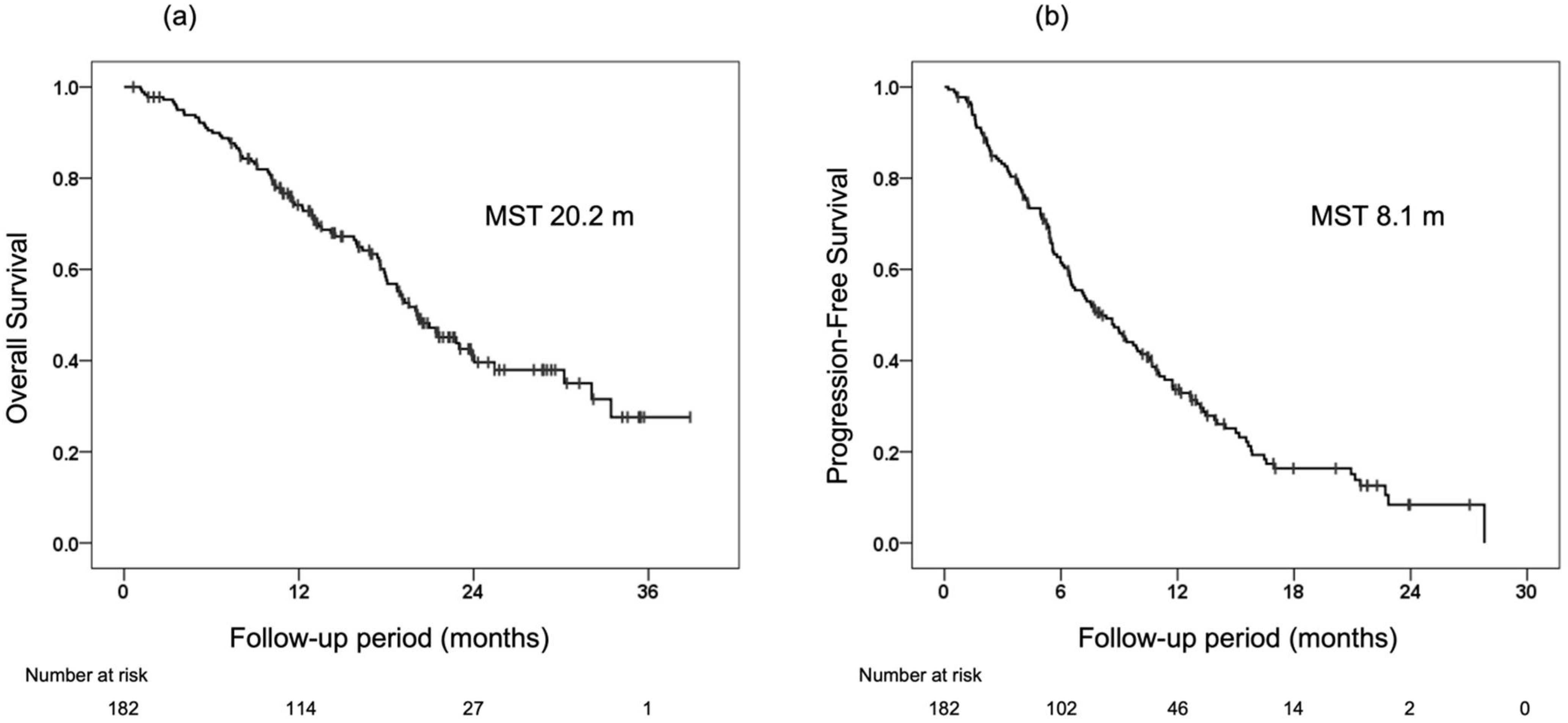
3.2. Treatment Response and Survival

The median OS and PFS of the 182 patients included in the study were 20.2 months and 8.1 months, respectively (Figure 1). Table 2 shows the radiological response at the first, second, and best times by RECIST and mRECIST evaluations. On the initial radiological response evaluation, 41 patients (23.7%) had OR, 102 (59.0%) had stable disease (SD), and 30 (17.3%) had progressive disease (PD) on RECIST evaluation (ORR 23.7%, DCR 82.7%), and 80 patients (47.6%) had OR, 60 (35.7%) had SD and 28 (16.7%) had PD on mRECIST evaluation (ORR 47.6%, DCR 83.3%). Similarly, good ORR and DCR were confirmed on both RECIST and mRECIST evaluations at the second and best response evaluations.

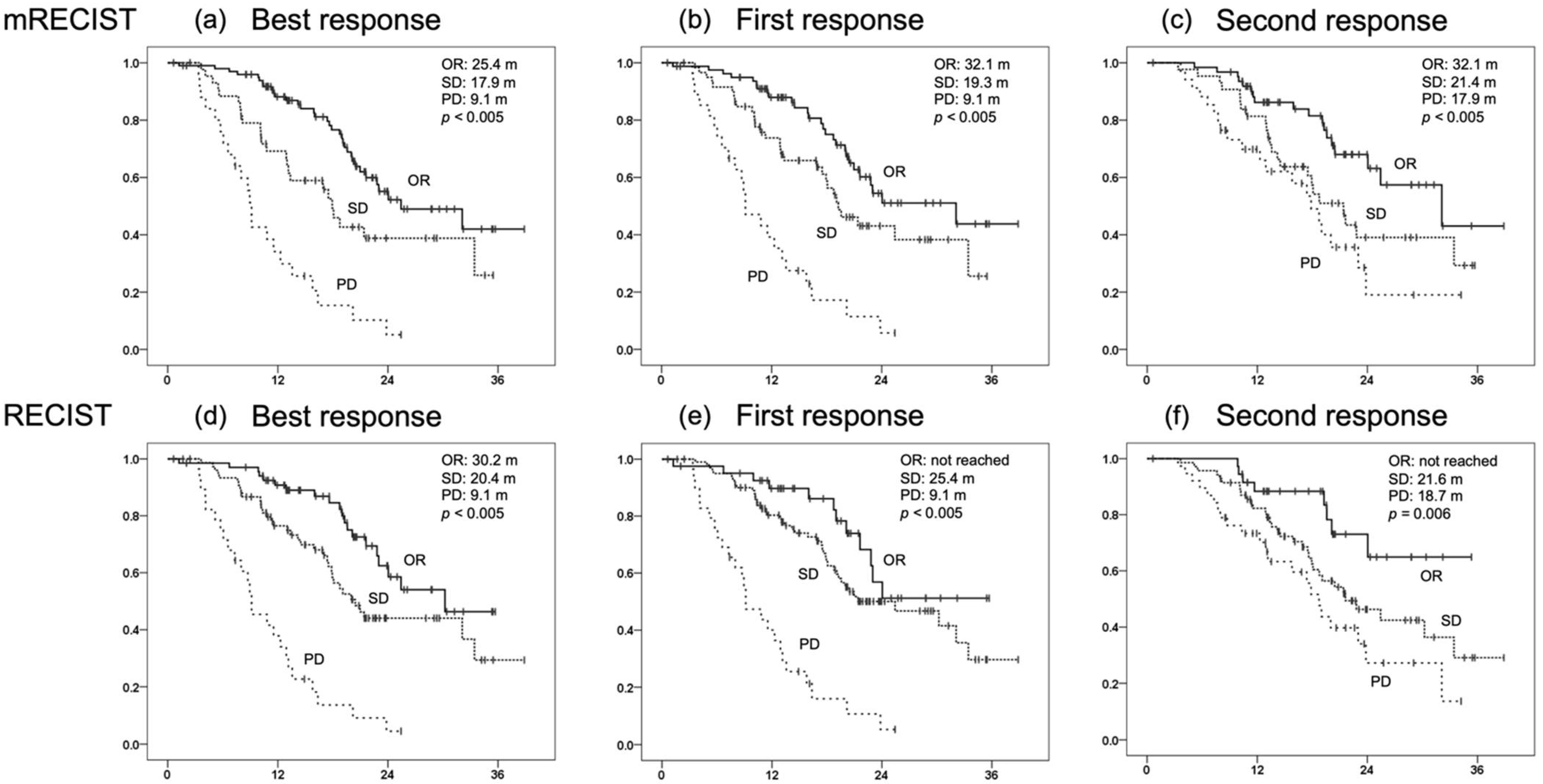
3.3. OS for Each Initial Radiological Response and Prognostic Factors for OS

The median OS by initial radiological response on RECIST was not reached in the OR group, but was 25.4 months in the SD group and 9.1 months in the PD group, while the median OS by initial radiological response on mRECIST was 32.1 months in the OR group, 19.3 months in the SD group, and 9.1 months in the PD group. Both RECIST and mRECIST evaluations showed significant OS stratification by response (RECIST: p < 0.005, mRECIST: p < 0.005). Similarly, the best and second radiological evaluations also showed significant stratification of OS for each response (Figure 2).
Next, the prognostic factors for OS were examined in patients treated with lenvatinib by univariate and multivariate analyses (Table 3). To avoid confounding factors, analyses were performed separately for RECIST and mRECIST. On multivariate analysis with factors including RECIST, etiology (hazard ratio, 0.605; 95% confidence interval, 0.380–0.962; p = 0.034), mALBI at initiation (hazard ratio, 0.409; 95% confidence interval, 0.249–0.674; p < 0.005), serum alpha-fetoprotein (AFP) level at initiation (hazard ratio, 0.409; 95% confidence interval, 0.251–0.667; p < 0.005), and initial radiological response on RECIST (hazard ratio, 0.369; 95% confidence interval, 0.197–0.691; p < 0.005) were independent prognostic factors for OS. On multivariate analysis of factors including mRECIST, the following were identified as independent prognostic factors for OS: mALBI at initiation (hazard ratio, 0.451; 95% confidence interval, 0.277–0.734; p < 0.005), serum AFP level at initiation (hazard ratio, 0.359; 95% confidence interval, 0.221–0.583; p < 0.005), and initial radiological response on mRECIST (hazard ratio, 0.378; 95% confidence interval, 0.234–0.611; p < 0.005). On both RECIST and mRECIST evaluations, good mALBI at initiation (1–2a), low AFP at initiation, and obtaining OR at the initial radiological response evaluation were extracted as independent prognostic factors for OS in lenvatinib.

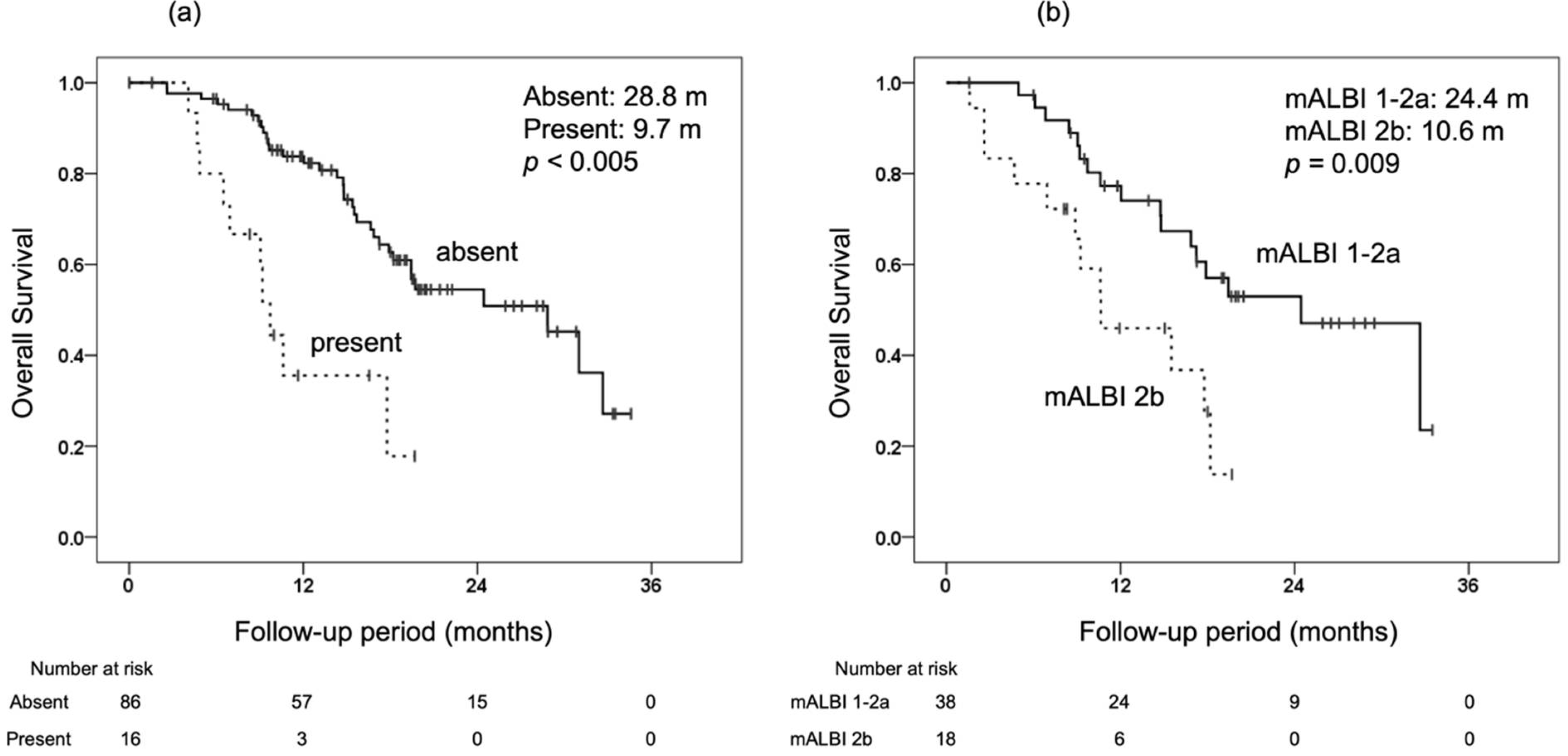
3.4. Prognostic Factors for OS in Patients with SD at the Initial Radiological Response Evaluation

Since it has been shown that obtaining OR at the initial radiological response evaluation contributes to longer OS, patients with SD at the initial radiological response evaluation were examined next. On univariate and multivariate analyses, the independent prognostic factors for OS from the initial response evaluation were examined separately for RECIST and mRECIST (Table 4). In patients with SD at the initial response evaluation on RECIST, macrovascular invasion (MVI) at the time of initial evaluation (hazard ratio, 0.347; 95% confidence interval, 0.143–0.843; p = 0.019) was the independent prognostic factor for OS from the initial evaluation on multivariate analysis. The median OS from the initial evaluation on RECIST was 28.8 months in the group without MVI at the initial evaluation and 9.7 months in the group with MVI (Figure 3). In patients with SD at the initial response evaluation on mRECIST, mALBI at the time of initial evaluation (hazard ratio, 0.381; 95% confidence interval, 0.156–0.932; p = 0.035) was an independent prognostic factor for OS from the initial evaluation on multivariate analysis. The median OS from the initial evaluation on mRECIST was 24.4 months in the group with mALBI of 1–2a at the initial evaluation and 10.6 months in the group with mALBI of 2b (Figure 3). On both RECIST and mRECIST evaluations, the second radiological response was not a prognostic factor on univariate analysis.

4. Discussion

In this study, the effect of radiological response on prognosis in patients with u-HCC who received lenvatinib as a first-line systemic treatment was investigated. The results showed that initial radiological evaluations based on either RECIST or mRECIST were each stratified with respect to OS, and that the initial radiological response was an independent prognostic factor for OS. In addition, stratification of OS by response was confirmed not only for the initial response, but also for the second and best responses.
Next, univariate and multivariate analyses of factors contributing to OS from the initial radiological evaluation in patients with SD at the initial response evaluation were performed. It was found that MVI at the time of the initial response evaluation was an independent prognostic factor for OS from the initial radiological evaluation on RECIST, and mALBI grade at the time of the initial response evaluation was an independent prognostic factor for OS from the initial radiological evaluation on mRECIST. On the other hand, the second radiological response was not a prognostic factor for OS from the initial radiological response based on either RECIST or mRECIST evaluations. These results suggest that, in patients with SD at the initial radiological evaluation, the decision to continue the current treatment or switch to the next treatment should be made with careful consideration of factors related to the tumor and hepatic reserve at the time of the initial radiological evaluation, rather than simply continuing and waiting for the second response evaluation.
Several predictors for the efficacy of lenvatinib (4-week relative dose intensity [19], AFP [20,21], ALBI grade [20,21,22], neutrophil-to-lymphocyte ratio [23], and occurrence of hypothyroidism [24]) have been reported previously. On radiological evaluation, obtaining OR by mRECIST evaluation has been reported to be an independent predictor of OS with other MTAs [25,26,27]. Kaneko et al. reported that early evaluation by RECIST 1.1 was useful for prognostic stratification in lenvatinib [28], and Kudo et al. reported that objective response by mRECIST evaluation was associated with OS in a multivariate analysis of responders to lenvatinib in the REFLECT trial [29]. Hiraoka et al. also reported that, when ECOG PS and hepatic reserve function permit, continuing lenvatinib beyond PD, especially in u-HCC patients who showed a hand–foot skin reaction during lenvatinib treatment, might be a good therapeutic option [30]. One of the characteristics of lenvatinib is that it has shorter time to response compared to other drugs, especially atezolizumab plus bevacizumab, which is becoming established as a first-line treatment for u-HCC. In order to evaluate the efficacy of lenvatinib effectively, it is very important to confirm the response early in the course of treatment, when the relative dose intensity can be relatively maintained and is less susceptible to intolerance and loss of hepatic reserve.
If the radiological evaluation shows OR, it is desirable to continue the treatment, as long as it is well tolerated because good OS prolongation can be expected. Conversely, if the radiological evaluation shows PD, it would be desirable to consider switching to the next treatment. However, if the radiological response shows SD, there is no clear consensus on whether to continue or to switch to the next treatment. In the REFLECT trial, lenvatinib showed good ORR, but PFS was limited to about 7.4 months. The major causes of this limitation are decreased tolerability, decreased hepatic reserve, and acquisition of tolerance. In order to prolong OS, which is the main goal in the treatment of HCC, we should be more careful about making decisions when the radiological response is SD. This study is the first to examine the direction of treatment using multivariate analysis of factors contributing to OS when the radiological response is SD, after confirming the stratification of OS by radiological response.
In this study, there was a clear difference in OS of 25.4 months for RECIST and 19.3 months for mRECIST in the group with SD on initial radiological evaluation, compared to the group with OR and PD. The mRECIST evaluation is a valid evaluation method for u-HCC, especially with an MTA with angiogenesis inhibition, and it has been used along with the RECIST evaluation for radiological evaluation of HCC. The reason for this may be that the mRECIST evaluation more sensitively shows antitumor effects, as reflected in the loss of staining compared to the RECIST evaluation. Kuzuya et al. indicated that radiological antitumor response by mRECIST (i.e., disappearance of arterial tumor enhancement) may not necessarily reflect tumor necrosis, especially soon after initiation of lenvatinib [31]; thus, careful judgment is needed when using mRECIST. Nevertheless, the SD evaluation by mRECIST (i.e., no disappearance of arterial tumor enhancement) may indicate that even the anti-tumor effect, which is the most important feature of lenvatinib, was not achieved, so more attention may be required in treatment planning than the SD evaluation by RECIST.
In recent years, with the increase of new drug therapies in u-HCC, the treatment paradigm has changed, and the selection, timing, and sequence of appropriate therapies have become major issues. The IMbrave150 trial demonstrated that atezolizumab plus bevacizumab combination therapy significantly improved median OS (not reached vs. 13.2 months; hazard ratio 0.58, p < 0.001), median PFS per RECIST version 1.1 (6.8 vs. 4.3 months; HR 0.59; p < 0.001), and ORR per RECIST version 1.1 (27 vs. 12%; p < 0.001) compared to sorafenib, and also maintained quality of life [16]. As a result, atezolizumab plus bevacizumab is expected to be a useful systemic therapy for u-HCC and is positioned as a first-line treatment, but the median PFS is limited to 6.9 months, as reported by the updated analysis of IMbrave150. Due to the favorable safety profile and quality of life of atezolizumab plus bevacizumab, it is expected that many patients will be able to maintain hepatic reserve after progression and move on to the next treatment, but the optimal sequence of atezolizumab plus bevacizumab after progression has not yet been established. Yoo et al. reported that second-line treatment with sorafenib and lenvatinib after progression on atezolizumab plus bevacizumab was as effective as these MTAs in the pivotal phase 3 trials [32]. The efficacy of sequential multidrug therapy in u-HCC has been reported in several studies and further investigation is needed, including atezolizumab plus bevacizumab [33,34]. Alsina et al. reported that, in a post hoc analysis of patients entered into the REFLECT trial, first-line lenvatinib followed by subsequent systemic therapy led to a longer OS and may provide greater survival benefit in patients who achieved OR to lenvatinib [35]. How to effectively manage lenvatinib, which has a particularly high response rate among MTAs, is considered to be a very important issue. A large-scale prospective observational study is currently being conducted in Japan to collect real-world data on sequential systemic drug therapy for HCC and is expected to establish effective treatment regimens through further data collection [36].
It is still unclear which systemic therapy to prefer as the next therapy after lenvatinib. One of the problems with lenvatinib is that it often requires dose reduction or withdrawal due to adverse events or loss of hepatic reserve, regardless of response. It is very important to determine the appropriate timing for switching to the next therapy while maintaining hepatic reserve without unreasonably continuing lenvatinib. In this study, responders showed clearly better OS as in previous reports, and we also observed a clear stratification of OS in patients with SD and PD. On the other hand, poor prognosis was observed in patients with SD who had poor hepatic reserve (mALBI grade 2b) or MVI at the initial evaluation. These results suggest that it may be possible to stratify patients with SD, especially by focusing on hepatic reserve and MVI at the initial evaluation, so that we can consider the appropriate timing of switching from lenvatinib for OS prolongation earlier. With the current availability of multiple systemic therapies for HCC, it may be necessary to consider switching to other MTAs even if SD is achieved with lenvatinib. In the near future, it is expected that further systemic treatment options will become available with the results of ongoing clinical trials focusing on immunotherapy. It is known that a certain number of patients are refractory to immunotherapy in terms of tumor microenvironment, but it has been suggested that the use of MTAs, including lenvatinib, in combination with immunotherapy or as the next treatment may lead to response. Therefore, the management of MTAs (especially lenvatinib) and the establishment of multidrug sequential therapy will continue to be important clinical issues. In the current paradigm shift in the systemic treatment of u-HCC, we need to continue to accumulate clinical cases to establish evidence.
This study had several limitations as a retrospective study with a small sample size, an insufficient observation period, a reduced starting dose in some patients, and time bias due to the timing of radiological evaluations (every 4–8 weeks). In addition, a prospective study with a longer observation period and a larger number of patients is needed to draw a more definitive conclusion on whether switching from lenvatinib actually leads to prolonged OS. Nevertheless, we believe that the results of this study will have an important impact on the decision-making process during treatment with lenvatinib.

5. Conclusions

Radiological response on both RECIST and mRECIST evaluations stratifies OS, and achieving objective response at the first evaluation is an independent prognostic factor for OS. In addition, if the initial evaluation shows SD, it may be important to consider factors related to the tumor and hepatic reserve when determining treatment strategy.

Author Contributions

K.A.: conceptualization, formal analysis, and writing—original draft; T.K.: conceptualization, review, and editing; M.K., Y.J., Y.S., R.M., S.M., S.Y., K.N., Y.A. (Yuwa Ando), Y.K. and Y.F.: data curation; K.K., S.U., H.F., A.O., T.N., E.M., W.O., M.Y., M.I., N.M., S.T., K.T., K.M., Y.H., H.K. (Hirotaka Kouno), H.K. (Hiroshi Kohno), T.M., N.N., M.N., H.H., Y.A. (Yasuyuki Aisaka), T.A. and A.H.: treated the patients; H.A.: review and editing. All authors have read and agreed to the published version of the manuscript.

Funding

This research received no external funding.

Institutional Review Board Statement

This study was approved by the Ethics Review Committee of Hiroshima University (project identification code number E-1226) on 26 June 2018. This was a retrospective analysis of records stored in a database and official approval was received based on the Guidelines for Clinical Research issued by the Ministry of Health, Labour and Welfare of Japan. All procedures complied with the Declaration of Helsinki.

Informed Consent Statement

Informed consent was obtained from all subjects involved in the study.

Data Availability Statement

The data that support the findings of this study are available from the corresponding author upon reasonable request.

Acknowledgments

The authors would like to thank the study participants and extend our gratitude to Kazuaki Chayama for instruction.

Conflicts of Interest

M. I. has received research funding from Bristol Myers Squibb and AbbVie. H. A. has received honoraria from Eisai and Bayer. The other authors declare no conflicts of interest.

References

  1. Forner, A.; Reig, M.; Bruix, J. Hepatocellular carcinoma. Lancet 2018, 391, 1301–1314. [Google Scholar] [CrossRef]
  2. Jemal, A.; Bray, F.; Center, M.M.; Ferlay, J.; Ward, E.; Forman, D. Global cancer statistics. CA Cancer J. Clin. 2011, 61, 69–90. [Google Scholar] [CrossRef] [PubMed] [Green Version]
  3. Llovet, J.M.; Real, M.I.; Montaña, X.; Planas, R.; Coll, S.; Aponte, J.; Ayuso, C.; Sala, M.; Muchart, J.; Sola, R.; et al. Arterial embolization or chemoembolization versus symptomatic treatment in patients with unresectable hepatocellular carcinoma: A randomised controlled trial. Lancet 2002, 359, 1734–1739. [Google Scholar] [CrossRef]
  4. Obi, S.; Yoshida, H.; Toune, R.; Unuma, T.; Kanda, M.; Sato, S.; Tateishi, R.; Teratani, T.; Shiina, S.; Omata, M. Combination therapy of intra-arterial 5-fluorouracil and systemic interferon-alpha for advanced hepatocellular carcinoma with portal venous invasion. Cancer 2006, 106, 1990–1997. [Google Scholar] [CrossRef]
  5. Tateishi, R.; Shiina, S.; Teratani, T.; Obi, S.; Sato, S.; Koike, Y.; Fujishima, T.; Yoshida, H.; Kawabe, T.; Omata, M. Percutaneous radiofrequency ablation for hepatocellular carcinoma. An analysis of 1000 cases. Cancer 2005, 103, 1201–1209. [Google Scholar] [CrossRef]
  6. Miyagawa, S.; Makuuchi, M.; Kawasaki, S.; Kakazu, T. Criteria for safe hepatic resection. Am. J. Surg. 1995, 169, 589–594. [Google Scholar] [CrossRef]
  7. Uka, K.; Aikata, H.; Takaki, S.; Miki, D.; Kawaoka, T.; Jeong, S.C.; Takahashi, S.; Toyota, N.; Ito, K.; Chayama, K. Pretreatment predictor of response, time to progression, and survival to intraarterial 5-fluorouracil/interferon combination therapy in patients with advanced hepatocellular carcinoma. J. Gastroenterol. 2007, 42, 845–853. [Google Scholar] [CrossRef]
  8. Llovet, J.M.; Ricci, S.; Mazzaferro, V.; Hilgard, P.; Gane, E.; Blanc, J.F.; Oliveira, A.C.; Santoro, A.; Raoul, J.L.; Forner, A.; et al. Sorafenib in advanced hepatocellular carcinoma. N. Engl. J. Med. 2008, 359, 378–390. [Google Scholar] [CrossRef]
  9. Kudo, M.; Finn, R.S.; Qin, S.; Han, K.H.; Ikeda, K.; Piscaglia, F.; Baron, A.; Park, J.W.; Han, G.; Jassem, J.; et al. Lenvatinib versus sorafenib in first-line treatment of patients with unresectable hepatocellular carcinoma: A randomised phase 3 non-inferiority trial. Lancet 2018, 391, 1163–1173. [Google Scholar] [CrossRef] [Green Version]
  10. Bruix, J.; Qin, S.; Merle, P.; Granito, A.; Huang, Y.H.; Bodoky, G.; Pracht, M.; Yokosuka, O.; Rosmorduc, O.; Breder, V.; et al. Regorafenib for patients with hepatocellular carcinoma who progressed on sorafenib treatment (RESORCE): A randomised, double-blind, placebo-controlled, phase 3 trial. Lancet 2017, 389, 56–66. [Google Scholar] [CrossRef] [Green Version]
  11. Zhu, A.X.; Finn, R.S.; Galle, P.R.; Llovet, J.M.; Kudo, M. Ramucirumab in advanced hepatocellular carcinoma in REACH-2: The true value of alpha-fetoprotein. Lancet Oncol. 2019, 20, e191. [Google Scholar] [CrossRef] [Green Version]
  12. Abou-Alfa, G.K.; Meyer, T.; Cheng, A.L.; El-Khoueiry, A.B.; Rimassa, L.; Ryoo, B.Y.; Cicin, I.; Merle, P.; Chen, Y.; Park, J.W.; et al. Cabozantinib in patients with advanced and progressing hepatocellular carcinoma. N. Engl. J. Med. 2018, 379, 54–63. [Google Scholar] [CrossRef] [PubMed]
  13. Finn, R.S.; Qin, S.; Ikeda, M.; Galle, P.R.; Ducreux, M.; Kim, T.Y.; Kudo, M.; Breder, V.; Merle, P.; Kaseb, A.O.; et al. Atezolizumab plus bevacizumab in unresectable hepatocellular carcinoma. N. Engl. J. Med. 2020, 382, 1894–1905. [Google Scholar] [CrossRef] [PubMed]
  14. Rizzo, A.; Dadduzio, V.; Ricci, A.D.; Massari, F.; Di Federico, A.; Gadaleta-Caldarola, G.; Brandi, G. Lenvatinib plus pembrolizumab: The next frontier for the treatment of hepatocellular carcinoma? Expert Opin. Investig. Drugs 2021, 1–8. [Google Scholar] [CrossRef]
  15. Pfister, D.; Nunez, N.G.; Pinyol, R.; Govaere, O.; Pinter, M.; Szydlowska, M.; Gupta, R.; Qiu, M.; Deczkowska, A.; Weiner, A.; et al. NASH limits anti-tumour surveillance in immunotherapy-treated HCC. Nature 2021, 592, 450–456. [Google Scholar] [CrossRef]
  16. Ding, Z.; Dong, Z.; Chen, Z.; Hong, J.; Yan, L.; Li, H.; Yao, S.; Yan, Y.; Yang, Y.; Yang, C.; et al. Viral status and efficacy of immunotherapy in hepatocellular carcinoma: A systematic review with meta-analysis. Front. Immunol. 2021, 12, 733530. [Google Scholar] [CrossRef]
  17. Johnson, P.J.; Berhane, S.; Kagebayashi, C.; Satomura, S.; Teng, M.; Reeves, H.L.; O’Beirne, J.; Fox, R.; Skowronska, A.; Palmer, D.; et al. Assessment of liver function in patients with hepatocellular carcinoma: A new evidence-based approach-the ALBI grade. J. Clin. Oncol. 2015, 33, 550–558. [Google Scholar] [CrossRef]
  18. Hiraoka, A.; Kumada, T.; Tsuji, K.; Takaguchi, K.; Itobayashi, E.; Kariyama, K.; Ochi, H.; Tajiri, K.; Hirooka, M.; Shimada, N.; et al. Validation of modified ALBI grade for more detailed assessing hepatic function in hepatocellular carcinoma patients: A multicenter analysis. Liver Cancer 2019, 8, 121–129. [Google Scholar] [CrossRef]
  19. Kirino, S.; Tsuchiya, K.; Kurosaki, M.; Kaneko, S.; Inada, K.; Yamashita, K.; Osawa, L.; Hayakawa, Y.; Sekiguchi, S.; Okada, M.; et al. Relative dose intensity over the first four weeks of lenvatinib therapy is a factor of favorable response and overall survival in patients with unresectable hepatocellular carcinoma. PLoS ONE 2020, 15, e0231828. [Google Scholar] [CrossRef] [Green Version]
  20. Saeki, I.; Yamasaki, T.; Yamashita, S.; Hanazono, T.; Urata, Y.; Furutani, T.; Yokoyama, Y.; Oishi, T.; Maeda, M.; Kimura, T.; et al. Early predictors of objective response in patients with hepatocellular carcinoma undergoing lenvatinib treatment. Cancers 2020, 12, 779. [Google Scholar] [CrossRef] [Green Version]
  21. Fuchigami, A.; Imai, Y.; Uchida, Y.; Uchiya, H.; Fujii, Y.; Nakazawa, M.; Ando, S.; Sugawara, K.; Nakayama, N.; Tomiya, T.; et al. Therapeutic efficacy of lenvatinib for patients with unresectable hepatocellular carcinoma based on the middle-term outcome. PLoS ONE 2020, 15, e0231427. [Google Scholar] [CrossRef] [PubMed]
  22. Hiraoka, A.; Kumada, T.; Atsukawa, M.; Hirooka, M.; Tsuji, K.; Ishikawa, T.; Takaguchi, K.; Kariyama, K.; Itobayashi, E.; Tajiri, K.; et al. Prognostic factor of lenvatinib for unresectable hepatocellular carcinoma in real-world conditions—Multicenter analysis. Cancer Med. 2019, 8, 3719–3728. [Google Scholar] [CrossRef] [Green Version]
  23. Nakano, M.; Kuromatsu, R.; Niizeki, T.; Okamura, S.; Iwamoto, H.; Shimose, S.; Shirono, T.; Noda, Y.; Kamachi, N.; Koga, H.; et al. Immunological inflammatory biomarkers as prognostic predictors for advanced hepatocellular carcinoma. ESMO Open 2021, 6, 100020. [Google Scholar] [CrossRef]
  24. Shomura, M.; Okabe, H.; Sato, E.; Fukai, K.; Shiraishi, K.; Hirose, S.; Tsuruya, K.; Arase, Y.; Anzai, K.; Kagawa, T. Hypothyroidism is a predictive factor for better clinical outcomes in patients with advanced hepatocellular carcinoma undergoing lenvatinib therapy. Cancers 2020, 12, 3078. [Google Scholar] [CrossRef]
  25. Lencioni, R.; Montal, R.; Torres, F.; Park, J.W.; Decaens, T.; Raoul, J.L.; Kudo, M.; Chang, C.; Rios, J.; Boige, V.; et al. Objective response by mRECIST as a predictor and potential surrogate end-point of overall survival in advanced HCC. J. Hepatol. 2017, 66, 1166–1172. [Google Scholar] [CrossRef] [PubMed] [Green Version]
  26. Meyer, T.; Palmer, D.H.; Cheng, A.L.; Hocke, J.; Loembe, A.B.; Yen, C.J. mRECIST to predict survival in advanced hepatocellular carcinoma: Analysis of two randomised phase II trials comparing nintedanib vs sorafenib. Liver Int. 2017, 37, 1047–1055. [Google Scholar] [CrossRef] [PubMed] [Green Version]
  27. Vincenzi, B.; Di Maio, M.; Silletta, M.; D’Onofrio, L.; Spoto, C.; Piccirillo, M.C.; Daniele, G.; Comito, F.; Maci, E.; Bronte, G.; et al. Prognostic relevance of objective response according to EASL criteria and mRECIST criteria in hepatocellular carcinoma patients treated with loco-regional therapies: A literature-based meta-analysis. PLoS ONE 2015, 10, e0133488. [Google Scholar] [CrossRef]
  28. Kaneko, S.; Tsuchiya, K.; Kurosaki, M.; Kirino, S.; Inada, K.; Yamashita, K.; Osawa, L.; Hayakawa, Y.; Sekiguchi, S.; Watakabe, K.; et al. Three criteria for radiological response on survival in patients with hepatocellular carcinoma treated with lenvatinib. Hepatol. Res. 2020, 50, 137–143. [Google Scholar] [CrossRef] [Green Version]
  29. Kudo, M. Objective response by mRECIST is an independent prognostic factor of overall survival in systemic therapy for hepatocellular carcinoma. Liver Cancer 2019, 8, 73–77. [Google Scholar] [CrossRef]
  30. Hiraoka, A.; Kumada, T.; Tada, T.; Kariyama, K.; Tani, J.; Fukunishi, S.; Atsukawa, M.; Hirooka, M.; Tsuji, K.; Ishikawa, T.; et al. What can be done to solve the unmet clinical need of hepatocellular carcinoma patients following lenvatinib failure. Liver Cancer 2021, 10, 115–125. [Google Scholar] [CrossRef]
  31. Kuzuya, T.; Ishigami, M.; Ito, T.; Ishizu, Y.; Honda, T.; Ishikawa, T.; Fujishiro, M. Favorable radiological antitumor response at 2 weeks after starting lenvatinib for patients with advanced hepatocellular carcinoma. Hepatol. Res. 2019, 50, 374–381. [Google Scholar] [CrossRef] [PubMed]
  32. Yoo, C.; Kim, J.H.; Ryu, M.H.; Park, S.R.; Lee, D.; Kim, K.M.; Shim, J.H.; Lim, Y.S.; Lee, H.C.; Lee, J.; et al. Clinical outcomes with multikinase Inhibitors after progression on first-line atezolizumab plus bevacizumab in patients with advanced hepatocellular carcinoma: A multinational multicenter retrospective study. Liver Cancer 2021, 10, 107–114. [Google Scholar] [CrossRef]
  33. Ogasawara, S.; Ooka, Y.; Itokawa, N.; Inoue, M.; Okabe, S.; Seki, A.; Haga, Y.; Obu, M.; Atsukawa, M.; Itobayashi, E.; et al. Sequential therapy with sorafenib and regorafenib for advanced hepatocellular carcinoma: A multicenter retrospective study in Japan. Investig. New Drugs 2020, 38, 172–180. [Google Scholar] [CrossRef] [PubMed]
  34. Ando, Y.; Kawaoka, T.; Suehiro, Y.; Yamaoka, K.; Kosaka, Y.; Uchikawa, S.; Kodama, K.; Morio, K.; Fujino, H.; Nakahara, T.; et al. Analysis of post-progression survival in patients with unresectable hepatocellular carcinoma treated with lenvatinib. Oncology 2020, 98, 787–797. [Google Scholar] [CrossRef]
  35. Alsina, A.; Kudo, M.; Vogel, A.; Cheng, A.L.; Tak, W.Y.; Ryoo, B.Y.; Evans, T.R.J.; Lopez, C.L.; Daniele, B.; Misir, S.; et al. Effects of subsequent systemic anticancer medication following first-line lenvatinib: A post hoc responder analysis from the phase 3 REFLECT study in unresectable hepatocellular carcinoma. Liver Cancer 2020, 9, 93–104. [Google Scholar] [CrossRef] [PubMed]
  36. Prospective Observational Study of Systemic Therapy for Unresectable HCC in Japan: Real World Data of Systemic Therapy for HCC; UMIN ID 000040488. Available online: https://upload.umin.ac.jp/cgi-open-bin/ctr/ctr_view.cgi?recptno=R000046213 (accessed on 21 October 2021).
Figure 1. Overall survival (OS) and progression-free survival (PFS) from the initiation of lenvatinib in the 182 patients included in this study. (a) OS from the initiation of lenvatinib. (b) PFS from the initiation of lenvatinib.
Cancers 14 00320 g001
Figure 2. Comparison of overall survival (OS) by response at the first, second, and best responses evaluated by Response Evaluation Criteria in Solid Tumors (RECIST) and modified Response Evaluation Criteria in Solid Tumors (mRECIST). (a) OS at the best response evaluated by mRECIST (OR 25.4 months, SD 17.9 months, PD 9.1 months, p < 0.005). (b) OS at the first response evaluated by mRECIST (OR 32.1 months, SD 19.3 months, PD 9.1 months, p < 0.005). (c) OS at the second response evaluated by mRECIST (OR 32.1 months, SD 21.4 months, PD 17.9 months, p < 0.005). (d) OS at the best response evaluated by RECIST (OR 30.2 months, SD 20.4 months, PD 9.1 months, p < 0.005). (e) OS at the first response evaluated by RECIST (OR not reached, SD 25.4 months, PD 9.1 months, p < 0.005). (f) OS at the second response evaluated by RECIST (OR not reached, SD 21.6 months, PD 18.7 months, p = 0.006).
Cancers 14 00320 g002
Figure 3. Comparison of overall survival (OS) from the initial objective evaluation of lenvatinib in patients with SD at the initial radiological response evaluation. (a) OS from the initial objective evaluation by RECIST with or without macroscopic vascular invasion (absent 28.8 months, present 9.7 months, p < 0.005). (b) OS from the initial objective evaluation by mRECIST by mALBI grade (mALBI 1–2a 24.4 months, mALBI 2b 10.6 months, p = 0.009).
Cancers 14 00320 g003
Table 1. Clinical characteristics at the initiation of lenvatinib (n = 182).
CharacteristicMedian (Range)
or Patients, n
Age, range, y74 (46–90)
Sex (male/female), n154/28
Weight (<60/>60 kg), n84/98
Performance status (0/1), n166/16
Etiology (HBV/HCV/HBV + HCV/NBNC), n22/62/1/97
History of non-systemic treatment (with/without), n146/36
Total bilirubin, range, mg/dL0.8 (0.3–2.1)
Albumin, range, g/dL3.9 (2.9–4.9)
Prothrombin activity, range, %90 (59–131)
Child–Pugh score (5/6), n126/56
mALBI grade (1/2a/2b), n80/51/51
Size of main tumor, range, mm24.0 (0.0–190.0)
Relative tumor volume (<50/≥50%), n166/16
Macroscopic vascular invasion (absent/present), n153/29
Extrahepatic metastasis (absent/present), n130/52
BCLC stage (B/C), n110/72
Serum AFP value, range, ng/mL20.2 (0.5–236900.0)
Serum DCP value, range, mAU/mL174.0 (13.0–1083990.0)
Observation period, range, months14.7 (0.6–38.9)
HBV, hepatitis B virus infection; HCV, hepatitis C virus infection; NBNC, non-B-non-C viral hepatitis; mALBI, modified albumin–bilirubin; BCLC, Barcelona Clinic liver cancer; AFP, alpha-fetoprotein; DCP, des-γ-carboxy prothrombin.
Table 2. Radiological responses to lenvatinib.
ResponseRECIST % (n)mRECIST % (n)
Best1st2ndBest1st2nd
CR4.0 (7)2.3 (4)2.1 (3)17.1 (29)7.7 (13)12.9 (18)
PR35.6 (62)21.4 (37)23.4 (34)42.4 (72)39.9 (67)32.1 (45)
SD44.3 (77)59.0 (102)48.3 (70)25.9 (44)35.7 (60)30.7 (43)
PD16.1 (28)17.3 (30)26.2 (38)14.7 (25)16.7 (28)24.3 (34)
ORR39.7 (69)23.7 (41)25.5 (37)59.4 (101)47.6 (80)45.0 (63)
DCR83.9 (146)82.7 (143)73.8 (107)85.3 (145)83.3 (140)75.7 (106)
RECIST, Response Evaluation Criteria in Solid Tumors; mRECIST, modified Response Evaluation Criteria in Solid Tumors; CR, complete response; PR, partial response; SD, stable disease; PD, progressive disease; ORR, overall response rate; DCR, disease control rate.
Table 3. Univariate and multivariate analyses of prognostic factors for overall survival.
FactorsUnivariate
p-Value
Multivariate
HR95% CIp-Value
Age (<74 vs. ≥74 years)0.302
Sex (male vs. female)0.279
Etiology (NBNC vs. viral)0.0100.6050.380–0.9620.034
History of non-systemic treatment (with vs. without)0.981
mALBI grade (1/2a vs. 2b)<0.0050.4090.249–0.674<0.005
Macroscopic vascular invasion (absent vs. present)<0.0050.8380.320–1.1290.113
Extrahepatic metastasis (absent vs. present)0.0100.6010.456–1.1990.221
Relative tumor volume (<50% vs. ≥50%)<0.0050.7400.377–1.8660.666
Serum AFP value (<400 vs. ≥400), ng/mL<0.0050.4090.251–0.667<0.005
Serum DCP value (<174 vs. ≥174), ng/mL0.133
Initial objective response by RECIST (OR vs. non-OR)0.0070.3690.197–0.691<0.005
Age (<74 vs. ≥74 years)0.302
Sex (male vs. female)0.279
Etiology (NBNC vs. viral)0.0100.6620.416–1.0550.083
History of non-systemic treatment (with vs. without)0.981
mALBI grade (1/2a vs. 2b)<0.0050.4510.277–0.734<0.005
Macroscopic vascular invasion (absent vs. present)<0.0050.8610.437–1.6970.666
Extrahepatic metastasis (absent vs. present)0.0100.7860.484–1.2780.332
Relative tumor volume (<50% vs. ≥50%)<0.0050.4880.215–1.1110.087
Serum AFP value (<400 vs. ≥400), ng/mL<0.0050.3590.221–0.583<0.005
Serum DCP value (<174 vs. ≥174), ng/mL0.133
Initial objective response by mRECIST (OR vs. non-OR)<0.0050.3780.234–0.611<0.005
NBNC, non-B-non-C viral hepatitis; mALBI, modified albumin–bilirubin; AFP, alpha-fetoprotein; DCP, des-γ-carboxy prothrombin; RECIST, Response Evaluation Criteria in Solid Tumors; mRECIST, modified Response Evaluation Criteria in Solid Tumors; OR, objective response.
Table 4. Univariate and multivariate analyses of prognostic factors for overall survival from initial objective evaluation in patients with SD at the initial radiological response evaluation.
FactorsUnivariate
p-Value
Multivariate
HR95% CIp-Value
Age (<74 vs. ≥74 years)0.444
Sex (female vs. male)0.072
Etiology (NBNC vs. viral)0.0160.5840.303–1.1240.107
History of non-systemic treatment (with vs. without)0.555
mALBI grade at initial objective evaluation (1/2a vs. 2b)0.0310.7430.352–1.5670.435
Decrease in AFP value up to initial objective evaluation
(yes vs. no)
0.482
Decrease in DCP value up to initial objective evaluation
(yes vs. no)
0.574
Relative dose intensity up to initial objective evaluation
(<0.8 vs. ≥0.8)
0.540
Macroscopic vascular invasion at initial objective evaluation
(absent vs. present)
<0.0050.3470.143–0.8430.019
Extrahepatic metastasis at initial objective evaluation
(absent vs. present)
0.169
Relative tumor volume at initial objective evaluation
(<50% vs. ≥50%)
0.0050.4640.158–1.3610.162
Second objective response by RECIST (OR vs. non-OR)0.225
Age (<74 vs. ≥74 years)0.946
Sex (female vs. male)0.542
Etiology (NBNC vs. viral)0.052
History of non-systemic treatment (with vs. without)0.911
mALBI grade at initial objective evaluation (1/2a vs. 2b)0.0090.3810.156–0.9320.035
Decrease in AFP value up to initial objective evaluation
(yes vs. no)
0.323
Decrease in DCP value up to initial objective evaluation
(yes vs. no)
0.848
Relative dose intensity up to initial objective evaluation
(<0.8 vs. ≥0.8)
0.302
Macroscopic vascular invasion at initial objective evaluation
(absent vs. present)
0.0130.6710.247–1.8240.435
Extrahepatic metastasis at initial objective evaluation
(absent vs. present)
0.212
Relative tumor volume at initial objective evaluation
(<50% vs. ≥50%)
<0.0050.2160.042–1.1140.067
Second objective response by mRECIST (OR vs. non-OR)0.443
SD, stable disease; NBNC, non-B-non-C viral hepatitis; mALBI, modified albumin–bilirubin; AFP, alpha-fetoprotein; DCP, des-γ-carboxy prothrombin; RECIST, Response Evaluation Criteria in Solid Tumors; mRECIST, modified Response Evaluation Criteria in Solid Tumors; OR, objective response.
Publisher’s Note: MDPI stays neutral with regard to jurisdictional claims in published maps and institutional affiliations.

