Next Issue
Volume 14, February-1
Previous Issue
Volume 14, January-1


Cancers, Volume 14, Issue 2 (January-2 2022) – 199 articles

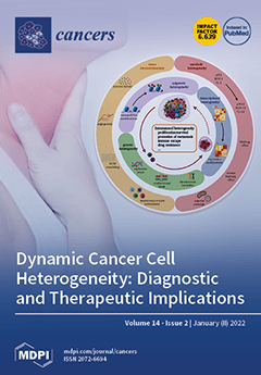
Cover Story (view full-size image): The complex architecture of cancer, resulting from intra-tumoral heterogeneity (ITH), remains the main obstacle to effective therapy. ITH can arise from genetic, epigenetic, and metabolic alterations in response to environmental pressures. This results in an uneven distribution of distinct tumor cell subpopulations across and within disease sites (spatial heterogeneity) and variations in the molecular composition of cancer cells across time (temporal heterogeneity). Recent technologies, such as liquid biopsies and spatial transcriptomics, offer considerable potential to assess the driving forces behind ITH. A better understanding of the dynamic heterogeneity of cancer could prove useful to overcome the limitations of precision medicine, as well as for new diagnostic approaches, and should lead to a constant reevaluation of present habits and fashions in concepts, diagnosis, and therapy. View this paper.
  • Issues are regarded as officially published after their release is announced to the table of contents alert mailing list.
  • You may sign up for e-mail alerts to receive table of contents of newly released issues.
  • PDF is the official format for papers published in both, html and pdf forms. To view the papers in pdf format, click on the "PDF Full-text" link, and use the free Adobe Reader to open them.
Order results
Result details
Section
Select all
Export citation of selected articles as:
14 pages, 1672 KiB  
Article
Comprehensive Approach to Distinguish Patients with Solid Tumors from Healthy Controls by Combining Androgen Receptor Mutation p.H875Y with Cell-Free DNA Methylation and Circulating miRNAs
by Elena Tomeva, Olivier J. Switzeny, Clemens Heitzinger, Berit Hippe and Alexander G. Haslberger
Cancers 2022, 14(2), 462; https://0-doi-org.brum.beds.ac.uk/10.3390/cancers14020462 - 17 Jan 2022
Cited by 17 | Viewed by 6138
Abstract
Liquid biopsy-based tests emerge progressively as an important tool for cancer diagnostics and management. Currently, researchers focus on a single biomarker type and one tumor entity. This study aimed to create a multi-analyte liquid biopsy test for the simultaneous detection of several solid [...] Read more.
Liquid biopsy-based tests emerge progressively as an important tool for cancer diagnostics and management. Currently, researchers focus on a single biomarker type and one tumor entity. This study aimed to create a multi-analyte liquid biopsy test for the simultaneous detection of several solid cancers. For this purpose, we analyzed cell-free DNA (cfDNA) mutations and methylation, as well as circulating miRNAs (miRNAs) in plasma samples from 97 patients with cancer (20 bladder, 9 brain, 30 breast, 28 colorectal, 29 lung, 19 ovarian, 12 pancreas, 27 prostate, 23 stomach) and 15 healthy controls via real-time qPCR. Androgen receptor p.H875Y mutation (AR) was detected for the first time in bladder, lung, stomach, ovarian, brain, and pancreas cancer, all together in 51.3% of all cancer samples and in none of the healthy controls. A discriminant function model, comprising cfDNA mutations (COSM10758, COSM18561), cfDNA methylation markers (MLH1, MDR1, GATA5, SFN) and miRNAs (miR-17-5p, miR-20a-5p, miR-21-5p, miR-26a-5p, miR-27a-3p, miR-29c-3p, miR-92a-3p, miR-101-3p, miR-133a-3p, miR-148b-3p, miR-155-5p, miR-195-5p) could further classify healthy and tumor samples with 95.4% accuracy, 97.9% sensitivity, 80% specificity. This multi-analyte liquid biopsy-based test may help improve the simultaneous detection of several cancer types and underlines the importance of combining genetic and epigenetic biomarkers. Full article
(This article belongs to the Special Issue Liquid Nucleic Acid-Based Biomarkers in Solid Tumors)
Show Figures

Graphical abstract

11 pages, 829 KiB  
Article
A Novel Urinary miRNA Biomarker for Early Detection of Colorectal Cancer
by Hiroyasu Iwasaki, Takaya Shimura, Mika Kitagawa, Tamaki Yamada, Ruriko Nishigaki, Shigeki Fukusada, Yusuke Okuda, Takahito Katano, Shin-ichi Horike and Hiromi Kataoka
Cancers 2022, 14(2), 461; https://0-doi-org.brum.beds.ac.uk/10.3390/cancers14020461 - 17 Jan 2022
Cited by 15 | Viewed by 3811
Abstract
Since noninvasive biomarkers as an alternative to invasive colonoscopy to detect colorectal cancer (CRC) are desired, we conducted this study to determine the urinary biomarker consisting of microRNAs (miRNAs). In total, 415 age- and sex-matched participants, including 206 patients with CRC and 209 [...] Read more.
Since noninvasive biomarkers as an alternative to invasive colonoscopy to detect colorectal cancer (CRC) are desired, we conducted this study to determine the urinary biomarker consisting of microRNAs (miRNAs). In total, 415 age- and sex-matched participants, including 206 patients with CRC and 209 healthy controls (HCs), were randomly divided into three groups: (1) the discovery cohort (CRC, n = 3; HC, n = 6); (2) the training cohort (140 pairs); and (3) the validation cohort (63 pairs). Among 11 urinary miRNAs with aberrant expressions between the two groups, miR-129-1-3p and miR-566 were significantly independent biomarkers that detect CRC. The panel consisting of two miRNAs could distinguish patients with CRC from HC participants with an area under the curve (AUC) = 0.811 in the training cohort. This panel showed good efficacy with an AUC = 0.868 in the validation cohort. This urinary biomarker combining miR-129-1-3p and miR-566 could detect even stage 0/I CRC effectively with an AUC = 0.845. Moreover, the expression levels of both miR-129-1-3p and miR-566 were significantly higher in primary tumor tissues than in adjacent normal tissue. Our established novel biomarker consisting of urinary miR-129-1-3p and miR-566 enables noninvasive and early detection of CRC. Full article
(This article belongs to the Special Issue Novel Biomarkers of Gastrointestinal Cancer)
Show Figures

Figure 1

17 pages, 3173 KiB  
Article
Molecular Landscape of the Coagulome of Oral Squamous Cell Carcinoma
by Marine Lottin, Simon Soudet, Julie Fercot, Floriane Racine, Julien Demagny, Jérémie Bettoni, Denis Chatelain, Marie-Antoinette Sevestre, Youcef Mammeri, Michele Lamuraglia, Antoine Galmiche and Zuzana Saidak
Cancers 2022, 14(2), 460; https://0-doi-org.brum.beds.ac.uk/10.3390/cancers14020460 - 17 Jan 2022
Cited by 11 | Viewed by 2909
Abstract
Background: Hemostatic complications, ranging from thromboembolism to bleeding, are a significant source of morbidity and mortality in cancer patients. The tumor coagulome represents the multiple genes and proteins that locally contribute to the equilibrium between coagulation and fibrinolysis. We aimed to study the [...] Read more.
Background: Hemostatic complications, ranging from thromboembolism to bleeding, are a significant source of morbidity and mortality in cancer patients. The tumor coagulome represents the multiple genes and proteins that locally contribute to the equilibrium between coagulation and fibrinolysis. We aimed to study the coagulome of Oral Squamous Cell Carcinoma (OSCC) and examine its link to the tumor microenvironment (TME). Methods: We used data from bulk tumor DNA/RNA-seq (The Cancer Genome Atlas), single-cell RNA-seq data and OSCC cells in culture. Results: Among all tumor types, OSCC was identified as the tumor with the highest mRNA expression levels of F3 (Tissue Factor, TF) and PLAU (urokinase type-plasminogen activator, uPA). Great inter- and intra-tumor heterogeneity were observed. Single-cell analyses showed the coexistence of subpopulations of pro-coagulant and pro-fibrinolytic cancer cells within individual tumors. Interestingly, OSCC with high F3 expressed higher levels of the key immune checkpoint molecules CD274/PD-L1, PDCD1LG2/PD-L2 and CD80, especially in tumor dendritic cells. In vitro studies confirmed the particularity of the OSCC coagulome and suggested that thrombin exerts indirect effects on OSCC cells. Conclusions: OSCC presents a specific coagulome. Further studies examining a possible negative modulation of the tumor’s adaptive immune response by the coagulation process are warranted. Full article
(This article belongs to the Special Issue Advances in Oral Cancer: From Pathology to Treatment)
Show Figures

Figure 1

20 pages, 3632 KiB  
Article
HELLS Is Negatively Regulated by Wild-Type P53 in Liver Cancer by a Mechanism Involving P21 and FOXM1
by Stefanie Schuller, Jan Sieker, Philip Riemenschneider, Bianca Köhler, Elisabeth Drucker, Sofia M. E. Weiler, Daniel Dauch, Carsten Sticht, Benjamin Goeppert, Stephanie Roessler, Silvia Ribback, Kai Breuhahn, Falko Fend, Frank Dombrowski, Kerstin Singer and Stephan Singer
Cancers 2022, 14(2), 459; https://0-doi-org.brum.beds.ac.uk/10.3390/cancers14020459 - 17 Jan 2022
Cited by 5 | Viewed by 3042
Abstract
The major tumor suppressor P53 (TP53) acts primarily as a transcription factor by activating or repressing subsets of its numerous target genes, resulting in different cellular outcomes (e.g., cell cycle arrest, apoptosis and senescence). P53-dependent gene regulation is linked to several [...] Read more.
The major tumor suppressor P53 (TP53) acts primarily as a transcription factor by activating or repressing subsets of its numerous target genes, resulting in different cellular outcomes (e.g., cell cycle arrest, apoptosis and senescence). P53-dependent gene regulation is linked to several aspects of chromatin remodeling; however, regulation of chromatin-modifying enzymes by P53 is poorly understood in hepatocarcinogenesis. Herein, we identified Helicase, lymphoid specific (HELLS), a major epigenetic regulator in liver cancer, as a strong and selective P53 repression target within the SNF2-like helicase family. The underlying regulatory mechanism involved P53-dependent induction of P21 (CDKN1A), leading to repression of Forkhead Box Protein M1 (FOXM1) that in turn resulted in downregulation of HELLS expression. Supporting our in vitro data, we found higher expression of HELLS in murine HCCs arising in a Trp53−/− background compared to Trp53+/+ HCCs as well as a strong and highly significant correlation between HELLS and FOXM1 expression in different HCC patient cohorts. Our data suggest that functional or mutational inactivation of P53 substantially contributes to overexpression of HELLS in HCC patients and indicates a previously unstudied aspect of P53′s ability to suppress liver cancer formation. Full article
(This article belongs to the Special Issue Cell Death, Inflammation, and Liver Cancer)
Show Figures

Figure 1

12 pages, 667 KiB  
Article
Blinatumomab as a Bridge Therapy for Hematopoietic Stem Cell Transplantation in Pediatric Refractory/Relapsed Acute Lymphoblastic Leukemia
by Katarzyna Pawinska-Wasikowska, Aleksandra Wieczorek, Walentyna Balwierz, Karolina Bukowska-Strakova, Marta Surman and Szymon Skoczen
Cancers 2022, 14(2), 458; https://0-doi-org.brum.beds.ac.uk/10.3390/cancers14020458 - 17 Jan 2022
Cited by 9 | Viewed by 3157
Abstract
Despite the progress that has been made in recent decades in the treatment of pediatric acute leukemias, e.g., converting acute lymphoblastic leukemia (ALL) from a fatal to a highly curable disease, 15–20% of children still relapse. Blinatumomab, a bispecific CD3/CD19 antibody construct, has [...] Read more.
Despite the progress that has been made in recent decades in the treatment of pediatric acute leukemias, e.g., converting acute lymphoblastic leukemia (ALL) from a fatal to a highly curable disease, 15–20% of children still relapse. Blinatumomab, a bispecific CD3/CD19 antibody construct, has been successfully used in relapsed/refractory r/r B-cell precursor ALL (BCP-ALL) as a bridge to hematopoietic stem cell transplantation (HSCT). We retrospectively assessed the efficacy and toxicity of blinatumomab in 13 children with r/r BCP-ALL. Between 2017 and 2021, thirteen children, aged 1–18 years, with r/r BCP-ALL were treated with blinatumomab. Two patients were administered blinatumomab for refractory relapse without complete remission (CR), one due to primary refractory disease, and ten patients were in CR with minimal residual disease (MRD) ≥ 10−3. The response rate in our cohort of patients was 85%, with subsequent feasible HSCT in 11 out of 13 children. Ten children reached MRD negativity after the first blinatumomab administration. The three-year OS for the study patients was 85% (Mantel–Cox, p < 0.001) and median follow-up was 24.5 (range: 1–47). All responders proceeded to HSCT and are alive in CR, and MRD negative. Although our study had some limitations with regard to its retrospective design and limited patient population, it clearly showed blinatumomab as not only a feasible but also an effective therapeutic option in pretreated children with r/r BCP-ALL, with a tolerable toxicity profile, paving the way for an HSCT procedure. Full article
(This article belongs to the Special Issue Advances in Acute Lymphoblastic Leukemia Therapy)
Show Figures

Figure 1

10 pages, 662 KiB  
Systematic Review
Laser Interstitial Thermal Therapy for Posterior Fossa Lesions: A Systematic Review and Analysis of Multi-Institutional Outcomes
by Mohammadmahdi Sabahi, Stephen J. Bordes, Edinson Najera, Alireza M. Mohammadi, Gene H. Barnett, Badih Adada and Hamid Borghei-Razavi
Cancers 2022, 14(2), 456; https://0-doi-org.brum.beds.ac.uk/10.3390/cancers14020456 - 17 Jan 2022
Cited by 6 | Viewed by 2303
Abstract
Background: Laser interstitial thermal therapy (LITT) has emerged as a treatment option for deep-seated primary and metastatic brain lesions; however, hardly any data exist regarding LITT for lesions of the posterior fossa. Methods: A quantitative systematic review was performed. Article selection was performed [...] Read more.
Background: Laser interstitial thermal therapy (LITT) has emerged as a treatment option for deep-seated primary and metastatic brain lesions; however, hardly any data exist regarding LITT for lesions of the posterior fossa. Methods: A quantitative systematic review was performed. Article selection was performed by searching MEDLINE (using PubMed), Scopus, and Cochrane electronic bibliographic databases. Inclusion criteria were studies assessing LITT on posterior fossa tumors. Results: 16 studies comprising 150 patients (76.1% female) with a mean age of 56.47 years between 2014 and 2021 were systematically reviewed for treatment outcomes and efficacy. Morbidity and mortality data could be extracted for 131 of the 150 patients. Death attributed to treatment failure, disease progression, recurrence, or postoperative complications occurred in 6.87% (9/131) of the pooled sample. Procedure-related complications, usually including new neurologic deficits, occurred in approximately 14.5% (19/131) of the pooled sample. Neurologic deficits improved with time in most cases, and 78.6% (103/131) of the pooled sample experienced no complications and progression-free survival at the time of last follow-up. Conclusions: LITT for lesions of the posterior fossa continues to show promising data. Future clinical cohort studies are required to further direct treatment recommendations. Full article
(This article belongs to the Special Issue Functional Neuro-Oncology)
Show Figures

Graphical abstract

16 pages, 14033 KiB  
Article
Biglycan Promotes Cancer Stem Cell Properties, NFκB Signaling and Metastatic Potential in Breast Cancer Cells
by Kanakaraju Manupati, Ritama Paul, Mingang Hao, Michael Haas, Zhaoqun Christine Bian, Tammy M. Holm, Jun-Lin Guan and Syn Kok Yeo
Cancers 2022, 14(2), 455; https://0-doi-org.brum.beds.ac.uk/10.3390/cancers14020455 - 17 Jan 2022
Cited by 8 | Viewed by 2952
Abstract
It is a major challenge to treat metastasis due to the presence of heterogenous BCSCs. Therefore, it is important to identify new molecular targets and their underlying molecular mechanisms in various BCSCs to improve treatment of breast cancer metastasis. Here, we performed RNA [...] Read more.
It is a major challenge to treat metastasis due to the presence of heterogenous BCSCs. Therefore, it is important to identify new molecular targets and their underlying molecular mechanisms in various BCSCs to improve treatment of breast cancer metastasis. Here, we performed RNA sequencing on two distinct co-existing BCSC populations, ALDH+ and CD29hi CD61+ from PyMT mammary tumor cells and detected upregulation of biglycan (BGN) in these BCSCs. Genetic depletion of BGN reduced BCSC proportions and tumorsphere formation. Furthermore, BCSC associated aggressive traits such as migration and invasion were significantly reduced by depletion of BGN. Glycolytic and mitochondrial metabolic assays also revealed that BCSCs exhibited decreased metabolism upon loss of BGN. BCSCs showed decreased activation of the NFκB transcription factor, p65, and phospho-IκB levels upon BGN ablation, indicating regulation of NFκB pathway by BGN. To further support our data, we also characterized CD24/CD44+ BCSCs from human luminal MCF-7 breast cancer cells. These CD24/CD44+ BCSCs similarly exhibited reduced tumorigenic phenotypes, metabolism and attenuation of NFκB pathway after knockdown of BGN. Finally, loss of BGN in ALDH+ and CD29hi CD61+ BCSCs showed decreased metastatic potential, suggesting BGN serves as an important therapeutic target in BCSCs for treating metastasis of breast cancer. Full article
(This article belongs to the Special Issue Signalling Pathways of Cancer Stem Cells)
Show Figures

Figure 1

15 pages, 1349 KiB  
Review
CD26/DPP4 as a Therapeutic Target in Nonalcoholic Steatohepatitis Associated Hepatocellular Carcinoma
by Sohji Nishina and Keisuke Hino
Cancers 2022, 14(2), 454; https://0-doi-org.brum.beds.ac.uk/10.3390/cancers14020454 - 17 Jan 2022
Cited by 9 | Viewed by 4622
Abstract
Hepatocellular carcinoma (HCC) is generally considered an “immune-cold” cancer since T cells are not observed abundantly in HCC tumor tissue. Combination therapy with immune checkpoint inhibitors and vascular endothelial growth factor (VEGF) inhibitors is currently recognized as a first-line systemic treatment for advanced-stage [...] Read more.
Hepatocellular carcinoma (HCC) is generally considered an “immune-cold” cancer since T cells are not observed abundantly in HCC tumor tissue. Combination therapy with immune checkpoint inhibitors and vascular endothelial growth factor (VEGF) inhibitors is currently recognized as a first-line systemic treatment for advanced-stage HCC. Immunologically, immune checkpoint inhibitors influence the recognition of cancer cells by T cells, and VEGF inhibitors influence the infiltration of T cells into tumors. However, no drugs that facilitate the trafficking of T cells toward tumors have been developed. Chemokines are promising agents that activate T cell trafficking. On the other hand, metabolic factors such as obesity and insulin resistance are considered risk factors for HCC development. CD26/dipeptidyl peptidase 4 (DPP4) functions as a serine protease, selectively cleaving polypeptides with a proline or alanine at the penultimate N-terminal position, such as chemokines. Recently, CD26/DPP4 has been reported to attenuate anticancer immunity via chemokine cleavage and to promote insulin resistance and inflammation in the liver and/or adipose tissue via dysregulation of macrophage M1/M2 polarization. In this review, we discuss the promotive roles of CD26/DPP4 in HCC development and progression and the potential of DPP4 inhibitors as therapeutic agents for HCC. Full article
(This article belongs to the Special Issue CD26 and Cancer)
Show Figures

Figure 1

14 pages, 2028 KiB  
Article
Autophagy Targeting and Hematological Mobilization in FLT3-ITD Acute Myeloid Leukemia Decrease Repopulating Capacity and Relapse by Inducing Apoptosis of Committed Leukemic Cells
by Marine Dupont, Mathilde Huart, Claire Lauvinerie, Audrey Bidet, Amélie Valérie Guitart, Arnaud Villacreces, Isabelle Vigon, Vanessa Desplat, Ali El Habhab, Arnaud Pigneux, Zoran Ivanovic, Philippe Brunet De la Grange, Pierre-Yves Dumas and Jean-Max Pasquet
Cancers 2022, 14(2), 453; https://0-doi-org.brum.beds.ac.uk/10.3390/cancers14020453 - 17 Jan 2022
Cited by 5 | Viewed by 2893
Abstract
Targeting FLT3-ITD in AML using TKI against FLT3 cannot prevent relapse even in the presence of complete remission, suggesting the resistance and/or the persistence of leukemic-initiating cells in the hematopoietic niche. By mimicking the hematopoietic niche condition with cultures at low oxygen concentrations, [...] Read more.
Targeting FLT3-ITD in AML using TKI against FLT3 cannot prevent relapse even in the presence of complete remission, suggesting the resistance and/or the persistence of leukemic-initiating cells in the hematopoietic niche. By mimicking the hematopoietic niche condition with cultures at low oxygen concentrations, we demonstrate in vitro that FLT3-ITD AML cells decrease their repopulating capacity when Vps34 is inhibited. Ex vivo, AML FLT3-ITD blasts treated with Vps34 inhibitors recovered proliferation more slowly due to an increase an apoptosis. In vivo, mice engrafted with FLT3-ITD AML MV4-11 cells have the invasion of the bone marrow and blood in 2 weeks. After 4 weeks of FLT3 TKI treatment with gilteritinib, the leukemic burden had strongly decreased and deep remission was observed. When treatment was discontinued, mice relapsed rapidly. In contrast, Vps34 inhibition strongly decreased the relapse rate, and even more so in association with mobilization by G-CSF and AMD3100. These results demonstrate that remission offers the therapeutic window for a regimen using Vps34 inhibition combined with mobilization to target persistent leukemic stem cells and thus decrease the relapse rate. Full article
(This article belongs to the Special Issue Targeting Autophagy for Cancer Treatment)
Show Figures

Figure 1

19 pages, 2971 KiB  
Article
Who’s Who? Discrimination of Human Breast Cancer Cell Lines by Raman and FTIR Microspectroscopy
by Inês P. Santos, Clara B. Martins, Luís A. E. Batista de Carvalho, Maria P. M. Marques and Ana L. M. Batista de Carvalho
Cancers 2022, 14(2), 452; https://0-doi-org.brum.beds.ac.uk/10.3390/cancers14020452 - 17 Jan 2022
Cited by 10 | Viewed by 2604
Abstract
(1) Breast cancer is presently the leading cause of death in women worldwide. This study aims at identifying molecular biomarkers of cancer in human breast cancer cells, in order to differentiate highly aggressive triple-negative from non-triple-negative cancers, as well as distinct triple-negative subtypes, [...] Read more.
(1) Breast cancer is presently the leading cause of death in women worldwide. This study aims at identifying molecular biomarkers of cancer in human breast cancer cells, in order to differentiate highly aggressive triple-negative from non-triple-negative cancers, as well as distinct triple-negative subtypes, which is currently an unmet clinical need paramount for an improved patient care. (2) Raman and FTIR (Fourier transform infrared) microspectroscopy state-of-the-art techniques were applied, as highly sensitive, specific and non-invasive methods for probing heterogeneous biological samples such as human cells. (3) Particular biochemical features of malignancy were unveiled based on the cells’ vibrational signature, upon principal component analysis of the data. This enabled discrimination between TNBC (triple-negative breast cancer) and non-TNBC, TNBC MSL (mesenchymal stem cell-like) and TNBC BL1 (basal-like 1) and TNBC BL1 highly metastatic and low-metastatic cell lines. This specific differentiation between distinct TNBC subtypes—mesenchymal from basal-like, and basal-like 1 with high-metastatic potential from basal-like 1 with low-metastatic potential—is a pioneer result, of potential high impact in cancer diagnosis and treatment. Full article
Show Figures

Graphical abstract

28 pages, 2999 KiB  
Review
Applications of Extracellular Vesicles in Triple-Negative Breast Cancer
by Frederic St-Denis-Bissonnette, Rachil Khoury, Karan Mediratta, Sara El-Sahli, Lisheng Wang and Jessie R. Lavoie
Cancers 2022, 14(2), 451; https://0-doi-org.brum.beds.ac.uk/10.3390/cancers14020451 - 17 Jan 2022
Cited by 16 | Viewed by 5464
Abstract
Triple-negative breast cancer (TNBC) is the most aggressive and refractory subtype of breast cancer, often occurring in younger patients with poor clinical prognosis. Given the current lack of specific targets for effective intervention, the development of better treatment strategies remains an unmet medical [...] Read more.
Triple-negative breast cancer (TNBC) is the most aggressive and refractory subtype of breast cancer, often occurring in younger patients with poor clinical prognosis. Given the current lack of specific targets for effective intervention, the development of better treatment strategies remains an unmet medical need. Over the last decade, the field of extracellular vesicles (EVs) has grown tremendously, offering immense potential for clinical diagnosis/prognosis and therapeutic applications. While TNBC-EVs have been shown to play an important role in tumorigenesis, chemoresistance and metastasis, they could be repurposed as potential biomarkers for TNBC diagnosis and prognosis. Furthermore, EVs from various cell types can be utilized as nanoscale drug delivery systems (NDDS) for TNBC treatment. Remarkably, EVs generated from specific immune cell subsets have been shown to delay solid tumour growth and reduce tumour burden, suggesting a new immunotherapy approach for TNBC. Intrinsically, EVs can cross the blood–brain barrier (BBB), which holds great potential to treat the brain metastases diagnosed in one third of TNBC patients that remains a substantial clinical challenge. In this review, we present the most recent applications of EVs in TNBC as diagnostic/prognostic biomarkers, nanoscale drug delivery systems and immunotherapeutic agents, as well as discuss the associated challenges and future directions of EVs in cancer immunotherapy. Full article
Show Figures

Figure 1

14 pages, 1481 KiB  
Review
Intra-Arterial Infusion Chemotherapy in Advanced Pancreatic Cancer: A Comprehensive Review
by Carmelo Laface, Mariarita Laforgia, Pasquale Molinari, Caterina Foti, Francesca Ambrogio, Cosmo Damiano Gadaleta and Girolamo Ranieri
Cancers 2022, 14(2), 450; https://0-doi-org.brum.beds.ac.uk/10.3390/cancers14020450 - 17 Jan 2022
Cited by 9 | Viewed by 3691
Abstract
Advanced pancreatic cancer (PC) has a very poor prognosis due to its chemoresistant nature. Nowadays, only a few therapeutic options are available for PC, and the most effective ones are characterized by low response rates (RRs), short progression-free survival and overall survival, and [...] Read more.
Advanced pancreatic cancer (PC) has a very poor prognosis due to its chemoresistant nature. Nowadays, only a few therapeutic options are available for PC, and the most effective ones are characterized by low response rates (RRs), short progression-free survival and overall survival, and severe toxicity. To improve clinical results, small series studies have evaluated loco-regional chemotherapy as a treatment option for PC, demonstrating its dose-dependent sensitivity towards the tumor. In fact, pancreatic arterial infusion (PAI) chemotherapy allows higher local concentrations of chemotherapeutic agents, sparing healthy tissues with a lower rate of adverse events compared to systemic chemotherapy. This therapeutic approach has already been evaluated in different types of tumors, especially in primary and metastatic liver cancers, with favourable results. With regard to advanced PC, a few clinical studies have investigated the safety and efficacy of PAI with promising results, especially in terms of RRs compared to systemic chemotherapy. However, clear evidence about its efficacy has not been established yet nor have the underlying mechanisms leading to its success. In this review, we aim to summarize the literature data on the clinical approaches to pancreatic arterial drug administration in terms of techniques, drug pharmacokinetics, and clinical outcomes for advanced PC. Full article
Show Figures

Figure 1

22 pages, 4439 KiB  
Article
Deciphering Biomarkers for Leptomeningeal Metastasis in Malignant Hemopathies (Lymphoma/Leukemia) Patients by Comprehensive Multipronged Proteomics Characterization of Cerebrospinal Fluid
by Pablo Juanes-Velasco, Norma Galicia, Elisa Pin, Ricardo Jara-Acevedo, Javier Carabias-Sánchez, Rodrigo García-Valiente, Quentin Lecrevisse, Carlos Eduardo Pedreira, Rafael Gongora, Jose Manuel Sanchez-Santos, Héctor Lorenzo-Gil, Alicia Landeira-Viñuela, Halin Bareke, Alberto Orfao, Peter Nilsson and Manuel Fuentes
Cancers 2022, 14(2), 449; https://0-doi-org.brum.beds.ac.uk/10.3390/cancers14020449 - 17 Jan 2022
Cited by 3 | Viewed by 2953
Abstract
In the present work, leptomeningeal disease, a very destructive form of systemic cancer, was characterized from several proteomics points of view. This pathology involves the invasion of the leptomeninges by malignant tumor cells. The tumor spreads to the central nervous system through the [...] Read more.
In the present work, leptomeningeal disease, a very destructive form of systemic cancer, was characterized from several proteomics points of view. This pathology involves the invasion of the leptomeninges by malignant tumor cells. The tumor spreads to the central nervous system through the cerebrospinal fluid (CSF) and has a very grim prognosis; the average life expectancy of patients who suffer it does not exceed 3 months. The early diagnosis of leptomeningeal disease is a challenge because, in most of the cases, it is an asymptomatic pathology. When the symptoms are clear, the disease is already in the very advanced stages and life expectancy is low. Consequently, there is a pressing need to determine useful CSF proteins to help in the diagnosis and/or prognosis of this disease. For this purpose, a systematic and exhaustive proteomics characterization of CSF by multipronged proteomics approaches was performed to determine different protein profiles as potential biomarkers. Proteins such as PTPRC, SERPINC1, sCD44, sCD14, ANPEP, SPP1, FCGR1A, C9, sCD19, and sCD34, among others, and their functional analysis, reveals that most of them are linked to the pathology and are not detected on normal CSF. Finally, a panel of biomarkers was verified by a prediction model for leptomeningeal disease, showing new insights into the research for potential biomarkers that are easy to translate into the clinic for the diagnosis of this devastating disease. Full article
(This article belongs to the Special Issue Clinical Proteomics in Proliferative Disorders)
Show Figures

Graphical abstract

20 pages, 7307 KiB  
Article
Identification of Tumor Antigens and Immune Subtypes for the Development of mRNA Vaccines and Individualized Immunotherapy in Soft Tissue Sarcoma
by Changwu Wu, Yingjuan Duan, Siming Gong, Georg Osterhoff, Sonja Kallendrusch and Nikolas Schopow
Cancers 2022, 14(2), 448; https://0-doi-org.brum.beds.ac.uk/10.3390/cancers14020448 - 17 Jan 2022
Cited by 4 | Viewed by 2993
Abstract
Soft tissue sarcomas (STS) are a rare disease with high recurrence rates and poor prognosis. Missing therapy options together with the high heterogeneity of this tumor type gives impetus to the development of individualized treatment approaches. This study identifies potential tumor antigens for [...] Read more.
Soft tissue sarcomas (STS) are a rare disease with high recurrence rates and poor prognosis. Missing therapy options together with the high heterogeneity of this tumor type gives impetus to the development of individualized treatment approaches. This study identifies potential tumor antigens for the development of mRNA tumor vaccines for STS and explores potential immune subtypes, stratifying patients for immunotherapy. RNA-sequencing data and clinical information were extracted from 189 STS samples from The Cancer Genome Atlas (TCGA) and microarray data were extracted from 103 STS samples from the Gene Expression Omnibus (GEO). Potential tumor antigens were identified using cBioportal, the Oncomine database, and prognostic analyses. Consensus clustering was used to define immune subtypes and immune gene modules, and graph learning-based dimensionality reduction analysis was used to depict the immune landscape. Finally, four potential tumor antigens were identified, each related to prognosis and antigen-presenting cell infiltration in STS: HLTF, ITGA10, PLCG1, and TTC3. Six immune subtypes and six gene modules were defined and validated in an independent cohort. The different immune subtypes have different molecular, cellular, and clinical characteristics. The immune landscape of STS reveals the immunity-related distribution of patients and intra-cluster heterogeneity of immune subtypes. This study provides a theoretical framework for STS mRNA vaccine development and the selection of patients for vaccination, and provides a reference for promoting individualized immunotherapy. Full article
(This article belongs to the Topic Application of Big Medical Data in Precision Medicine)
Show Figures

Figure 1

20 pages, 3643 KiB  
Article
Novel Risk Classification Based on Pyroptosis-Related Genes Defines Immune Microenvironment and Pharmaceutical Landscape for Hepatocellular Carcinoma
by Jianye Wang, Ying Wang, Marcella Steffani, Christian Stöß, Donna Ankerst, Helmut Friess, Norbert Hüser and Daniel Hartmann
Cancers 2022, 14(2), 447; https://0-doi-org.brum.beds.ac.uk/10.3390/cancers14020447 - 17 Jan 2022
Cited by 2 | Viewed by 2794
Abstract
Growing evidence has indicated that pyroptosis functions in the development of cancer. Nonetheless, specific roles of pyroptosis-related genes in tumor progression, immune response, prognosis, and immunotherapy have not been thoroughly elucidated. After a comprehensive evaluation of pyroptosis genes, unsupervised clustering was performed to [...] Read more.
Growing evidence has indicated that pyroptosis functions in the development of cancer. Nonetheless, specific roles of pyroptosis-related genes in tumor progression, immune response, prognosis, and immunotherapy have not been thoroughly elucidated. After a comprehensive evaluation of pyroptosis genes, unsupervised clustering was performed to generate three distinct clusters from hepatocellular carcinoma (HCC) samples. Three distinct pyroptosis-related molecular subtypes comprising three gene clusters that had differential prognostic effects on patient survival were then identified. Immune characteristics analyses revealed diversified immune cell infiltration among the subtypes. Two clusters served as immune-hot phenotypes associated with significantly poorer survival compared to a remaining third immune-cold cluster. Among these, the immune-hot clusters were characterized by abundant adaptive immune cell infiltration, active CD4+ and CD8+ T cells, high total leukocyte counts and tumor growth status, and lower Th17 cell and M2 macrophage densities. Then, risk scores indicated that low-risk patients were more sensitive to anti-tumor therapy. Subsequently, we found a significant correlation between pyroptosis and prognosis in HCC and that pyroptosis genes drive the heterogeneity of the tumor microenvironment. The risk scoring system, based on pyroptosis-related differentially expressed genes, was established to evaluate the individual outcomes and contribute to new insights into the molecular characterization of pyroptosis-related subtypes. Full article
(This article belongs to the Collection Cancer Biomarkers)
Show Figures

Graphical abstract

18 pages, 1778 KiB  
Review
Mucosa-Associated Lymphoid Tissue (MALT) Lymphoma in the Gastrointestinal Tract in the Modern Era
by Eri Ishikawa, Masanao Nakamura, Akira Satou, Kazuyuki Shimada and Shotaro Nakamura
Cancers 2022, 14(2), 446; https://0-doi-org.brum.beds.ac.uk/10.3390/cancers14020446 - 17 Jan 2022
Cited by 19 | Viewed by 6656
Abstract
Extranodal marginal zone lymphoma of mucosa-associated lymphoid tissue (MALT lymphoma) typically arises from sites such as the stomach, where there is no organized lymphoid tissue. Close associations between Helicobacter pylori and gastric MALT lymphoma or Campylobacter jejuni and immunoproliferative small intestinal disease (IPSID) [...] Read more.
Extranodal marginal zone lymphoma of mucosa-associated lymphoid tissue (MALT lymphoma) typically arises from sites such as the stomach, where there is no organized lymphoid tissue. Close associations between Helicobacter pylori and gastric MALT lymphoma or Campylobacter jejuni and immunoproliferative small intestinal disease (IPSID) have been established. A subset of tumors is associated with chromosomal rearrangement and/or genetic alterations. This disease often presents as localized disease, requiring diverse treatment approaches, from antibiotic therapy to radiotherapy and immunochemotherapy. Eradication therapy for H. pylori effectively cures gastric MALT lymphoma in most patients. However, treatment strategies for H. pylori-negative gastric MALT lymphoma are still challenging. In addition, the effectiveness of antibiotic therapy has been controversial in intestinal MALT lymphoma, except for IPSID. Endoscopic treatment has been noted to usually achieve complete remission in endoscopically resectable colorectal MALT lymphoma with localized disease. MALT lymphoma has been excluded from post-transplant lymphoproliferative disorders with the exception of Epstein–Barr virus (EBV)-positive marginal zone lymphoma (MZL). We also describe the expanding spectrum of EBV-negative MZL and a close association of the disease with the gastrointestinal tract. Full article
(This article belongs to the Special Issue Management of MALT Lymphoma)
Show Figures

Figure 1

13 pages, 935 KiB  
Article
A Few-Shot Learning Approach Assists in the Prognosis Prediction of Magnetic Resonance-Guided Focused Ultrasound for the Local Control of Bone Metastatic Lesions
by Fang-Chi Hsu, Hsin-Lun Lee, Yin-Ju Chen, Yao-An Shen, Yi-Chieh Tsai, Meng-Huang Wu, Chia-Chun Kuo, Long-Sheng Lu, Shauh-Der Yeh, Wen-Sheng Huang, Chia-Ning Shen and Jeng-Fong Chiou
Cancers 2022, 14(2), 445; https://0-doi-org.brum.beds.ac.uk/10.3390/cancers14020445 - 17 Jan 2022
Cited by 2 | Viewed by 2731
Abstract
Magnetic resonance-guided focused ultrasound surgery (MRgFUS) constitutes a noninvasive treatment strategy to ablate deep-seated bone metastases. However, limited evidence suggests that, although cytokines are influenced by thermal necrosis, there is still no cytokine threshold for clinical responses. A prediction model to approximate the [...] Read more.
Magnetic resonance-guided focused ultrasound surgery (MRgFUS) constitutes a noninvasive treatment strategy to ablate deep-seated bone metastases. However, limited evidence suggests that, although cytokines are influenced by thermal necrosis, there is still no cytokine threshold for clinical responses. A prediction model to approximate the postablation immune status on the basis of circulating cytokine activation is thus needed. IL-6 and IP-10, which are proinflammatory cytokines, decreased significantly during the acute phase. Wound-healing cytokines such as VEGF and PDGF increased after ablation, but the increase was not statistically significant. In this phase, IL-6, IL-13, IP-10, and eotaxin expression levels diminished the ongoing inflammatory progression in the treated sites. These cytokine changes also correlated with the response rate of primary tumor control after acute periods. The few-shot learning algorithm was applied to test the correlation between cytokine levels and local control (p = 0.036). The best-fitted model included IL-6, IL-13, IP-10, and eotaxin as cytokine parameters from the few-shot selection, and had an accuracy of 85.2%, sensitivity of 88.6%, and AUC of 0.95. The acceptable usage of this model may help predict the acute-phase prognosis of a patient with painful bone metastasis who underwent local MRgFUS. The application of machine learning in bone metastasis is equivalent or better than the current logistic regression. Full article
(This article belongs to the Special Issue Ultrasound-Based Treatments of Cancer)
Show Figures

Figure 1

15 pages, 1442 KiB  
Review
Clinical Significance of the Duality of Wnt/β-Catenin Signaling in Human Hepatocellular Carcinoma
by Tomoko Aoki, Naoshi Nishida and Masatoshi Kudo
Cancers 2022, 14(2), 444; https://0-doi-org.brum.beds.ac.uk/10.3390/cancers14020444 - 17 Jan 2022
Cited by 22 | Viewed by 3434
Abstract
Combination therapy with immune checkpoint inhibitors (ICIs) and vascular endothelial growth factor inhibitors has been approved as a first-line treatment for unresectable hepatocellular carcinoma (HCC), indicating a critical role of ICIs in the treatment of HCC. However, 20% of patients do not respond [...] Read more.
Combination therapy with immune checkpoint inhibitors (ICIs) and vascular endothelial growth factor inhibitors has been approved as a first-line treatment for unresectable hepatocellular carcinoma (HCC), indicating a critical role of ICIs in the treatment of HCC. However, 20% of patients do not respond effectively to ICIs; mutations in the activation of the Wnt/β-catenin pathway are known to contribute to primary resistance to ICIs. From this point of view, non-invasive detection of Wnt/β-catenin activation should be informative for the management of advanced HCC. Wnt/β-catenin mutations in HCC have a dual aspect, which results in two distinct tumor phenotypes. HCC with minimal vascular invasion, metastasis, and good prognosis is named the “Jekyll phenotype”, while the poorly differentiated HCC subset with frequent vascular invasion and metastasis, cancer stem cell features, and high serum Alpha fetoprotein levels, is named the “Hyde phenotype”. To differentiate these two HCC phenotypes, a combination of the hepatobiliary phase of gadolinium-ethoxybenzyl-diethylenetriamine (Gd-EOB-DTPA)-enhanced magnetic resonance imaging and fluoro-2-deoxy-D-glucose-PET/CT may be useful. The former is applicable for the detection of the Jekyll phenotype, as nodules present higher enhancement on the hepatobiliary phase, while the latter is likely to be informative for the detection of the Hyde phenotype by showing an increased glucose uptake. Full article
Show Figures

Figure 1

24 pages, 1884 KiB  
Review
Tumor Cell Infiltration into the Brain in Glioblastoma: From Mechanisms to Clinical Perspectives
by Fidan Seker-Polat, Nareg Pinarbasi Degirmenci, Ihsan Solaroglu and Tugba Bagci-Onder
Cancers 2022, 14(2), 443; https://0-doi-org.brum.beds.ac.uk/10.3390/cancers14020443 - 17 Jan 2022
Cited by 44 | Viewed by 7921
Abstract
Glioblastoma is the most common and malignant primary brain tumor, defined by its highly aggressive nature. Despite the advances in diagnostic and surgical techniques, and the development of novel therapies in the last decade, the prognosis for glioblastoma is still extremely poor. One [...] Read more.
Glioblastoma is the most common and malignant primary brain tumor, defined by its highly aggressive nature. Despite the advances in diagnostic and surgical techniques, and the development of novel therapies in the last decade, the prognosis for glioblastoma is still extremely poor. One major factor for the failure of existing therapeutic approaches is the highly invasive nature of glioblastomas. The extreme infiltrating capacity of tumor cells into the brain parenchyma makes complete surgical removal difficult; glioblastomas almost inevitably recur in a more therapy-resistant state, sometimes at distant sites in the brain. Therefore, there are major efforts to understand the molecular mechanisms underpinning glioblastoma invasion; however, there is no approved therapy directed against the invasive phenotype as of now. Here, we review the major molecular mechanisms of glioblastoma cell invasion, including the routes followed by glioblastoma cells, the interaction of tumor cells within the brain environment and the extracellular matrix components, and the roles of tumor cell adhesion and extracellular matrix remodeling. We also include a perspective of high-throughput approaches utilized to discover novel players for invasion and clinical targeting of invasive glioblastoma cells. Full article
(This article belongs to the Special Issue Research in Aggressive Brain Tumors: Biology and Precision Therapy)
Show Figures

Figure 1

29 pages, 3203 KiB  
Review
Keep Calm and Carry on with Extra Centrosomes
by Batuhan Mert Kalkan, Selahattin Can Ozcan, Nicholas J. Quintyne, Samantha L. Reed and Ceyda Acilan
Cancers 2022, 14(2), 442; https://0-doi-org.brum.beds.ac.uk/10.3390/cancers14020442 - 17 Jan 2022
Cited by 9 | Viewed by 4870
Abstract
Aberrations in the centrosome number and structure can readily be detected at all stages of tumor progression and are considered hallmarks of cancer. Centrosome anomalies are closely linked to chromosome instability and, therefore, are proposed to be one of the driving events of [...] Read more.
Aberrations in the centrosome number and structure can readily be detected at all stages of tumor progression and are considered hallmarks of cancer. Centrosome anomalies are closely linked to chromosome instability and, therefore, are proposed to be one of the driving events of tumor formation and progression. This concept, first posited by Boveri over 100 years ago, has been an area of interest to cancer researchers. We have now begun to understand the processes by which these numerical and structural anomalies may lead to cancer, and vice-versa: how key events that occur during carcinogenesis could lead to amplification of centrosomes. Despite the proliferative advantages that having extra centrosomes may confer, their presence can also lead to loss of essential genetic material as a result of segregational errors and cancer cells must deal with these deadly consequences. Here, we review recent advances in the current literature describing the mechanisms by which cancer cells amplify their centrosomes and the methods they employ to tolerate the presence of these anomalies, focusing particularly on centrosomal clustering. Full article
(This article belongs to the Special Issue Causes and Consequences of Chromosomal Instability in Cancer)
Show Figures

Figure 1

37 pages, 757 KiB  
Systematic Review
Efficacy and Safety of Immunotherapy for Cervical Cancer—A Systematic Review of Clinical Trials
by Mona W. Schmidt, Marco J. Battista, Marcus Schmidt, Monique Garcia, Timo Siepmann, Annette Hasenburg and Katharina Anic
Cancers 2022, 14(2), 441; https://0-doi-org.brum.beds.ac.uk/10.3390/cancers14020441 - 17 Jan 2022
Cited by 9 | Viewed by 4454
Abstract
Purpose: To systematically review the current body of evidence on the efficacy and safety of immunotherapy for cervical cancer (CC). Material and Methods: Medline, the Cochrane Central Register of Controlled Trials and Web of Science were searched for prospective trials assessing immunotherapy in [...] Read more.
Purpose: To systematically review the current body of evidence on the efficacy and safety of immunotherapy for cervical cancer (CC). Material and Methods: Medline, the Cochrane Central Register of Controlled Trials and Web of Science were searched for prospective trials assessing immunotherapy in CC patients in compliance with the Preferred Reporting Items for Systematic Reviews and Meta-Analyses guidelines. Full-text articles in English and German reporting outcomes of survival, response rates or safety were eligible. Results: Of 4655 screened studies, 51 were included (immune checkpoint inhibitors (ICI) n=20; therapeutic vaccines n = 25; adoptive cell transfer therapy n=9). Of these, one qualified as a phase III randomized controlled trial and demonstrated increased overall survival following treatment with pembrolizumab, chemotherapy and bevacizumab. A minority of studies included a control group (n = 7) or more than 50 patients (n = 15). Overall, response rates were low to moderate. No response to ICIs was seen in PD-L1 negative patients. However, few remarkable results were achieved in heavily pretreated patients. There were no safety concerns in any of the included studies. Conclusion: Strong evidence on the efficacy of strategies to treat recurrent or metastatic cervical cancer is currently limited to pembrolizumab in combination with chemotherapy and bevacizumab, which substantiates an urgent need for large confirmatory trials on alternative immunotherapies. Overall, there is sound evidence on the safety of immunotherapy in CC. Full article
(This article belongs to the Special Issue Immunotherapy of Cervical Cancer)
Show Figures

Figure 1

21 pages, 3171 KiB  
Review
Macrophages as a Therapeutic Target in Metastatic Prostate Cancer: A Way to Overcome Immunotherapy Resistance?
by Clara Martori, Lidia Sanchez-Moral, Tony Paul, Juan Carlos Pardo, Albert Font, Vicenç Ruiz de Porras and Maria-Rosa Sarrias
Cancers 2022, 14(2), 440; https://0-doi-org.brum.beds.ac.uk/10.3390/cancers14020440 - 16 Jan 2022
Cited by 17 | Viewed by 3999
Abstract
Prostate cancer (PC) is the most common malignancy and the fifth cause of cancer death in men. The treatment for localized or locally advanced stages offers a high probability of cure. Even though the therapeutic landscape has significantly improved over the last decade, [...] Read more.
Prostate cancer (PC) is the most common malignancy and the fifth cause of cancer death in men. The treatment for localized or locally advanced stages offers a high probability of cure. Even though the therapeutic landscape has significantly improved over the last decade, metastatic PC (mPC) still has a poor prognosis mainly due to the development of therapy resistance. In this context, the use of immunotherapy alone or in combination with other drugs has been explored in recent years. However, T-cell directed immune checkpoint inhibitors (ICIs) have shown limited activity with inconclusive results in mPC patients, most likely due to the highly immunosuppressive PC tumor microenvironment (TME). In this scenario, targeting macrophages, a highly abundant immunosuppressive cell type in the TME, could offer a new therapeutic strategy to improve immunotherapy efficacy. In this review, we summarize the growing field of macrophage-directed immunotherapies and discuss how these could be applied in the treatment of mPC, focusing on their combination with ICIs. Full article
(This article belongs to the Special Issue Tumor, Tumor-Associated Macrophages, and Therapy)
Show Figures

Figure 1

21 pages, 1737 KiB  
Article
A Signature of 14 Long Non-Coding RNAs (lncRNAs) as a Step towards Precision Diagnosis for NSCLC
by Anetta Sulewska, Jacek Niklinski, Radoslaw Charkiewicz, Piotr Karabowicz, Przemyslaw Biecek, Hubert Baniecki, Oksana Kowalczuk, Miroslaw Kozlowski, Patrycja Modzelewska, Piotr Majewski, Elzbieta Tryniszewska, Joanna Reszec, Zofia Dzieciol-Anikiej, Cezary Piwkowski, Robert Gryczka and Rodryg Ramlau
Cancers 2022, 14(2), 439; https://0-doi-org.brum.beds.ac.uk/10.3390/cancers14020439 - 16 Jan 2022
Cited by 10 | Viewed by 3583
Abstract
LncRNAs have arisen as new players in the world of non-coding RNA. Disrupted expression of these molecules can be tightly linked to the onset, promotion and progression of cancer. The present study estimated the usefulness of 14 lncRNAs (HAGLR, ADAMTS9-AS2, LINC00261, MCM3AP-AS1, TP53TG1, [...] Read more.
LncRNAs have arisen as new players in the world of non-coding RNA. Disrupted expression of these molecules can be tightly linked to the onset, promotion and progression of cancer. The present study estimated the usefulness of 14 lncRNAs (HAGLR, ADAMTS9-AS2, LINC00261, MCM3AP-AS1, TP53TG1, C14orf132, LINC00968, LINC00312, TP73-AS1, LOC344887, LINC00673, SOX2-OT, AFAP1-AS1, LOC730101) for early detection of non-small-cell lung cancer (NSCLC). The total RNA was isolated from paired fresh-frozen cancerous and noncancerous lung tissue from 92 NSCLC patients diagnosed with either adenocarcinoma (LUAD) or lung squamous cell carcinoma (LUSC). The expression level of lncRNAs was evaluated by a quantitative real-time PCR (qPCR). Based on Ct and delta Ct values, logistic regression and gradient boosting decision tree classifiers were built. The latter is a novel, advanced machine learning algorithm with great potential in medical science. The established predictive models showed that a set of 14 lncRNAs accurately discriminates cancerous from noncancerous lung tissues (AUC value of 0.98 ± 0.01) and NSCLC subtypes (AUC value of 0.84 ± 0.09), although the expression of a few molecules was statistically insignificant (SOX2-OT, AFAP1-AS1 and LOC730101 for tumor vs. normal tissue; and TP53TG1, C14orf132, LINC00968 and LOC730101 for LUAD vs. LUSC). However for subtypes discrimination, the simplified logistic regression model based on the four variables (delta Ct AFAP1-AS1, Ct SOX2-OT, Ct LINC00261, and delta Ct LINC00673) had even stronger diagnostic potential than the original one (AUC value of 0.88 ± 0.07). Our results demonstrate that the 14 lncRNA signature can be an auxiliary tool to endorse and complement the histological diagnosis of non-small-cell lung cancer. Full article
Show Figures

Figure 1

14 pages, 4352 KiB  
Systematic Review
The Value of Platelet-to-Lymphocyte Ratio as a Prognostic Marker in Cholangiocarcinoma: A Systematic Review and Meta-Analysis
by Dong Liu, Zoltan Czigany, Lara R. Heij, Stefan A. W. Bouwense, Ronald van Dam, Sven A. Lang, Tom F. Ulmer, Ulf P. Neumann and Jan Bednarsch
Cancers 2022, 14(2), 438; https://0-doi-org.brum.beds.ac.uk/10.3390/cancers14020438 - 16 Jan 2022
Cited by 18 | Viewed by 2717
Abstract
The platelet-to-lymphocyte ratio (PLR), an inflammatory parameter, has shown prognostic value in several malignancies. The aim of this meta-analysis was to determine the impact of pretreatment PLR on the oncological outcome in patients with cholangiocarcinoma (CCA). A systematic literature search has been carried [...] Read more.
The platelet-to-lymphocyte ratio (PLR), an inflammatory parameter, has shown prognostic value in several malignancies. The aim of this meta-analysis was to determine the impact of pretreatment PLR on the oncological outcome in patients with cholangiocarcinoma (CCA). A systematic literature search has been carried out in the PubMed and Google Scholar databases for pertinent papers published between January 2000 and August 2021. Within a random-effects model, the pooled hazard ratio (HR) and 95% confidence interval (CI) were calculated to investigate the relationships among the PLR, overall survival (OS), and disease-free survival (DFS). Subgroup analysis, sensitivity analysis, and publication bias were also conducted to further evaluate the relationship. A total of 20 articles comprising 5429 patients were included in this meta-analysis. Overall, the pooled outcomes revealed that a high PLR before treatment is associated with impaired OS (HR = 1.14; 95% CI = 1.06–1.24; p < 0.01) and DFS (HR = 1.57; 95% CI = 1.19–2.07; p < 0.01). Subgroup analysis revealed that this association is not influenced by the treatment modality (surgical vs. non-surgical), PLR cut-off values, or sample size of the included studies. An elevated pretreatment PLR is prognostic for the OS and DFS of CCA patients. More high-quality studies are required to investigate the pathophysiological basis of the observation and the prognostic value of the PLR in clinical management as well as for patient selection. Full article
(This article belongs to the Special Issue Perihilar Cholangiocarcinoma)
Show Figures

Figure 1

17 pages, 4009 KiB  
Article
Pathological Significance of GLUT-1 Expression in Breast Cancer Cells in Diabetic and Obese Patients: The French Guiana Study
by Valentin Suteau, John Bukasa-Kakamba, Beatrice Virjogh-Cenciu, Antoine Adenis, Nadia Sabbah and Kinan Drak Alsibai
Cancers 2022, 14(2), 437; https://0-doi-org.brum.beds.ac.uk/10.3390/cancers14020437 - 16 Jan 2022
Cited by 3 | Viewed by 2274
Abstract
The prevalence of obesity and type 2 diabetes is higher in French Guiana compared to mainland France. These metabolic disorders are associated with an increased risk of cancer. One of the factors involved is hyperinsulinemia that promotes the action of glucose transporter 1 [...] Read more.
The prevalence of obesity and type 2 diabetes is higher in French Guiana compared to mainland France. These metabolic disorders are associated with an increased risk of cancer. One of the factors involved is hyperinsulinemia that promotes the action of glucose transporter 1 (GLUT-1). The objective of this study is to characterize the expression of GLUT-1 in breast cancers cells in diabetic and obese patients compared to those who are not and to describe the clinical and histological prognostic factors of breast cancer in this population. We conducted a monocentric study including patients with breast cancer diagnosed between 2014 and 2020. Patients were classified into three groups: diabetes, obesity, and control group. The GLUT-1 expression was assessed by immunohistochemistry. In total, 199 patients were included in this study. The median age was 53.5 years, and the median tumor size was 2.8 cm. Luminal A was the most frequent molecular type (58.1%), followed by the triple-negative type (19.9%). The breast cancer in our population was characterized by a younger age at diagnosis, more aggressive molecular types, and larger tumor size. Thus, we suggest the advancement of the age of breast cancer screening in this territory. A total of 144 patients (31 diabetes, 22 obese, and 91 control group) were included for the study of GLUT-1 expression. Overexpression of GLUT-1 was observed in 60.4% of cases and in all carcinoma in situ lesions. GLUT-1 overexpression was associated with more aggressive cancers. This overexpression is correlated with high histological grade, high proliferation index, and aggressive molecular types. Our study found no difference in GLUT-1 expression between the diabetic or obese patients and the control group. These results highlight the potential role of GLUT-1 as a tumor metabolic prognostic marker and also as an interesting target therapy, independently of patient metabolic disorder. Full article
Show Figures

Figure 1

15 pages, 827 KiB  
Article
Clinical Impact of Prospective Whole Genome Sequencing in Sarcoma Patients
by Luuk J. Schipper, Kim Monkhorst, Kris G. Samsom, Linda J.W. Bosch, Petur Snaebjornsson, Hester van Boven, Paul Roepman, Lizet E. van der Kolk, Winan J. van Houdt, Winette T.A. van der Graaf, Gerrit A. Meijer and Emile E. Voest
Cancers 2022, 14(2), 436; https://0-doi-org.brum.beds.ac.uk/10.3390/cancers14020436 - 16 Jan 2022
Cited by 10 | Viewed by 3057
Abstract
With more than 70 different histological sarcoma subtypes, accurate classification can be challenging. Although characteristic genetic events can largely facilitate pathological assessment, large-scale molecular profiling generally is not part of regular diagnostic workflows for sarcoma patients. We hypothesized that whole genome sequencing (WGS) [...] Read more.
With more than 70 different histological sarcoma subtypes, accurate classification can be challenging. Although characteristic genetic events can largely facilitate pathological assessment, large-scale molecular profiling generally is not part of regular diagnostic workflows for sarcoma patients. We hypothesized that whole genome sequencing (WGS) optimizes clinical care of sarcoma patients by detection of diagnostic and actionable genomic characteristics, and of underlying hereditary conditions. WGS of tumor and germline DNA was incorporated in the diagnostic work-up of 83 patients with a (presumed) sarcomas in a tertiary referral center. Clinical follow-up data were collected prospectively to assess impact of WGS on clinical decision making. In 12/83 patients (14%), the genomic profile led to revision of cancer diagnosis, with change of treatment plan in eight. All twelve patients had undergone multiple tissue retrieval procedures and immunohistopathological assessments by regional and expert pathologists prior to WGS analysis. Actionable biomarkers with therapeutic potential were identified for 30/83 patients. Pathogenic germline variants were present in seven patients. In conclusion, unbiased genomic characterization with WGS identifies genomic biomarkers with direct clinical implications for sarcoma patients. Given the diagnostic complexity and high unmet need for new treatment opportunities in sarcoma patients, WGS can be an important extension of the diagnostic arsenal of pathologists. Full article
(This article belongs to the Special Issue New Directions for Treating Soft Tissue Sarcomas)
Show Figures

Figure 1

16 pages, 1973 KiB  
Article
Machine Learning Using Real-World and Translational Data to Improve Treatment Selection for NSCLC Patients Treated with Immunotherapy
by Arsela Prelaj, Mattia Boeri, Alessandro Robuschi, Roberto Ferrara, Claudia Proto, Giuseppe Lo Russo, Giulia Galli, Alessandro De Toma, Marta Brambilla, Mario Occhipinti, Sara Manglaviti, Teresa Beninato, Achille Bottiglieri, Giacomo Massa, Emma Zattarin, Rosaria Gallucci, Edoardo Gregorio Galli, Monica Ganzinelli, Gabriella Sozzi, Filippo G. M. de Braud, Marina Chiara Garassino, Marcello Restelli, Alessandra Laura Giulia Pedrocchi and Francesco Trovo'add Show full author list remove Hide full author list
Cancers 2022, 14(2), 435; https://0-doi-org.brum.beds.ac.uk/10.3390/cancers14020435 - 16 Jan 2022
Cited by 14 | Viewed by 3333
Abstract
(1) Background: In advanced non-small cell lung cancer (aNSCLC), programmed death ligand 1 (PD-L1) remains the only biomarker for candidate patients to immunotherapy (IO). This study aimed at using artificial intelligence (AI) and machine learning (ML) tools to improve response and efficacy predictions [...] Read more.
(1) Background: In advanced non-small cell lung cancer (aNSCLC), programmed death ligand 1 (PD-L1) remains the only biomarker for candidate patients to immunotherapy (IO). This study aimed at using artificial intelligence (AI) and machine learning (ML) tools to improve response and efficacy predictions in aNSCLC patients treated with IO. (2) Methods: Real world data and the blood microRNA signature classifier (MSC) were used. Patients were divided into responders (R) and non-responders (NR) to determine if the overall survival of the patients was likely to be shorter or longer than 24 months from baseline IO. (3) Results: One-hundred sixty-four out of 200 patients (i.e., only those ones with PD-L1 data available) were considered in the model, 73 (44.5%) were R and 91 (55.5%) NR. Overall, the best model was the linear regression (RL) and included 5 features. The model predicting R/NR of patients achieved accuracy ACC = 0.756, F1 score F1 = 0.722, and area under the ROC curve AUC = 0.82. LR was also the best-performing model in predicting patients with long survival (24 months OS), achieving ACC = 0.839, F1 = 0.908, and AUC = 0.87. (4) Conclusions: The results suggest that the integration of multifactorial data provided by ML techniques is a useful tool to select NSCLC patients as candidates for IO. Full article
(This article belongs to the Special Issue Cancer: Advances in T Cell-Based Clinical Immunotherapies)
Show Figures

Figure 1

23 pages, 1477 KiB  
Review
Coronary Artery Disease and Cancer: Treatment and Prognosis Regarding Gender Differences
by Stefan A. Lange and Holger Reinecke
Cancers 2022, 14(2), 434; https://0-doi-org.brum.beds.ac.uk/10.3390/cancers14020434 - 16 Jan 2022
Cited by 7 | Viewed by 4127
Abstract
Cardiovascular disease and cancer remain the leading causes of hospitalization and mortality in high-income countries. Survival after myocardial infarction has improved but there is still a difference in clinical outcome, mortality, and developing heart failure to the disadvantage of women with myocardial infarction. [...] Read more.
Cardiovascular disease and cancer remain the leading causes of hospitalization and mortality in high-income countries. Survival after myocardial infarction has improved but there is still a difference in clinical outcome, mortality, and developing heart failure to the disadvantage of women with myocardial infarction. Most major cardiology trials and registries have excluded patients with cancer. As a result, there is only very limited information on the effects of coronary artery disease in cancer patients. In particular, the outcomes in women with cancer and coronary artery disease and its management remain empiric. We reviewed studies of over 27 million patients with coronary artery disease and cancer. Our review focused on the most important types of cancer (breast, colon, lung, prostate) and hematological malignancies with particular attention to sex-specific differences in treatment and prognosis. Full article
(This article belongs to the Section Systematic Review or Meta-Analysis in Cancer Research)
Show Figures

Figure 1

16 pages, 548 KiB  
Review
Multimodal Management of Grade 1 and 2 Pancreatic Neuroendocrine Tumors
by Ugo Marchese, Martin Gaillard, Anna Pellat, Stylianos Tzedakis, Einas Abou Ali, Anthony Dohan, Maxime Barat, Philippe Soyer, David Fuks and Romain Coriat
Cancers 2022, 14(2), 433; https://0-doi-org.brum.beds.ac.uk/10.3390/cancers14020433 - 15 Jan 2022
Cited by 9 | Viewed by 4755
Abstract
Pancreatic neuroendocrine tumors (p-NETs) are rare tumors with a recent growing incidence. In the 2017 WHO classification, p-NETs are classified into well-differentiated (i.e., p-NETs grade 1 to 3) and poorly differentiated neuroendocrine carcinomas (i.e., p-NECs). P-NETs G1 and G2 are often non-functioning tumors, [...] Read more.
Pancreatic neuroendocrine tumors (p-NETs) are rare tumors with a recent growing incidence. In the 2017 WHO classification, p-NETs are classified into well-differentiated (i.e., p-NETs grade 1 to 3) and poorly differentiated neuroendocrine carcinomas (i.e., p-NECs). P-NETs G1 and G2 are often non-functioning tumors, of which the prognosis depends on the metastatic status. In the localized setting, p-NETs should be surgically managed, as no benefit for adjuvant chemotherapy has been demonstrated. Parenchymal sparing resection, including both duodenum and pancreas, are safe procedures in selected patients with reduced endocrine and exocrine long-term dysfunction. When the p-NET is benign or borderline malignant, this surgical option is associated with low rates of severe postoperative morbidity and in-hospital mortality. This narrative review offers comments, tips, and tricks from reviewing the available literature on these different options in order to clarify their indications. We also sum up the overall current data on p-NETs G1 and G2 management. Full article
Show Figures

Figure 1

9 pages, 1297 KiB  
Article
Optimal Surgical Extent in Patients with Unilateral Multifocal Papillary Thyroid Carcinoma
by Joohyun Woo and Hyungju Kwon
Cancers 2022, 14(2), 432; https://0-doi-org.brum.beds.ac.uk/10.3390/cancers14020432 - 15 Jan 2022
Cited by 3 | Viewed by 2392
Abstract
Multifocality increases the risk of recurrence in patients with papillary thyroid carcinoma (PTC); however, it is unclear whether multifocality justifies more extensive or aggressive surgical treatment. Here, we evaluated the effect of the operative extent on the recurrence-free survival (RFS) of patients with [...] Read more.
Multifocality increases the risk of recurrence in patients with papillary thyroid carcinoma (PTC); however, it is unclear whether multifocality justifies more extensive or aggressive surgical treatment. Here, we evaluated the effect of the operative extent on the recurrence-free survival (RFS) of patients with multifocal PTC. Between 2010 and 2019, 718 patients with unilateral multifocal PTC were enrolled; 115 patients (16.0%) underwent ipsilateral thyroid lobectomy, and 606 patients (84.0%) underwent total thyroidectomy. With a mean follow up of 5.2 years, RFS was comparable between the total thyroidectomy and lobectomy groups (p = 0.647) after adjusting for potential confounders. Multivariable Cox regression analysis also demonstrated that the operative extent was not an independent predictor of recurrence (HR 1.686, 95% CI: 0.321–8.852). Subgroup analyses further indicated that both total thyroidectomy and thyroid lobectomy resulted in comparable RFS for multifocal PTC patients with other high-risk factors, including tumor size > 1 cm (p = 0.711), lymph node metastasis (p = 0.536), and intermediate ATA risk of recurrence (p = 0.682). In conclusion, thyroid lobectomy was not associated with the risk of recurrence in patients with multifocal PTCs. Multifocality in PTC may not always require aggressive surgery. Full article
(This article belongs to the Special Issue Advances in the Diagnosis and Treatment of Thyroid Carcinoma)
Show Figures

Figure 1

Previous Issue
Back to TopTop