Article

Pathological Significance of GLUT-1 Expression in Breast Cancer Cells in Diabetic and Obese Patients: The French Guiana Study

by
Valentin Suteau
1,
John Bukasa-Kakamba
2,
Beatrice Virjogh-Cenciu
3,
Antoine Adenis
4,
Nadia Sabbah
2 and
Kinan Drak Alsibai
1,5,*
1
Department of Pathology, Cayenne Hospital Center André Rosemon, F-97306 Cayenne, French Guiana
2
Department of Endocrinology and Metabolic Diseases, Cayenne Hospital Center André Rosemon, F-97306 Cayenne, French Guiana
3
Department of Medicine, Hôpital de jour Adults, Cayenne Hospital Center André Rosemon, F-97306 Cayenne, French Guiana
4
Clinical Investigation Center Antilles French Guiana (CIC INSERM 1424), Cayenne Hospital Center André Rosemon, F-97306 Cayenne, French Guiana
5
Center of Biological Resources (CRB Amazonie), Cayenne Hospital Center André Rosemon, F-97306 Cayenne, French Guiana
*
Author to whom correspondence should be addressed.
Submission received: 28 October 2021 / Revised: 9 December 2021 / Accepted: 12 January 2022 / Published: 16 January 2022

Abstract

:

Simple Summary

This study describes the clinical, histological, and molecular features of breast cancer in French Guiana, and characterizes the expression of the tumor metabolic marker GLUT-1 in breast cancers cells in diabetic and obese patients compared to a control group. This study reveals an overall overexpression of GLUT-1 in 60% of invasive breast carcinomas and in all medullary pattern and carcinoma in situ lesions. Our results highlight the potential role of GLUT-1 as a tumor metabolic prognostic marker and also as an interesting target therapy, independently of patient metabolic disorder.

Abstract

The prevalence of obesity and type 2 diabetes is higher in French Guiana compared to mainland France. These metabolic disorders are associated with an increased risk of cancer. One of the factors involved is hyperinsulinemia that promotes the action of glucose transporter 1 (GLUT-1). The objective of this study is to characterize the expression of GLUT-1 in breast cancers cells in diabetic and obese patients compared to those who are not and to describe the clinical and histological prognostic factors of breast cancer in this population. We conducted a monocentric study including patients with breast cancer diagnosed between 2014 and 2020. Patients were classified into three groups: diabetes, obesity, and control group. The GLUT-1 expression was assessed by immunohistochemistry. In total, 199 patients were included in this study. The median age was 53.5 years, and the median tumor size was 2.8 cm. Luminal A was the most frequent molecular type (58.1%), followed by the triple-negative type (19.9%). The breast cancer in our population was characterized by a younger age at diagnosis, more aggressive molecular types, and larger tumor size. Thus, we suggest the advancement of the age of breast cancer screening in this territory. A total of 144 patients (31 diabetes, 22 obese, and 91 control group) were included for the study of GLUT-1 expression. Overexpression of GLUT-1 was observed in 60.4% of cases and in all carcinoma in situ lesions. GLUT-1 overexpression was associated with more aggressive cancers. This overexpression is correlated with high histological grade, high proliferation index, and aggressive molecular types. Our study found no difference in GLUT-1 expression between the diabetic or obese patients and the control group. These results highlight the potential role of GLUT-1 as a tumor metabolic prognostic marker and also as an interesting target therapy, independently of patient metabolic disorder.

1. Introduction

Obesity and type 2 diabetes are major public health problems in the world, with 600 and 415 million affected adults, respectively [1,2]. These two metabolic disorders are proven risk factors for many cancers, including breast cancer [3]. Since 2020, breast cancer has been the most common cancer in the world [4].
The mechanisms that explain the role of obesity and type 2 diabetes in carcinogenesis are poorly understood. One of the mechanisms is the state of hyperinsulinemia [3]. Hyperinsulinemia could participate in the deregulation of the energetic metabolism of cancers since cancer cells need more energy to proliferate [3,5]. The best-known deregulation mechanism of metabolism is the Warburg effect [6,7], which consists of a redirection of energy production by aerobic glycolysis that requires increased glucose requirements. Glucose transporter 1 (GLUT-1) is the tumor cell’s main means of obtaining glucose [8,9,10]. GLUT-1 is a transmembrane protein that catalyzes the entry of glucose into cells [11] and physiologically ensures the basal level of glucose necessary for their survival [12]. It is overexpressed in many cancers, including lung, esophageal, and breast cancers, and in hepatocellular carcinoma [13]. It has been shown that insulin upregulates the GLUT-1 protein [14]. Insulin leads to an increase in the translation of the mRNA coding for GLUT-1 [14]. The translocation of GLUT-1 transporter to the membrane, its site of action, induces an increase in intracellular glucose [15]. This upregulation of GLUT-1 by insulin is dose-dependent [16]. In addition, inhibition of GLUT-1 results in decreased glucose entry into cancer cells despite insulin, suggesting that GLUT-1 is the primary pathway for insulin-regulated glucose entry [17]. These effects may be partly dependent on the insulin-stimulated PI3K-Akt signaling pathway that regulates GLUT-1 [18,19,20]. Furthermore, preclinical data suggest that GLUT-1 inactivation is a viable therapeutic target and that its inhibition could explain the action of some anti-cancer therapies [10].
French Guiana is a European territory located in South America, with 288,090 inhabitants (Insee 2020) in an area of 86,500 km2. Guiana is divided into two zones: a zone mostly covered by primary forest with restricted access where 15% of the population lives and a coastal zone where the three main cities are located; Cayenne, Saint Laurent du Maroni, and Kourou.
The prevalence of metabolic disorders like obesity and type 2 diabetes in French Guiana are higher than in mainland France. The prevalence of diabetes is up to 9.3% in women, and the prevalence of obesity is estimated at 17.9% in adults and 22.1% in women [21]. Thus, type 2 diabetes and obesity are major public health problems in French Guiana. Moreover, breast cancer is the most common cancer in women in this territory [22]. However, breast cancer remains scarcely studied in this territory, particularly its histological, prognostic, and molecular characteristics [23,24].
In order to understand the mechanisms involved in breast cancer in diabetic and obese patients and to look for a potential target therapy, we will investigate whether the hyperinsulinemia state in diabetic or obese patients promotes more frequently GLUT-1 overexpressing cancers. In other words, we will search for a link between patient metabolic disorders and cancer metabolism.
The main objectives of this study are to characterize GLUT-1 expression in breast cancer in diabetic and obese patients compared to non-diabetic and non-obese patients, and to compare histological and prognostic factors between these three groups.

2. Materials and Methods

We conducted a monocentric retrospective study between January 2014 and December 2020 at Cayenne hospital Centre Andrée Rosemon (Cayenne, French Guiana, France) including women with a histological diagnosis of invasive breast cancer. This study was carried out using the computerized database of the Department of Pathology (Diamic software). A total of 199 patients were identified. Pathological medical data collected included: histological type, tumor size, modified Scarff Bloom and Richardson (SBR) histopronostic score [25], Ki67 proliferation index, hormone receptor (HR) status including estrogen receptors (ER) and progesterone receptors (PR), epidermal growth factor 2 (HER2) status, and metastatic status. The metastatic status was determined from the initial radiological and histological reports and/or the surgical specimen when available. Tumors were classified according to the latest WHO histological classification [26].
The other medical data collected at the time of histological diagnosis were: age at diagnosis, city of residence, body mass index (BMI), diabetes status, size of the tumor, and presence of metastases. These data were searched in the computerized (CORA software) and paper files of our hospital. When data were missing, the attending physician in charge of the patient was contacted.

2.1. Definition of Clinical Groups

Patients were classified into three groups according to their BMI and type 2 diabetes status: control group (non-obese, non-diabetic), diabetes group (type 2 diabetes, obese or non-obese), and obese group (obese non-diabetic).
To determine obese status, BMI was used and classified into four categories according to WHO criteria: thinness: (BMI < 18.5 kg/m2), normal weight: (BMI: 18.5–24.9 kg/m2), overweight: (BMI 25–29.9 kg/m2), and obesity (BMI ≥ 30 kg/m2). Obesity include three classes: class 1 (BMI 30–34.9 kg/m2), class 2 (BMI 35–39.9 kg/m2), and class 3 (BMI ≥ 40 kg/m2).
Type 2 diabetes is defined as a blood glucose level at any time of the day ≥2.00 g/L (11.1 mmol/L) or a fasting blood glucose level (i.e., no caloric intake for at least 8 h) ≥1.26 g/L (7.00 mmol/L) tested twice, or blood glucose level ≥2.00 g/L (11.1 mmol/L) measured two hours after 75 g of oral glucose administration [27].

2.2. Definition of Molecular Types of Breast Carcinoma

Breast cancers were classified into four molecular types according to their HR expression and HER2 status: two luminal subtypes; luminal A (HR+/HER2) and luminal B (HR+/HER2+); the HER2-enriched type (HR−/HER2+), and the triple-negative (TN) type (HR−/HER2−) [28].
Tumors were considered HR-positive when at least 10% of tumor cells expressed estrogen and/or progesterone receptors by immunohistochemistry (IHC) [29]. Breast cancers were considered HER2 positive according to the criteria defined by ASCO/CAP (American Society of Clinical Oncology/College of American Pathologists) in 2018 [30]: IHC score 3+ (defined as complete and intense membrane staining in >10% of tumor cells), or IHC score 2+ (weak/moderate complete membrane staining in >10% of cells; or intense complete membrane staining in ≤10% of tumor cells) with a dual-probe Fluorescence In Situ Hybridization (FISH) study showing HER2 gene amplification (defined as HER2 gene copy number ≥4 and <6, associated with a ratio of HER2 gene copy number to chromosome17 centromere number ≥2; or HER2 gene copy number ≥6).

2.3. GLUT-1 Immunohistochemistry Technique

An automated IHC technique was performed with the Leica Bond Max (Leica Biosystems GmbH, Wetzlar, DE) on 4% formalin-fixed, paraffin-embedded tissue (FFPE). FFPE sections of 3 µm were made with an HM 355S microtome (Microm Microtech France, Brignais, France) and then applied to adhesive slides. A primary monoclonal rabbit anti-GLUT-1 antibody (clone EP141; Epitomics—an Abcam company, Cambridge, UK) diluted at 1/300 was used. The antibody was tested on normal placental and mammary tissue. The rest of the manipulations were performed according to the manufacturer’s recommendations.

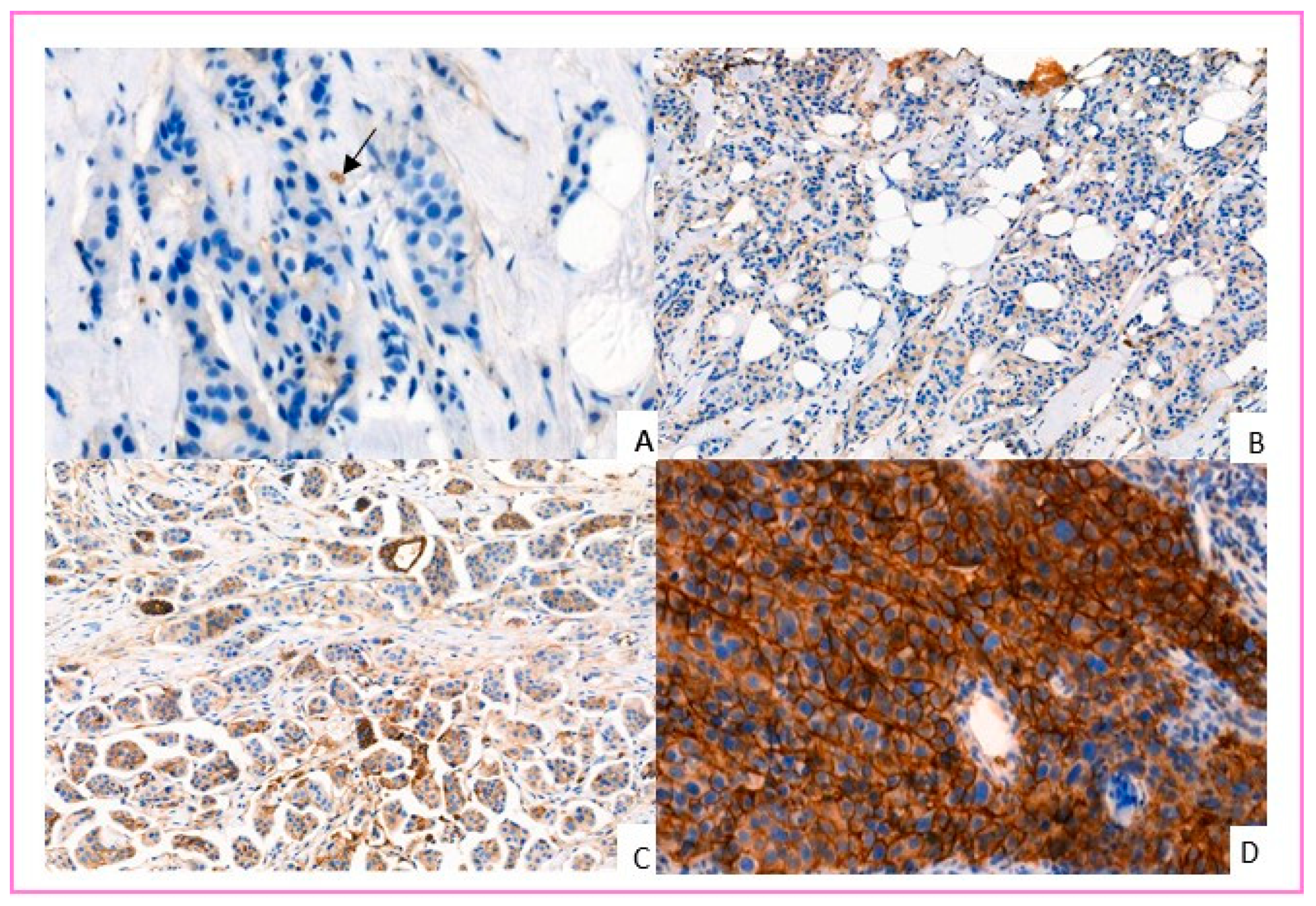
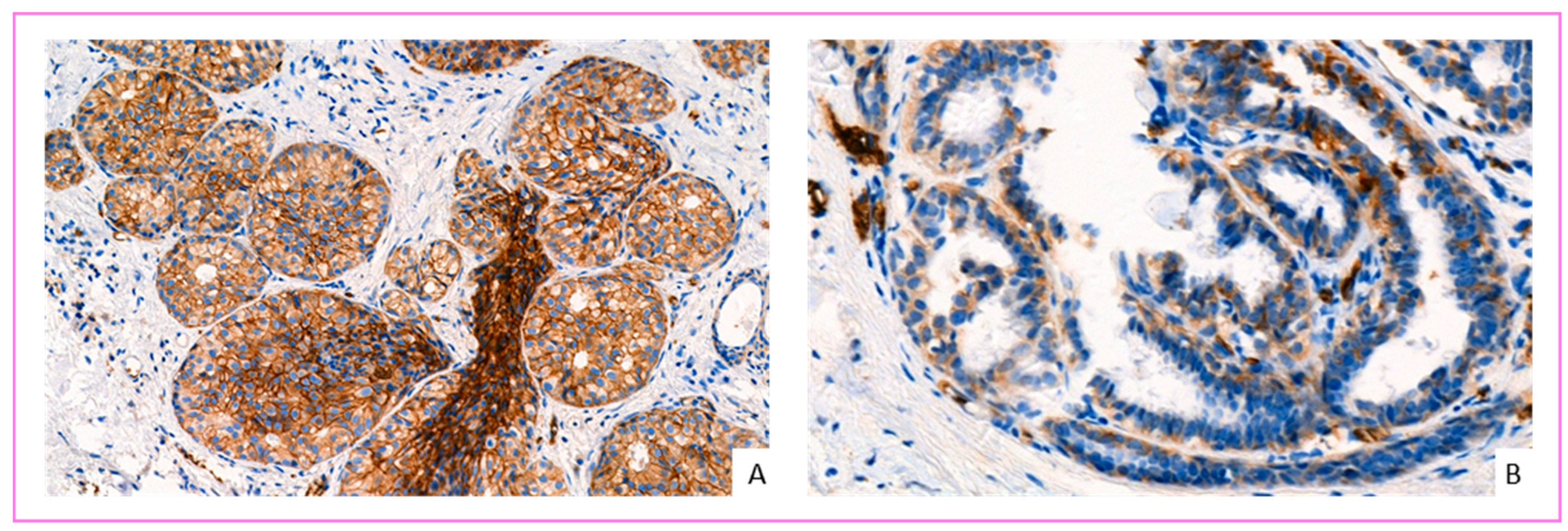
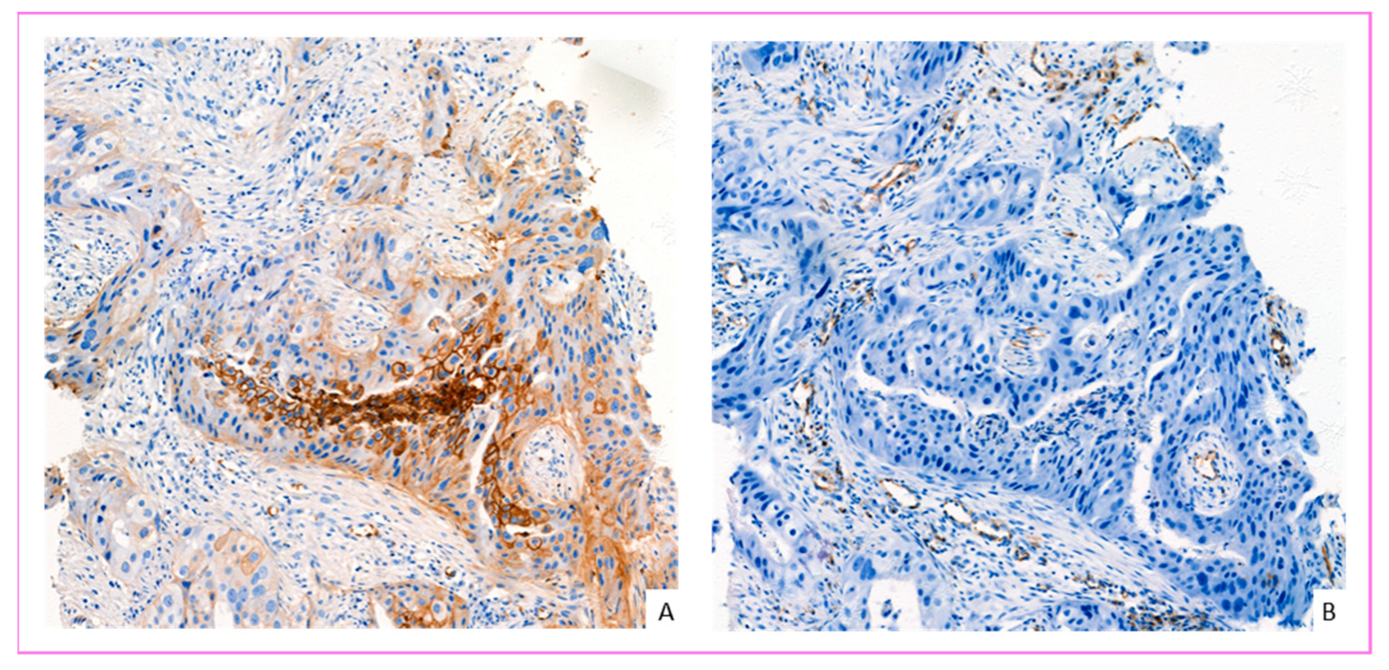
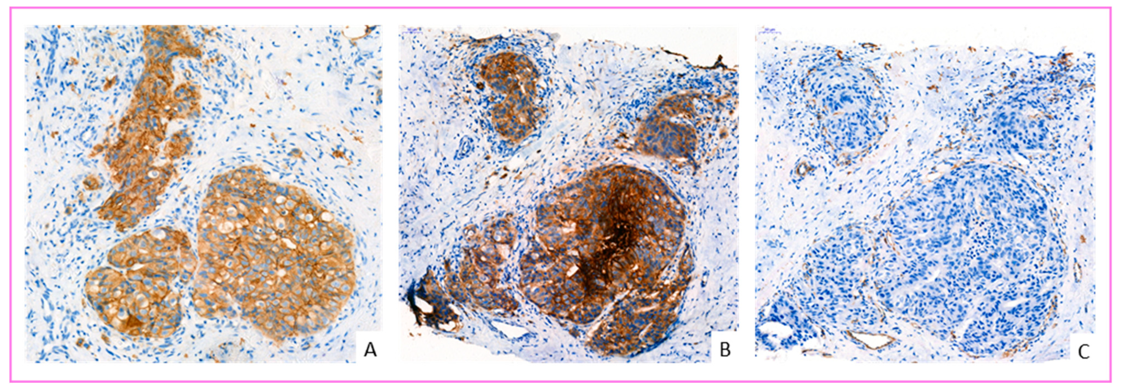
2.4. Evaluation of GLUT-1 IHC Labeling

GLUT-1 IHC images were obtained by slide scanner (Pannoramic 250, 3D histech, Budapest, Hungary) and interpreted on 3D histech software (caseViewer). Red blood cells were used as an internal positive control. Only membrane staining was considered positive. The evaluation was performed using a semi-quantitative score inspired by Sakashita et al. [31], which grades according to the proportion of GLUT-1 positive cells. We defined a four-grade IHC score: Score 0 negative (no membrane staining by tumor cells); Score 1+, weakly positive (membrane staining in <10% of tumor cells); Score 2+, moderately positive (membrane staining in ≥10 and <50% of tumor cells); Grade 3, strongly positive (membrane staining in ≥50% of tumor cells). Tumors classified as score 2+ or 3+ were considered to overexpress GLUT-1 (Figure 1). All cases were interpreted independently by two pathologists unaware of the patients’ associated medical data. In case of disagreement about GLUT-1 IHC score, cases were discussed until consensus was reached.

2.5. Exclusion Criteria

Patients were excluded from this series if they had a diagnosis of recurrent breast cancer, and the primary tumor was not diagnosed in our department. In a second step, patients were excluded for analysis of factors and comparison between the three groups if BMI at diagnosis was not known; and in the third step for GLUT-1 interpretation, if FFPE material was not available. If the patients presented a bifocal or bilateral carcinoma with similar histological and immunohistochemical characteristics, only one location was considered for GLUT-1 expression.

2.6. Statistical Analysis

Statistical analyses were performed using Excel software (version 2109, Microsoft Co., Redmond, WA, USA), the biostaTGV website [32] and STATA® software (version 13.1, StataCorp LP., College Station, TX, USA). Categorical, binary, or discrete variables with very few modalities were expressed as headcount and percentage. Quantitative variables that did not reveal a symmetrically shaped distribution were expressed as median, first and third quartile (Q1, Q3), and if relevant, minimum and maximum (min., max.). The χ test2 and Fisher’s exact test were performed to test for independence between two categorical variables. The Student’s t-test and the Wilcoxon–Mann–Whitney test were performed to test the independence between a qualitative and a quantitative variable. The statistical tests were two-tailed. The p values were considered significant at the 5% level. Missing data were excluded from the statistical analyses.

2.7. Ethical Statement

A privacy impact assessment (PIA) and authorization for this study were validated by the Data Protection Officer (DPO) of Cayenne Hospital Center. The non-opposition of the patients was signified by a clinician on the request document sent to the Pathology Department notifying that patient did not object to the use of her sample for scientific research according to the European Data Protection Regulation (GDPR).

3. Results

A total of 199 patients were selected; 190 were retained for analysis of general clinical and histological features; 162 for comparative histopronostic analysis of the three groups of patients; and 144 for IHC evaluation of GLUT-1 in these same groups. BMI was missed in 28 patients because 19 patients did not provide either a primary care physician or a telephone number, 8 left a number that was no longer assigned, and one refused to answer questions. Four patients had bifocal carcinoma, three had bilateral carcinoma. Only one specimen from each patient was selected due to similarities in histopronostic and molecular features.

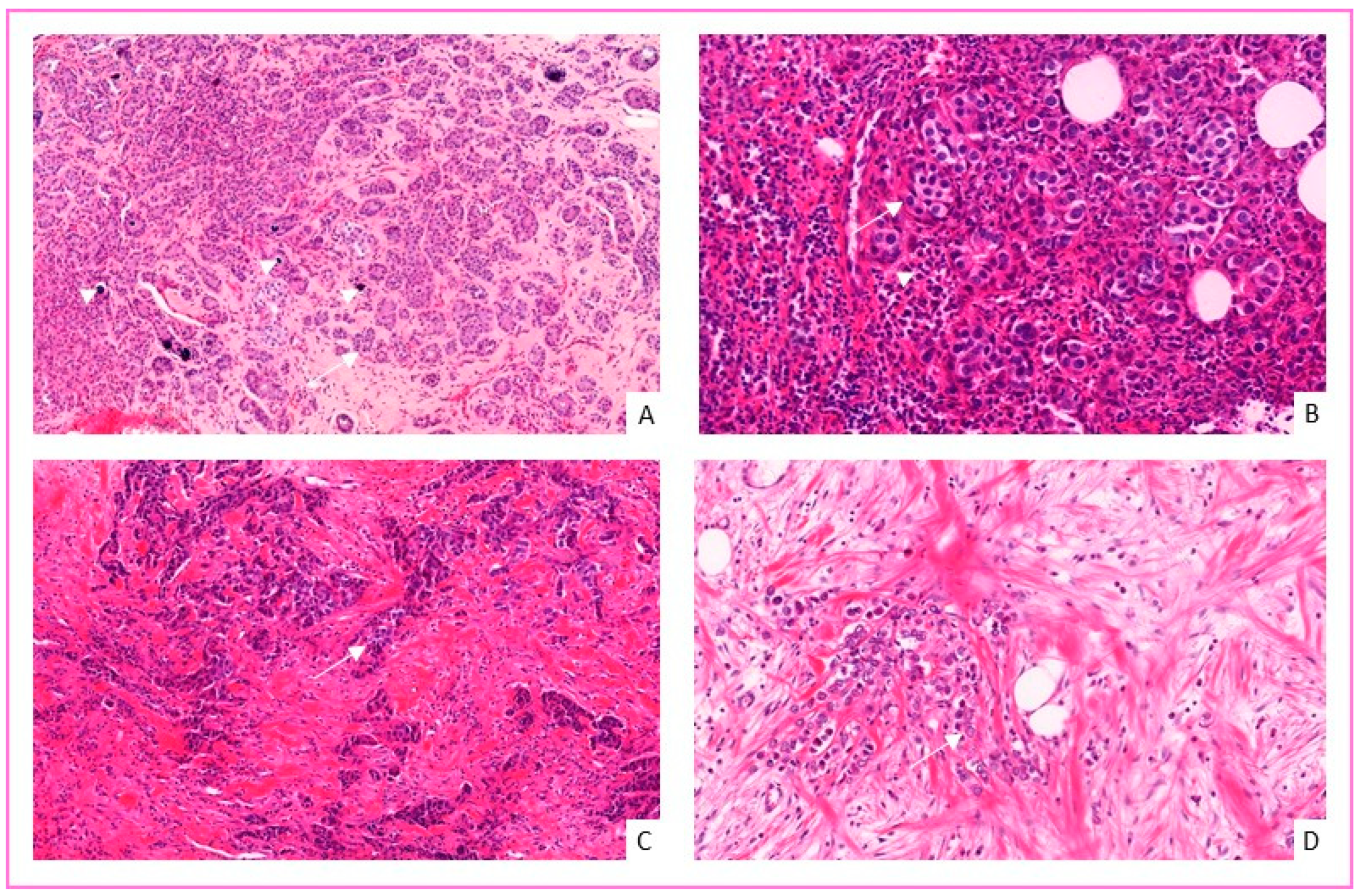
3.1. General Clinical and Histological Features of Breast Cancer in French Guiana

Data regarding age at diagnosis, city of residence, histological type, lymph node metastasis, or distant metastasis were available for all patients. BMI, tumor size, modified SBR histological grade, HR status, HER2 status, Ki67 proliferation index, and molecular group were available in 85.3, 69.5, 98.3, 99.5, 97.9, 96.3, and 97.9%, respectively. Clinical and histological data are summarized in Table 1. Figure 2 shows the incidence of breast cancer by the city of residence.
The incidence of breast cancer was highest in the main cities of the territory, where 85% of the population lives; the city of Cayenne and its surroundings (especially Matoury and Rémire-Montjoly), Kourou, and Saint Laurent du Maroni.
The median age at diagnosis was 53.5 years (Q1–Q3: [45.0; 63.0]) (min-max: [25; 92]). The median BMI at diagnosis was 26.6 (Q1–Q3: [23.1; 30.5]). (Additional data regarding age, tumor size and BMI are available in Table 2, Table 3 and Table 4, respectively). The median histological size was 2.8 cm (Q1–Q3: [1.6; 4.1]) (min.max: [0.1; 17]).
There were 57 tumors (30%) with lymph node metastasis at diagnosis and 13 tumors (6.8%) with distant metastasis. Regarding histological types, 172 (90.6%) were invasive carcinomas of no special type (NST), 6 of which were of medullary pattern, 11 (5.8%) invasive lobular carcinomas, 4 (2.1%) invasive papillary carcinomas, one (0.5%) invasive micropapillary carcinoma, one tubular carcinoma, and one mucinous carcinoma (Figure 2). The tumors were of modified SBR histological grade I in 18 (9.6%) cases; grade II in 117 (62.6%) cases; and grade III in 52 (27.8%) cases. HR status was positive in 131 (69.3%) patients, and HER2 was positive in 41 (22.0%) patients. The median Ki67 proliferation index was 20 (Q1–Q3: [10.0;40.0]). The molecular types of the tumors were luminal A in 108 cases (58.1%), luminal B in 21 cases (11.3%), HER2-enriched in 20 cases (10.7%), and TN in 37 cases (19.9%).

3.2. Histological and Prognostic Features of Breast Cancer in French Guiana

Of the 162 analyzed cases, 100 were in the control group, 36 in the diabetes group, and 26 in the obese group. Half of the patients in the diabetes group were obese (18/36; 50%). There were no significant differences between clinical groups and the presence of lymph node or distant metastasis at diagnosis, histological grade, HR, and HER2 status, Ki67 proliferation index, and molecular type. We found a trend for the presence of distant metastasis at diagnosis for diabetic patients (p = 0.067) and a trend for negative HR status for patients in the obese group (p = 0.061) compared to the control group (Table 5 summarizes the results of each analysis).
Data on tumor size, modified SBR histological grade, HR status, HER2 status, Ki67 proliferation index, and molecular type were available in 80, 98.8, 99.4, 99.4, 96.3, and 99.4%, respectively.

3.3. GLUT-1 Expression in Breast Cancer Cells

In total, 144 patients with FFPE available material were included for GLUT-1 IHC analysis, 91 were in the control group, 31 in the diabetes group, and 22 in the obese group. Data regarding tumor size, modified SBR histological grade, HR status, HER2 status, Ki67 proliferation index, and the molecular group were available in: 78.5, 99.3, 100, 100, 97.2, and 100%, respectively.
Epithelial and myoepithelial cells of the terminal ductulo-lobular units (TDLUs) never showed overexpression. TDLU cells were either negative (score 0) or slightly positives (1 to 9% positive cells; score 1+).
Our analyses revealed GLUT-1 overexpression (score 2+ or 3+) in 87/144 (60.4%). However, there was no significant difference between GLUT-1 overexpression and clinical groups (Table 6).
GLUT-1 overexpressed tumors were associated with high SBR histological grade III, higher Ki67 proliferation index, more aggressive molecular types, including HER2 positive and HR negative status (Figure 3). There was no significant correlation between GLUT-1 expression and tumor size or presence of lymph node or distant metastases (Table 7 and Table 8).
Interestingly, all invasive carcinomas NST with medullary pattern presented a strong overexpression of GLUT-1 with IHC score 3+. Some tumors showed heterogeneous expression with areas of high expression and areas of low or no expression.
More interestingly, it was noted that carcinoma in situ always showed GLUT-1 overexpression (n = 29) in the three patient groups, with a statistically significant difference compared to associated infiltrative contingent (p: 2.60 × 10⁻⁶, data not shown) (Figure 4A). In contrast, GLUT-1 overexpression was not observed in atypical and non-atypical epithelial hyperplasia (Figure 4B).
We did not find a significant difference for GLUT-1 expression between non-obese diabetic patients and the general population (p = 0.718; Table 9), and between obese and non-obese patients regardless of diabetes status (p = 0.890; Table 10).

4. Discussion

In this study, we described for the first time the clinical, histological, and molecular features of breast cancer in French Guiana. We then compared histological and prognostic factors between patients with and without type 2 diabetes and obesity. Finally, we evaluated the expression of GLUT-1 by IHC technique in these three groups.
French Guiana is inhabited by communities of different ethnic origins, including Creoles and Maroons of African origin, as well as Asian and European populations. Amerindians are the indigenous inhabitants of French Guiana and live mainly in the rainforest along the rivers of the hinterland, maintaining a traditional lifestyle.
The Department of Pathology of Cayenne Hospital Center is the only specialist laboratory in French Guiana, which analyses the majority of samples from the city of Cayenne and adjacent towns, as well as from the isolated communes’ health centers known as CDPS (Centres Délocalisés de Prévention et de Soins). Nevertheless, between 2014 and 2020, we had only 199 patients with invasive breast cancer, which is lower than the reported incidence in French Guiana [33]. In fact, some analyses from hospitals of Kourou and West Guiana in Saint Laurent du Maroni are sent to laboratories in mainland France, resulting in a false lower incidence in our study for these two cities.
In our study, the median age of the patients at the time of diagnosis was 53.5 years, compared to 63 years in mainland France (INCa data), 62 years in the United States of America (USA) [34], and 49 years in Saudi Arabia [35]. Our data corroborate those of a previous study [21]. The young age of the population in French Guiana could at least partly explain these differences. Indeed, the proportion of people in the 60–74 age group is almost three times lower in Guiana than in mainland France (7 and 18%, respectively, INSERM data).
The median tumor size at diagnosis was 2.8 cm. Furthermore, we observed that only 30.3% of the tumors were smaller than 2 cm, which is much lower than what is reported by studies on populations from USA or Poland (58.4 and 51.9%, respectively) [36,37], but closer to the Middle East population of Saudi Arabia (42.6%) [35].
The most frequent molecular group in our population was luminal A (58.1%), followed by TN (19.9%), then luminal B (11.3%), and finally HER2 enriched (10.3%). The frequency of the observed molecular groups was closer to those present in the Saudi population (luminal A: 58.5%, luminal B: 14%, enriched HER2: 11.5%, and TN: 16%) than to those of western populations that report less aggressive TN and enriched HER2 subtypes, in favor of luminal A (such as the USA population: Luminal A: 73.3%, Luminal B: 11.1%, TN: 10.9% and enriched HER2: 4.6%) [35,37,38]. Breast cancer in French Guiana was, therefore, more similar to the Middle Eastern population in terms of molecular type distribution, younger age, and larger size at diagnosis than the Western populations [35].
Our observation reveals an aggressive presentation of breast cancer at diagnosis in French Guiana. This may be due to a delay in diagnosis, suggesting the need to lower the screening age for breast cancer in this territory, as suggested in a previous study [21].
Moreover, we did not observe any significant difference between histopronostic factors and the three patient groups. Nevertheless, there was a trend for negative HR status in the obese group and the presence of distant metastases at diagnosis in the type 2 diabetes group. Our data are different from previous studies that report a lower Ki67 proliferation index and a higher proportion of lymph node metastases in patients with type 2 diabetes [39,40]. This difference may be due to the molecular breast cancer cells specificities of our population which were more aggressive. In agreement with the literature, we did not observe any significant difference between histological SBR grade, histological type, and molecular type in diabetic patients and the control group [40,41].
To our knowledge, this study is the first study that analyzed molecular type in obese patients with breast cancer. In the obese group, we observed a tendency for a negative HR status. The literature on this subject is discordant [42,43].
GLUT-1 is a uniportal transmembrane protein encoded in humans by the SLC2A1 gene [12,44]. Its main function is to catalyze the entry of glucose into cells [11], and it plays a key role in energy metabolism by providing basal glucose transport in cells. To this end, GLUT-1 is physiologically expressed, at least virtually, in all tissues, and importantly in erythrocytes and endothelial cells of the blood-brain barrier [44]. In this study, we observed that GLUT-1 was overexpressed in 60.4% of the tumors. This overexpression is correlated with high SBR histological grade, high proliferation index, more aggressive molecular types, HER2 positive, and HR negative status. However, this overexpression was not correlated with the obesity or diabetic status of patients.
The overexpression of GLUT-1 in more than 60% of invasive breast cancer in our population is in agreement with the literature, which reports between 35 and 70% of positivity [45,46,47,48,49,50]. The correlation between GLUT-1 overexpression, histological grade, proliferation index, aggressive molecular types, and negative RH status was previously reported [51]. Nevertheless, we observed for the first time an association between the overexpression of GLUT-1 and the positive HER2 status (p = 0.020). This result may be related to the molecular specificities of our population.
However, if 60% of breast cancers overexpress GLUT-1, we can hypothesize that cancer cells that do not overexpress GLUT-1 use another metabolic pathway or overexpress other glucose transporters than GLUT-1. Previous studies have shown that GLUT-3, GLUT-4, and GLUT-12 can be upregulated in breast cancer cells [8,9,52].
Surprisingly, all invasive carcinomas NST with medullary pattern showed high GLUT-1 overexpression (score 3+). These carcinomas are characterized by a stroma rich in tumor-infiltrating lymphocytes (TILs) and syncytial carcinomatous cells of high nuclear grade, with high mitotic activity. We hypothesize that increased glycolysis leads to the production of pro-inflammatory metabolites that influence the architecture of the tumor’s inflammatory microenvironment [53]. Alternatively, lactates produced by a shift in metabolism towards glycolysis may be an adaptation of the tumor to escape immune cells [54].
Interestingly, all carcinoma in situ (CIS) lesions showed GLUT-1 overexpression regardless of their nuclear grade (low grade, intermediate grade, and high grade), but we did not observe overexpression in atypical and non-atypical hyperplasia. This overexpression is independent of the status of adjacent infiltrating carcinoma cells. The absence of expression of GLUT-1 in atypical hyperplasia and the presence of the expression in CIS has been reported previously [49,55]. Furthermore, it has been suggested that this phenotype may be a strong selective advantage for CIS [56]. Our study suggests that a late loss of overexpression of GLUT-1 in breast cancer cells may occur in the invasive stage of tumor progression (Figure 5A), as observed previously with HER2 [57]. However, the spatial comparison using the vascular marker CD31 on the same section revealed the neoangiogenesis of the tumor stroma. The new vessels are located mainly around the invasive carcinoma clusters (Figure 5A). Moreover, a recent study demonstrated that GLUT1 is required for breast cancer formation and robust HER2-induced proliferation even under normoxic conditions [56] (Figure 6A–C). Thus, a further spatial correlation study of GLUT-1 and HER2 can be very informative about the regulation of GLUT1 by HER2.
Furthermore, some tumors showed heterogeneous staining of GLUT-1 with high and low staining areas in the same tumor, suggesting heterogeneity in energy metabolism. This has been highlighted as a property of some tumors [58]. In this study, the expression of GLUT-1 was quantified only at the protein level by immunohistochemistry. However, real-time PCR and digital PCR can give accurate copy numbers and are therefore much more reliable than immunohistochemistry.
We did not find any difference between patient groups and GLUT-1 expression. As many processes are involved in the regulation of GLUT-1, it is possible that other factors such as hypoxia play a prominent role [59]. Hypoxia factor 1a (HIF1a) induces GLUT-1 expression potentially through the PI3K/akt pathway [60]. Indeed, it has been shown that the more distant the cancer cells were from vessels, the more GLUT-1 was expressed [61]. In addition, HIF1a induces intratumoral angiogenesis, reduces mitochondrial metabolism, and promotes glycolysis [60]. Therefore, HIF1a would be involved in the energetic metabolism of cancers [54]. However, the expression of HIF1a in tumors occurs in 50% under conditions of normoxia [62]. This interaction between HIF1a and GLUT-1 could explain some cases of tumor heterogeneity. Thus, further study using hypoxia and vascular markers could be useful to define the oxygenation status of the tumor microenvironment and to investigate the role of hypoxia in GLUT-1 overexpression in breast cancer cells. Moreover, spatial comparison of hypoxia and vascular markers and GLUT-1 can inform if GLUT-1 expression is related to glycolytic phenotype of cancer cells and to obesity/diabetes status of the patients.
Previous studies revealed that in breast tumors, GLUT-1 overexpression on IHC was associated with high-grade histological tumors, with negative HR, TN molecular group, poor total survival, and progression-free survival [45,51,63]. Oh et al. showed that GLUT-1 had a role in TNBC tumorigenesis by inhibiting GLUT-1 and observed a decrease in cell growth, migration, and invasion [64]. GLUT-1 silencing also decreased EGFR activation and the MAPK cascade (c-Raf/MEK/ERK). An involvement in the epithelial–mesenchymal transition due to the observed decrease in CD44—itself correlated with this transition—was also suggested [56]. The deregulation of the energy system is one of the major characteristics of cancers [5]. Several types of regulatory changes have been described [47]. The most common is the Warburg effect [6,7], in which the tumor cell, instead of making energy available through oxidative phosphorylation, will use the aerobic glycolysis pathway, resulting in the formation of lactates and a relatively small amount of available energy (2 moles of ATP/mole of glucose). The tumor cell will therefore have particularly high glucose requirements. This apparent lack of efficiency allows the cell to produce metabolites necessary for its growth and proliferation. The tumor cell captures glucose in large part via GLUT-1 [64]. Thus, GLUT-1 is logically associated with more aggressive tumors [13].
Finally, GLUT-1 overexpression, frequently observed in invasive breast cancer, may present a prognostic factor, and also an interesting therapeutic target, independently of diabetic or obese status. We believe that a further clinical study in a larger cohort with a consensus on the evaluation of GLUT-1 expression in breast cancer cells is needed to determine a score to stratify patients eligible for promising GLUT-1 target therapy.

5. Conclusions

We have described for the first time the clinical, histological, and molecular features of breast cancer in French Guiana. The breast cancers in our population are characterized by a young age at diagnosis, aggressive molecular types, and large tumor size. Our results highlight the need to lower the screening age of breast cancer in French Guiana.
We also demonstrated an overexpression of GLUT-1 in 60% of invasive breast carcinomas and in all medullary pattern and carcinoma in situ lesions. Nevertheless, there was no significant difference in GLUT-1 expression between diabetic or obese patients and the control group. The overexpression of GLUT-1 observed in invasive breast cancer and in carcinoma in situ lesions may present a tumor metabolic prognostic marker and also an interesting target therapy, independently of patient metabolic disorder.

Author Contributions

Conceptualization, N.S. and K.D.A.; data curation, V.S.; formal analysis, V.S. and K.D.A.; investigation, V.S., J.B.-K., B.V.-C., A.A., N.S. and K.D.A.; methodology, N.S. and K.D.A.; project administration, A.A.; supervision, K.D.A.; validation, V.S., N.S. and K.D.A.; visualization, V.S., N.S. and K.D.A.; writing—original draft, V.S.; writing—review and editing, N.S. and K.D.A. All authors have read and agreed to the published version of the manuscript.

Funding

This research received no external funding.

Institutional Review Board Statement

The study was conducted according to the guidelines of the Declaration of Helsinki and approved by our Institutional Review Board. A Privacy Impact Assessment (PIA) and authorization for this study were validated by the Data Protection Officer (DPO) of Cayenne Hospital Center. The non-opposition of the patients was signified by a clinician on the request document sent to the Pathology Department notifying that patient “not object” to the use of her sample for scientific research according to the European Data Protection Regulation (GDPR).

Informed Consent Statement

Informed consent was obtained from the patients.

Data Availability Statement

The data used and analyzed during the current study are available in the manuscript text and tables. Further information could be obtained from the corresponding author on reasonable request.

Acknowledgments

The results presented in this manuscript are part of a thesis project of Valentin Suteau at Université des Antilles. We would like to thank the technical team of the Department of Pathology for the quality of the immunohistochemistry analysis, especially Ka Yu Chan, Veronique Louvel, Aline Masson, and Melissa Cherubin. We also thank Hugo Pouchin from CRB Amazonie for administrative aspects and Fabrice Quet for statistical advice.

Conflicts of Interest

The authors declare no conflict of interest.

References

  1. The GBD 2015 Obesity Collaborators. Health Effects of Overweight and Obesity in 195 Countries over 25 Years. N. Engl. J. Med. 2017, 377, 13–27. [Google Scholar] [CrossRef]
  2. Zheng, Y.; Ley, S.H.; Hu, F.B. Global aetiology and epidemiology of type 2 diabetes mellitus and its complications. Nat. Rev. Endocrinol. 2018, 14, 88–98. [Google Scholar] [CrossRef]
  3. Gallagher, E.J.; LeRoith, D. Hyperinsulinaemia in cancer. Nat. Cancer 2020, 20, 629–644. [Google Scholar] [CrossRef]
  4. Sung, H.; Ferlay, J.; Siegel, R.L.; Laversanne, M.; Soerjomataram, I.; Jemal, A.; Bray, F. Global Cancer Statistics 2020: GLOBOCAN Estimates of Incidence and Mortality Worldwide for 36 Cancers in 185 Countries. CA Cancer J. Clin. 2021, 71, 209–449. [Google Scholar] [CrossRef] [PubMed]
  5. Hanahan, D.; Weinberg, R.A. Hallmarks of Cancer: The Next Generation. Cell 2011, 144, 646–674. [Google Scholar] [CrossRef] [PubMed] [Green Version]
  6. Warburg, O.; Wind, F.; Negelein, E. The metabolism of tumors in the body. J. Gen. Physiol. 1927, 8, 519–530. [Google Scholar] [CrossRef] [PubMed] [Green Version]
  7. Warburg, O. On the Origin of Cancer Cells. Science 1956, 123, 309–314. [Google Scholar] [CrossRef]
  8. Kuo, M.-H.; Chang, W.-W.; Yeh, B.-W.; Chu, Y.-S.; Lee, Y.-C.; Lee, H.-T. Lee Glucose Transporter 3 Is Essential for the Survival of Breast Cancer Cells in the Brain. Cells 2019, 8, 1568. [Google Scholar] [CrossRef] [Green Version]
  9. Garrido, P.; Osorio, F.G.; Morán, J.; Cabello, E.; Alonso, A.; Freije, J.M.; González, C. Loss of GLUT4 Induces Metabolic Reprogramming and Impairs Viability of Breast Cancer Cells. J. Cell. Physiol. 2014, 230, 191–198. [Google Scholar] [CrossRef]
  10. Barbosa, A.M.; Martel, F. Targeting Glucose Transporters for Breast Cancer Therapy: The Effect of Natural and Synthetic Compounds. Cancers 2020, 12, 154. [Google Scholar] [CrossRef] [PubMed] [Green Version]
  11. Olson, A.L.; Pessin, J.E. Structure, function, and regulation of the mammalian facilitative glucose transporter gene family. Annu. Rev. Nutr. 1996, 16, 235–256. [Google Scholar] [CrossRef]
  12. Mueckler, M.; Caruso, C.; Baldwin, S.A.; Panico, M.; Blench, I.; Morris, H.R.; Allard, W.J.; Lienhard, G.E.; Lodish, H.F. Sequence and structure of a human glucose transporter. Science 1985, 229, 941–945. [Google Scholar] [CrossRef] [PubMed]
  13. Wang, J.; Ye, C.; Chen, C.; Xiong, H.; Xie, B.; Zhou, J.; Chen, Y.; Zheng, S.; Wang, L. Glucose transporter GLUT1 expression and clinical outcome in solid tumors: A systematic review and meta-analysis. Oncotarget 2017, 8, 16875–16886. [Google Scholar] [CrossRef] [Green Version]
  14. Taha, C.; Liu, Z.; Jin, J.; Al-Hasani, H.; Sonenberg, N.; Klip, A. Opposite translational control of GLUT1 and GLUT4 glucose transporter mRNAs in response to insulin. Role of mammalian target of rapamycin, protein kinase b, and phosphatidylinositol 3-kinase in GLUT1 mRNA translation. J. Biol. Chem. 1999, 274, 33085–33091. [Google Scholar] [CrossRef] [Green Version]
  15. Cifuentes, M.; García, M.A.; Arrabal, P.M.; Martínez, F.; Yañez, M.J.; Jara, N.; Weil, B.; Domínguez, D.; Medina, R.A.; Nualart, F. Insulin regulates GLUT1-mediated glucose transport in MG-63 human osteosarcoma cells. J. Cell. Physiol. 2011, 226, 1425–1432. [Google Scholar] [CrossRef] [PubMed]
  16. Laybutt, D.R.; Thompson, A.L.; Cooney, G.J.; Kraegen, E.W. Selective chronic regulation of GLUT1 and GLUT4 content by insulin, glucose, and lipid in rat cardiac muscle in vivo. Am. J. Physiol. Content 1997, 273, H1309–H1316. [Google Scholar] [CrossRef] [PubMed]
  17. Silva, C.; Andrade, N.; Guimarães, J.T.; Patrício, E.; Martel, F. The in vitro effect of the diabetes-associated markers insulin, leptin and oxidative stress on cellular characteristics promoting breast cancer progression is GLUT1-dependent. Eur. J. Pharmacol. 2021, 898, 173980. [Google Scholar] [CrossRef] [PubMed]
  18. Kang, C.; LeRoith, D.; Gallagher, E.J. Diabetes, Obesity, and Breast Cancer. Endocrinology 2018, 159, 3801–3812. [Google Scholar] [CrossRef] [Green Version]
  19. Melstrom, L.G.; Salabat, M.R.; Ding, X.-Z.; Milam, B.M.; Strouch, M.; Pelling, J.C.; Bentrem, D.J. Apigenin Inhibits the GLUT-1 Glucose Transporter and the Phosphoinositide 3-Kinase/Akt Pathway in Human Pancreatic Cancer Cells. Pancreas 2008, 37, 426–431. [Google Scholar] [CrossRef]
  20. Gonnella, R.; Santarelli, R.; Farina, A.; Granato, M.; D’Orazi, G.; Faggioni, A.; Cirone, M. Kaposi sarcoma associated herpesvirus (KSHV) induces AKT hyperphosphorylation, bortezomib-resistance and GLUT-1 plasma membrane exposure in THP-1 monocytic cell line. J. Exp. Clin. Cancer Res. 2013, 32, 79. [Google Scholar] [CrossRef] [Green Version]
  21. Daigre, J.-L.; Atallah, A.; Boissin, J.-L.; Jean-Baptiste, G.; Kangambega, P.; Chevalier, H.; Balkau, B.; Smadja, D.; Inamo, J. The prevalence of overweight and obesity, and distribution of waist circumference, in adults and children in the French Overseas Territories: The PODIUM survey. Diabetes Metab. 2012, 38, 404–411. [Google Scholar] [CrossRef] [PubMed]
  22. Carvalho, L.; Belliardo, S.; Andrieu, A.; de Maria, F.; Chatignoux, É. Incidence et mortalité des cancers en Guyane, 2007–2014. Synthèse de l’état des connaissances/incidence and mortality of cancers in French Guiana, 2007–2014. Summary of the state of knowledge. Bull. Epidémiol. Hebd. 2020, 36–37, 730–736. [Google Scholar]
  23. Roué, T.; Fior, A.; Plenet, J.; Belliardo, S.; Nacher, M. The epidemiology of breast cancer in French Guiana 2003–2006. SpringerPlus 2013, 2, 471. [Google Scholar] [CrossRef]
  24. Roué, T.; Labbé, S.; Belliardo, S.; Plenet, J.; Douine, M.; Nacher, M. Predictive Factors of the Survival of Women With Invasive Breast Cancer in French Guiana: The Burden of Health Inequalities. Clin. Breast Cancer 2016, 16, e113–e118. [Google Scholar] [CrossRef]
  25. Elston, C.W.; Ellis, I.O. Pathological prognostic factors in breast cancer. I. The value of histological grade in breast cancer: Expe-rience from a large study with long-term follow-up. Histopathology 1991, 19, 403–410. [Google Scholar] [CrossRef]
  26. Available online: https://tumourclassification.iarc.who.int/welcome/ (accessed on 10 October 2021).
  27. Drouin, P.; Blickle, J.F.; Charbonnel, B.; Eschwege, E.; Guillausseau, P.J.; Plouin, P.F.; Daninos, J.M.; Balarac, N.; Sauvanet, J.P. Diagnostic et classification du diabete sucré les nouveaux criteres. Diabetes Metab. 1999, 25, 12. [Google Scholar]
  28. Cheang, M.C.; Martin, M.; Nielsen, T.O.; Prat, A.; Voduc, D.; Rodriguez-Lescure, A.; Ruiz, A.; Chia, S.; Shepherd, L.; Ruiz-Borrego, M.; et al. Defining Breast Cancer Intrinsic Subtypes by Quantitative Receptor Expression. Oncologist 2015, 20, 474–482. [Google Scholar] [CrossRef] [Green Version]
  29. Iwamoto, T.; Booser, D.; Valero, V.; Murray, J.L.; Koenig, K.; Esteva, F.; Ueno, N.T.; Zhang, J.; Shi, W.; Qi, Y.; et al. Estrogen Receptor (ER) mRNA and ER-Related Gene Expression in Breast Cancers That Are 1% to 10% ER-Positive by Immunohistochemistry. J. Clin. Oncol. 2012, 30, 729–734. [Google Scholar] [CrossRef]
  30. Wolff, A.C.; Hammond, M.E.H.; Allison, K.H.; Harvey, B.E.; Mangu, P.B.; Bartlett, J.M.S.; Bilous, M.; Ellis, L.O.; Fitzgibbons, P.; Hanna, W.; et al. Human Epidermal Growth Factor Receptor 2 Testing in Breast Cancer: American Society of Clinical Oncology/College of American Pathologists Clinical Practice Guideline Focused Update. J. Clin. Oncol. 2018, 36, 2105–2122. [Google Scholar] [CrossRef] [Green Version]
  31. Sakashita, M.; Aoyama, N.; Minami, R.; Maekawa, S.; Kuroda, K.; Shirasaka, D.; Ichihara, T.; Kuroda, Y.; Maeda, S.; Kasuga, M. Glut1 expression in T1 and T2 stage colorectal carcinomas: Its relationship to clinicopathological features. Eur. J. Cancer 2001, 37, 204–209. [Google Scholar] [CrossRef]
  32. BiostaTGV-Statistiques en Ligne. Institut Pierre Louis d’Epidémiologie, Santé Publique UMR S 1136, INSERM et Sorbonne Université (UPMC). Available online: https://biostatgv.sentiweb.fr/ (accessed on 15 October 2021).
  33. Joachim, C.; Véronique-Baudin, J.; Desroziers, L.; Chatignoux, É.; Belliardo, S.; Plenet, J.; Macni, J.; Ulric-Gervaise, S.; Peruvien, J.; Bhakkan-Mambir, B.; et al. Gynaecological cancer in Caribbean women: Data from the French population-based cancer registries of Martinique, Guadeloupe and French Guiana (2007–2014). BMC Cancer 2020, 20, 643. [Google Scholar] [CrossRef]
  34. Howlader, N.; Noone, A.; Krapcho, M.; Miller, D.; Brest, A.; Yu, M.; Ruhl, J.; Tatalovich, Z.; Mariotto, A.; Lewis, D.R.; et al. SEER Cancer Statistics Review, 1975–2016; National Cancer Institute: Bethesda, MD, USA, 2019. Available online: https://seer.cancer.gov/archive/csr/1975_2016/ (accessed on 15 October 2021).
  35. Al-thoubaity, F.K. Molecular classification of breast cancer: A retrospective cohort study. Ann. Med. Surg. 2020, 49, 44–48. [Google Scholar] [CrossRef] [PubMed]
  36. Yang, X.R.; Sherman, M.E.; Rimm, D.L.; Lissowska, J.; Brinton, L.A.; Peplonska, B.; Hewitt, S.; Anderson, W.F.; Szeszenia-Dąbrowska, N.; Bardin-Mikolajczak, A.; et al. Differences in Risk Factors for Breast Cancer Molecular Subtypes in a Population-Based Study. Cancer Epidemiol. Biomark. Prev. 2007, 16, 439–443. [Google Scholar] [CrossRef] [PubMed] [Green Version]
  37. Howlader, N.; Noone, A.; Krapcho, M.; Miller, D.; Bishop, K.; Altekruse, S.; Kosary, C.L.; Yu, M.; Ruhl, J.; Tatalovich, Z.; et al. SEER Cancer Statistics Review, 1975–2013; National Cancer Institute: Bethesda, MD, USA, 2016.
  38. Cortet, M.; Bertaut, A.; Molinie, F.; Bara, S.; Beltjens, F.; Coutant, C.; Arveux, P. Trends in molecular subtypes of breast cancer: Description of incidence rates between 2007 and 2012 from three French registries. BMC Cancer 2018, 18, 161. [Google Scholar] [CrossRef] [PubMed] [Green Version]
  39. Hou, G.; Zhang, S.; Zhang, X.; Wang, P.; Hao, X.; Zhang, J. Clinical pathological characteristics and prognostic analysis of 1,013 breast cancer patients with diabetes. Breast Cancer Res. Treat. 2013, 137, 807–816. [Google Scholar] [CrossRef] [PubMed]
  40. Li, Z.; Luo, Y.; Gong, Y.; Liu, Y.; Qiu, W.; Tu, J. Clinical features and molecular phenotypes of breast cancer in patients with type-2 diabetes mellitus. Asian Pac. J. Cancer Prev. 2011, 12, 2183–2188. [Google Scholar]
  41. Bronsveld, H.K.; Jensen, V.; Vahl, P.; De Bruin, M.L.; Cornelissen, S.; Sanders, J.; Auvinen, A.; Haukka, J.; Andersen, M.; Vestergaard, P.; et al. Diabetes and Breast Cancer Subtypes. PLoS ONE 2017, 12, e0170084. [Google Scholar] [CrossRef] [PubMed]
  42. Turkoz, F.P.; Solak, M.; Petekkaya, I.; Keskin, O.; Kertmen, N.; Sarici, F.; Arik, Z.; Babacan, T.; Ozisik, Y.; Altundag, K. The prognostic impact of obesity on molecular subtypes of breast cancer in premenopausal women. J. BUON Off. J. Balk. Union Oncol. 2013, 18, 335–341. [Google Scholar]
  43. Nattenmüller, C.J.; Kriegsmann, M.; Sookthai, D.; Fortner, R.T.; Steffen, A.; Walter, B.; Johnson, T.; Kneisel, J.; Katzke, V.; Bergmann, M.; et al. Obesity as risk factor for subtypes of breast cancer: Results from a prospective cohort study. BMC Cancer 2018, 18, 616. [Google Scholar] [CrossRef]
  44. Uldry, M.; Thorens, B. The SLC2 family of facilitated hexose and polyol transporters. Pflüg. Arch. 2004, 447, 480–489. [Google Scholar] [CrossRef] [Green Version]
  45. Hussein, Y.R.; Bandyopadhyay, S.; Semaan, A.; Ahmed, Q.; Albashiti, B.; Jazaerly, T.; Nahleh, Z.; Ali-Fehmi, R. Glut-1 Expression Correlates with Basal-like Breast Cancer. Transl. Oncol. 2011, 4, 321–327. [Google Scholar] [CrossRef] [Green Version]
  46. Kuo, S.-J.; Wu, Y.-C.; Chen, C.-P.; Tseng, H.-S.; Chen, D.-R. Expression of Glucose Transporter-1 in Taiwanese Patients with Breast Carcinoma—A Preliminary Report. Kaohsiung J. Med. Sci. 2006, 22, 339–345. [Google Scholar] [CrossRef] [Green Version]
  47. Choi, J.; Kim, D.H.; Jung, W.H.; Koo, J.S. Metabolic interaction between cancer cells and stromal cells according to breast cancer molecular subtype. Breast Cancer Res. 2013, 15, R78. [Google Scholar] [CrossRef] [Green Version]
  48. Kang, S.S.; Chun, Y.K.; Hur, M.H.; Lee, H.K.; Kim, Y.J.; Hong, S.R.; Lee, J.H.; Lee, S.G.; Park, Y.K. Clinical Significance of Glucose Transporter 1 (GLUT1) Expression in Human Breast Carcinoma. Jpn. J. Cancer Res. 2002, 93, 1123–1128. [Google Scholar] [CrossRef]
  49. Jang, S.M.; Han, H.; Jang, K.-S.; Jun, Y.J.; Jang, S.-H.; Min, K.-W.; Chung, M.S.; Paik, S.S. The Glycolytic Phenotype is Correlated with Aggressiveness and Poor Prognosis in Invasive Ductal Carcinomas. J. Breast Cancer 2012, 15, 172–180. [Google Scholar] [CrossRef]
  50. Pinheiro, C.; Sousa, B.; Albergaria, A.; Paredes, J.; Dufloth, R.; Vieira, D.; Schmitt, F.; Baltazar, F. GLUT1 and CAIX expression profiles in breast cancer correlate with adverse prognostic factors and MCT1 overexpression. Histol. Histopathol. 2011, 26, 1279–1286. [Google Scholar] [PubMed]
  51. Deng, Y.; Zou, J.; Deng, T.; Liu, J. Clinicopathological and prognostic significance of GLUT1 in breast cancer: A meta-analysis. Medicine 2018, 97, e12961. [Google Scholar] [CrossRef] [PubMed]
  52. Rogers, S.; Macheda, M.; Docherty, S.E.; Carty, M.D.; Henderson, M.A.; Soeller, W.C.; Gibbs, E.M.; James, D.; Best, J. Identification of a novel glucose transporter-like protein—GLUT-12. Am. J. Physiol. Metab. 2002, 282, E733–E738. [Google Scholar] [CrossRef]
  53. Netea-Maier, R.T.; Smit, J.W.A.; Netea, M.G. Metabolic changes in tumor cells and tumor-associated macrophages: A mutual relationship. Cancer Lett. 2018, 413, 102–109. [Google Scholar] [CrossRef]
  54. Kim, J.; DeBerardinis, R.J. Mechanisms and Implications of Metabolic Heterogeneity in Cancer. Cell Metab. 2019, 30, 434–446. [Google Scholar] [CrossRef] [PubMed]
  55. Wang, L.; Zhang, S.; Wang, X. The Metabolic Mechanisms of Breast Cancer Metastasis. Front. Oncol. 2021, 10, 602416. [Google Scholar] [CrossRef] [PubMed]
  56. Wellberg, E.A.; Johnson, S.; Finlay-Schultz, J.; Lewis, A.S.; Terrell, K.L.; Sartorius, C.A.; Abel, E.D.; Muller, W.J.; Anderson, S.M. The glucose transporter GLUT1 is required for ErbB2-induced mammary tumorigenesis. Breast Cancer Res. 2016, 18, 131. [Google Scholar] [CrossRef] [Green Version]
  57. Allred, D.C.; Clark, G.M.; Tandon, A.K.; Molina, R.; Tormey, D.C.; Osborne, C.K.; Gilchrist, K.W.; Mansour, E.G.; Abeloff, M.; Eudey, L. HER-2/neu in node-negative breast cancer: Prognostic significance of overexpression influenced by the presence of in situ carcinoma. J. Clin. Oncol. J. Am. Soc. Clin. Oncol. 1992, 10, 599–605. [Google Scholar] [CrossRef]
  58. de la Cruz-López, K.G.; Castro-Muñoz, L.J.; Reyes-Hernández, D.O.; García-Carrancá, A.; Manzo-Merino, J. Lactate in the Regulation of Tumor Microenvironment and Therapeutic Approaches. Front. Oncol. 2019, 9, 1143. [Google Scholar] [CrossRef] [PubMed] [Green Version]
  59. Chen, C.-L.; Chu, J.-S.; Su, W.-C.; Huang, S.-C.; Lee, W.-Y. Hypoxia and metabolic phenotypes during breast carcinogenesis: Expression of HIF-1α, GLUT1, and CAIX. Virchows Arch. 2010, 457, 53–61. [Google Scholar] [CrossRef] [PubMed]
  60. Tirpe, A.A.; Gulei, D.; Ciortea, S.M.; Crivii, C.; Berindan-Neagoe, I. Hypoxia: Overview on Hypoxia-Mediated Mechanisms with a Focus on the Role of HIF Genes. Int. J. Mol. Sci. 2019, 20, 6140. [Google Scholar] [CrossRef] [Green Version]
  61. Mendez, L.E.; Manci, N.; Cantuaria, G.; Gomez-Marin, O.; Penalver, M.; Braunschweiger, P.; Nadji, M. Expression of Glucose Transporter-1 in Cervical Cancer and Its Precursors. Gynecol. Oncol. 2002, 86, 138–143. [Google Scholar] [CrossRef]
  62. Al Tameemi, W.; Dale, T.P.; Al-Jumaily, R.M.K.; Forsyth, N.R. Hypoxia-Modified Cancer Cell Metabolism. Front. Cell Dev. Biol. 2019, 7, 4. [Google Scholar] [CrossRef] [PubMed] [Green Version]
  63. Ganapathy, V.; Thangaraju, M.; Prasad, P.D. Nutrient transporters in cancer: Relevance to Warburg hypothesis and beyond. Pharmacol. Ther. 2009, 121, 29–40. [Google Scholar] [CrossRef] [PubMed]
  64. Oh, S.; Kim, H.; Nam, K.; Shin, I. Glut1 promotes cell proliferation, migration and invasion by regulating epidermal growth factor receptor and integrin signaling in triple-negative breast cancer cells. BMB Rep. 2017, 50, 132–137. [Google Scholar] [CrossRef] [PubMed]
Figure 1. GLUT-1 immunohistochemistry (IHC) score in invasive breast carcinoma. (A) Score 0: No membrane staining by tumor cells (positive internal control: red blood cells—black arrow). (B) Score 1+: Membrane staining in some tumor cells (<10%). (C) Score 2+: membrane staining in ≥10% and <50% of tumor cells. (D) Score 3+: membrane staining in ≥50–100% of tumor cells (chicken wire appearance) (IHC GLUT-1, scanned slides, magnification ×330).
Cancers 14 00437 g001
Figure 2. Examples of invasive breast carcinomas in diabetes or obese patients. (A) Invasive breast carcinoma NST SBR I shows tubular structures (arrow) consisting of cancer cells with moderate nuclear pleomorphism and low mitotic activity within a sclerotic but low inflammatory stroma (arrowheads: microcalcifications) (magnification ×200). (B) Invasive breast carcinoma NST with medullary features shows syncytial cell trabeculae with high nuclear pleomorphism within a TILs-rich stroma (arrowhead) (magnification ×330). (C) Invasive breast carcinoma NST SBR II shows no glandular structures (arrow) with moderate nuclear pleomorphism, and moderate mitotic activity, in a sclerotic stroma (magnification ×200). (D) Invasive lobular carcinoma shows cells organize in files (arrow) within an edematous stroma (magnification ×400).
Cancers 14 00437 g002
Figure 3. GLUT-1 IHC overexpression in invasive breast cancer cells. (A) invasive breast carcinoma NST of triple-negative molecular type, overexpressed GLUT-1 (score 3) (×240 magnification). (B) invasive breast carcinomas NST, HER2 enriched molecular type, overexpressed GLUT-1 (score 3) (×300 magnification).
Cancers 14 00437 g003
Figure 4. GLUT-1 IHC staining in ductal carcinoma in situ. (A) GLUT-1 overexpressed (score 3) in ductal carcinoma in situ of intermediate nuclear grade (magnification ×400). (B) Atypical hyperplasia: epithelial cells show weakly membrane staining in <10% of ductal cells (score 1) (magnification ×600).
Cancers 14 00437 g004
Figure 5. Spatial comparison between GLUT-1 expression and the vascular marker CD31 by immunohistochemistry on the same breast cancer section. (A) High-grade ductal carcinoma in situ with invasive area: this figure shows GLUT-1 expression in carcinoma in situ cells (center), which disappears in the invasive lesion (periphery) (B). This image shows the neoangiogenesis and vascularization of the tumor stroma by the CD31 endothelial cell marker. The new vessels are located mainly around the invasive carcinoma clusters (magnification ×200).
Cancers 14 00437 g005
Figure 6. Spatial comparison between GLUT-1 and HER2 overexpressed cancer cells, and the vascular marker CD31 by immunohistochemistry on the same breast cancer section. (A) This image shows overexpression of breast cancer cells by HER2 (score 3+). (B) The same cancer cells overexpress GLUT-1 (score 3). (C) This image shows neoangiogenesis and vascularization of the tumor stroma by CD31. The new vessels are located very close to the invasive carcinoma clusters (magnification ×200).
Cancers 14 00437 g006
Table 1. General clinical and histological features at diagnosis (n = 190).
Age (year) †
(n = 190)
53.5 [45.0;63.0]Modified SBR
(n = 187)
BMI (kg/m2) †
(n = 162)
26.6 [23.1;30.5]I18 (9.6%)
II117 (62.6%)
Tumor size (cm) †
(n = 190)
2.8 [1.6;4.1]III52 (27.8%)
HR+ *
(n = 189)
131 (69.3%)
M+ *
(n = 190)
13 (6.8%)
N+ *
(n = 190)
57 (30.0%)HER2+ *
(n = 186)
41 (22.0%)
Histological types
(n = 190)
Ki67 †
(n = 183)
20 [10;40]
NST172 (90.6%)Molecular Groups *
CLI11 (5.8%)(n = 186)
CPI4 (2.1%)Luminal A108 (58.1%)
Tubular1 (0.5%)Luminal B21 (11.3%)
Micro-papillary1HER2 enriched20 (10.7%)
Mucinous1TNBC37 (19.9%)
*: number (%); † median [Q1, Q3]; M+: distant metastasis; N+: regional lymph node metastasis; NST: non-specific invasive breast carcinoma: CLI: invasive lobular carcinoma; IPC: invasive papillary carcinoma; Micro. Pap: invasive micro papillary carcinoma; TNBC: triple-negative breast carcinoma.
Table 2. Distribution of patients by age group (n = 190).
Age Groups (Years)Nb. (%)
<3510 (5.2)
35–4963 (33.2)
50–6471 (37.4)
65–7423 (12.1)
≥7523 (12.1)
Table 3. Tumor size at diagnosis (n = 132).
Size (cm)Nb. (%)
<2 cm40 (30.3%)
2–5 cm66 (50%)
>5 cm26 (19.7%)
Table 4. Distribution of BMI in the population (n = 162).
CategoryBMI (kg/m2)Nb. (%)
Thinness<18.52 (1.2)
Normal weight[18.5–24.9]55 (34.0)
Overweight[25–29.9]61 (37.7)
Obesity including:≥3044 (27.1)
Obesity class 1[30–34.9]27 (16.7)
Obesity class 2[35–39.9]12 (7.4)
Obesity class 3≥405 (3.0)
Table 5. Clinicopathological comparisons of diabetic and obese patients with the control group.
ParametersTotal
(n = 162)
Control
(n = 100)
Diabetes
(n = 36)
p ValueObese
(n = 26)
p Value
Tumor size (cm) †2.8 [1.8;4.5]
(n = 129)
2.8 [1.9;4.2]
(n = 76)
2.5 [1.5;4.0]
(n = 32)
0.3703.6 [2.0;7.0]
(n = 21)
0.378
M+ * 0.067 0.632
Yes13 (8.0)5 (5.0)6 (16.7) 2 (8.3)
No149 (92.0)95 (95.0)30 (83.3) 24 (91.7)
N+ * 0.157 0.161
Yes56 (34.6)35 (35.0)8 (22,2) 13 (50.0)
No106 (65.4)65 (65.0)28 (77.8) 13 (50.0)
SBR modified * 0.246 0.329
I11 (6.9)6 (6.1)2 (5.6) 3 (12.0)
II103 (64.3)69 (69.7)20 (55.5) 14 (56.0)
III46 (28.8)24 (24.2)14 (38.9) 8 (32.0)
RH * 0.203 0.061
Positive112 (69.6)75 (75.0)23 (63.9) 14 (56.0)
Negatives49 (30.4)25 (25.0)13 (36.1) 11 (44.0)
HER2 * 0.904 0.672
Positive38 (23.6)24 (24.0)9 (25.0) 5 (20.0)
Negative123 (76.4)76 (76.0)27 (75.0) 20 (80.0)
Ki67 †20 [20;40]
(n= 156)
20 [10;35]
(n = 97)
20 [10;40]
(n = 35)
0.66627.5 [15;52]
(n = 24)
0.082
Molecular Groups * (Molecular Groups) 0.780 0.256
Luminal A90 (55.9)59 (59.0)19 (52.8) 12 (48.0)
Luminal B23 (14.3)16 (16.0)5 (13.9) 2 (8.0)
HER2 enriched16 (9.9)9 (9.0)4 (11.1) 3 (12.0)
TNBC32 (19.9)16 (16.0)8 (22.2) 8 (32.0)
*: number (%); † median [Q1, Q3]; M+: distant metastasis; N+: regional node metastasis.
Table 6. Comparison of GLUT-1 expression between the diabetic and obese patients and the control group.
Total
Nb. (%)
Control Group
Nb. (%)
Diabetes
Nb. (%)
p ValueObese
Nb. (%)
p Value
Overexpression of GLUT-1 0.732 0.833
Yes87 (60.4)56 (61.5)18 (58.1) 13 (59.1)
No57 (39.6)35 (38.5)13 (41.9) 9 (40.9)
Total144 (100)91 (63.2)31 (21.5) 22 (15.3)
Table 7. Comparison of clinicopathological data with GLUT-1 expression (n = 144).
GLUT-1
Overexpressing
GLUT-1
Not Overexpressed
p Value
Size (cm) †2.5 [2.0;4.6]
(n = 69)
3 [1.8;5.0]
(n = 44)
0.594
M+ * 0.344
Yes5 (5.7)6 (10.5)
No82 (94.3)51 (89.5)
N+ * 0.887
Yes30 (34.5)19 (51.4)
No57 (65.5)38 (48.6)
Modified SBR 0.000420
I6 (7.0)4 (7.0)
II49 (57.0)48 (84.2)
III31 (36.0)5 (8.8)
RH * 0.000131
Positive50 (57.5)52 (91.2)
Negative37 (42.5)5 (8.8)
HER2 * 0.020
Positive27 (31.0)8 (14.0)
Negative60 (69.0)49 (86)
Ki67 †28 [16;60]
(n = 86)
15 [10;20]
(n = 54)
4.51 × 10⁻⁶
Molecular Groups * 2.91 × 10⁻⁶
Luminal A35 (40.2)47 (82.5)
Luminal B16 (18.4)5 (8.8)
HER2 enriched12 (13.8)3 (5.3)
Triple negative24 (27.6)2 (3.4)
†: median [Q1, Q3]; *: number (%).
Table 8. Comparison of clinicopathological data and GLUT-1 expression between the diabetic and obese patients and the control group.
GLUT-1 Over.Total
(n = 144)
Control
(n = 91)
Diabetes
(n = 31)
Value of pObese
(n = 22)
Value of p
Tumor size (cm) †
Yes2.5 [2.0;4.6]
(n = 69)
2.8 [1.8;4.1]
(n = 44)
3.5 [1.7;4.1]
(n = 15)
0.7805.2 [3.1;11.5]
(n = 10)
0.106
No3.0 [1.8;5.0]
(n = 44)
2.5 [1.9;5.0]
(n = 25)
2.4 [2.0;4.0]
(n = 12)
0.6962.6 [2.3;4.9]
(n = 10)
0.855
M+ * 1 1
Yes5 (45.4)2 (40.0)2 (50.0) 1 (33.3)
No6 (54.6)3 (60.0)2 (50.0) 2 (66.7)
N+ * 0.677 0.735
Yes30183 9
No19123 4
Modified SBRYes 0.270 0.125
I 632 1
II 49368 5
III 31168 7
No 0.209 1
I 430 1
II 483010 8
III 523 0
HR+ * 0.574 0.254
Yes50 (49.0)37 (52,1)9 (45.0) 9 (69.2)
No52 (51.0)34 (47.9)11 (55.0) 4 (30.8)
HER2+ * 1 1
Yes27 (49.0)17 (77.3)6 (75.0) 4 (80.0)
No8 (22.9)5 (22.7)2 (25.0) 1 (20.0)
Ki67 †
Yes28 [16;60]
(n = 86)
22 [15;54]
(n = 55)
35 [20;56]
(n = 18)
0.19533 [25;65]
(n = 13)
0.144
No15 [10;20]
(n = 54)
15 [10;20]
(n = 33)
15 [10;18]
(n = 13)
0.64119 [12;38]
(n = 8)
0.185
GLUT-1 Over. GLUT-1 overexpression; †: median [Q1, Q3]; *: number (%); M+: presence of distant metastasis; N+: presence of regional lymph node metastasis; RH+: RH positive status; HER2+: HER2 positive status.
Table 9. Comparison of GLUT-1 overexpression between non-obese diabetic patients and the general population.
Non-Obese Diabetics
No. (%)
Rest of the Population
No. (%)
Total
No. (%)
p Value
0.718
GLUT-1 overexpressing9 (56.2)78 (60.9)87 (60.4)
GLUT-1 not overexpressed7 (43.8)50 (39.1)57 (39.6)
Total No. %16 (11.1)128 (88.9)144 (100)
Table 10. Comparison of GLUT-1 overexpression between obese and non-obese patients regardless of diabetes status.
Obese
No. (%)
Non-Obese
No. %
Total
No. (%)
p Value
0.890
GLUT-1 overexpressing22 (59.5)65 (60.7)87 (60.4)
GLUT-1 not overexpressed15 (40.5)42 (39.3)57 (39.6)
Total No. %37 (25.7)107 (74.3)144 (100)
Publisher’s Note: MDPI stays neutral with regard to jurisdictional claims in published maps and institutional affiliations.

