Article

Evolution of Free Volumes in Polycrystalline BaGa2O4 Ceramics Doped with Eu3+ Ions

1
Specialized Computer Systems Department, Lviv Polytechnic National University, 12 Bandera Str., 79013 Lviv, Ukraine
2
Department of Electronics and Computer Technologies, Ivan Franko National University of Lviv, 50 Dragomanov Str., 79005 Lviv, Ukraine
3
Institute of Solid State Physics, University of Latvia, Kengaraga 8, LV-1063 Riga, Latvia
*
Authors to whom correspondence should be addressed.
Submission received: 26 October 2021 / Revised: 28 November 2021 / Accepted: 3 December 2021 / Published: 5 December 2021

Abstract

:
BaGa2O4 ceramics doped with Eu3+ ions (1, 3 and 4 mol.%) were obtained by solid-phase sintering. The phase composition and microstructural features of ceramics were investigated using X-ray diffraction and scanning electron microscopy in comparison with energy-dispersive methods. Here, it is shown that undoped and Eu3+-doped BaGa2O4 ceramics are characterized by a developed structure of grains, grain boundaries and pores. Additional phases are mainly localized near grain boundaries creating additional defects. The evolution of defect-related extended free volumes in BaGa2O4 ceramics due to the increase in the content of Eu3+ ions was studied using the positron annihilation lifetime spectroscopy technique. It is established that the increase in the number of Eu3+ ions in the basic BaGa2O4 matrix leads to the agglomeration of free-volume defects with their subsequent fragmentation. The presence of Eu3+ ions results in the expansion of nanosized pores and an increase in their number with their future fragmentation.

1. Introduction

BaGa2O4 ceramics are considered to be promising material for use as an insulator in optoelectronic devices [1,2], as a secondary coating for plasma panels [3,4,5], etc. [6]. The doping of impurities in the form of rare-earth ions leads to the expansion of the functional properties of such ceramics [7]. Spinel compounds doped with transition metal ions are promising due to the number of ways to tailor the luminescence spectrum by adjusting the impurity content or by influencing the localization of the dopant in the structure.
The investigation of materials similar to BaGa2O4 ceramics in both undoped and doped forms is mainly focused on the study of their luminescent properties [1,8,9,10,11,12,13,14]. Ceramics are a double oxide belonging to the quadrilateral frame topologies that exist in multiple polymorphs, which is important for luminescence studies because it exhibits luminescence without the inclusion of rare-earth ions. However, the most interesting are rare-earth-doped BaGa2O4 ceramics. Most investigations of such material are limited to X-ray diffraction (XRD), optical studies, etc. [15,16,17,18,19]. Nevertheless, the doping of such ceramics not only leads to the modification of their luminescent properties and changes in phase composition, but also causes structural transformation [20,21]. This significantly transforms the inner structure of ceramics forming additional defect-related free volumes [22,23,24,25,26,27].
One promising method (as an alternative and addition to traditional ones) for studying the free volume in solids of different structural types is positron annihilation lifetime (PAL) spectroscopy [28,29]. This technique has long been considered for the structural analysis of functional materials such as ceramics [30,31,32], glass [33,34], polymers [35,36], nanocomposites [37,38], etc. Scientific groups that are actively working in this direction have presented different approaches to the analysis of the PAL spectra of ceramic materials and their decomposition into different numbers of components [39,40,41]. We have also conducted a lot of research using the PAL method to study changes in free volume in spinel ceramics [42], chalcogenide glasses [43] under the influence of technological modification, etc. It was shown that functional ceramics are characterized by the decomposition of the PAL spectrum into two, three and four components depending on the developed porous structure of materials. Knowledge about the mechanisms of inner free-volume transformations is crucial for facilitating advances in the design and control of ceramics microstructure. This is required to optimize properties that enable efficient scintillation features in various applications.
It was shown that for functional ceramic materials two PAL channels are possible: the capturing of positrons by bulk defects with short and medium positron lifetimes, as well as the channel of decay of ortho-positronium (o-Ps) atoms with a long lifetime. The short-term component reflects the microstructural features of the main phase, the middle one is connected with defect-related voids near grain boundaries and the lifetime of the long-term component can estimate the transformation of nanopores. However, the study of BaGa2O4 ceramics using the PAL method has not yet been conducted. Therefore, the goal of this work was to study the transformations of inner free volumes (extended defects at grain boundaries and nanopores) in BaGa2O4 ceramics doped with different amounts of Eu3+ ions using the PAL method in combination with XRD and scanning electron microscopy (SEM) techniques.

2. Materials and Methods

Samples of pure BaGa2O4 ceramics and those with Eu3+ impurities were prepared using high-temperature solid-phase synthesis. Powders of BaCO3 and Ga2O3 oxides with purities of 99.99% were taken as starting components for synthesis. Isopropyl CH3-CH(OH)-CH3 alcohol was also used in the sintering of BaGa2O4-based ceramics [44].
The oxide mixtures were taken at a stoichiometric ratio of 1:1 mol per 2 g of raw material. The content of the Eu3+ ions (Eu2O3 with a purity of 99.99%) was determined to replace the equivalent molar content of Ga and Ba. The amount of Eu3+ ions was 0, 1, 3 and 4 mol.%. The oxide mixture was stirred thoroughly in an agate solution for 6 h with the addition of isopropyl alcohol. The obtained raw material was air-dried for 1 h at a temperature of 80 °C. After that, the raw material was pressed under a pressure of 150 kg/cm2, obtaining workpieces with a diameter of 6 mm and a thickness of 1.5 mm. The blanks were placed on a platinum substrate and annealed in a furnace at 1200 °C for 12 h in air. In the next stage, annealing was performed at 1300 °C for 5 h. The final ceramic samples were in the form of tablets with a diameter of 4 mm and a thickness of 1 mm.
X-ray examinations were performed using a STOE STADI P (STOE & Cie GmbH, Darmstadt, Germany) diffractometer. The source of the X-rays was a copper anode tube with Cu Kα1 radiation (λ = 1.5406 Å). The investigated range of diffraction angles used was from 2 to 100° with a minimum measurement step of 0.005°. The microstructural study of grains, grain boundaries and pores in the BaGa2O4 ceramics doped with Eu3+ ions was investigated using a high-resolution ZEISS Ultra Plus SEM (Carl Zeiss Microscopy Deutschland GmbH, Zeiss, Germany) with two secondary electron detection systems including elementary compositions analysis.
The ORTEC system with a 22Na isotope as a positron source was used for PAL measurements. Investigations were performed at 22 °C and a relative humidity of 35% for two identical ceramic samples placed in a sandwich configuration as described in [45].
The measured PAL spectra were calculated using LT software [46] in a three-component fitting procedure (lifetimes τ1, τ2, τ3 and intensities I1, I2, I3) for spinel ceramics with a branched porous structure [45]. Parameters, such as the average lifetime of positrons τav, the lifetime of positrons in defect-free bulk τb and the rate of positron trapping in defects κd, were obtained using a two-stage positron trapping model [45]. We also determined the τ2τb difference (which describes the size of the free-volume defects where positrons are trapped) and the τ2b ratio (which correlates with the nature of defects). Using the lifetime of the third component, the radii of the nanopores (R3) were determined using the Tao–Eldrup model [47].

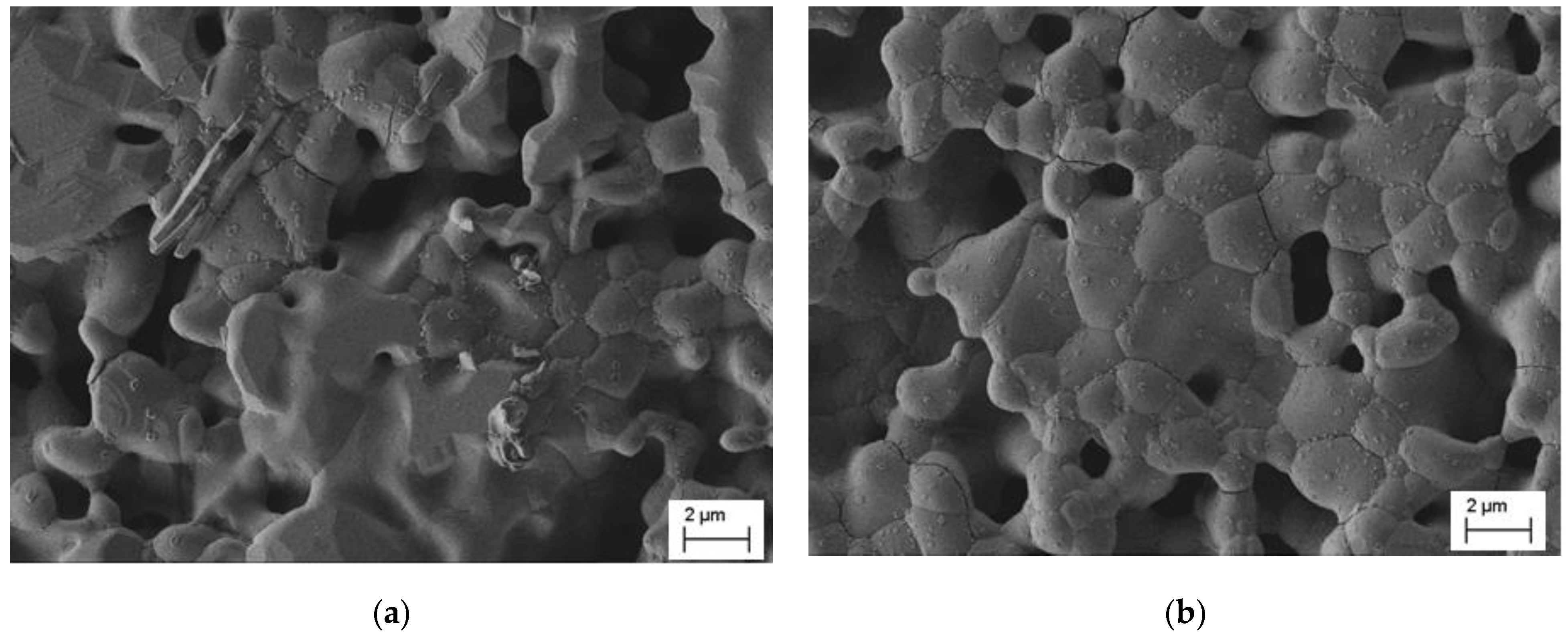
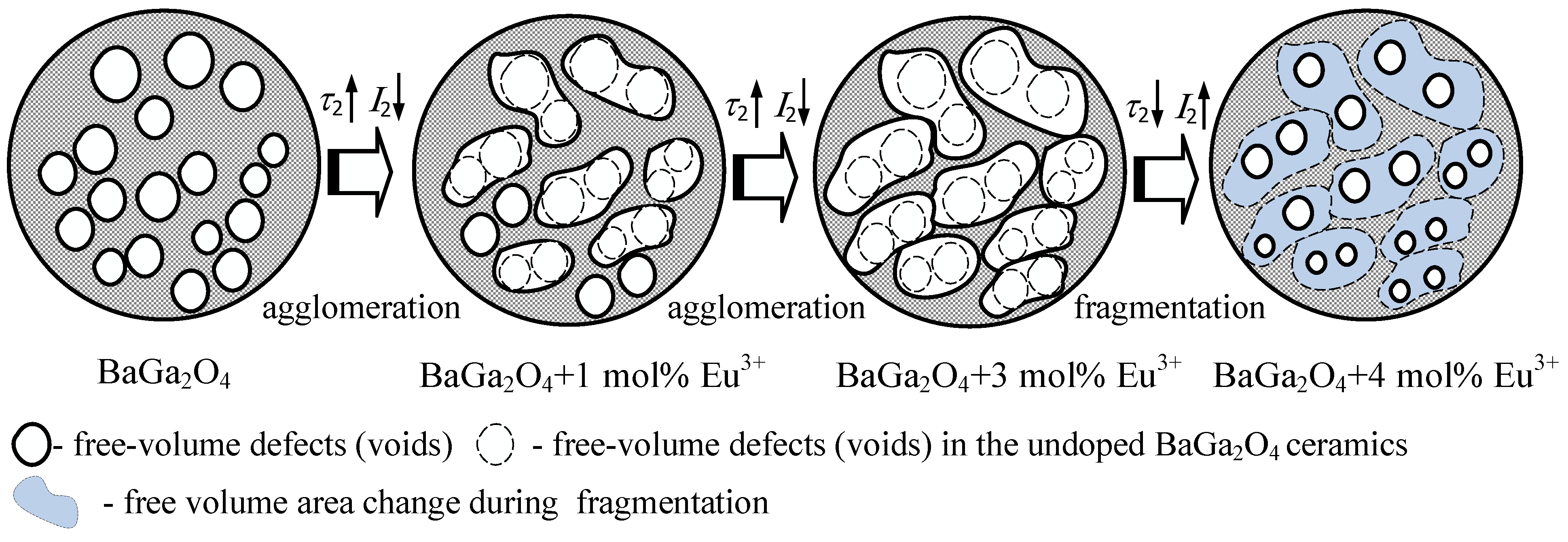
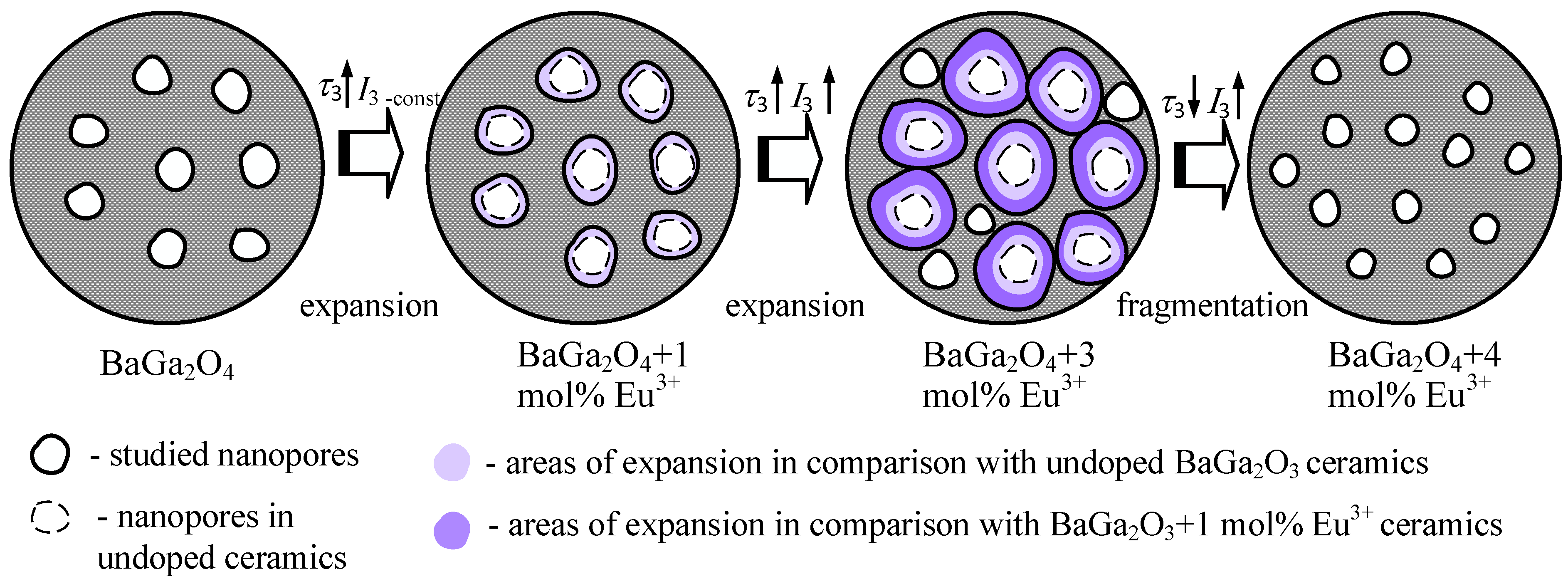
3. Results and Discussion

According to the results of the XRD investigations [44], the initial undoped BaGa2O4 ceramics contain three phases: 34.2 wt.% of the main BaGa2O4 phase, 58.5 wt.% of the additional Ba2.84Ga11.32O19.82 phase and 7.3 wt.% of the Ga2O3 phases. The BaGa2O4 ceramics with 1 mol.% Eu3+ ions are single-phase. The BaGa2O4 ceramics with a 3 and 4 mol.% impurity of Eu3+ ions contain two phases: 97.3 and 96.7 wt.% of the BaGa2O4 phase as well as 2.7 and 3.3 wt.% of the additional Eu3GaO6 phase, respectively. The results of the detailed XRD analysis using Rietveld’s method are represented in Table 1.
Generalized XRD patterns for undoped BaGa2O4 ceramics and those doped with Eu3+ ions, in comparison with the diffraction pattern of BaGa2O4 in the ICSD reference data file, are shown in Figure 1. The availability of the main phase in the studied ceramics indicates peaks at the reflection angle 2θ of 28.12°.
As can be seen from the SEM images, both undoped BaGa2O4 ceramics (Figure 2a) as well as ceramics with 1 mol.% Eu3+ ions (Figure 2b) have a fairly branched structure of grains, grain boundaries and pores of different forms and shapes [45]. Samples of these ceramics are characterized by a small grain size and homogeneity in the morphology of the structure. The presence of Eu3+ ions leads to an increase in the pore size of the initial BaGa2O4 ceramics, the formation of grain agglomerates and their further growth. In addition, in both cases there is the formation of micro- and nanosized crystals on the grain surface.
In addition, the elemental composition of the studied ceramic samples was evaluated using the method of energy-dispersion X-ray (EDX) spectroscopy for selected areas (Figure 3).
It is known that the accuracy of the EDX technique depends on the nature of the sample and other factors so only clearly defined peaks were analyzed. As can be seen from Figure 3a, elements in the BaGa2O4 compound are evenly distributed over the volume of crystallites, and there are reflexes of the main elements of the material. However, in the sample of BaGa2O4 ceramics doped with 1 mol.% Eu3+ ions (Figure 3b), their percentage was not established (despite the presence of Eu-reflexes). Obviously, such a small number of Eu3+ ions is not enough to identify the selected area. For BaGa2O4 ceramics with 3 mol.% Eu3+ ions, the presence of the latter increases to 0.74% (Figure 3c), while a further rise in the content of impurities (to 4 mol.% Eu3+ ions) leads to a significant increase in the material to 4.88% (Figure 3d, Table 2). Thus, the obtained results indicate the successful incorporation of Eu3+ ions into the BaGa2O4 matrix, especially if their concentration exceeds 1 mol.%.
Obviously, the presence of different amounts of Eu3+ ions in the ceramic BaGa2O4 matrix will modify their inner free volume. Extended defects formed in ceramic materials and mainly localized near grain boundaries are also due to the release of additional phases. The PAL method was used to study such defect-related free volumes in undoped and Eu3+-doped BaGa2O4 ceramics.
The fitting parameters of PAL spectra for the studied ceramics calculated within a three-component procedure are presented in Table 3. It is established that with rising Eu3+ content in the BaGa2O4 matrix, an increase in the lifetime τ1 and intensity I1 of the first short-term component is observed. Most probably, the structure of the mail phase of the ceramics is improved. However, when the Eu3+ ions increase to 4 mol.%, the opposite trend occurs. The lifetime τ2 of the second component increases and intensity I2 decreases with Eu3+ content (to 3 mol.%), while a further rise in Eu3+ ions (to 4 mol.%) leads to a decreased τ2 and an increased I2.
As noted in [42,45], the lifetime τ2 of the second component should be associated with the capture of positrons by defect-related free volumes. According to XRD data, undoped BaGa2O4 ceramics contain a large number of additional phases. As shown by the SEM method, these phases are unevenly distributed in the volume of the ceramic and are mainly localized near the grain boundaries. The extracted phases play the role of special centers of positron capture in the volume of ceramics. Since undoped BaGa2O4 ceramics contain two additional phases, positrons in such samples will be better captured.
The lifetime τ2 correlates with the size of the free-volume defects, where positrons are captured, and the intensity correlates with their number. Thus, when the Eu3+ content increases to 3 mol.%, the agglomeration of free-volume defects occurs, while the supersaturation of the BaGa2O4 matrix with Eu3+ (up to 4 mol.%) leads to their fragmentation. The identified trends (Table 4) correlate with the positron trapping parameters calculated within the two-state positron trapping model [45]. Schematically, the evolution of defect-related free volumes near grain boundaries with increasing Eu3+ ions in BaGa2O4 ceramics is shown in Figure 4.
As mentioned above, the lifetime of the second component correlates with the size of the free-volume defect, while its intensity is defined by the amount of these defects. The agglomeration process is reflected in the increase in the second component’s lifetime and in the decrease in its intensity. Solid lines in Figure 4 show existing voids in the sample, and dotted lines indicate voids in the initial ceramics. This schematically illustrates the process when two smaller voids are joined to form a larger one.
As can be seen from Table 3, lifetime τ2 increases from 0.424 ns in BaGa2O4 ceramics to 0.450 ns in BaGa2O4 ceramics containing 1 mol.% Eu3+ ions, and intensity decreases from 14.9 to 13.2%.In Figure 4, it is schematically demonstrated that most voids were agglomerated, though there were some left unchanged. Generally, the number of voids is smaller and the overall volume of the voids is larger. For BaGa2O4 ceramics doped with 3 mol.% Eu3+ ions, agglomeration is also the case, and the lifetime of the second component keeps increasing up until 0.550 ns, whereas intensity goes down to 7.9%. It is schematically shown that the voids in BaGa2O4 ceramics with 1 mol.% Eu3+ ions were unchanged; in BaGa2O4 ceramics with 3 mol.% Eu3+ ions, they were involved in agglomeration. That is why the number of voids decreased even more and the overall void volume became larger. For BaGa2O4 ceramics with 4 mol.% Eu3+ we observe the opposite—fragmentation of free-volume defects occurs. These changes are manifested in the decrease in τ2 lifetime and the increase in I2 intensity. In Figure 4, the region where free-volume voids change compared to BaGa2O4 ceramics with 3 mol.% Eu3+ ions due to fragmentation is shown in blue. The intensity I2 for BaGa2O4 ceramics with 4 mol.% Eu3+ ions is close to the I2 for undoped BaGa2O4 ceramics, but the lifetime τ2 is shorter. This is evidence of the smaller overall size of free-volume defects in BaGa2O4 ceramics with 4 mol.% Eu3+ ions.
The third long-term component of the PAL spectrum (the second channel of positron annihilation with lifetime τ3 and intensity I3) is associated with the o-Ps decaying in nanopores [45]. As can be seen from Table 3, lifetime τ3 and intensity I3 increase with Eu3+ content in the BaGa2O4 ceramics (to 3 mol.%). However, when the amount of Eu3+ ions rises to 4 mol.%, the lifetime τ3 decreases and the intensity I3 continues to increase. It is obvious that the presence of Eu3+ ions in the ceramic matrix leads to the expansion of nanosized pores and an increase in their number. However, the supersaturation of ceramics with doped ions (to 4 mol.%) results in the fragmentation of nanopores. The obtained trends are reflected in the radius of nanopores R3 (Table 4) calculated using the Tao–Eldrup model. This process is schematically shown in Figure 5.
Adding 1 mol.% Eu3+ to the BaGa2O4 ceramics is reflected in the increased lifetime of the third component (up to 2.289 ns) at a constant intensity I3 as compared to the undoped ceramics. This confirms the process of nanopore volume expansion. Further increase in Eu3+ content up to 3 mol.% facilitates an increase in both lifetime τ3 and intensity I3, pointing at the continuation of the nanopore expansion process with simultaneous new pore creation. However, for BaGa2O4 ceramics with 4 mol.% Eu3+ ions, the opposite fragmentation process takes place, which can be described as a decreased lifetime of the third component and its increased intensity. Total nanopore volume is smaller than it is for undoped ceramics (see Table 3), thus indicating the excess character of such Eu3+ doping.
Thus, PAL spectroscopy can be a tool that allows us to assess the ability of Eu penetration into ceramic grains. The sites of Eu3+ ions define the positron trapping rate, and related structural transformations can be analyzed using positron component parameters, which change depending on the concentration of doping ions. This method can also be used to determine the size of nanopores in undoped and doped ceramic materials.

4. Conclusions

The structural features and evolution of free-volume defects in BaGa2O4 ceramics obtained by solid-phase synthesis from the initial BaCO3 and Ga2O3 components with the addition of different amounts of Eu2O3 content (1, 3 and 4 mol.%) were investigated. The structural features of ceramics were studied using XRD as well as SEM with an EDX elemental analysis. It is established that, according to the quantitative analysis of the elemental composition, samples of the undoped BaGa2O4 ceramics have the largest deviations from the stoichiometric composition; they have three phases. Such processes are apparently caused by the evaporation of the constituent synthesis powders during the annealing process at high temperatures. The detected signs correlate with the XRD data.
Additional phases in ceramics are mainly localized near the grain boundaries and create defective centers for positron capture studied by PAL spectroscopy. Analyzing the second component of the PAL spectra for the undoped and Eu3+-doped BaGa2O4 ceramics, it was shown that an increase in Eu3+ content from 1 to 3 mol.% leads to the agglomeration of free-volume defects near the grain boundaries of ceramics. At the same time, nanopores in ceramics expand and their number increases. Further increase in the content of Eu3+ ions is accompanied with the fragmentation of both free-volume defects and nanopores.
Results obtained by PAL spectroscopy can serve as a research base for the development of independent complementary methods for studying nanosized free volumes in ceramic materials, including neutron and heavy ion irradiated MgAl2O4 spinels [22,27,48], Si3N4 [49], Ge3N4 [50] and AlN [51,52,53], which are especially promising as diagnostic materials for nuclear applications. Other interesting and very important areas are the understanding of porosity, its development and transformation in electrochemical and other devices for energy conversion [54,55,56,57,58,59,60,61,62,63,64].

Author Contributions

Writing—original draft preparation, H.K.; writing—review and editing, I.K., V.P. and A.I.P.; investigation of ceramics, H.K., Y.K., A.L. and I.K.; treatment of experimental results, H.K. and Y.K.; project administration, A.I.P. All authors have read and agreed to the published version of the manuscript.

Funding

This work was supported by the Ministry of Education and Science of Ukraine (project for young researchers No. 0119U100435) for H.K and Y.K.; the National Research Foundation of Ukraine (project 2020.02/0217) for I.K. and H.K. as well as by the Latvian research council via the Latvian National Research Program under the topic “High-Energy Physics and Accelerator Technologies”, Agreement No: VPP-IZM-CERN-2020/1-0002, for V.P. and A.I.P.

Institutional Review Board Statement

Not applicable.

Informed Consent Statement

Not applicable.

Data Availability Statement

The data presented in this study are available on request from the corresponding author.

Acknowledgments

H.K. and Y.K. would like to thank A. Ingram for assistance in PAL experiments. The authors thank E.A. Kotomin and M. Brik for the many useful discussions. The research was (partly) performed in the Institute of Solid State Physics, University of Latvia ISSP UL. ISSP UL as the Center of Excellence is supported through the Framework Program for European universities Union Horizon 2020, H2020-WIDESPREAD-01–2016–2017-TeamingPhase2 under Grant Agreement No. 739508, CAMART2 project.

Conflicts of Interest

The authors declare no conflict of interest.

References

  1. Noto, L.L.; Poelman, D.; Orante-Barrón, V.R.; Swart, H.C.; Mathevula, L.E.; Nyenge, R.; Chithambo, M.; Mothudi, B.M.; Dhlamini, M.S. Photoluminescence and thermoluminescence properties of BaGa2O4. Phys. B Condens. Matter 2018, 535, 268–271. [Google Scholar] [CrossRef]
  2. Kodu, M.; Avarmaa, T.; Mändar, H.; Jaaniso, R. Pulsed Laser Deposition of BaGa2O4. Appl. Phys. A 2008, 93, 801–805. [Google Scholar] [CrossRef]
  3. Gust, D.; Moore, T.A.; Moore, A.L. Solar Fuels via Artificial Photosynthesis. Acc. Chem. Res. 2009, 42, 1890–1898. [Google Scholar] [CrossRef]
  4. Skillen, N.; Robertson, P.K.J. Artificial photosynthesis. In Solar Energy; World Scientific Publishing Co. Pte. Ltd.: Singapore, 2016; pp. 205–241. [Google Scholar] [CrossRef]
  5. Acuña, W.; Tellez, J.F.; Macías, M.A.; Roussel, P.; Ricote, S.; Gauthier, G.H. Synthesis and Characterization of BaGa2O4 and Ba3Co2O6(CO3)0.6 Compounds in the Search of Alternative Materials for Proton Ceramic Fuel Cell (PCFC). Solid State Sci. 2017, 71, 61–68. [Google Scholar] [CrossRef]
  6. Guérineau, T.; Strutynski, C.; Skopak, T.; Morency, S.; Hanafi, A.; Calzavara, F.; Ledemi, Y.; Danto, S.; Cardinal, T.; Messaddeq, Y.; et al. Extended Germano-Gallate Fiber Drawing Domain: From Germanates to Gallates Optical Fibers. Opt. Mater. Express 2019, 9, 2437–2445. [Google Scholar] [CrossRef] [Green Version]
  7. Gonçalves, J.M.; Munoz, R.A.; Angnes, L. Materials for Optical, Magnetic and Electronic Devices. J. Mater. Chem. C 2021, 9, 8708–8717. [Google Scholar] [CrossRef]
  8. Nakauchi, D.; Okada, G.; Kawaguchi, N.; Yanagida, T. Luminescent and Scintillation Properties of Eu-Doped (Ba, Sr)Al2O4 Crystals. Opt. Mater. 2019, 87, 58–62. [Google Scholar] [CrossRef]
  9. Khattab, T.A.; Abd El-Aziz, M.; Abdelrahman, M.S.; El-Zawahry, M.; Kamel, S. Development of Long-persistent Photoluminescent Epoxy Resin Immobilized with Europium (II)-doped Strontium Aluminate. Luminescence 2020, 35, 478–485. [Google Scholar] [CrossRef]
  10. Yu, L.; den Engelsen, D.; Gorobez, J.; Fern, G.R.; Ireland, T.G.; Frampton, C.; Silver, J. Crystal Structure, Photoluminescence and Cathodoluminescence of Sr1-xCaxAl2O4 Doped with Eu2+. Opt. Mater. Express 2019, 9, 2175–2195. [Google Scholar] [CrossRef]
  11. Maphiri, V.M.; Mhlongo, M.R.; Hlatshwayo, T.T.; Motaung, T.E.; Koao, L.F.; Motloung, S.V. Citrate Sol-Gel Synthesis of BaAl2O4:X% Cu2+ (0 ≤ x ≤ 1) Nano-Phosphors: Structural, Morphological and Photoluminescence Properties. Opt. Mater. 2020, 109, 110244. [Google Scholar] [CrossRef]
  12. den Engelsen, D.; Fern, G.R.; Ireland, T.G.; Yang, F.; Silver, J. Photoluminescence and Cathodoluminescence of BaAl2O4:Eu2+ and Undoped BaAl2O4: Evidence for F-Centres. Opt. Mater. Express 2020, 10, 1962–1980. [Google Scholar] [CrossRef]
  13. den Engelsen, D.; Fern, G.R.; Ireland, T.G.; Silver, J. Laser-Activated Luminescence of BaAl2O4:Eu. ECS J. Solid State Sci. Technol. 2020, 9, 026001. [Google Scholar] [CrossRef]
  14. Shivaramu, N.J.; Coetsee, E.; Roos, W.D.; Nagabhushana, K.R.; Swart, H.C. Charge Carrier Trapping Processes in Un-Doped and BaAl2O4:Eu3+ Nanophosphor for Thermoluminescent Dosimeter Applications. J. Phys. D Appl. Phys. 2020, 53, 475305. [Google Scholar] [CrossRef]
  15. Xie, Q.; Li, B.; He, X.; Zhang, M.; Chen, Y.; Zeng, Q. Correlation of Structure, Tunable Colors, and Lifetimes of (Sr, Ca, Ba)Al2O4:Eu2+, Dy3+ Phosphors. Materials 2017, 10, 1198. [Google Scholar] [CrossRef] [PubMed] [Green Version]
  16. Wang, S.; Wang, Y.; Gao, H.; Li, J.; Fang, L.; Yu, X.; Tang, S.; Zhao, X.; Sun, G. Synthesis and Characterization of BaAl2O4: Ce and Mn-Ce-Co-Doped BaAl2O4 Composite Materials by a Modified Polyacrylamide Gel Method and Prediction of Photocatalytic Activity Using Artificial Neural Network (ANN) Algorithm. Optik 2020, 221, 165363. [Google Scholar] [CrossRef]
  17. Malkamäki, M.; Bos, A.J.J.; Dorenbos, P.; Lastusaari, M.; Rodrigues, L.C.V.; Swart, H.C.; Hölsä, J. Persistent Luminescence Excitation Spectroscopy of BaAl2O4:Eu2+,Dy3+. Phys. B Condens. Matter 2020, 593, 411947. [Google Scholar] [CrossRef]
  18. Nakauchi, D.; Okada, G.; Kato, T.; Kawaguchi, N.; Yanagida, T. Crystal Growth and Scintillation Properties of Eu:BaAl2O4 Crystals. Radiat. Meas. 2020, 135, 106365. [Google Scholar] [CrossRef]
  19. Vrankić, M.; Šarić, A.; Bosnar, S.; Pajić, D.; Dragović, J.; Altomare, A.; Falcicchio, A.; Popović, J.; Jurić, M.; Petravić, M.; et al. Magnetic Oxygen Stored in Quasi-1D Form within BaAl2O4 Lattice. Sci. Rep. 2019, 9, 15158. [Google Scholar] [CrossRef] [PubMed] [Green Version]
  20. Lisovskii, S.; Meganov, A.; Khrustov, V.R.; Ivanov, V. Ultraviolet Cathodoluminescence of Pure Zinc Aluminate ZnAl2O4. J. Phys. Conf. Ser. 2019, 1410, 012089. [Google Scholar] [CrossRef]
  21. Valiev, D.; Khasanov, O.; Dvilis, E.; Stepanov, S.; Paygin, V.; Ilela, A. Structural and Spectroscopic Characterization of Tb3+-Doped MgAl2O4 Spinel Ceramics Fabricated by Spark Plasma Sintering Technique. Phys. Status Solidi (B) 2020, 257, 1900471. [Google Scholar] [CrossRef]
  22. Lushchik, A.; Feldbach, E.; Kotomin, E.A.; Kudryavtseva, I.; Kuzovkov, V.N.; Popov, A.I.; Seeman, V.; Shablonin, E. Distinctive Features of Diffusion-Controlled Radiation Defect Recombination in Stoichiometric Magnesium Aluminate Spinel Single Crystals and Transparent Polycrystalline Ceramics. Sci. Rep. 2020, 10, 7810. [Google Scholar] [CrossRef] [PubMed]
  23. Mironova-Ulmane, N.; Popov, A.I.; Krieke, G.; Antuzevics, A.; Skvortsova, V.; Elsts, E.; Sarakovskis, A. Low-Temperature Studies of Cr3+ Ions in Natural and Neutron-Irradiated g-Al Spinel. Low Temp. Phys. 2020, 46, 1154–1159. [Google Scholar] [CrossRef]
  24. Platonenko, A.; Gryaznov, D.; Kotomin, E.A.; Lushchik, A.; Seeman, V.; Popov, A.I. Hybrid Density Functional Calculations of Hyperfine Coupling Tensor for Hole-Type Defects in MgAl2O4. Nucl. Instrum. Methods Phys. Res. Sect. B Beam Interact. Mater. At. 2020, 464, 60–64. [Google Scholar] [CrossRef]
  25. Li, Q.; Liu, T.; Xu, X.; Wang, X.; Guo, R.; Jiao, X.; Lu, Y. Study on the Optical Spectra of MgAl2O4 with Oxygen Vacancies. Mater. Technol. 2021, 36, 279–285. [Google Scholar] [CrossRef]
  26. Li, Q.; Liu, T.; Xu, X.; Guo, R.; Jiao, X.; Wang, X.; Lu, Y. Study of Cation Vacancies with Localized Hole States in MgAl2O4 Crystals. J. Phys. Chem. Solids 2020, 145, 109542. [Google Scholar] [CrossRef]
  27. Seeman, V.; Feldbach, E.; Kärner, T.; Maaroos, A.; Mironova-Ulmane, N.; Popov, A.I.; Shablonin, E.; Vasil’chenko, E.; Lushchik, A. Fast-Neutron-Induced and as-Grown Structural Defects in Magnesium Aluminate Spinel Crystals with Different Stoichiometry. Opt. Mater. 2019, 91, 42–49. [Google Scholar] [CrossRef]
  28. Gholami, Y.H.; Yuan, H.; Wilks, M.Q.; Josephson, L.; el Fakhri, G.; Normandin, M.D.; Kuncic, Z. Positron Annihilation Localization by Nanoscale Magnetization. Sci. Rep. 2020, 10, 20262. [Google Scholar] [CrossRef]
  29. Rementeria, R.; Domínguez-Reyes, R.; Capdevila, C.; Garcia-Mateo, C.; Caballero, F.G. Positron Annihilation Spectroscopy Study of Carbon-Vacancy Interaction in Low-Temperature Bainite. Sci. Rep. 2020, 10, 487. [Google Scholar] [CrossRef] [Green Version]
  30. Dlubek, G.; Kilburn, D.; Bondarenko, V.; Pionteck, J.; Krause-Rehberg, R.; Alam, M.A. Positron Annihilation: A Unique Method for Studying Polymers. Macromol. Symp. 2004, 210, 11–20. [Google Scholar] [CrossRef]
  31. Dai, H.Y.; Liu, H.Z.; Peng, K.; Ye, F.J.; Li, T.; Chen, J.; Chen, Z.P. Correlation between Vacancy Defects and Magnetic Properties of the GdMn1-xZnxO3 Multiferroic Ceramics Studied by Positron Annihilation. Mater. Res. Bull. 2019, 119, 110565. [Google Scholar] [CrossRef]
  32. Barad, D.; Mange, P.L.; Jani, K.K.; Mukherjee, S.; Ahmed, M.; Kumar, S.; Dolia, S.N.; Pandit, R.; Raval, P.Y.; Modi, K.B.; et al. Ca2+-Substitution Effect on the Defect Structural Changes in the Quadruple Perovskite Series Ca1+xCu3-xTi4O12 Studied by Positron Annihilation and Complementary Methods. Ceram. Int. 2021, 47, 2631–2640. [Google Scholar] [CrossRef]
  33. Ghanem, A.; Mohamed, K. Effect of Gamma-Ray on Producing Induced Colour Centres and on Positron Annihilation Lifetime of Bismuth-Doped Zinc Sodium Borate Glasses. Arab. J. Nucl. Sci. Appl. 2021, 54, 1–9. [Google Scholar] [CrossRef]
  34. Ashok, J.; Kostrzewa, M.; Ingram, A.; Reddy, M.S.; Kumar, V.R.; Gandhi, Y.; Veeraiah, N. Free Volume Estimation in Au and Ag Mixed Sodium Antimonate Glass Ceramics by Means of Positron Annihilation. Phys. B Condens. Matter 2019, 570, 266–273. [Google Scholar] [CrossRef]
  35. El-Gamal, S.; Elsayed, M. Positron Annihilation and Electrical Studies on the Influence of Loading Magnesia Nanoribbons on PVA-PVP Blend. Polym. Test. 2020, 89, 106681. [Google Scholar] [CrossRef]
  36. Zhang, H.J.; Sellaiyan, S.; Sako, K.; Uedono, A.; Taniguchi, Y.; Hayashi, K. Effect of Free-Volume Holes on Static Mechanical Properties of Epoxy Resins Studied by Positron Annihilation and PVT Experiments. Polymer 2020, 190, 122225. [Google Scholar] [CrossRef]
  37. Biswas, D.; Rajan, A.; Kabi, S.; Das, A.S.; Singh, L.S.; Nambissan, P.M.G. Structural Defects Characterization of Silver-Phosphate Glass Nanocomposites by Positron Annihilation and Related Experimental Studies. Mater. Charact. 2019, 158, 109928. [Google Scholar] [CrossRef]
  38. Fan, J.; Zhou, W.; Wang, Q.; Chu, Z.; Yang, L.; Yang, L.; Sun, J.; Zhao, L.; Xu, J.; Liang, Y.; et al. Structure Dependence of Water Vapor Permeation in Polymer Nanocomposite Membranes Investigated by Positron Annihilation Lifetime Spectroscopy. J. Membr. Sci. 2018, 549, 581–587. [Google Scholar] [CrossRef]
  39. Sato, K.; Tamiya, R.; Xu, Q.; Tsuchida, H.; Yoshiie, T. Detection of Deuterium Trapping Sites in Tungsten by Thermal Desorption Spectroscopy and Positron Annihilation Spectroscopy. Nucl. Mater. Energy 2016, 9, 554–559. [Google Scholar] [CrossRef] [Green Version]
  40. Wang, Z.F.; Wang, B.; Yang, Y.R.; Hu, C.P. Correlations between Gas Permeation and Free-Volume Hole Properties of Polyurethane Membranes. Eur. Polym. J. 2003, 39, 2345–2349. [Google Scholar] [CrossRef]
  41. Melikhova, O.; Kuriplach, J.; Prochazka, I.; Cizek, J.; Hou, M.; Zhurkin, E.; Pisov, S. Simulation of Positron Annihilation Response to Mechanical Deformation of Nanostructured Ni3Al. Appl. Surf. Sci. 2008, 255, 157–159. [Google Scholar] [CrossRef]
  42. Shpotyuk, O.; Balitska, V.; Brunner, M.; Hadzaman, I.; Klym, H. Thermally-Induced Electronic Relaxation in Structurally-Modified Cu0.1Ni0.8Co0.2Mn1.9O4 Spinel Ceramics. Phys. B Condens. Matter 2015, 459, 116–121. [Google Scholar] [CrossRef]
  43. Klym, H.; Ingram, A.; Shpotyuk, O.; Karbovnyk, I. Influence of CsCl Addition on the Nanostructured Voids and Optical Properties of 80GeS2-20Ga2S3 Glasses. Opt. Mater. 2016, 59, 39–42. [Google Scholar] [CrossRef]
  44. Kostiv, Y.; Luchechko, A.; Klym, H.; Karbovnyk, I.; Sadovyi, B.; Zaremba, O.; Kravets, O. Structural properties of polycrystalline BaGa2O4 Ceramics Doped with Eu3+ Ions. In Proceedings of the XIth International Scientific and Practical Conference on Electronics and Information Technologies, Lviv, Ukraine, 16–18 September 2019; pp. 307–311. [Google Scholar] [CrossRef]
  45. Klym, H.; Ingram, A.; Hadzaman, I.; Shpotyuk, O. Evolution of Porous Structure and Free-volume Entities in Magnesium Aluminate Spinel Ceramics. Ceram. Int. 2014, 40, 8561–8567. [Google Scholar] [CrossRef]
  46. Kansy, J.; Giebel, D. Study of Defect Structure with New Software for Numerical Analysis of PAL Spectra. J. Phys. Conf. Ser. 2011, 265, 0102030. [Google Scholar] [CrossRef] [Green Version]
  47. Goworek, T. Comments on the Relation: Positronium Lifetime—Free Volume Size Parameters of the Tao-Eldrup Model. Chem. Phys. Lett. 2002, 366, 184–187. [Google Scholar] [CrossRef]
  48. Lushchik, A.; Dolgov, S.; Feldbach, E.; Pareja, R.; Popov, A.I.; Shablonin, E.; Seeman, V. Creation and Thermal Annealing of Structural Defects in Neutron-Irradiated MgAl2O4 Single Crystals. Nucl. Instrum. Methods Phys. Res. Sect. B Beam Interact. Mater. At. 2018, 435, 31–37. [Google Scholar] [CrossRef]
  49. Feldbach, E.; Museur, L.; Krasnenko, V.; Zerr, A.; Kitaura, M.; Kanaev, A. Defects Induced by He+ Irradiation in γ-Si3N4. J. Lumin. 2021, 237, 118132. [Google Scholar] [CrossRef]
  50. Feldbach, E.; Zerr, A.; Museur, L.; Kitaura, M.; Manthilake, G.; Tessier, F.; Krasnenko, V.; Kanaev, A. Electronic Band Transitions in γ-Ge3N4. Electron. Mater. Lett. 2021, 17, 315–323. [Google Scholar] [CrossRef]
  51. Kozlovskiy, A.; Kenzhina, I.; Zdorovets, M.V. Optical and Structural Properties of AlN Ceramics Irradiated with Heavy Ions. Opt. Mater. 2019, 91, 130–137. [Google Scholar] [CrossRef]
  52. Zdorovets, M.V.; Dukenbayev, K.; Kozlovskiy, A.L. Study of Helium Swelling in Nitride Ceramics at Different Irradiation Temperatures. Materials 2019, 12, 2415. [Google Scholar] [CrossRef] [PubMed] [Green Version]
  53. Kozlovskiy, A.; Kenzhina, I.; Dukenbayev, K.; Zdorovets, M. Influence of He-ion Irradiation of Ceramic AlN. Vacuum 2019, 163, 45–51. [Google Scholar] [CrossRef]
  54. Kurteeva, A.A.; Bogdanovich, N.M.; Bronin, D.I.; Porotnikova, N.M.; Vdovin, G.K.; Pankratov, A.A.; Beresnev, S.M.; Kuz’mina, L.A. Options for Adjustment of Microstructure and Conductivity of Cathodic Substrates of La(Sr)MnO3. Russ. J. Electrochem. 2010, 46, 811–819. [Google Scholar] [CrossRef]
  55. Porotnikova, N.M.; Eremin, V.A.; Farlenkov, A.S.; Kurumchin, E.K.; Sherstobitova, E.A.; Kochubey, D.I.; Ananyev, M.V. Effect of AO Segregation on Catalytical Activity of La0.7A0.3MnO3±δ (A = Ca, Sr, Ba) Regarding Oxygen Reduction Reaction. Catal. Lett. 2018, 148, 2839–2847. [Google Scholar] [CrossRef]
  56. Osinkin, D.A.; Khodimchuk, A.V.; Porotnikova, N.M.; Bogdanovich, N.M.; Fetisov, A.V.; Ananyev, M.V. Rate-Determining Steps of Oxygen Surface Exchange Kinetics on Sr2Fe1.5Mo0.5O6−δ. Energies 2020, 13, 250. [Google Scholar] [CrossRef] [Green Version]
  57. Suchikova, Y.O. Sulfide Passivation of Indium Phosphide Porous Surfaces. J. Nano-Electron. Phys. 2017, 9, 01006. [Google Scholar] [CrossRef]
  58. Suchikova, J.A. Synthesis of Indium Nitride Epitaxial Layers on a Substrate of Porous Indium Phosphide. J. Nano-Electron. Phys. 2015, 7, 03017. [Google Scholar]
  59. Shlimas, D.; Kenzhina, I.; Zdorovets, M. Study of the Use of Ionizing Radiation to Improve the Efficiency of Performance of Nickel Nanostructures as Anodes of Lithium-ion Batteries. Mater. Res. Express 2019, 6, 055026. [Google Scholar] [CrossRef]
  60. Rumiantseva, Y.; Melnichuk, I.; Garashchenko, V.; Zaporozhets, O.; Turkevich, V.; Bushlya, V. Influence of cBN Content, Al2O3 and Si3N4 Additives and Their Morphology on Microstructure, Properties, and Wear of PCBN with NbN Binder. Ceram. Int. 2020, 46, 22230–22238. [Google Scholar] [CrossRef]
  61. Olenych, I.B.; Aksimentyeva, O.I.; Monastyrskii, L.S.; Horbenko, Y.Y.; Partyka, M.V. Electrical and Photoelectrical Properties of Reduced Graphene Oxide—Porous Silicon Nanostructures. Nanoscale Res. Lett. 2017, 12, 272. [Google Scholar] [CrossRef]
  62. Luchechko, A.; Zhydachevskyy, Y.; Ubizskii, S.; Kravets, O.; Popov, A.I.; Rogulis, U.; Elsts, E.; Bulur, E.; Suchocki, A. Afterglow, TL and OSL Properties of Mn2+-doped ZnGa2O4 Phosphor. Sci. Rep. 2019, 9, 9544. [Google Scholar] [CrossRef]
  63. Dimza, V.; Popov, A.I.; Lāce, L.; Kundzins, M.; Kundzins, K.; Antonova, M.; Livins, M. Effects of Mn Doping on Dielectric Properties of Ferroelectric Relaxor PLZT Ceramics. Curr. Appl. Phys. 2017, 17, 169–173. [Google Scholar] [CrossRef]
  64. Bystrova, A.; Dekhtyar, Y.D.; Popov, A.; Coutinho, J.; Bystrov, V. Modified Hydroxyapatite Structure and Properties: Modeling and Synchrotron Data Analysis of Modified Hydroxyapatite Structure. Ferroelectrics 2015, 475, 135–147. [Google Scholar] [CrossRef]
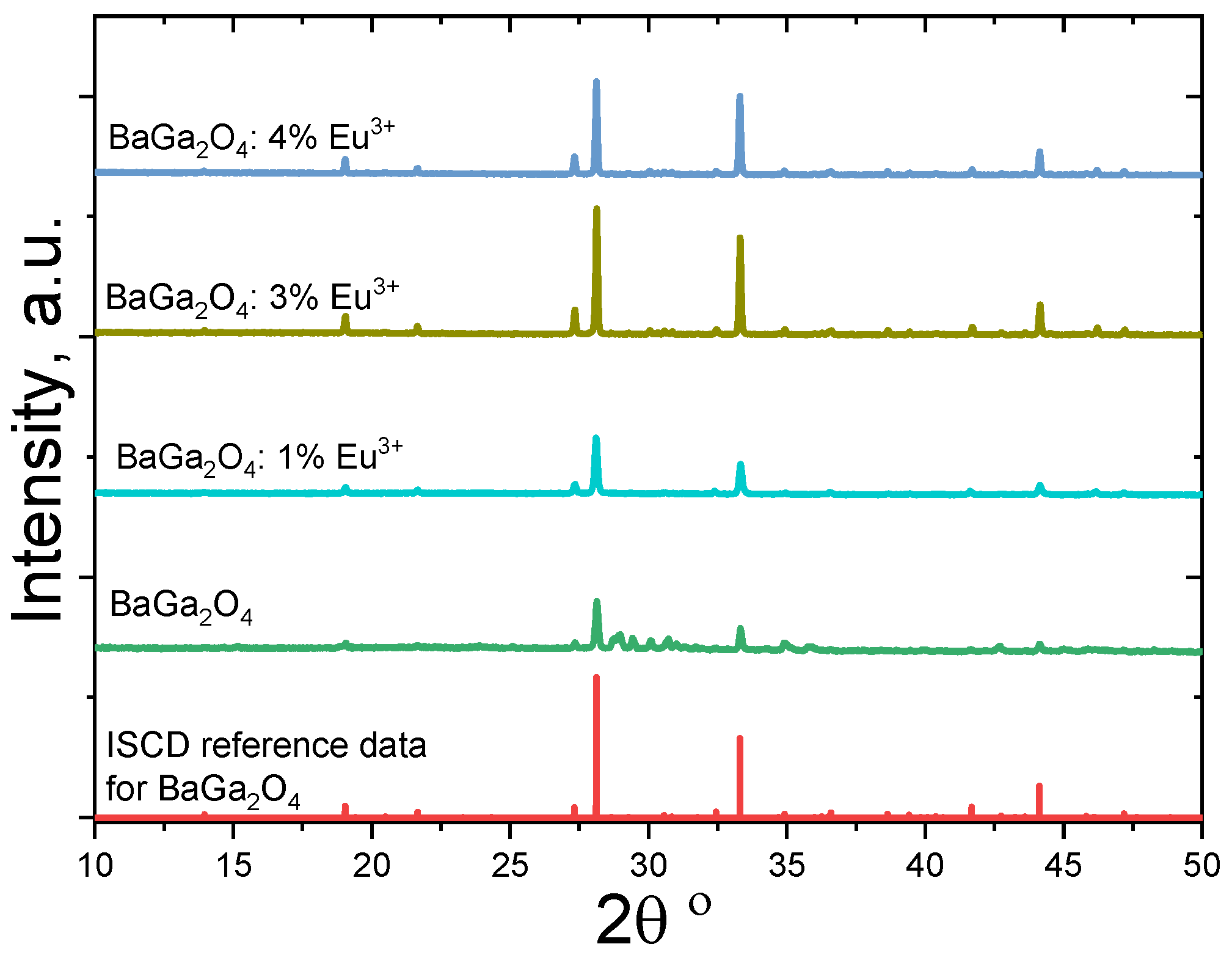
Figure 1. XRD patterns for undoped and Eu3+-doped BaGa2O4 ceramics in comparison with ICSD reference data for BaGa2O4.
Crystals 11 01515 g001
Figure 2. SEM images for undoped (a) and Eu3+-doped (b) BaGa2O4 ceramics.
Crystals 11 01515 g002
Figure 3. Microstructure of the selected areas and elemental composition of the undoped BaGa2O4 ceramics (a) and those doped with 1 mol.% (b), 3 mol.% (c) and 4 mol.% of Eu3+ ions (d).
Crystals 11 01515 g003
Figure 4. Diagram explaining the evolution of defect-related voids in BaGa2O4 ceramics caused by Eu3+ doping.
Crystals 11 01515 g004
Figure 5. Diagram explaining the evolution of nanopores in BaGa2O4 ceramics caused by Eu3+ doping.
Crystals 11 01515 g005
Table 1. Phase composition and Rietveld refined parameters for undoped and Eu3+-doped BaGa2O4 ceramics.
SampleNumber of PhasesPhases, Lattice Parameters,
Weight Fraction
BaGa2O43 phases:
BaGa2O4,
Ba2.84Ga11.32O19.82
and Ga2O3
BaGa2O4:
a = 18.619(1) Å, c = 8.670(1) Å
fraction 34.2 wt.%
Ba2.84Ga11.32O19.82:
a = 15.807(1) Å, b = 11.687(1) Å, c = 5.136(1) Å, β = 107.62(1) Å, fraction 58.5 wt.%
Ga2O3:
a = 12.213(4) Å, b = 3.042(1) Å,
c = 5.810(2) Å, β = 103.65(3) Å,
fraction 7.3 wt.%
BaGa2O4 + 1 mol.% Eu3+1 phase:
(Ba,Eu)Ga2O4
(Ba,Eu)Ga2O4:
a = 18.605(1) Å, c = 8.670(1) Å,
fraction 100 wt.%
BaGa2O4 + 3 mol.% Eu3+2 phases:
BaGa2O4,
Eu3GaO6
BaGa2O4:
a = 18.620(1) Å, c = 8.658 (1) Å,
fraction 97.3 wt.%
Eu3GaO6:
a = 9.026 (2) Å, b = 11.344(2) Å,
c = 5.496(1) Å, fraction 2.7 wt.%
BaGa2O4 + 4 mol.% Eu3+2 phases:
BaGa2O4,
Eu3GaO6
BaGa2O4:
a = 18.622(1) Å, c = 8.658(1) Å,
fraction 96.7 wt.%
Eu3GaO6:
a = 9.031(2) Å, b = 11.343(2) Å, c = 5.497(1) Å, fraction 3.3 wt.%
Table 2. Elemental composition of the undoped and Eu3+-doped BaGa2O4 ceramics.
SampleMass, %Atom, %
OGaBaEuOGaBaEu
BaGa2O4163430-592813-
BaGa2O4 + 1 mol.% Eu3+171629-711514-
BaGa2O4 + 3 mol.% Eu3+1630440.745925150.28
BaGa2O4 + 4 mol.% Eu3+1625324.886122151.98
Table 3. Lifetimes and intensities for undoped and Eu3+-doped BaGa2O4 ceramics obtained in three-component fitting procedure.
Sampleτ1 (±0.002),
ns
I1 (±0.1),
%
τ2 (±0.001),
ns
I2 (±0.1),
%
τ3 (±0.001),
ns
I3 (±0.1),
%
BaGa2O40.20083.30.42414.92.1961.8
BaGa2O4 + 1 mol.% Eu3+0.20685.00.45013.22.2891.8
BaGa2O4 + 3 mol.% Eu3+0.21289.90.5507.92.3902.2
BaGa2O4 + 4 mol.% Eu3+0.20183.30.41114.42.1572.4
Table 4. Positron trapping parameters and nanopore radius for undoped and Eu3+-doped BaGa2O4 ceramics.
Sampleτav, nsτb, nsκd, ns−1τ2τb, nsτ2/τbR3, nm
BaGa2O40.2340.2180.400.211.950.306
BaGa2O4 + 1 mol.% Eu3+0.2390.2220.350.232.020.314
BaGa2O4 + 3 mol.% Eu3+0.2400.2230.230.332.460.322
BaGa2O4 + 4 mol.% Eu3+0.2320.2180.370.191.890.302
Publisher’s Note: MDPI stays neutral with regard to jurisdictional claims in published maps and institutional affiliations.

