
Facile Two-Step Deposition of Calcium Oxalate Film on Dolomite to Improve Acid Rain Resistance


1
Institute of Cultural Heritage and History of Science & Technology, University of Science and Technology Beijing, Beijing 100083, China
2
Institute of Archaeology, Chinese Academy of Social Sciences, Beijing 100101, China
3
International Joint Research Laboratory of Environmental and Social Archaeology, Shandong University, Qingdao 266237, China
*
Authors to whom correspondence should be addressed.
Crystals 2022, 12(5), 734; https://0-doi-org.brum.beds.ac.uk/10.3390/cryst12050734
Submission received: 6 May 2022
/
Revised: 17 May 2022
/
Accepted: 18 May 2022
/
Published: 20 May 2022
(This article belongs to the Special Issue Corrosion, Coatings and Crystals)
Abstract
:The deposition of a calcium oxalate layer on dolomite demonstrates potential application in stone culture heritage conservation. However, due to insufficient coverage and the presence of cracks, the film’s usefulness is restricted. In this investigation, we used a simple two-step procedure to create a cohesive and uncracked film. The findings show that the protective layer provides better coverage of the dolomite surface without causing cracks and significantly improves acid resistance. Furthermore, after the simple two-step treatment, the color and adhesive strength of dolomite substrates remained nearly unchanged.
1. Introduction
Marble is sedimentary rock composed of calcite and dolomite generated from recrystallization at elevated temperatures and pressures. It has been employed extensively in the production of numerous stone artifacts, particularly in China [1]. The artifacts are a vital aspect of cultural heritage because they contain a wealth of information about ancient societies [2,3,4]. Unfortunately, because of its chemical composition and internal structure, marble is prone to aging and degradation, which can result in cracks, separation, and discoloration [5,6,7]. Physical, chemical and biological factors have been found to imperil stone artifacts, and one of the principal weathering processes is the solution process in acid rain [8,9,10]. Several materials for the preservation of stone artifacts have been developed, but CaC2O4·H2O has received a lot of attention because of its excellent compatibility with carbonate rock and low acid rain solubility [11]. Various solutions, such as oxalic acid, ammonium oxalate, ammonium salt of oxamate and diethyl oxalate, have been successfully treated on the surface of white Carrara marble and biomicritic limestone to generate CaC2O4·H2O films [12,13,14,15]. Because of its safety, versatility and eco-friendliness, applying ammonium oxalate ((NH4)2C2O4) solution to the surface of stone artifacts is one of the most often used ways to form CaC2O4·H2O films [16]. This aqueous solution is applied as a poultice, resulting in the controlled partial transformation of calcium carbonate into calcium oxalate via a heterogeneous reaction from carbonate stones [17]. The dissolving rate of dolomite and the concentration of calcium ions at the reaction interface are crucial for forming the CaC2O4·H2O film with improved substrate coverage during the reaction process [18].
Thick structure formation, altering the reaction parameters and adding additives were explored as ways to improve the acid resistance properties of the CaC2O4·H2O film [19,20,21]. Although promising results were obtained, most of them cannot be used to safeguard stone relics, since the results were acquired by lowering the pH of the solution, increasing the reaction temperature and utilizing an electro-mediated approach [22,23,24]. As a result, adding an external composite to the CaC2O4·H2O film is regarded as a potential strategy for improving film performance [25]. Research in this field started with incorporating the nano-calcium oxalate in a silica matrix to generate a thin film that is compatible with the building stone [26]. Then, a more comprehensive treatment was recommended, with strengthening, hydrophobic and self-cleaning properties, and the change in aesthetic value, pore size distribution and capillary water absorption coefficient ranged within acceptable limits [27]. However, the hydrophobic properties of silica have potential harmful effects on stone caves, as underground water cannot pass through the protective coating during capillary ascent, especially when the solution contains soluble salts [28]. Graziani et al. used inorganic materials to improve substrate coverage and acid resistance. After treating the stone sample with ammonium oxalate, they brushed diammonium hydrogen phosphate and calcium chloride on the sample surface. The results show that the second treatment provides better substrate coverage and significantly improves acid rain resistance [29]. Due to its stronger affinity to carbonate rock surface than water, adding ethanol to diammonium hydrogen phosphate solution has also been observed to improve substrate coverage [30]. Double application of a 0.1 M DAP + 0.1 mM CaCl2 solution, also containing 0.5 wt % ethanol, during the first application was found to provide significant protection on massive marble samples [31]. The second treatment at a low concentration would create two overlying layers of reduced thickness, thus possibly reducing the drying stresses while improving coverage of the surface [29].
In conclusion, adding calcium ions and utilizing ethanol solution during the two-step treatment strategy can improve the substrate coverage of the treated samples. However, in the cultural heritage field, the preparation and application of the CaC2O4·H2O ethanol solution have yet to be described as a second treatment step for improving the substrate coverage of the protective coating. Thus, we devised a facile two-step approach for synthesizing the CaC2O4·H2O film (see Supplementary Materials Figures S1 and S2). Our research goals were as follows: (1) to investigate the effects of the second treatment approach with the CaC2O4·H2O ethanol solution on the topographic characteristics of the CaC2O4·H2O film; (2) to study the effects of the second treatment approach on the properties of the dolomite substrate; (3) to compare the acid rain resistance properties of the treated samples.
2. Experimental Section
2.1. Materials
The following materials were used for the sample preparation and simulated acid rain dissolution test: cotton wool [32], ammonium oxalate ((NH4)2C2O4), calcium acetate (Ca(CH3COO)2·H2O), dimethyl oxalate (C4H6O4), sodium hydroxide (NaOH), ethanol (C2H5OH), nitric acid (65% HNO3) and sulfuric acid (70% H2SO4). All chemicals were acquired from Aladdin-reagent Co., Ltd. and were of analytical grade. The dolomite substrate (Angera stone) of the dimensions of 25 × 25 × 10 mm was obtained from the local market and polished with SiC paper (180#), then cleaned with deionized water and baked for 24 h at 110 °C to remove residual moisture. A total of 13 specimens were synthesized. For XRD, Raman, FT-IR, SEM and AFM analyses, one set of samples was sliced, glued or gold-coated. The second set of samples was evaluated for color change, surface mass change and acid rain resistance properties. First, the untreated substrate was taken into consideration (from now on indicated as “UT”). Results were compared with the ammonium oxalate treatment sample (named AO). The concentration of duplicate samples was chosen to avoid drying cracks or residues and labeled according to the concentration of calcium oxalate solution (0.02, 0.04, 0.06, 0.08 and 0.10 mol/L), such as 0.02 mol/L CA, 0.04 mol/L CA, 0.06 mol/L CA, 0.08 mol/L CA and 0.10 mol/L CA. Descriptions of the samples and the analyses performed are presented in Table 1.
2.2. Synthesis and Film Deposition
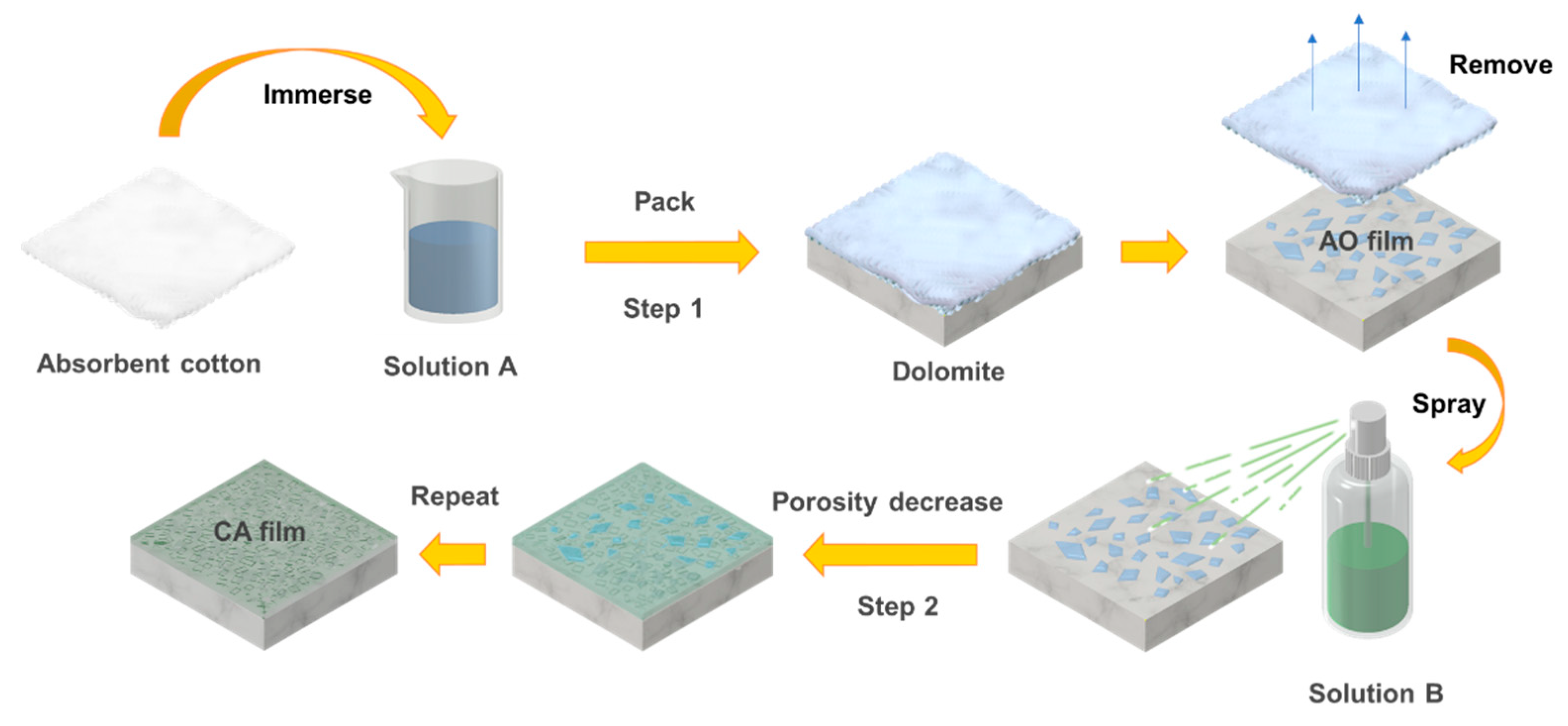
Two flasks were labeled A and B for the coating solution preparation. Solution A consisted of 10 mL of 0.1 M (NH4)2C2O4 in water and was stirred at room temperature for 30 min. The result was a reasonable amount of solution that had a minor impact on the dolomite surface [33]. Then, two solutions were prepared: (i) 0.176 g Ca(CH3COO)2·H2O dissolved in 10 mL water was stirred at room temperature for 30 min, and (ii) 0.118 g C4H6O4 and 0.008 g NaOH dissolved in 40 mL ethanol and stirred at room temperature for 30 min. The two solutions were mixed in a flask and stirred for 30 min to prepare Solution B (0.02 mol/L CaC2O4·H2O). The hydrolysis rate of C4H6O4 was restrained by NaOH at room temperature [34].
The facile two-step deposition resulted in a coherent and uncracked CaC2O4·H2O film. The cotton wool with Solution A was poulticed to the dolomite substrate surface and sealed, then stored at room temperature for 24 h, as shown in Figure 1 [35]. Following the reaction, Solution B was sprayed on the treated dolomite surface in concentrations of 0.02, 0.04, 0.06, 0.08 and 0.10 mol/L, and dried at room temperature (see Supplementary Materials Figure S3). After being sprayed four times, the film’s coverage area was increased. On the CA sample, a homogenous interconnected network of newly formed CaC2O4·H2O crystals was found.
2.3. Characterization and Analysis Method
X-ray diffraction (XRD, Bruker D8 Advance) analysis was employed to characterize the change in dolomite substrate, with Cu Kα radiation at 45 kV and 200 mA in a scanning range of 10–70(2θ) with a scanning speed of 1°/min and step size of 0.02°. The samples were cut to couple with the diffractometer camera for analysis.
The Raman spectrum (Raman, Horiba LabRAM HR) of the protection film was obtained to analyze the crystallinity degree. The laser wavelength was 785 nm, the objective lens was 50 xe and the spatial resolution was 1 μm.
Fourier transform infrared (Thermo Fisher Nicolet is 5) spectra of the film were measured to evaluate the residue content. The experiment set the acquisition mode to attenuated total reflection (ATR). The spectral range of 4000–500 cm−1 was selected, the spectral resolution was 4 cm−1 and the number of scans was 16.
A field emission scanning electron microscope with energy dispersive X-ray spectrometry (SEM-EDS, Tescan Vega3) was used to characterize the effectiveness of the film. Backscattered electron images of the sample were obtained at an accelerating voltage of 20 kV.
Atomic force microscopy (AFM, Bruker Nano Dimension Edge) observation was performed on the film exterior surface in air and tapping modes. In this test, the whole sample was used.
Colorimetric measurements (CM-26D, Konica Minolta) were carried out on the sample surface at five spots to evaluate the aesthetic effect of the two-step deposition by comparing the untreated and treated specimens. The lightness variable ΔL * and the difference in color saturation ΔC * were obtained by comparing L *, and C * values were achieved from the untreated and treated specimens. The color change ΔE * was expressed in the CIE L *a *b * space according to the Formula (1)
The surface adhesion strength of the specimens was tested by the Scotch Tape Test (STT) at five areas. The acid resistance of the specimens was evaluated by simulating the rain dissolution process learned from Graziani’s references [33]. The dripping apparatus consists of one line of nozzles. A solution composed of HNO3 and H2SO4 (1:1, pH = 4.0) was dripped onto the sample, and the period of dripping was 2 h. Considering the annual average rainfall in Shandong province (720 mm), the number of high pollution days and the size of the specimens (25 × 20 mm2), the sample was submitted to an average volume of simulated rain of about 750 mL, which corresponded to about 5 years of acid rain in Shandong province.
3. Results and Discussion
3.1. Characterization of the Film
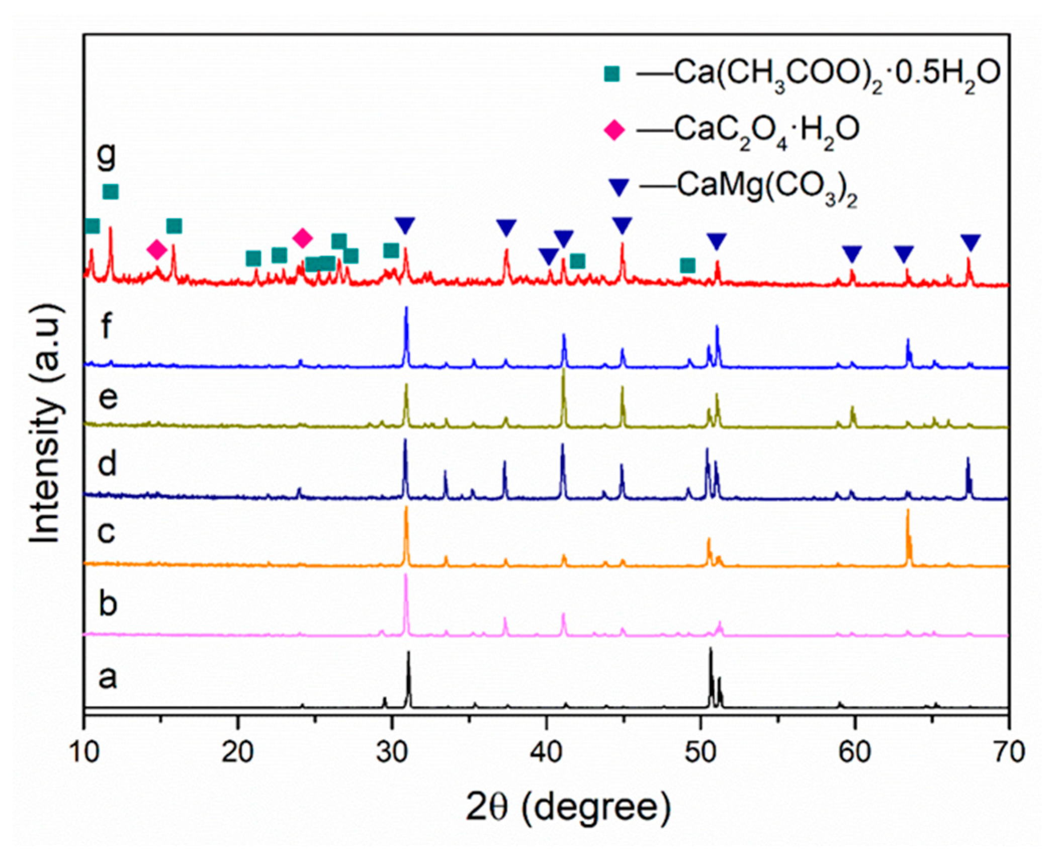
We studied the composition of these samples by X-ray diffraction (XRD) (Figure 2). A series of characteristic diffraction peaks (2θ = 30.92, 33.48, 50.45, 63.40) of dolomite were found in the XRD pattern of each sample. We can thus conclude that the two-step deposition process has a negligible effect on the intensity of dolomite reflection. Furthermore, most of the samples did not show the characteristic peaks of the CaC2O4·H2O phases, indicating that the newly formed phases in the treated dolomite had a low constituent. Ca(CH3COO)2·0.5H2O can also be found in the XRD patterns of the CA sample when the concentration of Solution B was higher than 0.06 mol/L.
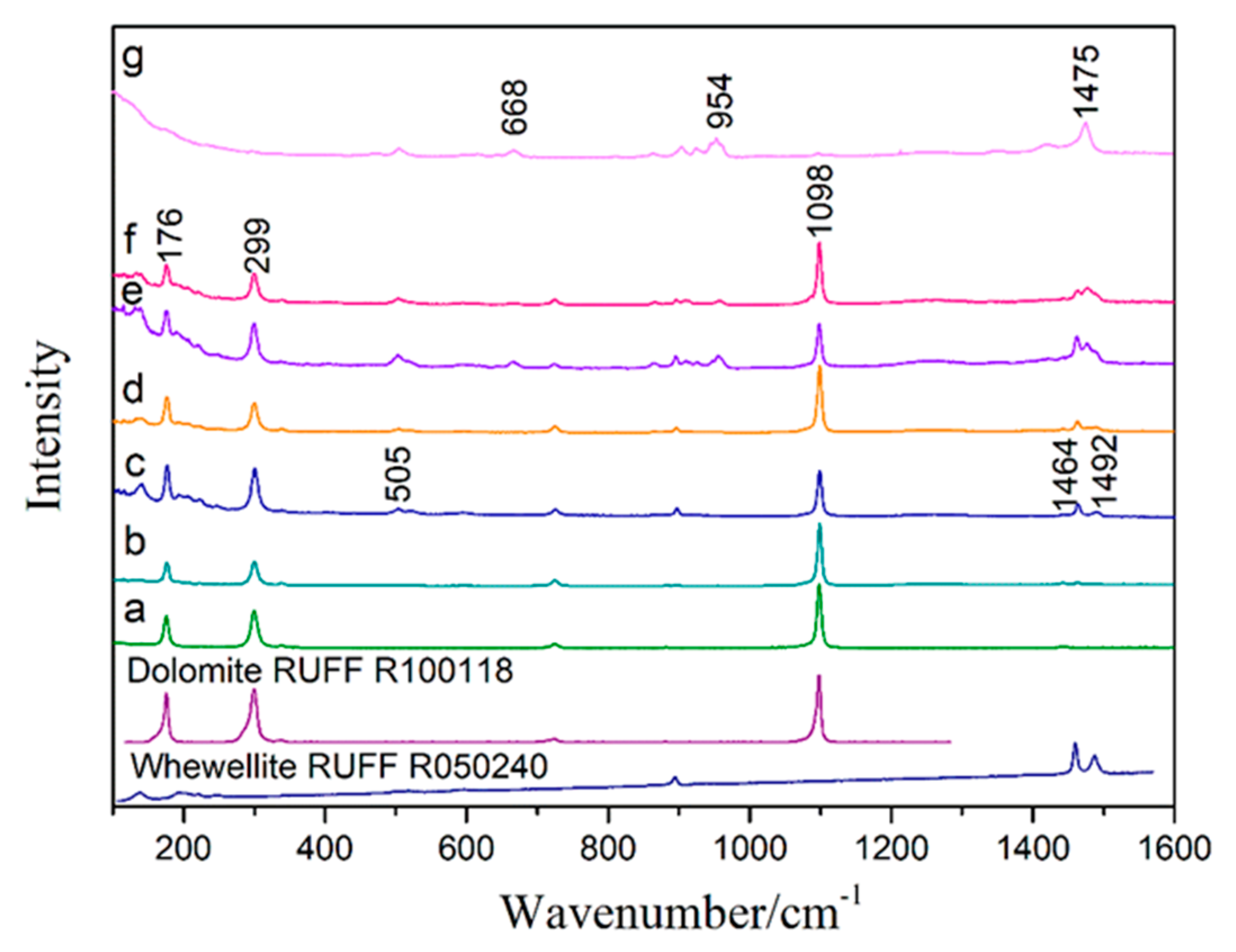
The Raman spectra of the CA sample treated with different concentrations of Solution B are compared in Figure 3. Analysis of the untreated sample showed sharp peaks at 176 cm−1, 299 cm−1 and 1098 cm−1 due to the dolomite [31]. New bands at 505 cm−1, 1464 cm−1 and 1492 cm−1, which are characteristic of whewellite, appeared after the Solution A treatment [36]. Weddellite (1475 cm−1) was formed when Solution B was introduced at a relatively high concentration (0.06–0.10 mol/L), and unreacted Ca(CH3COO)2·0.5H2O was also found at 668 cm−1 and 954 cm−1 [37,38,39]. Weddellite is unstable in environmental conditions and will reduce the acid-resistance properties of protective material, as previously described [40]. Thus, Solution B performs better at low concentrations (0.02–0.04 mol/L). Furthermore, the mineral glushinskite (MgC2O4·2H2O) was not identified in the dolomite surface layer. The high Mg concentrations required for glushinskite precipitation are limited not only by the whewellite precipitates, but also by the low concentration of (NH4)2C2O4 solution in step one [41].
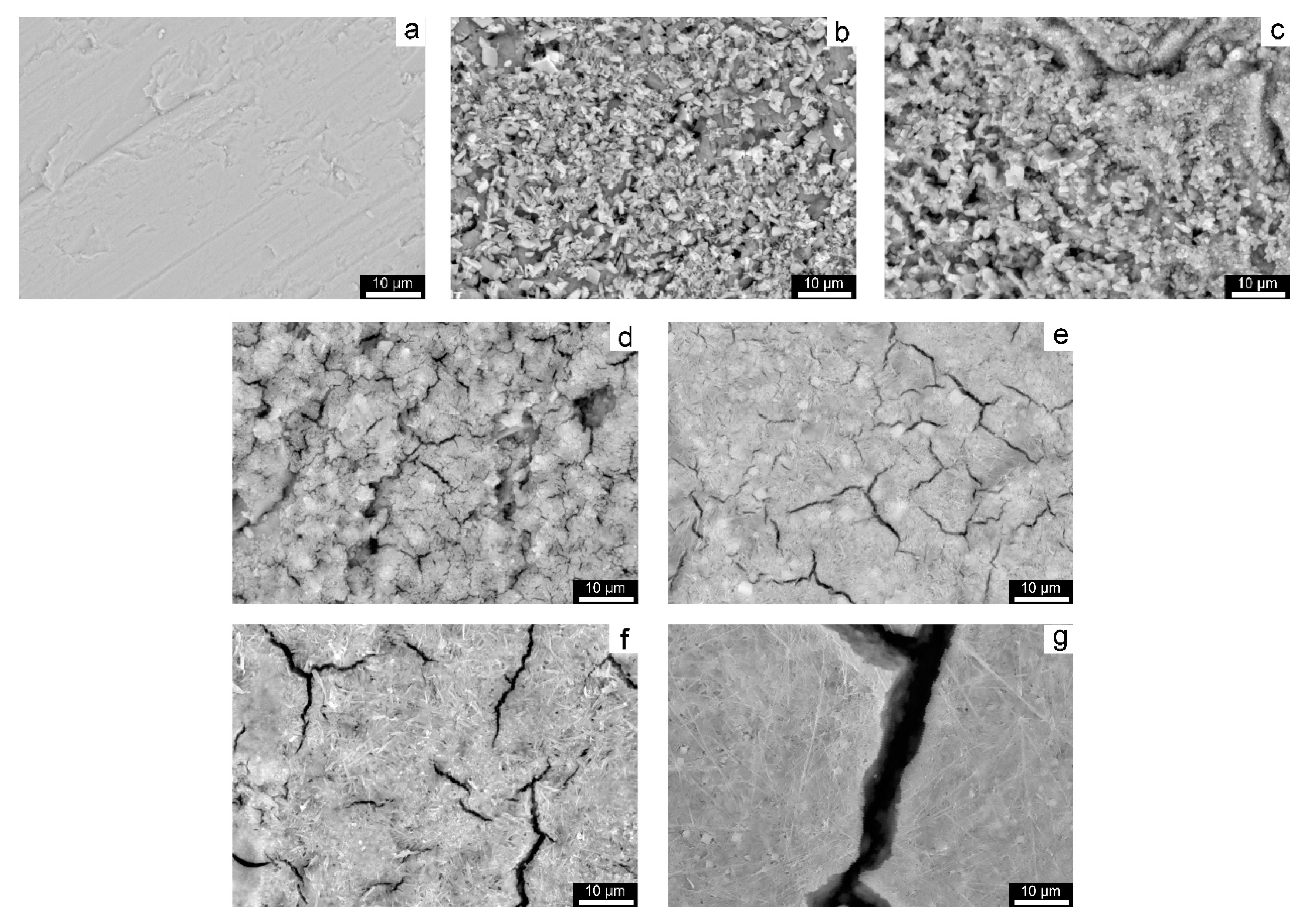
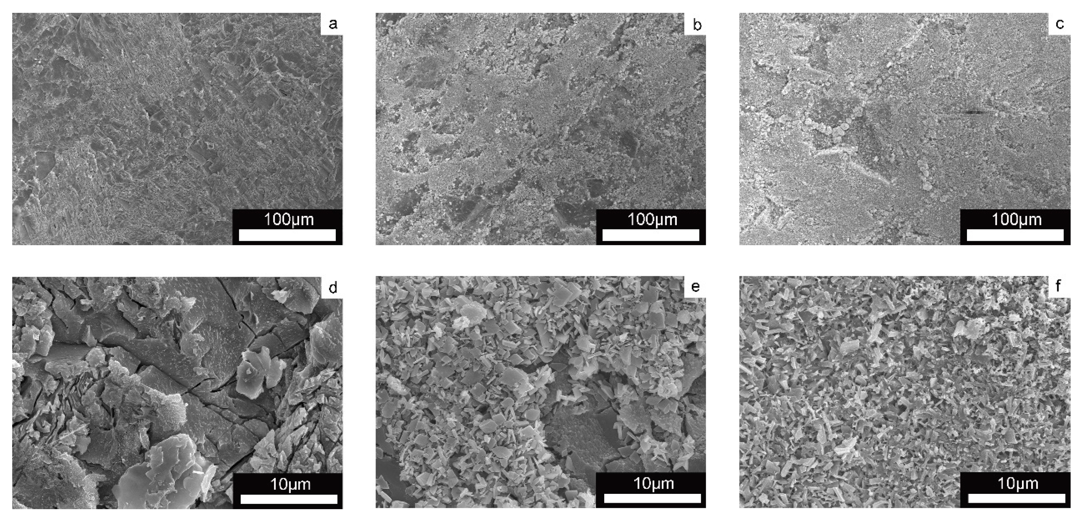
One of the main purposes of the two-step deposition strategy is to improve the film coverage area on the substrate. To see the actual effect, we investigated the microscopic morphology of the treated sample by SEM (Figure 4). On all treated dolomite surfaces, a layer of the newly developed phase was observed. Comparing Figure 4b,c, the effect on the crystal structure of the film can be seen. An irregular CaC2O4·H2O crystal was formed by the treatment of 0.02 mol/L of Solution B (pH = 7), indicating that CaC2O4·H2O crystals develop randomly under higher pH conditions. According to Ruiz-Agudo, the CaC2O4·H2O crystal habit in this condition is predominantly equant, and the precipitation usually forms irregular aggregates [42]. Furthermore, it was discovered that the two-step deposition considerably improves the surface coverage of dolomite by the CaC2O4·H2O coating. The sample treated with Solution B at a relatively high concentration was more prone to cracking than the other samples, which may be subjected to drying stress [43]. Thus, a relatively low concentration of Solution B was used to treat the sample to create an uncracked film and improve surface coverage.
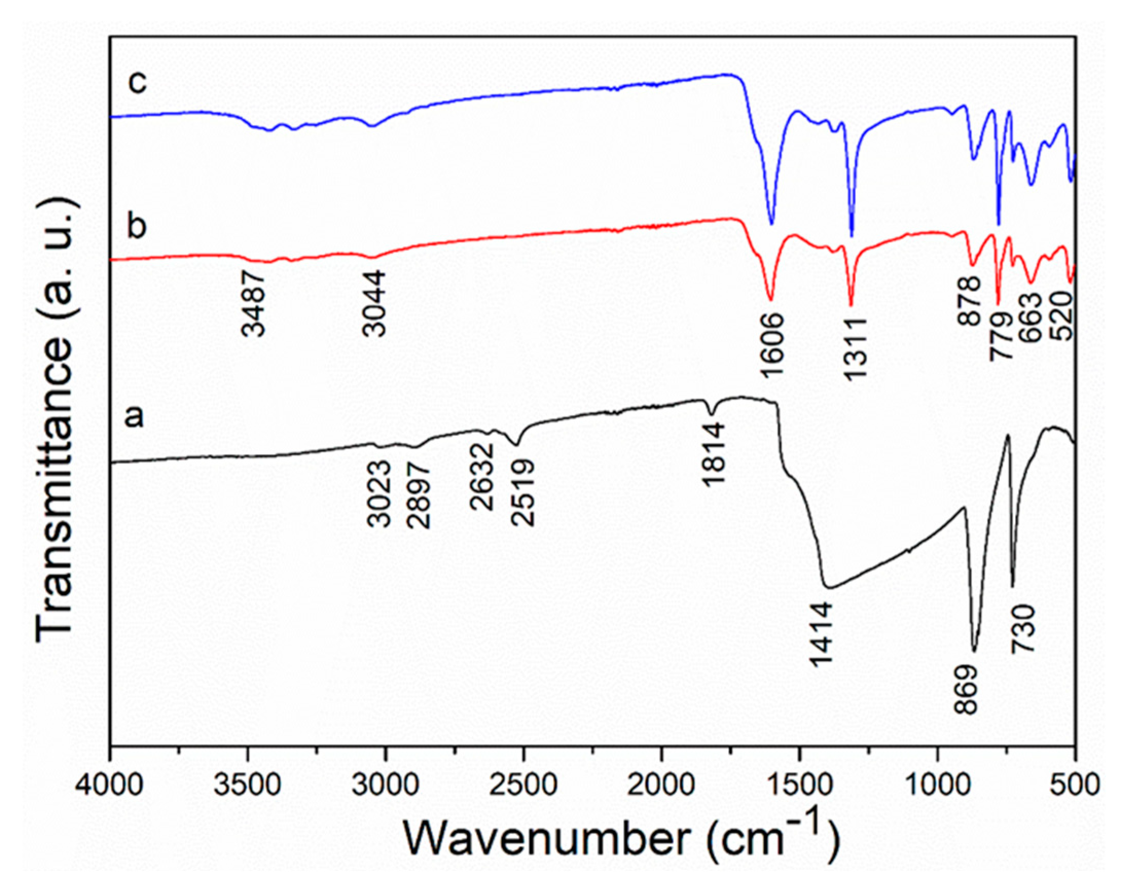
FT-IR spectra of the untreated stone and treated sample were performed to investigate the influence of the two-step deposition process in the functional group of the CaC2O4·H2O film (Figure 5). A contribution from the dolomite was observed at 730cm−1, 869 cm−1 and 1814 cm−1 in the untreated sample (Figure 5a), and another contribution from CaCO3 was detected at 1414 cm−1. Carboxylate groups at 520 cm−1, 663 cm−1, 779 cm−1, 1311 cm−1 and1606 cm−1, and broad vibration bands of −OH groups at 3044 cm−1 and 3487 cm−1, confirm the formation of CaC2O4·H2O phases after the surface treatment (Figure 5b) [44,45,46,47,48,49]. These bands were relatively strong after the two-step deposition process (Figure 5c), which is consistent with the film’s thick structure. In addition, the oxalate group in CaC2O4·H2O was retained before and after treatment with Solution B. In a relatively low concentration (0.02 mol/L), the solution almost has no effect on the mineralogical composition of the AO film.
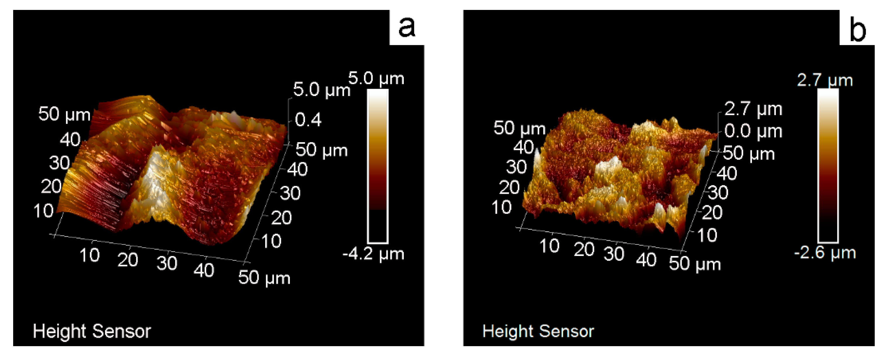
AFM observations (Figure 6) were conducted to measure the roughness of AO and CA specimens (0.02 mol/L). The roughness of AO specimens ranged from −4.2 to 5.0 μm, whereas that of CA specimens ranged from −2.6 to 2.7 μm, according to the height profiles obtained. Surface roughness was reduced, and a homogenous layer was formed as a result of the two-step deposition procedure. Based on the AFM measurements, we can conclude that Solution B improves the coverage of calcium oxalate on dolomite and smooths the surface of the (NH4)2C2O4-treated sample.
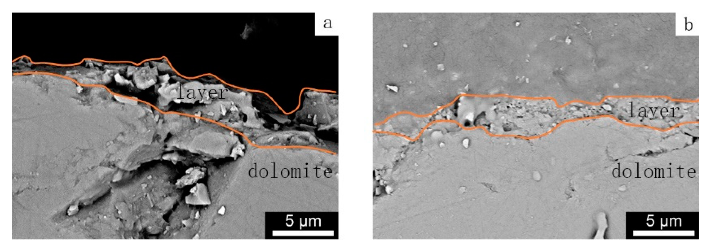
Surface roughness is closely related to the layer structure. To assess the alteration, a direct SEM examination of cross-sectioned samples of treated dolomite was carried out (Figure 7). Roughly, the thickness of the CA layer (3 μm) was similar to the AO layer under the proposed experimental conditions. Furthermore, no obvious warping between the CA layer and the dolomite substrate indicates good compatibility and combination. Consequently, Solution B reduces the AO layer’s porosity.
3.2. Property Assessment of the Film
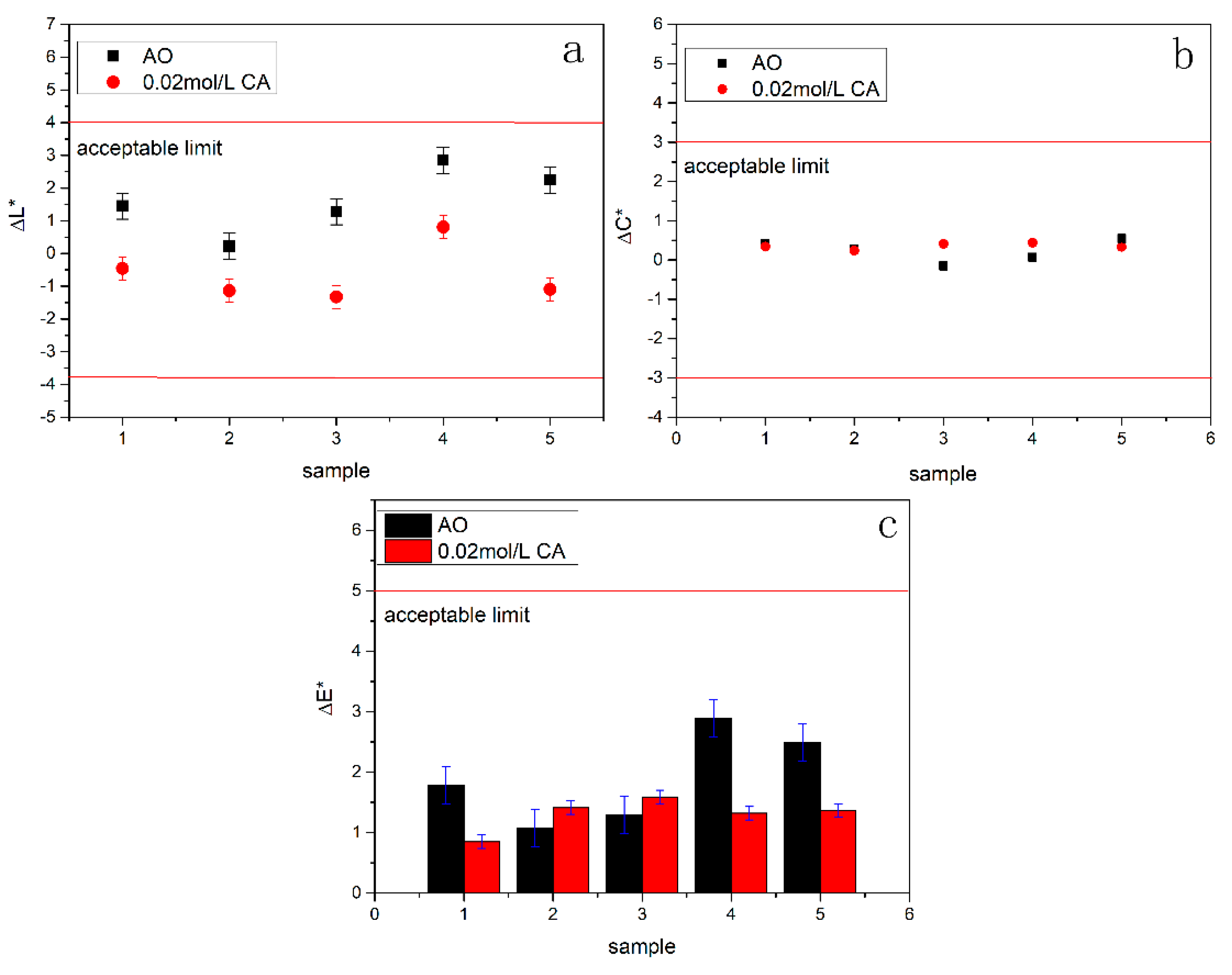
Considering that the treatment of 0.02 mol/L of Solution B can form a simple and coherent calcium oxalate film on dolomite, we chose this sample to evaluate the properties of the film. To investigate the impact of the two-step deposition procedure on the aesthetic properties of the stone object, possible color changes were assessed (Figure 8), and all the data are shown in Table S1 (see Supplementary Materials). The color change could not be visually observed after each treatment because it was below the perceptibility threshold [50]. ΔL * unit changes were sometimes slightly higher than ΔC* unit changes. Furthermore, the decreases in ΔL * and ΔE * appeared to be more pronounced in the AO specimen than in the CA specimen, and the chromatic coordinates of the sample trends was more constant. Such a color change is largely due to the different morphology of the CaC2O4·H2O film.
To investigate the compatibility of each treatment, an STT test was performed. The results suggest that the amount of material released from the surface of both the AO and CA sample was notably less (Figure 9). This also implies that the formation of CaC2O4·H2O increased the cohesiveness of the dolomite surface [51]. The consolidation effect of ammonium oxalate was widely reported, even though the calcium oxalate layer formed after treatment has been found incomplete [52]. Such depositing of the CaC2O4·H2O layer efficiently lowered the nucleation energy barrier and worked as a seed layer to accelerate the crystal aggregate in metastable solution [53,54]. As a result, the seed layer had a significant impact on the second treatment’s consolidation properties. It should also be noted that the CA sample’s surface mass change was similar to the untreated surface. The exfoliation of subsurface dolomite could barely be induced by the inner stress of the surface layer.
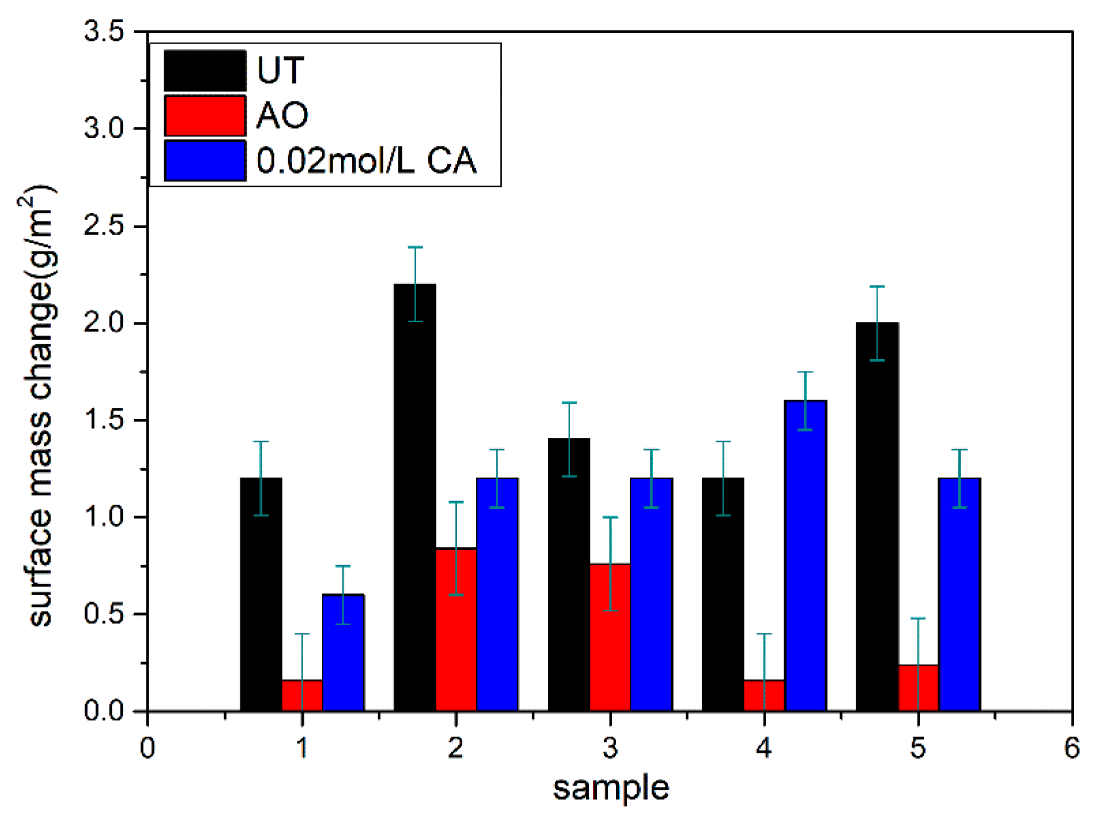
The simulated acid rain (HNO3:H2SO4 = 1:1) resistance results indicate that the two-step deposition of the CaC2O4·H2O film is effective against acid dissolution due to the increased substrate coverage (Figure 10). Surface etching was detected in the untreated sample, and the same phenomenon also appeared in the AO sample after the acid attack. This indicated that the acid rain did not destroy the protective layer, but it had a significant impact on the dolomite. Meanwhile, the dissolution of the treated and untreated specimens was evaluated by measuring the pH value of the runoff solution. Due to the dolomite reacting with the sulfuric acid to form calcium sulfide in the UT sample, the solution pH value changed noticeably (increased to 4.9). The immersion tests appeared to have less impact on the sample AO, with lower variations in the solution pH value (increased to 4.5). The change in the pH value was negligible in the 0.02 mol/L CA sample (increased to 4.3). Therefore, the protective efficiency increased after Solution B was applied to the AO sample surface.
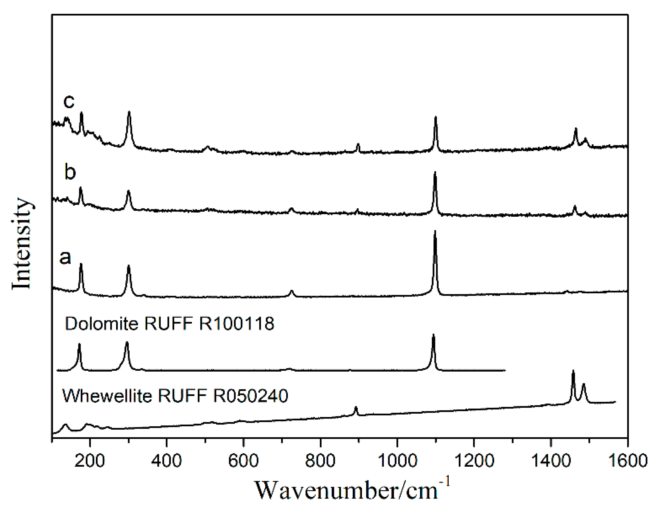
Raman spectra were used to examine the chemical composition of the samples, as shown in Figure 11. The predominant component of specimens is dolomite, and the peaks ascribed to whewellite can be found in b and c specimens. Corrosion products, such as Ca(NO3)2 and CaSO4·H2O, were difficult to detect, and may have been removed from the sample surface by acid rain. The Raman spectra are consistent with the SEM analysis.
4. Conclusions
A thick and continuous CaC2O4·H2O protective film was synthesized on a dolomite surface by a two-step deposition process. The introduction of the solution containing Ca(CH3COO)2·H2O, C4H6O4, NaOH and C2H5OH to the (NH4)2C2O4-treated sample increased the substrate coverage while decreasing the porosity of the film. Chromatic value and surface mass loss were almost unchanged in the treated samples. The CA sample showed a stronger acid rain resistance than the AO sample, according to the results of the simulated acid rain test. After the facile two-step treatment, the CaC2O4·H2O film had little effect on the chromatic value and adhesion strength of the dolomite substrates.
Although the two-step deposition procedure required more time to manufacture a protective layer, this research raises the potential for the external deposition of an inorganic film onto dolomite without the need for a chemical reaction. More research is needed to adapt the whole procedure, such as first precipitating a CaCO3 layer and then synthesizing a protective layer. Furthermore, the durability of the whewellite layer and, therefore, the degree of dolomite protection should be evaluated.
Supplementary Materials
The following supporting information can be downloaded at: https://0-www-mdpi-com.brum.beds.ac.uk/article/10.3390/cryst12050734/s1, Figure S1: The SEM of dolomite after different composition solution B treatment (a) calcium acetate + dimethyl oxalate, (b) calcium acetate + dimethyl oxalate + nitric acid, (c) calcium acetate + dimethyl oxalate + sodium hydroxide; Figure S2: The SEM of dolomite after different additive content solution B treatment (a) 0.0001mol, (b) 0.0002mol, (c) 0.0003mol, (d) 0.0004mol, (e) 0.0005mol; Figure S3 The SEM of dolomite after solution B treatment (a) spray, (b) poultice, (c) brush; Table S1: Chromatic value of sample.
Author Contributions
Conceptualization, J.Z.; methodology, J.Z. and Y.G.; software, Y.G.; resources, S.W.; writing—original draft preparation, J.Z.; writing—review and editing, H.H. and F.W.; funding acquisition, S.W. and Q.M. All authors have read and agreed to the published version of the manuscript.
Funding
This research was funded by the Beijing Municipal Administration of Cultural Heritage (PXM2020-039208-000005).
Institutional Review Board Statement
Not applicable.
Informed Consent Statement
Not applicable.
Data Availability Statement
Not applicable.
Acknowledgments
The authors want to thank the Beijing Stone Carving Art Museum for the supply of material support for this work.
Conflicts of Interest
The authors declare that they have no known competing financial interest or personal relationships that could have appeared to influence the work reported in this paper.
References
- Cai, W.K.; Liu, J.H.; Zhou, C.H.; Keeling, J.; Glasmacher, U.A. Structure, genesis and resources efficiency of dolomite. Chem. Geol. 2021, 573, 120191. [Google Scholar] [CrossRef]
- Liu, X.; Koestler, R.J.; Warscheid, T.; Katayama, Y.; Gu, J.D. Microbial deterioration and sustainable conservation of stone monuments and buildings. Nat. Sustain. 2020, 3, 991–1004. [Google Scholar] [CrossRef]
- David, M.E.; Ion, R.M.; Grigorescu, R.M.; Lancu, L.; Andrei, E.R. Nanomaterials used in conservation and restoration of cultural heritage: An up-to-date overview. Materials 2020, 13, 2064. [Google Scholar] [CrossRef] [PubMed]
- Fidanza, M.R.; Caneva, G. Natural biocides for the conservation of stone cultural heritage: A review. J. Cult. Herit. 2019, 38, 271–286. [Google Scholar] [CrossRef]
- Charola, A.E.; Ware, R. Acid deposition and the deterioration of stone: A brief review of a broad topic. Geol. Soc. Lond. Spec. Publ. 2002, 205, 393–406. [Google Scholar] [CrossRef]
- Morillas, H.; Maguregui, M.; Gallego-Cartagena, E.; Marcaida, I.; Carral, N.; Madariaga, J.M. The influence of marine environment on the conservation state of Built Heritage: An overview study. Sci. Total Environ. 2020, 745, 140899. [Google Scholar] [CrossRef]
- Vidal, F.; Vicente, R.; Silva, J.M. Review of environmental and air pollution impacts on built heritage: 10 questions on corrosion and soiling effects for urban intervention. J. Cult. Herit. 2019, 37, 273–295. [Google Scholar] [CrossRef]
- Bonazza, A.; Messina, P.; Sabbioni, C.; Grossi, C.M.; Brimblecombe, P. Mapping the impact of climate change on surface recession of carbonate buildings in Europe. Sci. Total Environ. 2009, 407, 2039–2050. [Google Scholar] [CrossRef]
- Turkington, A.V.; Paradise, T.R. Sandstone weathering: A century of research and innovation. Geomorphology 2005, 67, 229–253. [Google Scholar] [CrossRef]
- Dakal, T.C.; Cameotra, S.S. Microbially induced deterioration of architectural heritages: Routes and mechanisms involved. Environ. Sci. Eur. 2012, 24, 1–13. [Google Scholar] [CrossRef] [Green Version]
- Rampazzi, L. Calcium oxalate films on works of art: A review. J. Cult. Herit. 2019, 40, 195–214. [Google Scholar] [CrossRef]
- King, H.E.; Mattner, D.C.; Plümper, O.; Geisler, T.; Putnis, A. Forming cohesive calcium oxalate layers on marble surfaces for stone conservation. Cryst. Growth Des. 2014, 14, 3910–3917. [Google Scholar] [CrossRef]
- Mudronja, D.; Vanmeert, F.; Hellemans, K.; Fazinic, S.; Janssens, K.; Tibljas, D.; Rogosic, M.; Jakovljevic, S. Efficiency of applying ammonium oxalate for protection of monumental limestone by poultice, immersion and brushing methods. Appl. Phys. A 2013, 111, 109–119. [Google Scholar] [CrossRef] [Green Version]
- Conti, C.; Aliatis, I.; Casati, M.; Colombo, C.; Matteini, M.; Negrotti, R.; Realini, M.; Zerbi, G. Diethyl oxalate as a new potential conservation product for decayed carbonatic substrates. J. Cult. Herit. 2014, 15, 336–338. [Google Scholar] [CrossRef]
- Maiore, L.; Aragoni, M.C.; Carcangiu, G.; Cocco, O.; Isaia, F.; Lippolis, V.; Meloni, P.; Murru, A.; Slawin, A.M.Z.; Tuveri, E.; et al. Oxamate salts as novel agents for the restoration of marble and limestone substrates: Case study of ammonium N-phenyloxamate. New J. Chem. 2016, 40, 2768–2774. [Google Scholar] [CrossRef] [Green Version]
- Dreyfuss, T. Artificially induced calcium oxalate on limestone in urban environments-new findings. J. Cult. Herit. 2020, 42, 56–63. [Google Scholar] [CrossRef]
- Aragoni, M.C.; Giacopetti, L.; Arca, M.; Carcangiu, G.; Columbu, S.; Gimeno, D.; Isaia, F.; Lippolis, V.; Meloni, P.; Ezquerra, A.N.; et al. Ammonium monoethyloxalate (AmEtOx): A new agent for the conservation of carbonate stone substrates. New J. Chem. 2021, 45, 5327–5339. [Google Scholar] [CrossRef]
- Ruiz-Agudo, E.; Putnis, C.V.; Putnis, A. Coupled dissolution and precipitation at mineral–fluid interfaces. Chem. Geol. 2014, 383, 132–146. [Google Scholar] [CrossRef]
- Ding, Z.; Fang, Y.; Su, J.F.; Hong, S.; Dong, B. In situ precipitation for the surface treatment and repair of cement-based materials. J. Adhes. Sci. Technol. 2020, 34, 1233–1240. [Google Scholar] [CrossRef]
- Naidu, S.; Blair, J.; Scherer, G.W. Acid-resistant coatings on marble. J. Am. Ceram. Soc. 2016, 99, 3421–3428. [Google Scholar] [CrossRef]
- Burgos-Cara, A.; Rodríguez-Navarro, C.; Ortega-Huertas, M.; Ruiz-Agudo, E. Bioinspired Alkoxysilane Conservation Treatments for Building Materials Based on Amorphous Calcium Carbonate and Oxalate Nanoparticles. ACS Appl. Nano Mater. 2019, 2, 4954–4967. [Google Scholar] [CrossRef]
- Burgos-Cara, A.; Putnis, C.V.; Ortega-Huertas, M.; Ruiz-Agudo, E. Influence of pH and citrate on the formation of oxalate layers on calcite revealed by in situ nanoscale imaging. Crystengcomm 2017, 19, 3420–3429. [Google Scholar] [CrossRef]
- Burgos-Cara, A.; Ruiz-Agudo, E.; Rodriguez-Navarro, C. Effectiveness of oxalic acid treatments for the protection of marble surfaces. Mater. Des. 2017, 115, 82–92. [Google Scholar] [CrossRef]
- Meloni, P.; Manca, F.; Carcangiu, G. Marble protection: An inorganic electrokinetic approach. Appl. Surf. Sci. 2013, 273, 377–385. [Google Scholar] [CrossRef]
- Osticioli, I.; Botticelli, G.; Matteini, P.; Siano, S.; Pini, R.; Matteini, M. Micro-Raman analysis on the combined use of ammonium oxalate and ammonium phosphate for the consolidation and protection of carbonate stone artifacts. J. Raman Spectrosc. 2017, 48, 966–971. [Google Scholar] [CrossRef]
- Verganelaki, A.; Kapridaki, C.; Maravelaki-Kalaitzaki, P. Modified tetraethoxysilane with nanocalcium oxalate in one-pot synthesis for protection of building materials. Ind. Eng. Chem. Res. 2015, 54, 7195–7206. [Google Scholar] [CrossRef]
- Kapridaki, C.; Verganelaki, A.; Dimitriadou, P.; Maravelaki-Kalaitzaki, P. Conservation of monuments by a three-layered compatible treatment of teos-nano-calcium oxalate consolidant and teos-pdms-TiO2 hydrophobic/photoactive hybrid nanomaterials. Materials 2018, 11, 684. [Google Scholar] [CrossRef] [Green Version]
- Zhang, H.; Liu, Q.; Liu, T.; Zhang, B. The preservation damage of hydrophobic polymer coating materials in conservation of stone relics. Prog. Org. Coat. 2013, 76, 1127–1134. [Google Scholar] [CrossRef]
- Graziani, G.; Sassoni, E.; Franzoni, E.; Scherer, G.W. Hydroxyapatite coatings for marble protection: Optimization of calcite covering and acid resistance. Appl. Surf. Sci. 2016, 368, 241–257. [Google Scholar] [CrossRef]
- Sassoni, E.; Graziani, G.; Franzoni, E.; Scherer, G.W. Calcium phosphate coatings for marble conservation: Influence of ethanol and isopropanol addition to the precipitation medium on the coating microstructure and performance. Corros. Sci. 2018, 136, 255–267. [Google Scholar] [CrossRef]
- Graziani, G.; Sassoni, E.; Scherer, G.W.; Franzoni, E. Resistance to simulated rain of hydroxyapatite- and calcium oxalate-based coatings for protection of marble against corrosion. Corros. Sci. 2017, 127, 168–174. [Google Scholar] [CrossRef]
- Liu, Y.; Yang, F.; Zuo, G.; Zhang, R.; Wei, G.; Ma, Q. Protection of the surface weathering stone artworks by a chemical conversion method. Constr. Build. Mater. 2018, 182, 210–214. [Google Scholar] [CrossRef]
- Liu, Q.; Zhang, B.J. Syntheses of a novel nanomaterial for conservation of historic stones inspired by nature. Mater. Lett. 2007, 61, 4976–4979. [Google Scholar] [CrossRef]
- Hajir, M.; Graf, R.; Tremel, W. Stable amorphous calcium oxalate: Synthesis and potential intermediate in biomineralization. Chem. Commun. 2014, 50, 6534–6536. [Google Scholar] [CrossRef] [PubMed]
- Doherty, B.; Pamplona, M.; Selvaggi, R.; Miliani, C.; Matteini, M.; Sgamellotti, A.; Brunetti, B. Efficiency and resistance of the artificial oxalate protection treatment on marble against chemical weathering. Appl. Surf. Sci. 2007, 253, 4477–4484. [Google Scholar] [CrossRef]
- Sun, J.; Wu, Z.; Cheng, H.; Zhang, Z.; Frost, R.L. A Raman spectroscopic comparison of calcite and dolomite. Spectrochim. Acta Part A Mol. Biomol. Spectrosc. 2014, 117, 158–162. [Google Scholar] [CrossRef] [Green Version]
- Frost, R.L.; Weier, M.L. Thermal treatment of whewellite-a thermal analysis and Raman spectroscopic study. Thermochim. Acta 2004, 409, 79–85. [Google Scholar] [CrossRef] [Green Version]
- Wróbel-Kwiatkowska, M.; Żuk, M.; Szopa, J.; Dymińska, L.; Mączka, M.; Hanuza, J. Poly-3-hydroxy butyric acid interaction with the transgenic flax fibers: FT-IR and Raman spectra of the composite extracted from a GM flax. Spectrochim. Acta Part A Mol. Biomol. Spectrosc. 2009, 73, 286–294. [Google Scholar] [CrossRef]
- Neal, A.L.; Kabengi, N.; Grider, A.; Bertsch, P.M. Can the soil bacterium cupriavidus necator sense ZnO nanomaterials and aqueous Zn2+ differentially? Nanotoxicology 2012, 6, 371–380. [Google Scholar] [CrossRef]
- Zoppi, A.; Lofrumento, C.; Mendes, N.F.C.; Castellucci, E. Metal oxalates in paints: A Raman investigation on the relative reactivities of different pigments to oxalic acid solutions. Anal. Bioanal. Chem. 2010, 397, 841–849. [Google Scholar] [CrossRef]
- Conti, C.; Casati, M.; Colombo, C.; Realini, M.; Brambilla, L.; Zerbi, G. Phase transformation of calcium oxalate dihydrate-monohydrate: Effects of relative humidity and new spectroscopic data. Spectrochim. Acta Part A Mol. Biomol. 2014, 128, 413–419. [Google Scholar] [CrossRef] [PubMed]
- Perez-Rodriguez, J.L.; Duran, A.; Centeno, M.A.; Matinez-Blanes, J.M.; Robador, M.D. Thermal analysis of monument patina containing hydrated calcium oxalates. Thermochim. Acta 2011, 512, 5–12. [Google Scholar] [CrossRef]
- Ruiz-Agudo, E.; álvarez-Lloret, P.; Putnis, C.V.; Rodriguez-Navarro, A.B.; Putnis, A. Influence of chemical and structural factors on the calcite-calcium oxalate transformation. Crystengcomm 2013, 15, 9968–9979. [Google Scholar] [CrossRef]
- Sassoni, E. Hydroxyapatite and other calcium phosphates for the conservation of cultural heritage: A Review. Materials 2018, 11, 557. [Google Scholar] [CrossRef] [PubMed] [Green Version]
- Wei, W.; Huang, H.; Cui, J.; Wei, Z. Evaluation of amorphous calcium phosphate as an advantageous solid-phase extraction adsorbent for analysis of oxalic acid in plant xylem saps by RP-HPLC. J. Liq. Chromatogr. Relat. Technol. 2014, 37, 2667–2680. [Google Scholar] [CrossRef]
- Braconnier, L.; Clémenon, I.; Legens, C.; Moizan, V.; Diehl, F.; Pillière, H.; Echegut, P.; Meneses, D.D.S.; Schuurman, Y. An X-ray diffractometer coupled with diffuse reflectance infrared Fourier transform spectroscopy and gas chromatography for in situ and in operando characterization: An innovative analytical laboratory instrument. J. Appl. Crystallogr. 2012, 46, 262–266. [Google Scholar] [CrossRef]
- Cruz-May, T.N.; Herrera, A.; Rodríguez-Hernández, J.; Basulto-Martínez, M.; Flores-Tapia, J.P.; Quintana, P. Structural and morphological characterization of kidney stones in patients from the Yucatan Maya population. J. Mol. Struct. 2021, 1235, 130267. [Google Scholar] [CrossRef]
- Valarmathi, D.; Abraham, L.; Gunasekaran, S. Growth of calcium oxalate monohydrate crystal by gel method and its spectroscopic analysis. Indian J. Pure Appl. Phys. 2010, 48, 36–48. [Google Scholar]
- So, R.T.; Blair, N.E.; Masterson, A.L. Carbonate mineral identification and quantification in sediment matrices using diffuse reflectance infrared Fourier transform spectroscopy. Environ. Chem. Lett. 2020, 18, 1725–1730. [Google Scholar] [CrossRef]
- Rodrigues, J.D.; Grossi, A. Indicators and ratings for the compatibility assessment of conservation actions. J. Cult. Herit. 2007, 8, 32–43. [Google Scholar] [CrossRef]
- Sassoni, E.; Graziani, G.; Franzoni, E. Repair of sugaring marble by ammonium phosphate: Comparison with ethyl silicate and ammonium oxalate and pilot application to historic artifact. Mater. Des. 2018, 88, 1145–1157. [Google Scholar] [CrossRef]
- Charola, A.E.; Centeno, S.A.; Normandin, K. The New York Public Library: Protective Treatment for Sugaring Marble. J. Archit. Conserv. 2010, 16, 29–44. [Google Scholar] [CrossRef]
- Sun, H.; Luo, M.; Weng, W.; Cheng, K.; Du, P.; Shen, G.; Han, G. Room-temperature preparation of ZnO nanosheets grown on Si substrates by a seed-layer assisted solution route. Nanotechnology 2008, 19, 125603. [Google Scholar] [CrossRef] [PubMed]
- Pitt, K.; Mitchell, G.P.; Ray, A.; Heywood, B.R.; Hounslow, M.J. Micro-mechanical model of calcium oxalate monohydrate aggregation in supersaturated solutions: Effect of crystal form and seed concentration. J. Cryst. Growth 2012, 361, 176–188. [Google Scholar] [CrossRef]
Figure 1.
Schematic diagram of the CaC2O4·H2O film deposition by a two-step process.

Figure 2.
XRD patterns of treated and untreated samples: (a) UT, (b) AO, (c) 0.02 mol/L CA, (d) 0.04 mol/L CA, (e) 0.06 mol/L CA, (f) 0.08 mol/L CA, (g) 0.10 mol/L CA.
Figure 2.
XRD patterns of treated and untreated samples: (a) UT, (b) AO, (c) 0.02 mol/L CA, (d) 0.04 mol/L CA, (e) 0.06 mol/L CA, (f) 0.08 mol/L CA, (g) 0.10 mol/L CA.

Figure 3.
Raman spectra of untreated and treated samples: (a) UT, (b) AO, (c) 0.02 mol/L CA, (d) 0.04 mol/L CA, (e) 0.06 mol/L CA, (f) 0.08 mol/L CA, (g) 0.10 mol/L CA.
Figure 3.
Raman spectra of untreated and treated samples: (a) UT, (b) AO, (c) 0.02 mol/L CA, (d) 0.04 mol/L CA, (e) 0.06 mol/L CA, (f) 0.08 mol/L CA, (g) 0.10 mol/L CA.

Figure 4.
SEM image of untreated and treated samples: (a) UT, (b) AO, (c) 0.02 mol/L CA, (d) 0.04 mol/L CA, (e) 0.06 mol/L CA, (f) 0.08 mol/L CA, (g) 0.10 mol/L CA.
Figure 4.
SEM image of untreated and treated samples: (a) UT, (b) AO, (c) 0.02 mol/L CA, (d) 0.04 mol/L CA, (e) 0.06 mol/L CA, (f) 0.08 mol/L CA, (g) 0.10 mol/L CA.

Figure 5.
FT-IR spectra of the untreated and treated samples: (a) UT, (b) AO, (c) 0.02 mol/L CA.

Figure 6.
AFM images of the treated samples: (a) AO, (b) 0.02 mol/L CA.

Figure 7.
Cross-section of the treated samples: (a) AO, (b) 0.02 mol/L CA.

Figure 8.
Chromatic change in sample: (a) ΔL * value, (b) ΔC * value, (c) ΔE * value.

Figure 9.
Surface mass change in the sample.

Figure 10.
SEM images after UT (a,d), AO (b,e) and 0.02 mol/L CA (c,f) were treated with 750 mL acid solution (HNO3:H2SO4 = 1:1), with an initial pH of 4.0.
Figure 10.
SEM images after UT (a,d), AO (b,e) and 0.02 mol/L CA (c,f) were treated with 750 mL acid solution (HNO3:H2SO4 = 1:1), with an initial pH of 4.0.

Figure 11.
Raman spectra of the specimens: (a) UT, (b) AO, and (c) 0.02 mol/L CA were treated with 750 mL acid solution (HNO3:H2SO4 = 1:1) with an initial pH of 4.0.
Figure 11.
Raman spectra of the specimens: (a) UT, (b) AO, and (c) 0.02 mol/L CA were treated with 750 mL acid solution (HNO3:H2SO4 = 1:1) with an initial pH of 4.0.


Table 1.
Characteristics of the samples.
| Sample | Type | Analysis Description |
|---|---|---|
| 1 | UT | XRD, Raman, SEM (surface), FTIR, Chromatic value (five spots) |
| 2 | AO | XRD, Raman, SEM (surface), SEM (cross-section), FTIR, AFM, Chromatic value (five spots) |
| 3 | 0.02 mol/L CA | XRD, Raman, SEM (surface), SEM (cross-section), FTIR, AFM, Chromatic value (five spots) |
| 4 | 0.04 mol/L CA | XRD, Raman, SEM (surface) |
| 5 | 0.06 mol/L CA | XRD, Raman, SEM (surface) |
| 6 | 0.08 mol/L CA | XRD, Raman, SEM (surface) |
| 7 | 0.10 mol/L CA | XRD, Raman, SEM (surface) |
| 8 | UT | STT (five areas) |
| 9 | AO | STT (five areas) |
| 10 | 0.02 mol/L CA | STT (five areas) |
| 11 | UT | Simulated acid rain resistance |
| 12 | AO | Simulated acid rain resistance |
| 13 | 0.02 mol/L CA | Simulated acid rain resistance |
Publisher’s Note: MDPI stays neutral with regard to jurisdictional claims in published maps and institutional affiliations. |
© 2022 by the authors. Licensee MDPI, Basel, Switzerland. This article is an open access article distributed under the terms and conditions of the Creative Commons Attribution (CC BY) license (https://creativecommons.org/licenses/by/4.0/).
Share and Cite
MDPI and ACS Style
Zha, J.; Gu, Y.; Wei, S.; Han, H.; Wang, F.; Ma, Q. Facile Two-Step Deposition of Calcium Oxalate Film on Dolomite to Improve Acid Rain Resistance. Crystals 2022, 12, 734. https://0-doi-org.brum.beds.ac.uk/10.3390/cryst12050734
AMA Style
Zha J, Gu Y, Wei S, Han H, Wang F, Ma Q. Facile Two-Step Deposition of Calcium Oxalate Film on Dolomite to Improve Acid Rain Resistance. Crystals. 2022; 12(5):734. https://0-doi-org.brum.beds.ac.uk/10.3390/cryst12050734
Chicago/Turabian StyleZha, Jianrui, Yaoqi Gu, Shuya Wei, Huarui Han, Feng Wang, and Qinglin Ma. 2022. "Facile Two-Step Deposition of Calcium Oxalate Film on Dolomite to Improve Acid Rain Resistance" Crystals 12, no. 5: 734. https://0-doi-org.brum.beds.ac.uk/10.3390/cryst12050734
Note that from the first issue of 2016, this journal uses article numbers instead of page numbers. See further details here.