
The Role of Cations and Anions in the Formation of Crystallization Oligomers in Protein Solutions as Revealed by Combination of Small-Angle X-ray Scattering and Molecular Dynamics
 , , ,
, , ,
Abstract
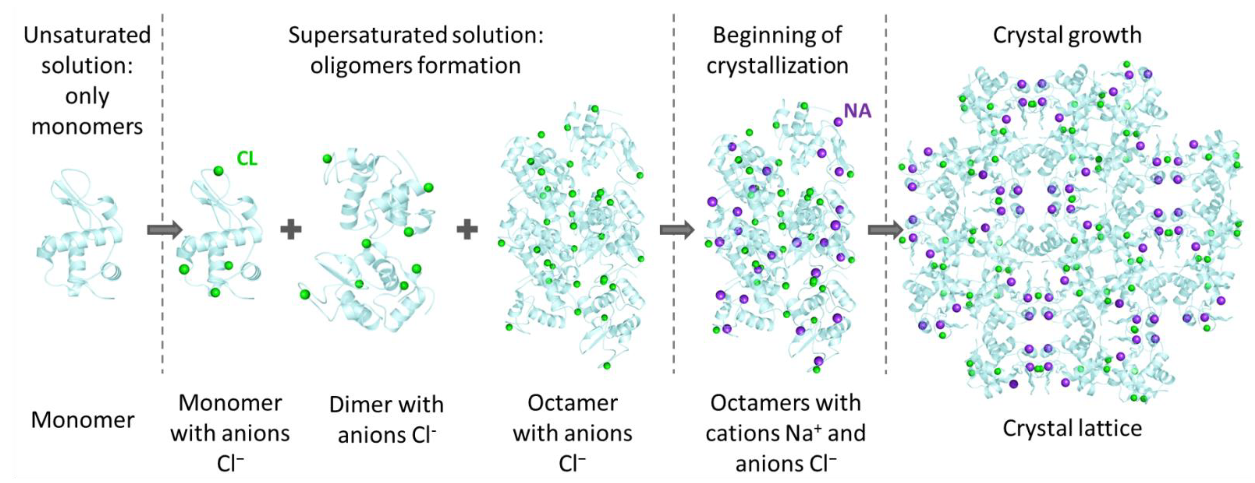
:1. Introduction
2. Materials and Methods
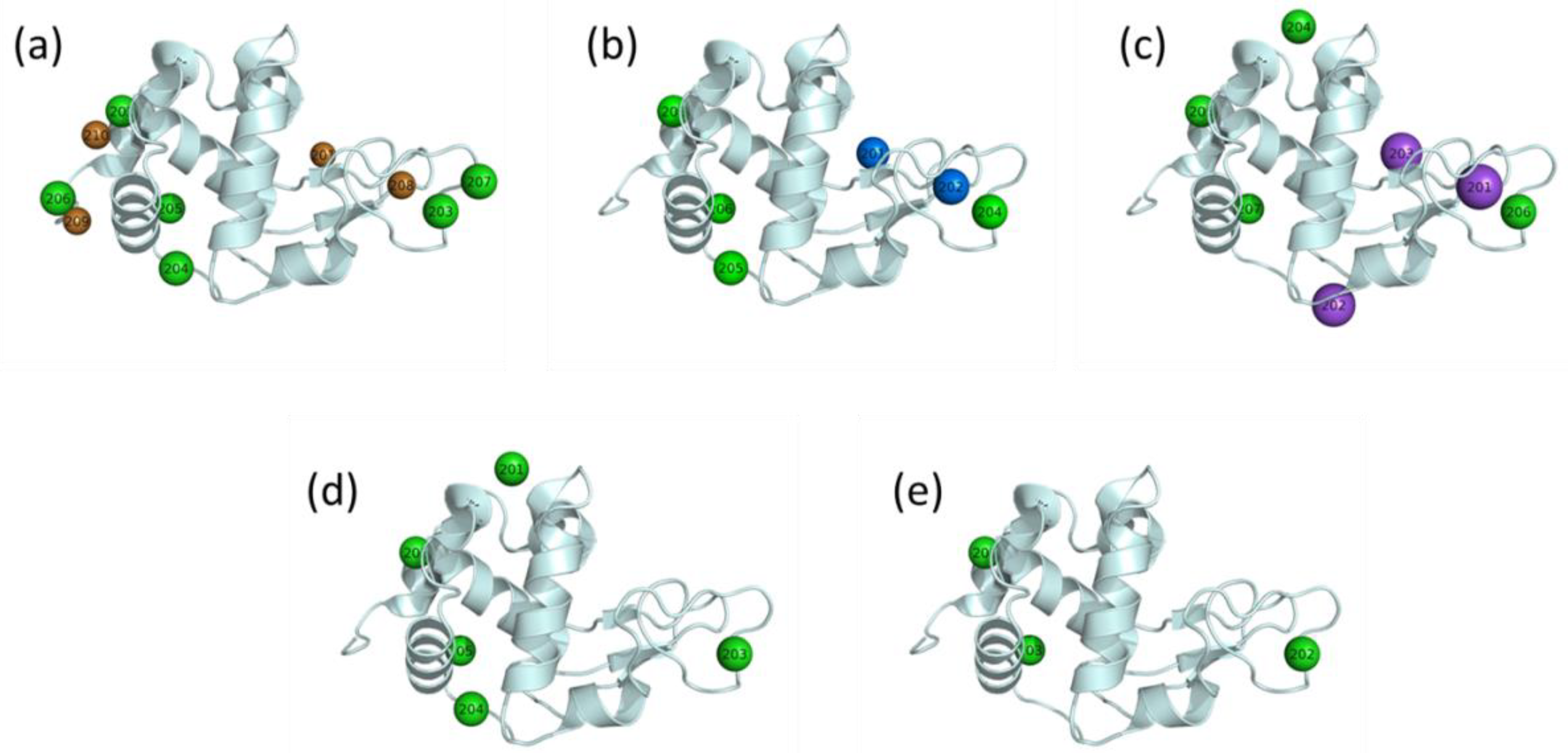
- Without any ions (4WLD);
- Only with cations (one Na+ ion per molecule, 4WLD);
- Only with anions (four Cl− ions per molecule, 6QWY);
- With all ions (three Na+ and four Cl− ions per molecule, 6QWY).
3. Results and Discussion
4. Conclusions
Supplementary Materials
Author Contributions
Funding
Institutional Review Board Statement
Data Availability Statement
Acknowledgments
Conflicts of Interest
References
- Ries-Kautt, M.M.; Ducruix, A.F. Relative effectiveness of various ions on the solubility and crystal growth of lysozyme. J. Biol. Chem. 1989, 264, 745–748. [Google Scholar] [CrossRef]
- Ducruix, A.; Guilloteau, J.P.; Riès-Kautta, M.; Tardieu, A. Protein interactions as seen by solution X-ray scattering prior to crystallogenesis. J. Cryst. Growth 1996, 168, 28–39. [Google Scholar] [CrossRef]
- Hofmeister, F. Zur Lehre von der Wirkung der Salze. Arch. Exp. Pathol. Pharmakol 1888, 24, 247–260. [Google Scholar] [CrossRef] [Green Version]
- Kovalchuk, M.V.; Blagov, A.E.; Dyakova, Y.A.; Gruzinov, A.Y.; Marchenkova, M.A.; Peters, G.S.; Pisarevsky, Y.V.; Timofeev, V.I.; Volkov, V.V. Investigation of the Initial Crystallization Stage in Lysozyme Solutions by Small-Angle X-ray Scattering. Cryst. Growth Des. 2016, 16, 1792–1797. [Google Scholar] [CrossRef]
- Marchenkova, M.A.; Volkov, V.V.; Blagov, A.E.; Dyakova, Y.A.; Ilina, K.B.; Tereschenko, E.Y.; Timofeev, V.I.; Pisarevsky, Y.V.; Kovalchuk, M.V. In situ study of the state of lysozyme molecules at the very early stage of the crystallization process by small-angle X-ray scattering. Crystallogr. Rep. 2016, 61, 5–10. [Google Scholar] [CrossRef]
- Boikova, A.S.; D’yakova, Y.A.; Il’ina, K.B.; Konarev, P.V.; Kryukova, A.E.; Marchenkova, M.A.; Blagov, A.E.; Pisarevskii, Y.V.; Koval’chuk, M.V. Small-angle X-ray scattering study of the influence of solvent replacement (from H2O to D2O) on the initial crystallization stage of tetragonal lysozyme. Crystallogr. Rep. 2017, 62, 837. [Google Scholar] [CrossRef]
- Marchenkova, M.A.; Kuranova, I.P.; Timofeev, V.I.; Boikova, A.S.; Dorovatovskii, P.V.; Dyakova, Y.A.; Ilina, K.B.; Pisarevskiy, Y.V.; Kovalchuk, M.V. The binding of precipitant ions in the tetragonal crystals of hen egg white lysozyme. J. Biomol. Struct. Dyn. 2020, 38, 5159–5172. [Google Scholar] [CrossRef]
- Kordonskaya, Y.V.; Timofeev, V.I.; Dyakova, Y.A.; Marchenkova, M.A.; Pisarevsky, Y.V.; Podshivalov, D.D.; Kovalchuk, M.V. Study of the Behavior of Lysozyme Oligomers in Solutions by the Molecular Dynamics Method. Crystallogr. Rep. 2018, 63, 947–950. [Google Scholar] [CrossRef]
- Kordonskaya, Y.V.; Marchenkova, M.A.; Timofeev, V.I.; Dyakova, Y.A.; Pisarevsky, Y.V.; Kovalchuk, M.V. Precipitant ions influence on lysozyme oligomers stability investigated by molecular dynamics simulation at different temperatures. J. Biomol. Struct. Dyn. 2021, 39, 7223–7230. [Google Scholar] [CrossRef]
- Kordonskaya, Y.V.; Timofeev, V.I.; Dyakova, Y.A.; Marchenkova, M.A.; Pisarevsky, Y.V.; Kovalchuk, M.V. Free energy change during the formation of crystalline contact between lysozyme monomers under different physical and chemical conditions. Crystals 2021, 11, 1121. [Google Scholar] [CrossRef]
- Blanchet, C.E.; Spilotros, A.; Schwemmer, F.; Graewert, M.A.; Kikhney, A.; Jeffries, C.M.; Franke, D.; Mark, D.; Zengerle, R.; Cipriani, F.; et al. Versatile sample environments and automation for biological solution X-ray scattering experiments at the P12 beamline (PETRA III, DESY). J. Appl. Cryst. 2015, 48, 431–443. [Google Scholar] [CrossRef] [PubMed] [Green Version]
- Franke, D.; Petoukhov, M.V.; Konarev, P.V.; Panjkovich, A.; Tuukkanen, A.; Mertens, H.D.T.; Kikhney, A.G.; Hajizadeh, N.R.; Franklin, J.M.; Jeffries, C.M.; et al. ATSAS 2.8: A comprehensive data analysis suite for small-angle scattering from macromolecular solutions. J. Appl. Cryst. 2017, 50, 1212–1225. [Google Scholar] [CrossRef] [PubMed] [Green Version]
- Konarev, P.V.; Volkov, V.V.; Sokolova, A.V.; Koch, M.H.J.; Svergun, D.I. PRIMUS: A Windows PC-based system for small-angle scattering data analysis. J. Appl. Crystallogr. 2003, 36, 1277–1282. [Google Scholar] [CrossRef]
- Barberato, C.; Henri, M.; Koch, J.; Svergun, D.; Barberato, C.; Koch, M.H.J. CRYSOL–a program to evaluate X-ray solution scattering of biological macromolecules from atomic coordinates. J. Appl. Cryst. 1995, 28, 768–773. [Google Scholar] [CrossRef]
- Schrödinger, L.; DeLano, W. PyMOL. 2015. Available online: http://www.pymol.org/pymol (accessed on 18 April 2022).
- Dolinsky, T.J.; Nielsen, J.E.; McCammon, J.A.; Baker, N.A. PDB2PQR: An automated pipeline for the setup of Poisson-Boltzmann electrostatics calculations. Nucleic Acids Res. 2004, 32, W665–W667. [Google Scholar] [CrossRef]
- Lindorff-Larsen, K.; Piana, S.; Palmo, K.; Maragakis, P.; Klepeis, J.L.; Dror, R.O.; Shaw, D.E. Improved side-chain torsion potentials for the Amber ff99SB protein force field. Proteins Struct. Funct. Bioinform. 2010, 78, 1950–1958. [Google Scholar] [CrossRef] [PubMed] [Green Version]
- Van Der Spoel, D.; Lindahl, E.; Hess, B.; Groenhof, G.; Mark, A.E.; Berendsen, H.J.C. GROMACS: Fast, flexible, and free. J. Comput. Chem. 2005, 26, 1701–1718. [Google Scholar] [CrossRef]
- Horn, H.W.; Swope, W.C.; Pitera, J.W.; Madura, J.D.; Dick, T.J.; Hura, G.L.; Head-Gordon, T. Development of an improved four-site water model for biomolecular simulations: TIP4P-Ew. J. Chem. Phys. 2004, 120, 9665–9678. [Google Scholar] [CrossRef]
- Berendsen, H.J.C.; Postma, J.P.M.; Van Gunsteren, W.F.; Dinola, A.; Haak, J.R. Molecular dynamics with coupling to an external bath. J. Chem. Phys. 1984, 81, 3684–3690. [Google Scholar] [CrossRef] [Green Version]
- Parrinello, M.; Rahman, A. Strain fluctuations and elastic constants. J. Chem. Phys. 1982, 76, 2662–2666. [Google Scholar] [CrossRef]
- Van Gunsteren, W.F.; Berendsen, H.J.C. A Leap-Frog Algorithm for Stochastic Dynamics. Mol. Simul. 1988, 1, 173–185. [Google Scholar] [CrossRef]
- Marchenkova, M.A.; Konarev, P.V.; Boikova, A.S.; Ilina, K.B.; Pisarevsky, Y.V.; Kovalchuk, M.V. Influence of Chlorides of Mono- and Divalent Metals on the Oligomeric Composition of Lysozyme Crystallization Solutions and Further Crystal Growth. Crystallogr. Rep. 2021, 66, 751–757. [Google Scholar] [CrossRef]
- Dyakova, Y.A.; Boikova, A.S.; Ilina, K.B.; Konarev, P.V.; Marchenkova, M.A.; Pisarevsky, Y.V.; Timofeev, V.I.; Kovalchuk, M.V. Study of the Influence of a Precipitant Cation on the Formation of Oligomers in Crystallization Solutions of Lysozyme Protein. Crystallogr. Rep. 2019, 64, 11–15. [Google Scholar] [CrossRef]


| Protein Concentration, mg/mL | Precipitant | Measurement Number | Rg, Å | Monomers, % | Dimers, % | Octamers, % | χ2 |
|---|---|---|---|---|---|---|---|
| 20 | - | 1 | 14.37 ± 0.2 | 100 | - | - | 3.17 |
| LiCl | 1 | 19.6 ± 0.2 | 78.8 ± 0.4 | 18.8 ± 0.4 | 2.4 ± 0.1 | 1.27 | |
| 19.7 ± 0.2 | 80.0 ± 0.4 | 17.5 ± 0.4 | 2.5 ± 0.1 | 1.18 | |||
| LiCl | 2 | 19.4 ± 0.2 | 79.9 ± 0.4 | 17.8 ± 0.4 | 2.3 ± 0.1 | 1.31 | |
| 19.5 ± 0.2 | 81.1 ± 0.4 | 16.5 ± 0.4 | 2.4 ± 0.1 | 1.17 | |||
| NaCl | 1 | 18.8 ± 0.2 | 81.9 ± 0.4 | 16.2 ± 0.4 | 1.9 ± 0.1 | 1.08 | |
| 18.9 ± 0.2 | 83.4 ± 0.4 | 14.7 ± 0.4 | 1.9 ± 0.1 | 1.02 | |||
| NaCl | 2 | 19.3 ± 0.2 | 82.6 ± 0.4 | 15.0 ± 0.4 | 2.4 ± 0.1 | 1.10 | |
| 19.4 ± 0.2 | 84.1 ± 0.4 | 13.5 ± 0.4 | 2.4 ± 0.1 | 1.06 | |||
| KCl | 1 | 19.5 ± 0.2 | 82.6 ± 0.4 | 14.8 ± 0.4 | 2.6 ± 0.1 | 1.06 | |
| 19.6 ± 0.2 | 83.2 ± 0.4 | 14.3 ± 0.4 | 2.5 ± 0.1 | 1.04 | |||
| KCl | 2 | 18.8 ± 0.2 | 84.4 ± 0.4 | 13.6 ± 0.4 | 2.0 ± 0.1 | 1.05 | |
| 18.9 ± 0.2 | 85.0 ± 0.4 | 13.0 ± 0.4 | 2.0 ± 0.1 | 1.04 | |||
| NiCl2 | 1 | 19.0 ± 0.2 | 86.6 ± 0.4 | 11.1 ± 0.4 | 2.3 ± 0.1 | 1.09 | |
| 19.2 ± 0.2 | 89.8 ± 0.4 | 7.7 ± 0.4 | 2.5 ± 0.1 | 1.02 | |||
| NiCl2 | 2 | 19.1 ± 0.2 | 87.0 ± 0.4 | 10.5 ± 0.4 | 2.5 ± 0.1 | 1.06 | |
| 19.3 ± 0.2 | 90.2 ± 0.4 | 7.1 ± 0.4 | 2.7 ± 0.1 | 1.00 | |||
| CuCl2 | 1 | 18.2 ± 0.2 | 90.6 ± 0.4 | 7.7 ± 0.4 | 1.7 ± 0.1 | 1.05 | |
| 18.1 ± 0.2 | 88.6 ± 0.4 | 9.8 ± 0.4 | 1.6 ± 0.1 | 1.03 | |||
| CuCl2 | 2 | 18.9 ± 0.2 | 94.4 ± 0.4 | 3.2 ± 0.4 | 2.4 ± 0.1 | 1.05 | |
| 18.8 ± 0.2 | 92.4 ± 0.4 | 5.3 ± 0.4 | 2.3 ± 0.1 | 1.02 | |||
| 40 | - | 1 | 14.37 ± 0.2 | 100 | - | - | 3.11 |
| LiCl | 1 | 23.1 ± 0.2 | 74.9 ± 0.4 | 18.2 ± 0.4 | 6.9 ± 0.1 | 1.91 | |
| 23.2 ± 0.2 | 75.6 ± 0.4 | 17.6 ± 0.4 | 6.8 ± 0.1 | 1.52 | |||
| LiCl | 2 | 23.0 ± 0.2 | 74.8 ± 0.4 | 18.5 ± 0.4 | 6.7 ± 0.1 | 1.83 | |
| 23.1 ± 0.2 | 75.5 ± 0.4 | 17.9 ± 0.4 | 6.6 ± 0.1 | 1.57 | |||
| NaCl | 1 | 21.9 ± 0.2 | 77.5 ± 0.4 | 17.2 ± 0.4 | 5.3 ± 0.1 | 1.49 | |
| 22.0 ± 0.2 | 78.5 ± 0.4 | 16.4 ± 0.4 | 5.1 ± 0.1 | 1.31 | |||
| NaCl | 2 | 21.9 ± 0.2 | 77.8 ± 0.4 | 16.9 ± 0.4 | 5.3 ± 0.1 | 1.55 | |
| 22.0 ± 0.2 | 78.7 ± 0.4 | 16.1 ± 0.4 | 5.2 ± 0.1 | 1.37 | |||
| KCl | 1 | 21.7 ± 0.2 | 80.5 ± 0.4 | 14.5 ± 0.4 | 5.0 ± 0.1 | 1.28 | |
| 21.8 ± 0.2 | 80.8 ± 0.4 | 14.4 ± 0.4 | 4.8 ± 0.1 | 1.23 | |||
| KCl | 2 | 21.4 ± 0.2 | 80.4 ± 0.4 | 15.0 ± 0.4 | 4.6 ± 0.1 | 1.18 | |
| 21.5 ± 0.2 | 80.6 ± 0.4 | 14.9 ± 0.4 | 4.5 ± 0.1 | 1.24 | |||
| NiCl2 | 1 | 21.9 ± 0.2 | 83.7 ± 0.4 | 11.0 ± 0.4 | 5.3 ± 0.1 | 1.22 | |
| 22.1 ± 0.2 | 86.7 ± 0.4 | 7.9 ± 0.4 | 5.4 ± 0.1 | 1.04 | |||
| NiCl2 | 2 | 21.9 ± 0.2 | 83.7 ± 0.4 | 11.0 ± 0.4 | 5.3 ± 0.1 | 1.22 | |
| 22.1 ± 0.2 | 86.7 ± 0.4 | 7.9 ± 0.4 | 5.4 ± 0.1 | 1.04 | |||
| CuCl2 | 1 | 20.9 ± 0.2 | 92.0 ± 0.4 | 3.8 ± 0.4 | 4.2 ± 0.1 | 1.30 | |
| 20.9 ± 0.2 | 89.8 ± 0.4 | 6.1 ± 0.4 | 4.1 ± 0.1 | 1.19 | |||
| CuCl2 | 2 | 20.9 ± 0.2 | 91.5 ± 0.4 | 4.4 ± 0.4 | 4.1 ± 0.1 | 1.21 | |
| 20.9 ± 0.2 | 89.8 ± 0.4 | 6.1 ± 0.4 | 4.1 ± 0.1 | 1.19 |
| Protein Concentration, mg/mL | Measurement Number | χ2 for the Structures with Cations and Anions (Set1) | χ2 for the Structures with Cations (Set2) | χ2 for the Structures with Anions (Set3) | χ2 for the Structures without Ions (Set4) | χ2 for the Structures from (Set3 + Set4) |
|---|---|---|---|---|---|---|
| 20 | 1 | 1.0504 | 1.0414 | 1.0397 | 1.0341 | 1.0339 |
| 2 | 1.0489 | 1.041 | 1.0389 | 1.0329 | 1.0327 | |
| 40 | 1 | 1.3043 | 1.2653 | 1.2556 | 1.2266 | 1.2254 |
| 2 | 1.2067 | 1.1703 | 1.1596 | 1.1352 | 1.1340 |
| Oligomer Type | With Cations and Anions | With Cations | With Anions | Without Ions |
|---|---|---|---|---|
| Dimer | 0.116 | 0.141 | 0.130 | 0.156 |
| Octamer | 0.272 | 0.179 | 0.182 | 0.210 |
Publisher’s Note: MDPI stays neutral with regard to jurisdictional claims in published maps and institutional affiliations. |
© 2022 by the authors. Licensee MDPI, Basel, Switzerland. This article is an open access article distributed under the terms and conditions of the Creative Commons Attribution (CC BY) license (https://creativecommons.org/licenses/by/4.0/).
Share and Cite
Marchenkova, M.A.; Konarev, P.V.; Kordonskaya, Y.V.; Ilina, K.B.; Pisarevsky, Y.V.; Soldatov, A.V.; Timofeev, V.I.; Kovalchuk, M.V. The Role of Cations and Anions in the Formation of Crystallization Oligomers in Protein Solutions as Revealed by Combination of Small-Angle X-ray Scattering and Molecular Dynamics. Crystals 2022, 12, 751. https://0-doi-org.brum.beds.ac.uk/10.3390/cryst12060751
Marchenkova MA, Konarev PV, Kordonskaya YV, Ilina KB, Pisarevsky YV, Soldatov AV, Timofeev VI, Kovalchuk MV. The Role of Cations and Anions in the Formation of Crystallization Oligomers in Protein Solutions as Revealed by Combination of Small-Angle X-ray Scattering and Molecular Dynamics. Crystals. 2022; 12(6):751. https://0-doi-org.brum.beds.ac.uk/10.3390/cryst12060751
Chicago/Turabian StyleMarchenkova, Margarita A., Petr V. Konarev, Yuliya V. Kordonskaya, Kseniia B. Ilina, Yury V. Pisarevsky, Alexander V. Soldatov, Vladimir I. Timofeev, and Mikhail V. Kovalchuk. 2022. "The Role of Cations and Anions in the Formation of Crystallization Oligomers in Protein Solutions as Revealed by Combination of Small-Angle X-ray Scattering and Molecular Dynamics" Crystals 12, no. 6: 751. https://0-doi-org.brum.beds.ac.uk/10.3390/cryst12060751