
A Study of Song Dynasty Polychrome Statue-Making Techniques and Materials in the Sage Mother Hall of the Jinci Temple, Shanxi, China

Abstract
:1. Introduction
2. Materials and Methods
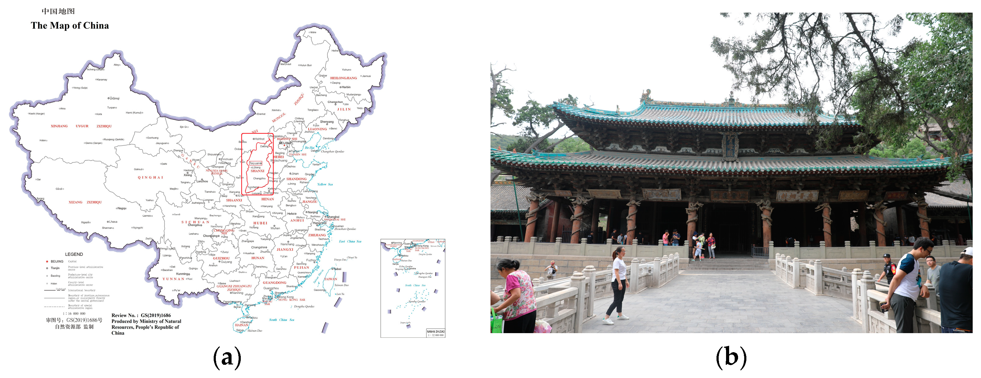
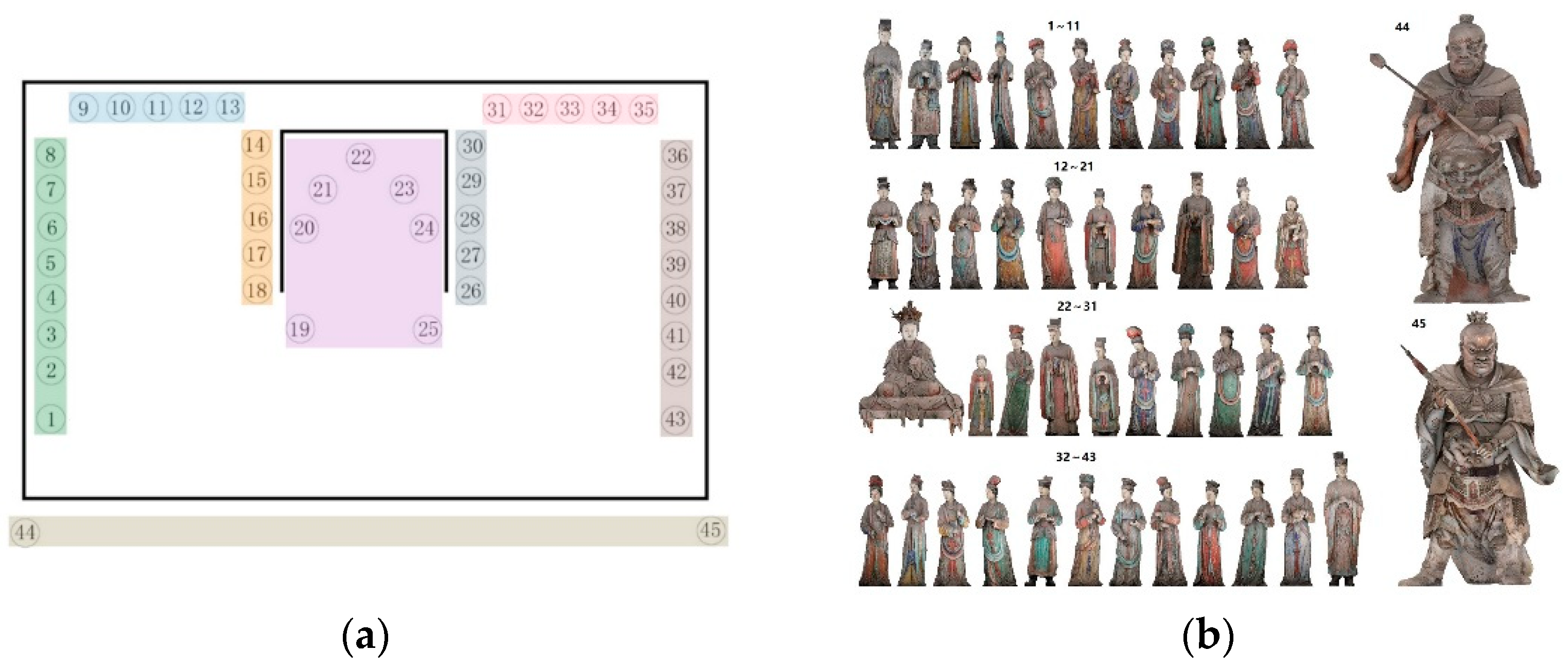
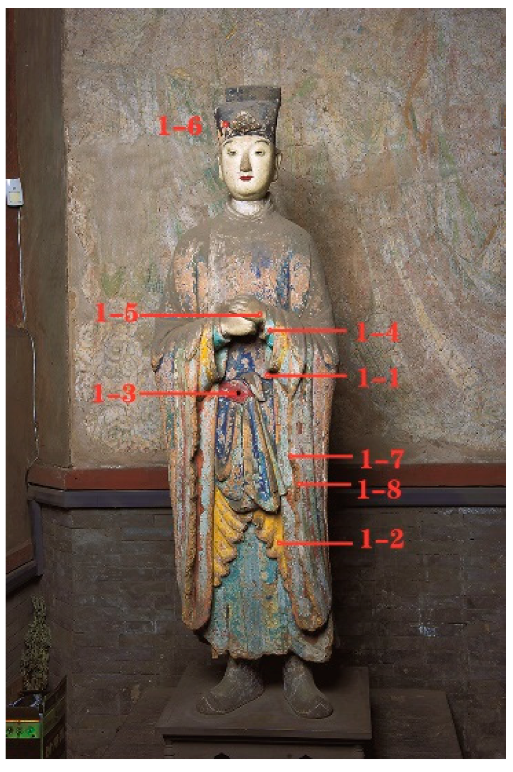
2.1. Sample Collection
2.2. Instrumentation and Measurements
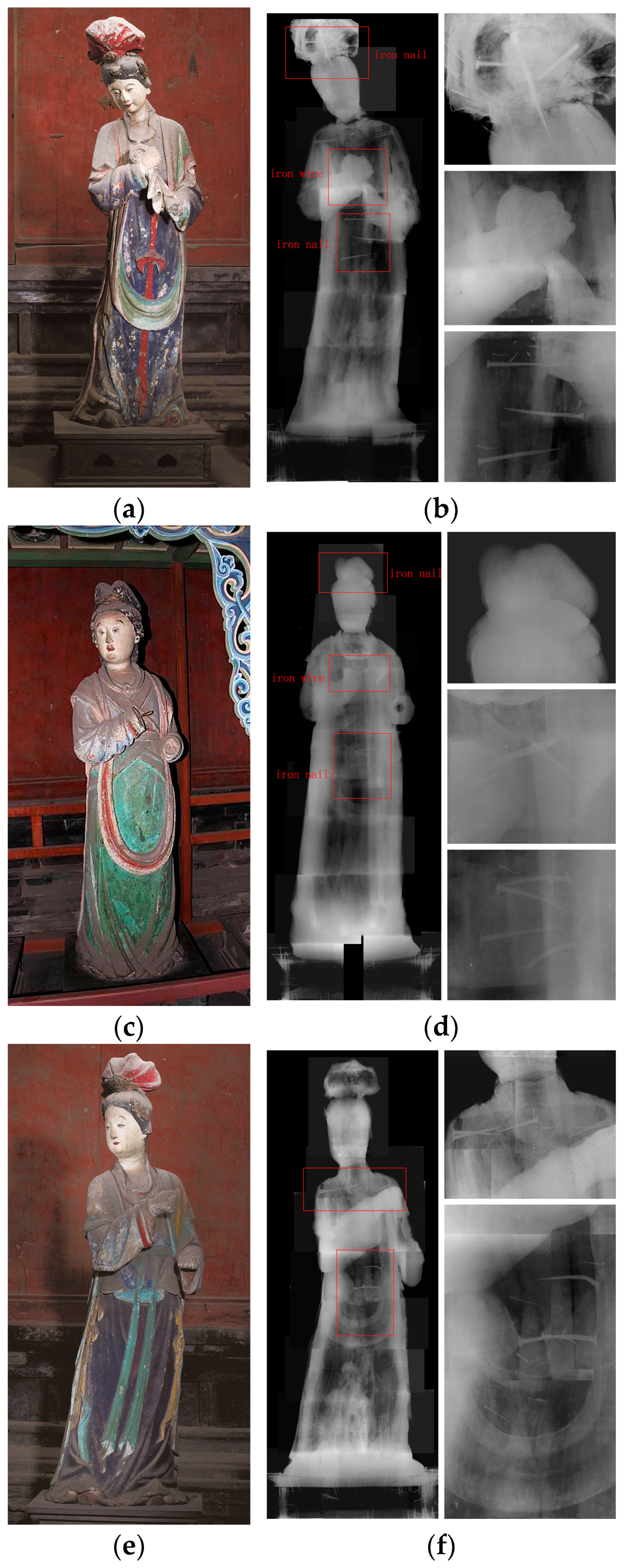
2.2.1. Non-Invasive In Situ Study
2.2.2. Laboratory Studies on Selected Samples
3. Results
3.1. Surface Elemental Information of Polychrome Statue
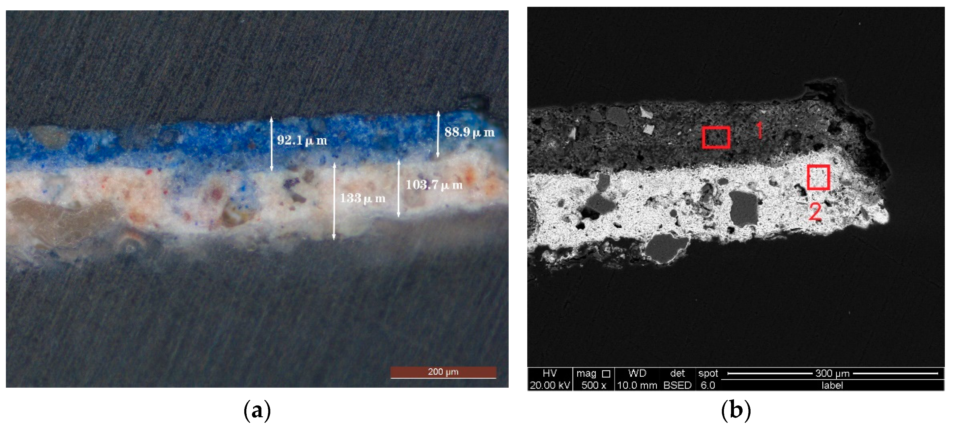
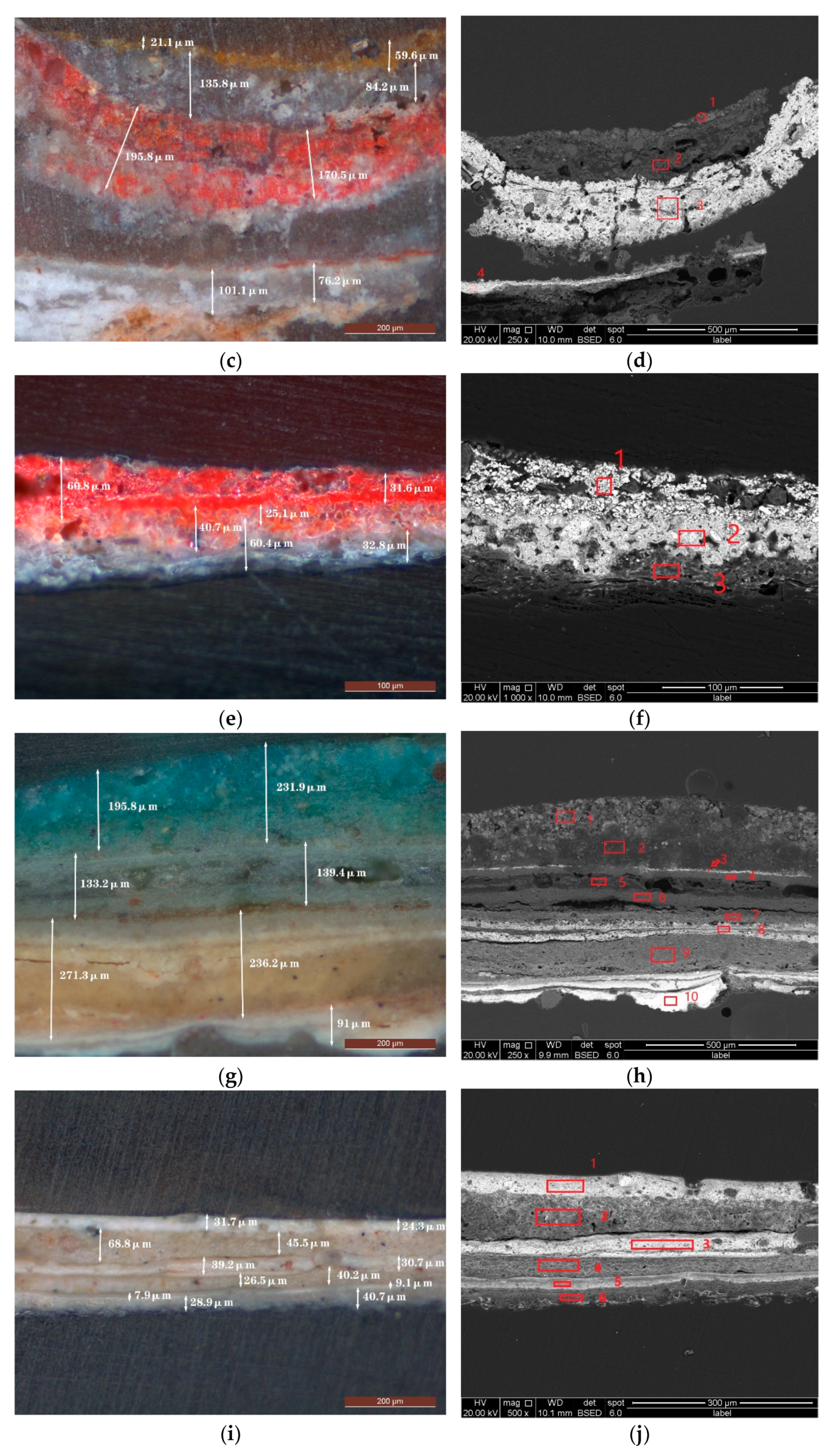
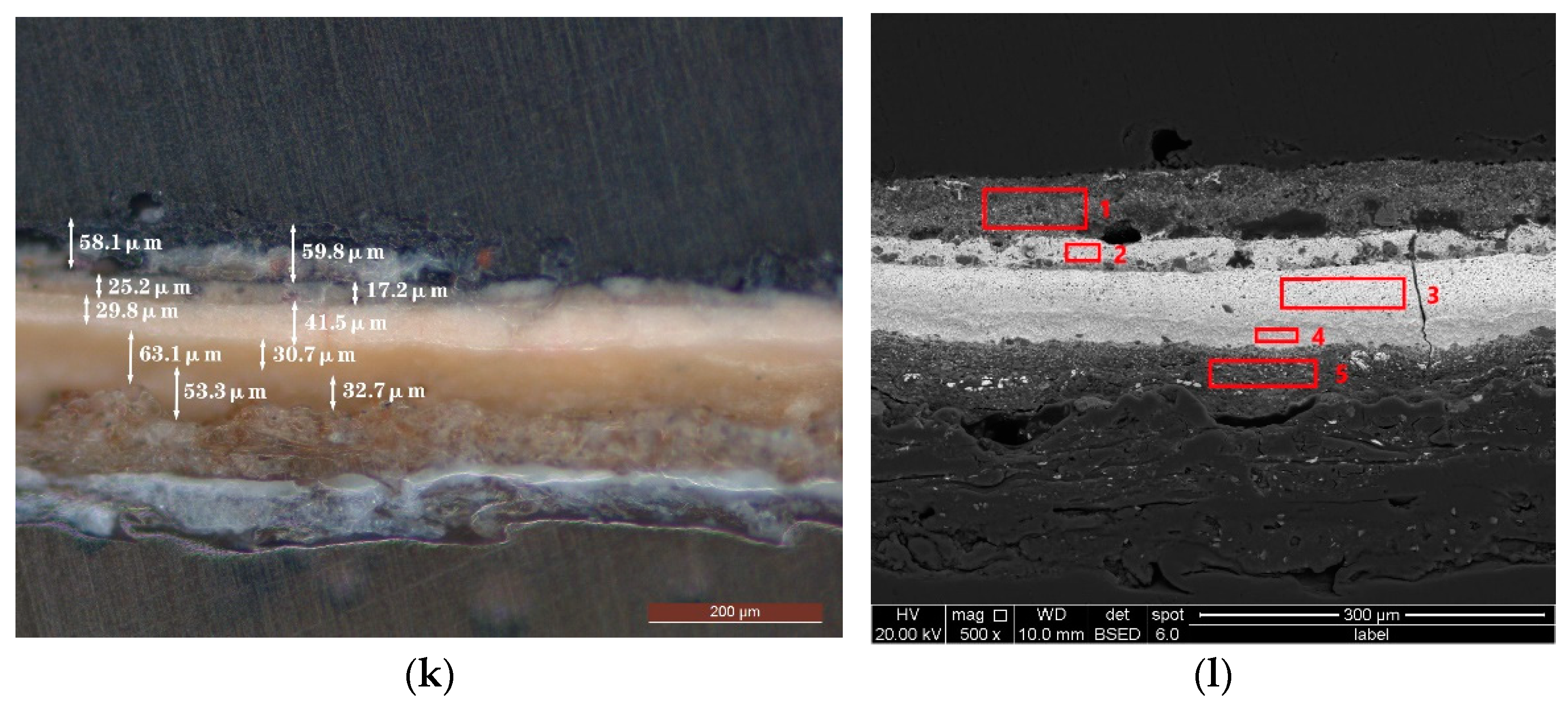
3.2. Microstructure of the Polychrome Statues
3.3. Internal Structure of Polychrome Statues
3.4. Analysis of Plaster Layer
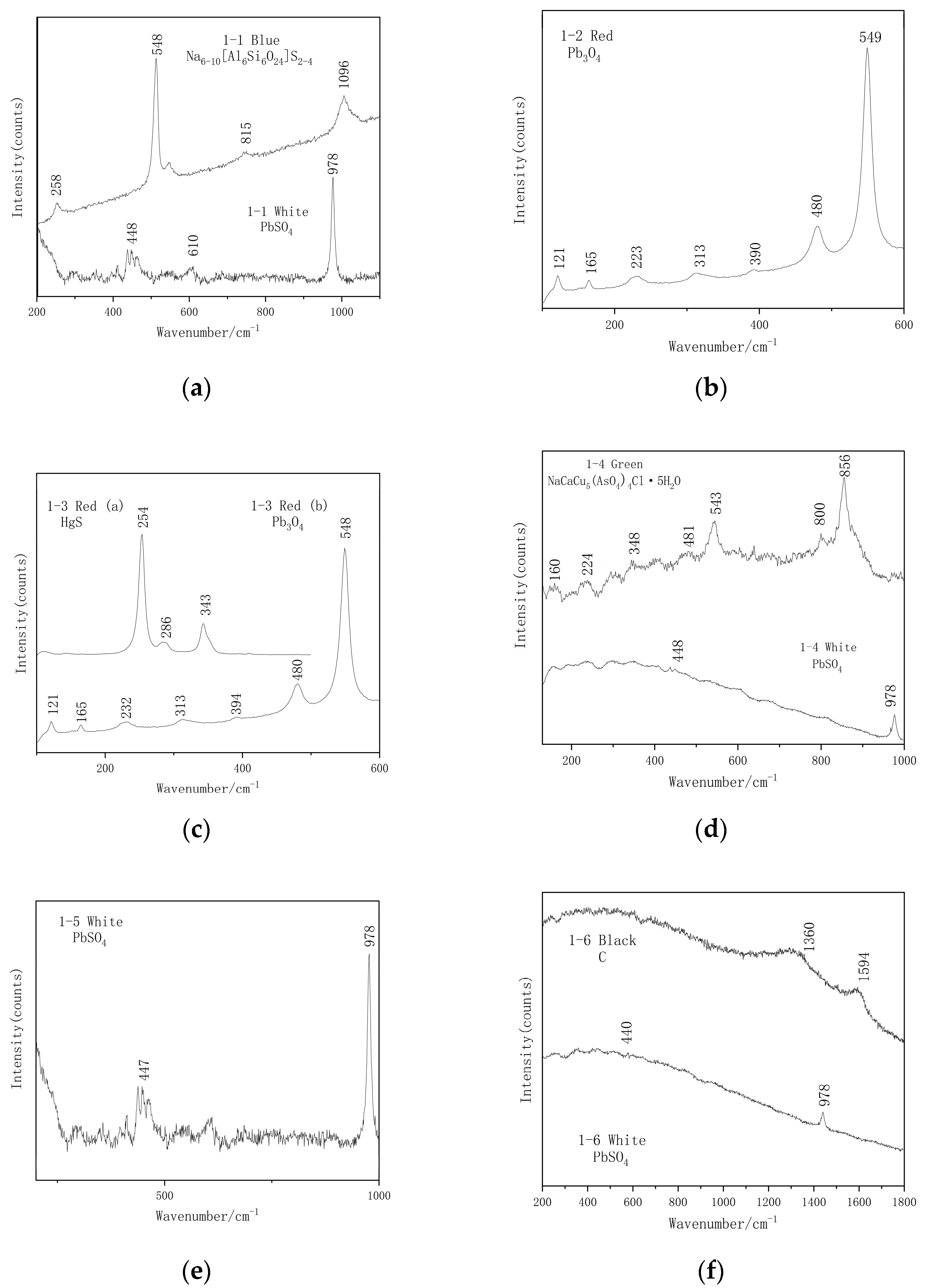
3.5. Analysis of Pigment Layers
4. Discussion
4.1. The Production Process of Polychrome Statues
4.2. The Pigments of Polychrome Statues
4.2.1. Red Pigments
4.2.2. Blue Pigments
4.2.3. Green Pigments
4.3. Multilayered Repainting
5. Conclusions
Author Contributions
Funding
Institutional Review Board Statement
Informed Consent Statement
Data Availability Statement
Acknowledgments
Conflicts of Interest
References
- Ran, X. Analysis of the Layout Axis of Jinci in Taiyuan, Shanxi Province. Adv. Mater. Res. 2015, 1065, 2601–2604. [Google Scholar] [CrossRef]
- Murray, J.K. The Divine Nature of Power: Chinese Ritual Architecture at the Sacred Site of Jinci. J. Asian Stud. 2011, 70, 535–536. [Google Scholar] [CrossRef]
- Smith, G.; Derbyshire, A.; Clark, R. In situ spectroscopic detection of lead sulphide on a blackened manuscript illumination by Raman microscopy. Stud. Conserv. 2002, 47, 250–256. [Google Scholar]
- Burgio, L.; Clark, R.J.; Firth, S. Raman spectroscopy as a means for the identification of plattnerite (PbO2), of lead pigments and of their degradation products. Analyst 2001, 126, 222–227. [Google Scholar] [CrossRef] [PubMed]
- Chen, X.L.; Yang, Q. Micro-Raman spectroscopy study of three green pigments containing copper and arsenic. Sci. Conserv. Archaeol. 2015, 27, 84–89. [Google Scholar] [CrossRef]
- Casali, F. X-ray and neutron digital radiography and computed tomography for cultural heritage. In Physical Techniques in the Study of Art, Archaeology and Cultural Heritage; Elsevier: Amsterdam, The Netherlands, 2006; pp. 41–123. [Google Scholar]
- Kanngießer, B.; Malzer, W.; Mantouvalou, I.; Sokaras, D.; Karydas, A.G. A deep view in cultural heritage—Confocal micro X-ray spectroscopy for depth resolved elemental analysis. Appl. Phys. A 2012, 106, 325–338. [Google Scholar] [CrossRef]
- Casali, F.; Bettuzzi, M.; Brancaccio, R.; Morigi, M.P. New X-ray digital radiography and computed tomography for cultural heritage. In Science for Cultural Heritage: Technological Innovation and Case Studies in Marine and Land Archaeology in the Adriatic Region and Inland; World Scientific Publishing Co. Pte Ltd.: Singapore, 2010; pp. 85–99. [Google Scholar]
- Singha, M.R.; Manib, B.R. Characterization of Pigments and Binders in Mural Painting Fragments from Bezeklik, China. Indian J. Hist. Sci 2019, 54, 348–360. [Google Scholar]
- Bell, I.M.; Clark, R.J.H.; Gibbs, P.J. Raman spectroscopic library of natural and synthetic pigments (pre- ≈ 1850 AD). Spectrochim. Acta Part A Mol. Biomol. Spectrosc. 1997, 53, 2159–2179. [Google Scholar] [CrossRef]
- Burgio, L.; Clark, R.J.H. Library of FT-Raman spectra of pigments, minerals, pigment media and varnishes, and supplement to existing library of Raman spectra of pigments with visible excitation. Spectrochim. Acta A 2001, 57, 1491–1521. [Google Scholar] [CrossRef]
- Frost, R.; Weier, M.; Williams Leverett, P.; Kloprogge, J. Raman spectroscopy of the sampleite group of minerals. J. Raman Spectrosc. 2007, 38, 574–583. [Google Scholar] [CrossRef] [Green Version]
- Whitfield, R.; Whitfield, S.; Agnew, N. Cave Temples of Mogao at Dunhuang: Art History on the Silk Road; Getty Publications: Los Angeles, CA, USA, 2015. [Google Scholar]
- Andrea, A.J. The silk road in world history: A review essay. Asian Rev. World Hist. 2014, 2, 105–127. [Google Scholar] [CrossRef]
- Song, J.; Xiang, W.; Yan, S.; Zhou, W.; Ma, L. Craftsmanship and materials: Painted Bodhisattva sculptures in the Fengguo Temple dated to the year 1020 in Yi County, Northeast China. Herit. Sci. 2021, 9, 1–19. [Google Scholar] [CrossRef]
- Ma, Z.F.; Wang, W.F.; Li, Y.H.; Li, B.; Cai, G.T.; Hou, B.L. Study on the material, process and disease analysis of the colorful sculpture of Baixiang Pagoda of Northern Song Dynasty in Wenzhou Museum. Dunhuang Res. 2002, 57–63, 115. [Google Scholar]
- Aze, S.; Vallet, J.M.; Baronnet, A.; Grauby, O. The fading of red lead pigment in wall paintings: Tracking the physico-chemical transformations by means of complementary micro-analysis techniques. Eur. J. Mineral. 2006, 18, 835–843. [Google Scholar] [CrossRef]
- Clark, R.J.H.; Curri, M.L.; Laganara, C. Raman microscopy: The identification of lapis lazuli on medieval pottery fragments from the south of Italy. Spectrochim. Acta Part A Mol. Biomol. Spectrosc. 1997, 53, 597–603. [Google Scholar] [CrossRef]
- Gaetani, M.C.; Santamaria, U.; Seccaroni, C. The use of Egyptian blue and lapis lazuli in the Middle Ages—The wall paintings of the San Saba Church in Rome. Stud. Conserv. 2004, 49, 13–22. [Google Scholar] [CrossRef]
- Zhang, Y.; Wang, J.; Liu, H.; Wang, X.; Zhang, S. Integrated analysis of pigments on murals and sculptures in Mogao Grottoes. Anal. Lett. 2015, 48, 2400–2413. [Google Scholar] [CrossRef]
- Liu, Z.; Han, Y.; Han, L.; Cheng, Y.; Ma, Y.; Fang, L. Micro-Raman analysis of the pigments on painted pottery figurines from two tombs of the Northern Wei Dynasty in Luoyang. Spectrochim. Acta Part A Mol. Biomol. Spectrosc. 2013, 109, 42–46. [Google Scholar] [CrossRef]
- Del Federico, E.; Shöfberger, W.; Schelvis, J.; Kapetanaki, S.; Tyne, L.; Jerschow, A. Insight into framework destruction in ultramarine pigments. Inorg. Chem. 2006, 45, 1270–1276. [Google Scholar] [CrossRef]
- Li, Z.; Wang, L.; Ma, Q.; Mei, J. A scientific study of the pigments in the wall paintings at Jokhang Monastery in Lhasa, Tibet, China. Herit. Sci. 2014, 2, 1–6. [Google Scholar] [CrossRef] [Green Version]
- Ajò, D.; Casellato, U.; Fiorin, E.; Vigato, P.A. A study of painting materials and technique by SEM-EDS microscopy, X-ray diffraction, micro FT-IR and photoluminescence spectroscopy. J. Cult. Herit. 2004, 5, 333–348. [Google Scholar] [CrossRef]
- Wang, L.Q.; Ma, Y.N.; Zhang, Y.X.; Zhao, X.; He, Q.J.; Guo, J.Y.; Ren, H.T. Pigment identification of Sleeping Buddha at World Cutural Heritage Dazu Rock Carvings with μ-Raman spectroscopy and Related Research. Spectrosc. Spectr. Anal. 2020, 40, 3199–3204. [Google Scholar]
- Wang, C.C.; Li, Z.h.M.; Wang, X.N.; Ma, Q.L. Scientifc study of the Song Dynasty polychrome Arhat statues from the Magic Clif Monastery in Jinan. Sci. Conserv. Archaeol. 2018, 30, 37–47. [Google Scholar] [CrossRef]
- Li, Z.M.; Wang, L.L.; Chen, H.L.; Ma, Q.L. Degradation of emerald green: Scientifc studieson multi-polychrome Vairocana statue in Dazu rock carvings, Chongqing, China. Herit. Sci. 2020, 8, 64. [Google Scholar] [CrossRef]
- Keune, K.; Boon, J.; Boitelle, R.; Shimadzu, Y. Degradation of Emerald green in oil paint and its contribution to the rapid change in colour of the Descente des vaches (1834–1835) painted by Théodore Rousseau. Stud. Conserv. 2013, 58, 199–210. [Google Scholar] [CrossRef]
- Holakooei, P.; Karimy, A.H.; Nafisi, G. Lammerite as a degradation product of emerald green: Scientific studies on a rural Persian wall painting. Stud. Conserv. 2018, 63, 391–402. [Google Scholar] [CrossRef]
- Ondruš, P.; Veselovský, F.; Hloušek, J.; Skála, R.; Vavěín, I.; Frýda, J.; Čejka, J.; Gabašová, A. Secondary minerals of the Jáchymov (Joachimsthal) ore district Sekundární minerály jáchymovského rudního revíru (Czech summary). J. Czech Geol. Soc. 1997, 42, 3–69. [Google Scholar]
- Tang, D.; Wang, C.; Nie, J.; Chen, R.; Niu, Q.; Kan, H.; Chen, B.; Perera, F.; Taiyuan, C.D.C. Health benefits of improving air quality in Taiyuan, China. Environ. Int. 2014, 73, 235–242. [Google Scholar] [CrossRef] [Green Version]


| Number | Samples | Analysis Method |
|---|---|---|
| 1-1 | Blue pigment | RAM, SEM-EDS |
| 1-2 | Yellow pigment | RAM, SEM-EDS |
| 1-3 | Red pigments | RAM, SEM-EDS |
| 1-4 | Green pigment | RAM, SEM-EDS |
| 1-5 | White pigments | RAM, SEM-EDS |
| 1-6 | Black pigment | RAM, SEM-EDS |
| 1-7 | Coarse clay layer | XRD |
| 1-8 | Fine clay layer | XRD |
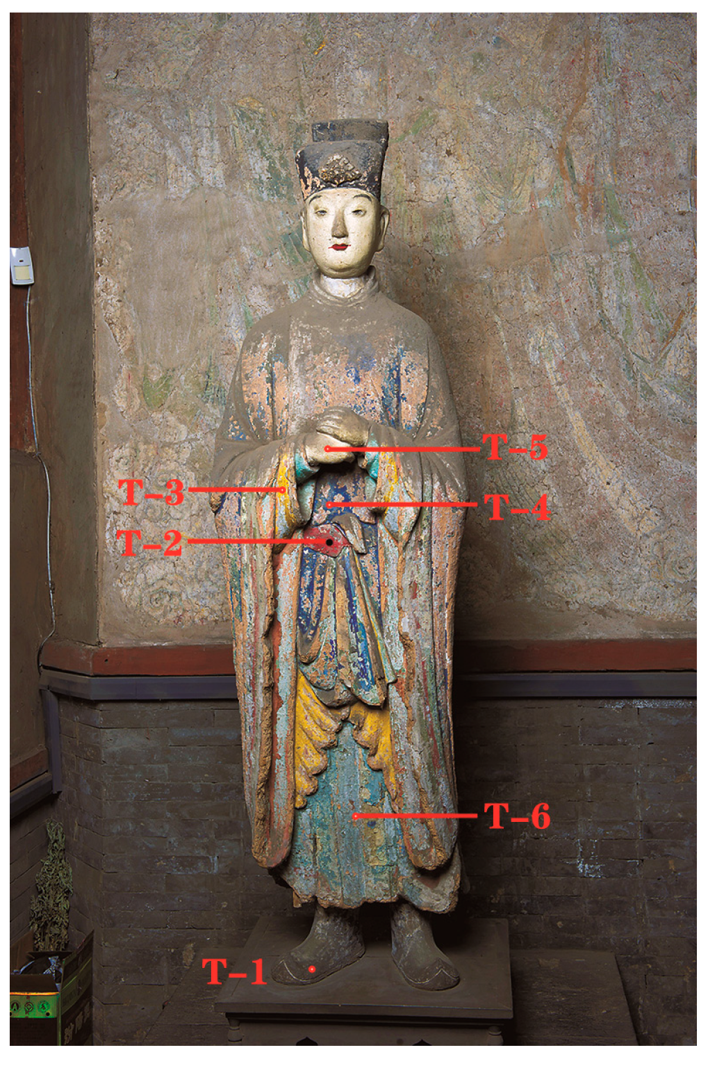
| Color | Point Number | Pb | As | Hg | Cu | Fe | Ca | S |
|---|---|---|---|---|---|---|---|---|
| Black | T-1 | 0.1% | 0% | 0% | 0.1% | 8.3% | 51.0% | 40.4% |
| Red | T-2 | 33.5% | 8.3% | 39.9% | 1.4% | 1.2% | 0.4% | 15.3% |
| Yellow | T-3 | 30.4% | 1.6% | 0% | 13.6% | 31.1% | 3.2% | 20.1% |
| Blue | T-4 | 10.3% | 0% | 0.8% | 63.6% | 3.3% | 5.2% | 16.9% |
| White | T-5 | 77.2% | 6.2% | 0% | 0.1% | 1.6% | 1.3% | 13.6% |
| Green | T-6 | 26.5% | 4.2% | 0% | 22.6% | 2.3% | 10.6% | 33.7% |
| Sample Number | Test Number | Elemental Content (wt%) | Thickness (um) | ||||||||||
|---|---|---|---|---|---|---|---|---|---|---|---|---|---|
| Na | Al | Si | S | Cl | Ca | Cu | Pb | Fe | Hg | As | |||
| 1-1 | 1 | 8.6 | 21.4 | 35.6 | 23.5 | 2.0 | 1.4 | 7.5 | - | - | - | - | 89~92 |
| 1-1 | 2 | - | - | - | 17.9 | - | - | - | 82.1 | - | - | - | 104~133 |
| 1-2 | 1 | - | 11.1 | 16.9 | 4.9 | - | 1.9 | - | - | 56.4 | - | 8.8 | 21~60 |
| 1-2 | 2 | - | 44.3 | 48.5 | - | - | 4.0 | - | - | 3.2 | - | - | 84~136 |
| 1-2 | 3 | - | - | 2.6 | - | - | 1.9 | 4.3 | 88.6 | 2.6 | - | - | 171~196 |
| 1-2 | 4 | 1.6 | - | - | 8.6 | 2.8 | 4.7 | - | 82.3 | - | - | - | 72~101 |
| 1-3 | 1 | - | - | - | 10.7 | - | - | - | - | - | 89.3 | - | 32~61 |
| 1-3 | 2 | - | - | - | 12.6 | - | - | - | 82.6 | 4.8 | - | - | 25~41 |
| 1-3 | 3 | - | 19.0 | 21.2 | 8.8 | - | 7.4 | - | 41.5 | 2.1 | - | - | 33~60 |
| 1-4 | 1 | - | 13.06 | 21.3 | 2.0 | 3.2 | 2.9 | 34.1 | - | 1 | - | 21.9 | 196~232 |
| 1-4 | 2 | 5.0 | 15.7 | 28.6 | 12.2 | 3.6 | - | 20.5 | - | 3.2 | - | 11.2 | |
| 1-4 | 3 | - | - | - | 8.7 | 3.6 | - | 3.9 | 63.2 | - | - | 20.6 | 133~139 |
| 1-4 | 4 | 3.9 | 29.7 | 33.5 | 20.6 | - | 8.3 | 4.0 | - | - | - | - | |
| 1-4 | 5 | - | 10.0 | 12.2 | 34.6 | 14.7 | 10.9 | 17.6 | - | - | - | - | |
| 1-4 | 6 | 1.9 | 28.0 | 41.1 | - | - | 2.1 | 14.0 | - | 2.3 | - | 8.5 | |
| 1-4 | 7 | - | 17.8 | 33.5 | 18.5 | 1.7 | - | 7.8 | - | 2.9 | - | 17.8 | 236~271 |
| 1-4 | 8 | - | 8.9 | 19.7 | 17.1 | - | - | 25.9 | 28.4 | - | - | - | |
| 1-4 | 9 | 4.5 | 26.8 | 50.4 | - | - | 3.2 | - | - | 2.0 | - | 13.1 | |
| 1-4 | 10 | - | - | - | 8.3 | - | 1.6 | - | 90.1 | - | - | - | ≈91 |
| 1-5 | 1 | - | - | - | 11.3 | - | - | - | 88.7 | - | - | - | 24~32 |
| 1-5 | 2 | - | 15.8 | 30.1 | 7.1 | - | 12.5 | - | 31.3 | 3.2 | - | - | 46~69 |
| 1-5 | 3 | - | - | - | 19.2 | - | - | - | 80.8 | - | - | - | 31~39 |
| 1-5 | 4 | - | 21.7 | 24.7 | 12.4 | - | 1.8 | - | 39.4 | - | - | - | 27~40 |
| 1-5 | 5 | - | 1.8 | 2.8 | 21.4 | - | 1.4 | - | 58.0 | 14.6 | - | - | 8~9 |
| 1-5 | 6 | - | 30.2 | 37.9 | 6.3 | - | 1.1 | - | 24.5 | - | - | - | 29~41 |
| 1-6 | 1 | - | 13.5 | 22.7 | 11.7 | - | 8.5 | - | 43.6 | - | - | - | 58~60 |
| 1-6 | 2 | - | - | - | 19.4 | - | 2.1 | - | 78.5 | - | - | - | 17~25 |
| 1-6 | 3 | - | - | - | 19.9 | - | - | - | 80.1 | - | - | - | 30~42 |
| 1-6 | 4 | - | 33.1 | 40.8 | - | - | - | - | 26.1 | - | - | - | ≈63 |
| 1-6 | 5 | - | 33.3 | 35.5 | 4.3 | - | 2.2 | - | 8.7 | 16.0 | - | - | |
| Sample Number | Sample Name | Phase | |
|---|---|---|---|
| 1-7 | Coarse mud layer | quartz, sanidine, calcite | |
| 1-8 | Fine clay layer | quartz, kaolinite, calcite | |
 |  | ||
| 1-7 XRD profile | 1-8 XRD profile | ||
Publisher’s Note: MDPI stays neutral with regard to jurisdictional claims in published maps and institutional affiliations. |
© 2022 by the authors. Licensee MDPI, Basel, Switzerland. This article is an open access article distributed under the terms and conditions of the Creative Commons Attribution (CC BY) license (https://creativecommons.org/licenses/by/4.0/).
Share and Cite
Li, J.; Zha, J.; Pan, X.; Zhao, T.; Li, J.; Guo, H. A Study of Song Dynasty Polychrome Statue-Making Techniques and Materials in the Sage Mother Hall of the Jinci Temple, Shanxi, China. Crystals 2022, 12, 1003. https://0-doi-org.brum.beds.ac.uk/10.3390/cryst12071003
Li J, Zha J, Pan X, Zhao T, Li J, Guo H. A Study of Song Dynasty Polychrome Statue-Making Techniques and Materials in the Sage Mother Hall of the Jinci Temple, Shanxi, China. Crystals. 2022; 12(7):1003. https://0-doi-org.brum.beds.ac.uk/10.3390/cryst12071003
Chicago/Turabian StyleLi, Jizhang, Jianrui Zha, Xiaoxuan Pan, Tao Zhao, Jinfang Li, and Hong Guo. 2022. "A Study of Song Dynasty Polychrome Statue-Making Techniques and Materials in the Sage Mother Hall of the Jinci Temple, Shanxi, China" Crystals 12, no. 7: 1003. https://0-doi-org.brum.beds.ac.uk/10.3390/cryst12071003