Article

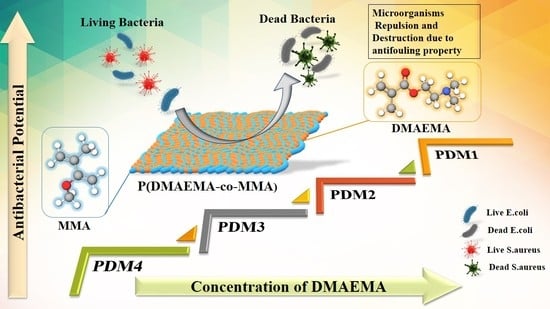
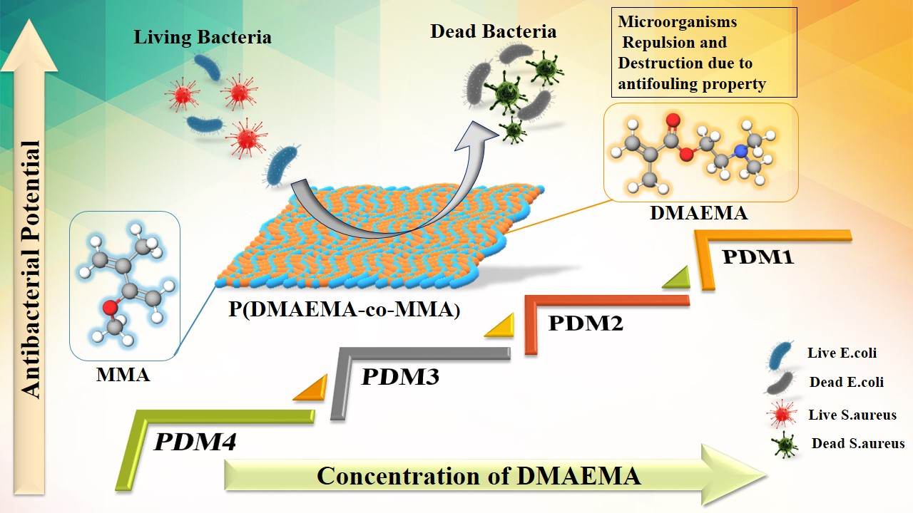
Antibacterial Amphiphilic Copolymers of Dimethylamino Ethyl Methacrylate and Methyl Methacrylate to Control Biofilm Adhesion for Antifouling Applications

1
Department of Chemistry, School of Natural Sciences, National University of Sciences and Technology, H-12, Islamabad 44000, Pakistan
2
Polymer Research Lab, School of Chemical and Material Engineering (SCME), National University of Sciences and Technology, H-12, Islamabad 44000, Pakistan
*
Author to whom correspondence should be addressed.
Submission received: 2 October 2020 / Revised: 17 December 2020 / Accepted: 20 December 2020 / Published: 9 January 2021
(This article belongs to the Collection Antibacterial Activity of Polymeric Materials)

Abstract

:
Amphiphilic copolymers are recognized as important biomaterials and used as antibacterial agents due to their effective inhibition of bacterial growth. In current study, the amphiphilic copolymers of P(DMAEMA-co-MMA) were synthesized using free radical polymerization by varying the concentrations of hydrophilic monomer 2-dimethylamino ethylmethacrylate (DMAEMA) and hydrophobic monomer methyl methacrylate (MMA) having PDI value of 1.65–1.93. The DMAEMA monomer, through ternary amine with antibacterial property optimized copolymers, P(DMAEMA-co-MMA), compositions to control biofilm adhesion. Antibacterial activity of synthesized copolymers was elucidated against Gram-positive Staphylococcus aureus (ATCC 6538) and Gram-negative Escherchia coli (ATCC 8739) by disk diffusion method, and zones of inhibition were measured. The desired composition that was PDM1 copolymer had shown good zones of inhibition i.e., 19 ± 0.33 mm and 20 ± 0.33 mm for E. coli and S. aureus respectively. The PDM1 and PDM2 have exhibited significant control over bacterial biofilm adhesion as tested by six well plate method. SEM study of bacterial biofilm formation has illustrated that these copolymers act in a similar fashion like cationic biocide. These compositions viz. PDM1 and PDM2, may be useful in development of bioreactors, sensors, surgical equipment and drug delivery devices.

Graphical Abstract

1. Introduction

Biofouling is the growth of microbes on exterior of material that initiated by protein adsorption or microorganisms on the surface of substrate that is a ubiquitous challenge for a number of bio medical applications [1,2]. Biofouling also occurs on different prosthetic devices, surgical equipment, protective apparel, sensors, drug delivery devices, contact lenses, medical implants, and bioreactors that causes adverse effects on human health [3,4]. The unregulated attachment of microbes on implant materials surface is an undesirable phenomenon that causes infection and degradation of the function of medical instruments [5,6]. In medical devices antifouling materials are originated by intermolecular interaction between extracellular biomolecules and the designed surfaces [7,8,9]. Most commonly used antimicrobial reagents for bio medical applications can be classified into four specific categories that include organic agents such as formaldehyde and isothiazolones, oxidants that include chlorine and peroxides, electrophilic agents such as mercury, copper and silver, cationic active compounds such as quaternary ammonium and chlorhexidine [10,11]. Use of antimicrobial polymers for biomedical applications has become more significant due to several advantages such as non-volatility, chemically stability, and skin impermeability [3,12]. Different antifouling functional groups such as quaternary ammonium group, flouro group, 2-acrylamido-2-methylpropanesulfonic acid (AMPS) and sulfobetaine offer a way to amend the polymer for certain end applications afterwards [13]. The quaternization of polymeric materials played crucial role in the formulation of new antifouling polymers [14,15]. Polymeric ammonium quaternary compounds have been widely used for antibacterial applications due to the properties such as low volatility, high chemical stability, low toxicity and low skin irritation potential [11,12]. Along with several advantages of quaternary ammonium group to be used as antibacterial agent, there arise some complications during synthesis due to steric and electrostatic effects to obtain a complete quaternization [16,17]. These materials are promising to maintain antibacterial activity and to reduce the risk of toxicity [18]. In one of the study, copolymerization of HEMA with a methacrylic monomer bearing a thiazole side group susceptible to quaternization was carried out, copolymers exhibited significant activity versus Gram-positive (S. aureus) and Gram-negative (P. aeruginosa and E. coli) bacteria [19]. Antimicrobial activity of copolymers increased with increasing of the cationic unit content [19]. Amphiphilic polymers had significant advantage over cationic polymers due to their improved biocidal activity as can be effective against Gram-positive and Gram negative bacteria [14,20]. In another study, synthesis of maleic anhydride and 4-methyl-1-pentene was used to produce amphiphilic copolymers with high antibiotic properties imitating natural antimicrobial peptides [15]. The copolymer obtained was modified by grafting with 3-(DMAPA), which was further improvised to create polycationic copolymers by quaternizing methyl iodide then dodecyl iodide. Antimicrobial properties of the synthesized copolymers have been tested against E. coli and S. aureus, showed nominal antibacterial activity against Gram-positive bacteria [15]. Lowe et al. copolymerized DMAEMA monomer with different hydrophobic monomers included; octyl, cyclohexyl, butyl and ethyl methacrylate that improved antibacterial activity of materials [21]. DMAEMA monomer was copolymerized with hydrophobic monomer enhanced antibacterial activity that depend upon suitable spacer length of hydrophobic segment [22]. In one of previous work, amphiphilic copolymers showed worthy antibacterial activity against Gram-positive bacteria and poor activity against Gram-negative bacteria. Xu et al. investigated cationic polymers as the main classes of materials against bacteria through the membrane-lysis mechanism. They studied antibacterial effects of linear and cyclic monomers of cyclic poly(2-(dimethylamino)ethyl methacrylate) (PDMAEMA) based copolymers with various components by the intra-chain click cyclization of α-alkyne-ω-azido via atom transfer radical polymerization [23]. In another study of amphiphilic polymers of thiazole ring 2-(2-(4-methylthiazol-5-yl)ethoxy)carbonyl)oxy)ethyl methacrylate monomer (MTZ) and non-hemotoxic poly(ethylene glycol) side chains (poly(ethylene glycol) methyl ether methacrylate (PEGMA) observed that longer hydrophobic chain, octyl were much more hemotoxic than their corresponding butylated copolymers [24].
In this study we report a facile approach to enhance the antimicrobial activity of amphiphilic copolymers by varying concentration of hydrophilic and hydrophobic monomers. These amphiphilic copolymers were synthesized via free radical polymerization with presence of initiator. Desired antimicrobial properties were achieved by t-amine functional group of DMAEMA that endowed low biofilm adhesion. Antibacterial assay was performed against standard Gram-positive S. aureus (ATCC 6538), and Gram-negative E. coli (ATCC 8739) through disk diffusion method in terms of inhibitory zone diameters (mm). These copolymers showed effective results for Gram-positive and Gram-negative bacteria, especially PDM1 besides PDM2. Amphiphilic copolymers PDM1 and PDM2 had shown low adhesion of microorganisms and mode of action against Gram-positive and Gram-negative bacteria were studied by SEM. PDM1 and PDM2, both compositions are suitable for the design of material with low adhesion of microorganisms needed, especially for antifouling surfaces. Schematic presentation of the synthesized P(DMAEMA-co-MMA) copolymers and the antifouling mechanism is shown in Figure 1.

2. Materials and Methodology

2.1. Materials

All chemicals were of analytical grade and used in chemical synthesis without further purification. Dimethylamino ethyl methacrylate (DMAEMA, 98%) and Trpytosoy broth (TSB) (Sigma-Aldrich, Humburg, Germany), Methyl methacrylate (MMA, 99%), 2,2-Azobisisobutyronitrile (AIBN, 98%), N,N-Dimethyl formamide (DMF, 99%) (Sigma Aldrich, St. Louis, MO, USA), Ethanol, Paraformaldehyde (PFA) and NaOH were acquired from Sigma Aldrich, Germany while water acquired from 18Ωmill pore RO Plant, was used in reaction. Phospate Buffer Solution (PBS) (VWR, Amersco, Leuven, Belgium), Mueller-Hinton agar (MHA) (Daejung, Shiheung, Korea) and BG11 medium (Scharlau, Barcelona, Spain) were used for algae growth. For the bioassays, E. coli (ATTC 8739) and S. aureus (ATCC 6538) were employed as representative strains.

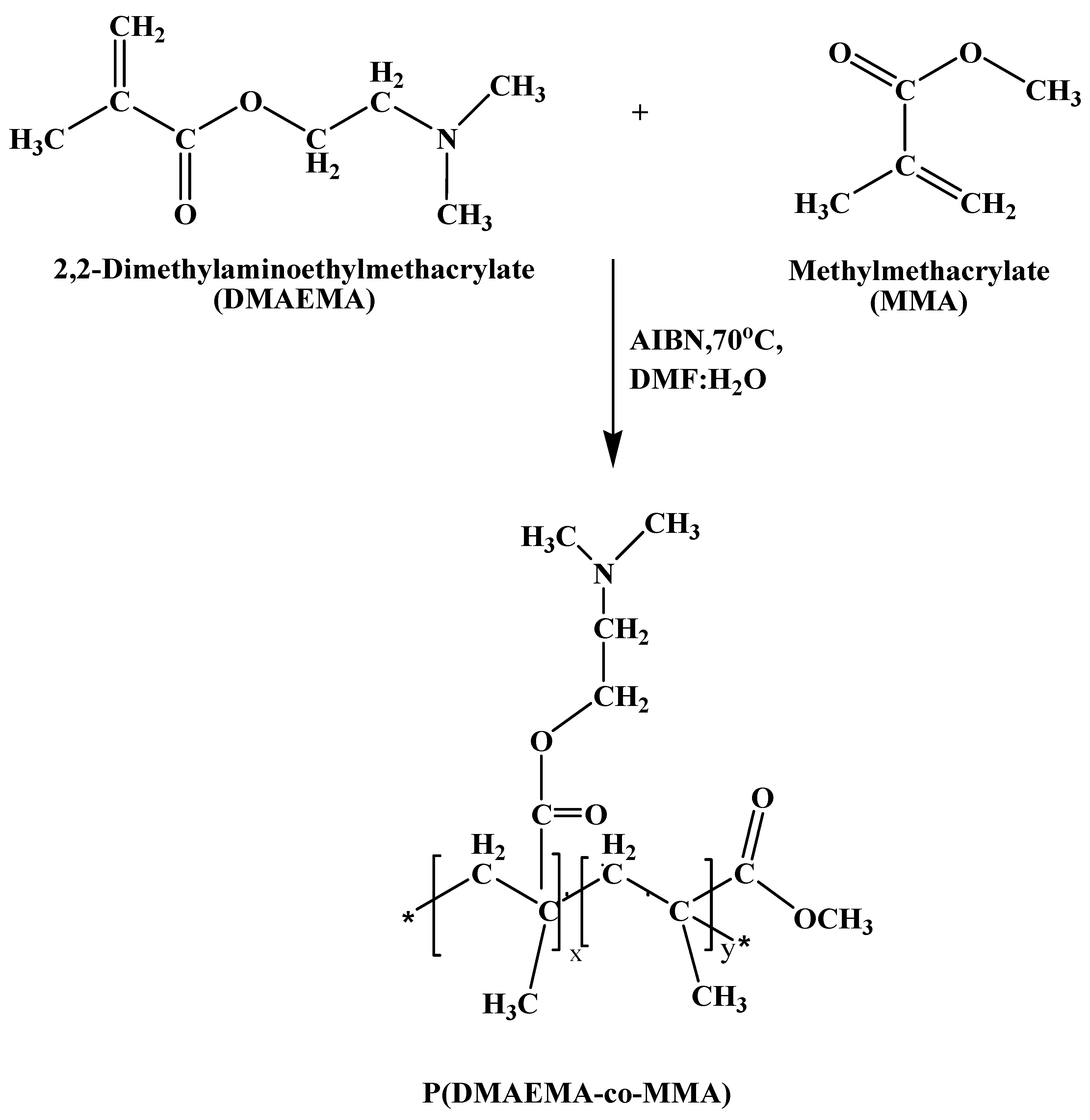
2.2. Synthesis of Copolymer P(DMAEMA-co-MMA)

Copolymerization between both components like DMAEMA and MMA was performed by free radical polymerization in the various proportions as shown in Table 1 [25]. For synthesis of PDM1 copolymer, DMAEMA (10 g, 63.6 mmol) and MMA (10 g, 99.8 mmol) were dissolved into 200 mL DMF under inert atmosphere with continuous stirring in polymerization reactor (IKA Eurostar200-P4). AIBN (0.2 g, 1.22 mmol) was introduced into the reaction flask, where reaction was permitted to proceed for 5 h with continuous stirring under nitrogen purging through schlenk line at 70 °C temperature. Synthesized copolymers P(DMAEMA-co-MMA) were recovered by freeze-drying and yield of synthesized copolymers was 62–65%. Same procedure was repeated for synthesis of PDM2, PDM3 andPDM4, while employing quantities of both monomers mentioned in Table 1.

2.3. Characterization

Infrared spectroscopy was performed by Bruker ALPHATIR spectrometer (Germany) at the rate of 20 scans per minute to investigate functional groups of copolymer. The 1H NMR spectra was verified by a Bruker Advance 400 spectrometer and operated by 400MHz, with CDCl3 (3 mg/mL) solvent. GPC was conducted to determine molecular weight using DMF as the eluent for observing the monomer conversion. DMF-GPC was recorded at a water 1515 system equipped with three HR waters columns (HR4, HR3 and HR1). This system consisted an Isocratic pump and a RI detector. Further calculations with (DMF were performed containing LiBr (0.01 M) as an eluting agent at a run rate of 1.0 mL/min. Different copolymers of different molecular weights were standardized with polystyrene. SEM analysis (JEOLJSM-6490LA) was performed to check normal and distorted bacterial growth on the polymeric materials at 1 µm. Earlier to SEM examination, the specimens were made moisture free and analysis was performed at 10 kV accelerating voltage.

2.4. Antibacterial Bioassay

Antibacterial activity against both bacteria E. coli and S. aureus was performed by disk diffusion method [25,26]. Bacterial cultures had been activated before performing antibacterial activity, and bacteria were streaked at freshly prepared Muller Hinton Agar (MHA) [26]. These agar plates were put into oven at 37 °C for 24 h. Colony from the new development was mixed into saline and optical density (O.D) set 0.5 by using McFarland standard after centrifuging. MHA agar was put into petri dish in sterile conditions by using a Bunsen burner in streamline flow hood. 100 µL from 0.5 O.D culture media was poured into the centre of new MHA plate and streaked by using cotton swab [18,27]. Culture was absorbed on the medium and samples of 8 mm size were placed on a plate, and prepared petri dishes were placed for 24 h at 37 °C. Zones of inhibition were measured around each polymeric sample and these experiments repeated three times [12,13]. These results were presented as a mean ± standard deviation and T-test used for determination of statistical importance (* p < 0.05, ** p < 0.01, *** p < 0.001, **** p < 0.0001) [28].

2.5. Biofilm Formation Test

Biofilm development test was done for E. coli and S. aureus in six well plates. All antifouling polymers cut into square shape (0.5 × 0.5 cm2). In six well plates, two controlled wells contained 2 mL of tryptosoy broth (TSB), two growth wells contained 1 mL of TSB with PMMA polymer and two treated wells contained 1 mL of TSB with antifouling copolymers [20,29]. Six-well plates of four copolymers were completely wrapped by food packing sheet and incubated at 37 °C for 24 h. Afterward plates were removed from incubator and bring into laminar flow hood. Samples were washed carefully with phosphate buffer solution (PBS) (1/100 mL) to remove unattached cells and subjected them to SEM fixation. 100–200 µL of 4% para formaldehyde (PFA) was placed on washed samples and allowed to dry for 30–40 min [30,31]. Samples were washed with 25%, 50%, 70% and 100% concentrations of ethanol. After drying, samples were preserved at −4 °C until subjected to SEM analysis [32].

3. Results and Discussion

Copolymers of both monomers MMA and DMAEMA are shown in Figure 2. P(DMAEMA-co-MMA) was made by free radical polymerization, using AIBN initiator and DMF solvent at 70 °C under inert medium [33,34,35]. Chemical reaction of both monomers MMA and DMAEMA is shown in Figure 3. Different copolymers were synthesized by varying concentration of both monomers and characterize by FTIR, 1HNMR and GPC.

3.1. FTIR Analysis

PMMA and PDMAEMA homopolymers are characterized by FTIR, as shown in Figure 4a. Here spectrum for PMMA showed absorption bands at 2997 cm−1 due to the stretching vibrations of –CH2– and –CH3 groups [32]. In PMMA spectrum absorption band at 1730 cm−1 exhibited stretching vibration of C = O group of ester [32]. In PDMAEMA spectrum band at 1730 cm−1 showed C = O group and band at 2842 cm−1 attributed to C-H stretching vibration of N(CH3)2 moieties [33,34]. In both acrylate homopolymers band at 1150–1250 cm−1 showed stretching vibration of C-O-C [35]. Copolymerization of MMA and DMAEMA confirmed and showed in Figure 4b. These copolymers from PDM1 to PDM4 contained the characteristic bands of DMAEMA and MMA. In each spectrum, band at 1020 cm−1 for C–N stretching vibration of tertiary amine confirms presence of DMAEMA segment into copolymers. Bands at 1730 cm−1 for C = O of ester corresponded to MMA moiety in the copolymers [35]. In copolymers PDM1 to PDM4 concentration of DMAEMA decreases and band intensity of C-N group also reduced. On the other hand, concentration of MMA increase from PDM1 to PDM4 hence signal intensity at 1730 cm−1 was enhanced [36]. In both Figure 4a,b bands appeared at 1450 cm−1 due to bending vibration of –CH2 group. DMAEMA is hydrophilic in nature and it absorb moisture and band due to O-H group at 3000–3500 cm−1 in Figure 2a.
All copolymers showed characteristic band for C-O-C at 1150–1250 cm−1 and a broad band at 3300–3400 cm−1 due to -OH group was clearly observed in PDM1 to PDM3 due to the moisture absorption by DMAEMA. In PDM4, –OH band disappeared because of very low concentration of DMAEMA and higher content of MMA.

3.2. 1HNMR Analysis

Chemical structure of amphiphilic copolymers PDM1 have been shown in Figure 5. The 1HNMR spectrum of –OCH2 of PDMAEMA exhibited a peak at 4.1 (c) ppm. The 1HNMR (h) peak at 3.6 ppm for –OCH3 that corresponds for PMMA [37]. The 1HNMR showed that molar ratio of PDMAEMA and PMMA was 1:1, which was determined using the integration of (c) and (h) peaks, which was equivalent to 2 and 3 protons respectively. In PDMAEMA segment, dimethylamino groups showed two sharp peaks at 2.26 (e) ppm for two protons of methyl group and 2.54 (d) ppm for six protons of amino group. Magnetic resonances at 1.0 ppm and 1.33 ppm are associated with the methyl protons of main chains while two signals around 1.8 ppm are assigned to the methyl group [38]. These 1HNMR results have confirmed that copolymerization was successfully executed.

3.3. GPC Results

The molecular weight of synthesized copolymers with changing concentration of both monomers confirm the controlled feature of polymers number average molecular weights (Mn, GPC) of polymers, as shown in Figure 6 [22]. Random copolymer synthesis and monomer conversion was resulted in high molecular weight polymers [39]. Polydispersity index (PDI), Mw/Mn values characterize samples ranged from 1.65 to 1.93 for free radical polymerization [40]. The symmetrical nature of the GPC curves of four copolymers the inexistence of an irreversible termination of both monomers DMAEMA and MMA [36]. Traces of symmetric GPC distribution showed the uniformity of the copolymers via free radical polymerization. [30]. These unimodal curves showed that the polymerization was completed successfully and that there was no unreacted monomers in the reaction product.

3.4. Antibacterial Bioassay

Amphiphilic copolymer of P(DMAEMA-co-MMA) are partially soluble in water, PDMAEMA is water soluble and PMMA is water insoluble due to hydrophilic and hydrophobic nature [41]. Antibacterial action was tested by disk diffusion method (DD) and zone of inhibitions were measured against both type of bacteria [42]. Bacterial bond of E. coli and S. aureus on surface of polymers did not significantly depend upon the molecular weight of polymers, but it dependent on the elementary process of bactericidal action of polymers [12]. PMMA has hydrophobic nature and not antibacterial action, while its copolymers with DMAEMA presented antimicrobial activity against S. aureus and E. coli [12]. These amphiphilic copolymers PDM1 to PDM4 have shown different zones of inhibitions alongside Gram-positive and Gram-negative bacteria in Figure 7a,b. These amphiphilic copolymers played profound effect on the antibacterial activity [43]. PDM1 has more concentration of DMAEMA monomer; hence demonstrated higher activity against bacteria because it had higher charge density due to amino groups [44]. Furthermore, concluding the biocidal efficacy of those copolymers for Gram-positive bacteria is greater than the Gram-negative, which is also consistent with the outcome stated by Ignatova et al. [45]. Since, the Gram-positive bacteria have cell wall that made up of only peptidoglycan the diffusion for the cationic polyelectrolytes with hydrophobic group are easier [43,46]. For Gram-negative bacteria (E. coli) it is however more complicated to diffuse over the cell wall while cells are surrounded by another outside membrane [43,46,47].

3.5. Biofilm Adhesion Studies by SEM

Antibacterial activities of all copolymers with different concentrations of DMAEMA were assessed against Gram-negative bacteria and Gram-positive bacteria which commonly cause biofilm on materials [11]. Biofouling resistance of synthesized copolymer materials against S. aureus and E. coli biofilm formation was studied by six well plate method for incubation time of 24 h. Bacterial cell had negative charge on the surface so easily attached on the cationic surface while its antibacterial activity was enhanced by molecular weight [43,48]. Antibacterial activity also influenced by the spacer length due to conformation charge density on the polymers [13]. As well as DMAEMA monomer had positive charge because ammonia group copolymerized and MMA had no charge with hydrophobic in nature [11]. Adhesion of Gram-positive bacteria S. aureus on the surface of copolymers shown in Figure 8 [6]. The increase in antibacterial activity increase with charge density by polymerization and assumed more adhesion due to negative charged bacterial cell surface, increase dispersal through the cell wall. These cationic polymers fix to the cytoplasmic membrane, interruption the cytoplasmic membrane, discharge of intracellular elements and bacterial cells mortality [6,49,50].
These amphiphilic polymers destabilize the surface of E. coli by interchange with cations of materials that cause rapture of cells as shown in Figure 9 [6]. Copolymer antibacterial action mechanism occurred by (a) bacterial cell surface adsorption, (b) cell wall diffusion, (c) cytoplasmic membrane adsorption, (d) cytoplasmic disruption, (e) leakage of cellular components, and (f) cell death [6,16]. Here more positive charge present on polymers PDM1 and PDM2 that caused interactions between polymers and bacteria. So it is a critical factor, and further action disrupted the cell wall, fluid leaked, cell raptured and death occurred [47,51]. The tertiary amine present in DMAEMA caused disruption of the bacterial cell wall.

4. Conclusions

Various copolymers (PDM1, PDM2, PDM3 and PDM4) were successfully prepared by free radical polymerization in the presence of AIBN initiator by employing different ratios of DMAEMA and MMA monomers. These copolymers were characterized by FTIR, 1HNMR, and GPC, which exhibited high antimicrobial activities against E. coli and S. aureus. Bare PMMA showed no antibacterial activity. Copolymer of P(DMAEMA-co-MMA), PDM1 showed maximum biocidal activity with an inhibition zone of 19 mm and 20 mm against E. coli and S. aureus respectively. In these copolymers, PDM1 and PDM2 had a higher concentration of DMAEMA; therefore, it showed greater antibacterial activities as compared to PDM3 and PDM4. This greater activity is attributed to the presence of amine groups along the chain length of the DMAEMA segment. Adhesion of microorganisms on the surface and biofilm formation decreased with an increase in the molar ratio of DMAEMA due to the presence of positive charges responsible for biocidal action. In SEM analysis of biofilm, the control and rupture of E. coli cell membrane was observed in PDM1 and cell disruption of S. aureus was observed in PDM2. Thus, PDM1 and PDM2 copolymers are potential candidates for antifouling applications with controlled biofilm formation.

Author Contributions

Conceptualization, experimental work and original draft preparation, S.M.; this research coordinated as a principal investigator, N.M.A.; edit writing and review and editing, N.M.A., A.M. and M.I. All authors have read and agreed to the published version of the manuscript.

Funding

Authors are grateful to NUST Research Directorate for research support. Shehla Mushtaq and Nasir M. Ahmad acknowledge HEC, NRPU through Project No. 3526 for financial support and funding.

Conflicts of Interest

No conflict of interest is declared by the authors.

References

  1. Xue, Y.; Xiao, H.; Zhang, Y. Antimicrobial polymeric materials with quaternary ammonium and phosphonium salts. Int. J. Mol. Sci. 2015, 16, 3626–3655. [Google Scholar] [CrossRef] [PubMed] [Green Version]
  2. Tejero, R.; Gutiérrez, B.; López, D.; López-Fabal, F.; Gómez-Garcés, J.L.; Muñoz-Bonilla, A.; Fernández-García, M. Tailoring macromolecular structure of cationic polymers towards efficient contact active antimicrobial surfaces. Polymers (Basel) 2018, 10, 241. [Google Scholar] [CrossRef] [PubMed] [Green Version]
  3. Huang, K.; Yang, C.; Huang, S.; Chen, C.; Lu, Y. Recent Advances in Antimicrobial Polymers. Int. J. Mol. Sci. 2016, 17, 1578. [Google Scholar] [CrossRef] [PubMed] [Green Version]
  4. Damodaran, V.B.; Murthy, N.S. Bio-inspired strategies for designing antifouling biomaterials. Biomater. Res. 2016, 1–11. [Google Scholar] [CrossRef] [PubMed] [Green Version]
  5. Callow, M.E.; Fletcher, R.L. The influence of low surface energy materials on bioadhesion—A review. Int. Biodeterior. Biodegrad. 1994, 34, 333–348. [Google Scholar] [CrossRef]
  6. Timofeeva, L.; Kleshcheva, N. Antimicrobial polymers: Mechanism of action, factors of activity, and applications. Appl. Microbiol. Biotechnol. 2011, 89, 475–492. [Google Scholar] [CrossRef]
  7. Liu, L.; Li, W.; Liu, Q. Recent development of antifouling polymers: Structure, evaluation, and biomedical applications in nano /micro-structures. Wiley Interdiscip. Rev. Nanomed. Nanobiotechnol. 2014, 6. [Google Scholar] [CrossRef]
  8. Abbasi, F.; Mirzadeh, H.; Katbab, A. Modification of polysiloxane polymers for biomedical applications: A review. Polym. Int. 2001, 1287. [Google Scholar] [CrossRef]
  9. Sun, W.; Liu, W.; Wu, Z.; Chen, H. Chemical Surface Modification of Polymeric Biomaterials for Biomedical Applications. Macromol. Rapid Commun. 2020, 41, 1900430. [Google Scholar] [CrossRef]
  10. Hons, J.C. The Development of Novel Antifouling Materials: A Multi-Disciplinary Approach. Ph.D. Thesis, Dublin City University, Dublin, Ireland, 2011. [Google Scholar]
  11. Lu, G.; Wu, D.; Fu, R. Studies on the synthesis and antibacterial activities of polymeric quaternary ammonium salts from dimethylaminoethyl methacrylate. React. Funct. Polym. 2007, 67, 355–366. [Google Scholar] [CrossRef]
  12. Elena, P.; Miri, K. Formation of contact active antimicrobial surfaces by covalent grafting of quaternary ammonium compounds. Colloids Surf. B Biointerfaces 2018, 169, 195–205. [Google Scholar] [CrossRef] [PubMed]
  13. Kenawy, E.R.; Worley, S.D.; Broughton, R. The Chemistry and Applications of Antimicrobial Polymers: A State-of-the-Art Review. Biomacromolecules 2007, 8. [Google Scholar] [CrossRef]
  14. Carmona-Ribeiro, A.M.; de Melo Carrasco, L.D. Cationic antimicrobial polymers and their assemblies. Int. J. Mol. Sci. 2013, 14, 9906–9946. [Google Scholar] [CrossRef] [PubMed] [Green Version]
  15. Szkudlarek, M.; Heine, E.; Keul, H.; Beginn, U.; Möller, M. Synthesis, characterization, and antimicrobial properties of peptides mimicking copolymers of maleic anhydride and 4-methyl-1-pentene. Int. J. Mol. Sci. 2018, 19, 2617. [Google Scholar] [CrossRef] [PubMed] [Green Version]
  16. Ren, W.; Cheng, W.; Wang, G.; Liu, Y. Developments in antimicrobial polymers. J. Polym. Sci. Part A Polym. Chem. 2017, 55, 632–639. [Google Scholar] [CrossRef]
  17. Wu, C.; Zhou, Y.; Wang, H.; Hu, J.; Wang, X. Formation of antifouling functional coating from deposition of a zwitterionic-co-nonionic polymer via “grafting to” approach. J. Saudi Chem. Soc. 2019. [Google Scholar] [CrossRef]
  18. Alamri, A.; El-newehy, M.H.; Al-deyab, S.S. Biocidal polymers: Synthesis and antimicrobial properties of benzaldehyde derivatives immobilized onto amine-terminated polyacrylonitrile. Chem. Cent. J. 2012, 6, 1. [Google Scholar] [CrossRef] [Green Version]
  19. Muñoz-Bonilla, A.; López, D.; Fernández-García, M. Providing antibacterial activity to poly(2-hydroxy ethyl methacrylate) by copolymerization with a methacrylic thiazolium derivative. Int. J. Mol. Sci. 2018, 19, 4120. [Google Scholar] [CrossRef] [Green Version]
  20. Taylor, P.; Zhou, Z.; Calabrese, D.R.; Taylor, W.; Finlay, J.A.; Callow, M.E.; Callow, J.A.; Fischer, D.; Kramer, E.J.; Ober, C.K. Amphiphilic triblock copolymers with PEGylated hydrocarbon structures as environmentally friendly marine antifouling and fouling-release coatings. Biofouling J. Bioadhesion Biofilm 2014, 37–41. [Google Scholar] [CrossRef]
  21. Ward, M.; Sanchez, M.; Elasri, M.O.; Lowe, A.B. Antimicrobial activity of statistical polymethacrylic sulfopropylbetaines against gram-positive and gramnegative bacteria. J. Appl. Polym. Sci. 2006, 101, 1036–1041. [Google Scholar] [CrossRef]
  22. Wang, F.P.; Yuan, T.; Li, W.X.; Zhang, J.Y.; Wang, Q.Z. Synthesis and characterization of amphiphilic copolymer poly [2-(dimethylamino)ethyl methacrylate-co-methyl methacrylate]. Adv. Mater. Res. 2014, 936, 776–779. [Google Scholar] [CrossRef]
  23. Xu, J.; Pu, L.; Ma, J.; Kumar, S.K.; Duan, H. Antibacterial properties of synthesized cyclic and linear cationic copolymers. Polym. Chem. 2020, 11, 6632–6639. [Google Scholar] [CrossRef]
  24. Cuervo-Rodríguez, R.; Muñoz-Bonilla, A.; López-Fabal, F.; Fernández-García, M. Hemolytic and antimicrobial activities of a series of cationic amphiphilic copolymers comprised of same centered comonomers with thiazole moieties and polyethylene glycol derivatives. Polymers (Basel) 2020, 12, 972. [Google Scholar] [CrossRef] [PubMed]
  25. Martí, M.; Frígols, B.; Serrano-Aroca, A. Antimicrobial Characterization of Advanced Materials for Bioengineering Applications. JoVE 2018, 1–10. [Google Scholar] [CrossRef] [PubMed] [Green Version]
  26. Newport, S.; Differ, B.; Hoelzer, K.; Cummings, K.J.; Warnick, L.D.; Schukken, Y.H.; Siler, J.D.; Davis, M.A.; Besser, T.E.; Wiedmann, M. Agar Disk Diffusion and Automated Microbroth Dilution Produce Similar Antimicrobial Susceptibility Testing Results. Foodborne Pathog. Dis. 2011, 8. [Google Scholar] [CrossRef] [Green Version]
  27. Li, B.; Logan, B.E. Bacterial adhesion to glass and metal-oxide surfaces. Colloids Surf. B Biointerfaces 2004, 36, 81–90. [Google Scholar] [CrossRef]
  28. Zhou, L.; Zhang, P.; Chen, Z.; Cai, S.; Jing, T.; Fan, H.; Mo, F.; Zhang, J.; Lin, R. Preparation, characterization, and evaluation of amphotericin B-loaded MPEG-PCL-g-PEI micelles for local treatment of oral candida albicans. Int. J. Nanomed. 2017, 12, 4269–4283. [Google Scholar] [CrossRef] [Green Version]
  29. Mai, T.; Rakhmatullina, E.; Bleek, K.; Boye, S.; Yuan, J.; Völkel, A.; Gräwert, M.; Cheaib, Z.; Eick, S.; Günter, C.; et al. Poly(ethylene oxide)- b -poly(3-sulfopropyl methacrylate) block copolymers for calcium phosphate mineralization and biofilm inhibition. Biomacromolecules 2014, 15, 3901–3914. [Google Scholar] [CrossRef]
  30. Chen, Q.; Li, S.; Feng, Z.; Wang, M.; Cai, C.; Wang, J.; Zhang, L. Poly(2-(diethylamino)ethyl methacrylate)-based, ph-responsive, copolymeric mixed micelles for targeting anticancer drug control release. Int. J. Nanomed. 2017, 12, 6857–6870. [Google Scholar] [CrossRef] [Green Version]
  31. Khan, F.; Hashmi, M.U.; Khalid, N.; Hayat, M.Q.; Ikram, A.; Janjua, H.A. Controlled assembly of silver nano-fluid in Heliotropium crispum extract: A potent anti-biofilm and bactericidal formulation. Appl. Surf. Sci. 2016, 387, 317–331. [Google Scholar] [CrossRef]
  32. Degirmenci, M. Synthesis and characterization of novel well-defined end-functional macrophotoinitiator of poly(MMA) by ATRP. J. Macromol. Sci. Pure Appl. Chem. 2005, 42 A, 21–30. [Google Scholar] [CrossRef]
  33. Zielińska, D.; Stawski, D.; Komisarczyk, A. Producing a poly(N,N-dimethylaminoethyl methacrylate) nonwoven by using the blowing out method. Text. Res. J. 2016, 86, 1837–1846. [Google Scholar] [CrossRef]
  34. Okten, N.S.; Canakci, C.C.; Orakdogen, N. Hertzian elasticity and triggered swelling kinetics of poly(amino ester)-based gel beads with controlled hydrophilicity and functionality: A mild and convenient synthesis via dropwise freezing into cryogenic liquid. Eur. Polym. J. 2019, 114, 176–188. [Google Scholar] [CrossRef]
  35. Shen Preparation and Characterization of PMMA and its Derivative via RAFT Technique in the Presence of Disulfide as a Source of Chain Transfer Agent. J. Membr. Sep. Technol. 2012, 117–128. [CrossRef]
  36. Huang, Y.; Yong, P.; Chen, Y.; Gao, Y.; Xu, W.; Lv, Y.; Yang, L.; Reis, R.L.; Pirraco, R.P.; Chen, J. Micellization and gelatinization in aqueous media of pH- and thermo-responsive amphiphilic ABC (PMMA82-b-PDMAEMA150- b -PNIPAM65) triblock copolymer synthesized by consecutive RAFT polymerization. RSC Adv. 2017, 7, 28711–28722. [Google Scholar] [CrossRef] [Green Version]
  37. Park, J.A.; Cho, K.Y.; Han, C.H.; Nam, A.; Kim, J.H.; Lee, S.H.; Choi, J.W. Quaternized Amphiphilic Block Copolymers/Graphene Oxide and a Poly(vinyl alcohol) Coating Layer on Graphene Oxide/Poly(vinylidene fluoride) Electrospun Nanofibers for Superhydrophilic and Antibacterial Properties. Sci. Rep. 2019, 9, 1–13. [Google Scholar] [CrossRef] [PubMed] [Green Version]
  38. Roy, S.G.; Bauri, K.; Pal, S.; Goswami, A.; Madras, G.; De, P. Synthesis, characterization and thermal degradation of dual temperature- and pH-sensitive RAFT-made copolymers of N,N-(dimethylamino)ethyl methacrylate and methyl methacrylate. Polym. Int. 2013, 62, 463–473. [Google Scholar] [CrossRef]
  39. Georges, M.K.; Veregin, R.P.N.; Kazmaier, P.M.; Hamer, G.K. Narrow Molecular Weight Resins by a Free-Radical Polymerization Process. Macromolecules 1993, 26, 2987–2988. [Google Scholar] [CrossRef]
  40. Iwasaki, T.; Yoshida, J.I. Free radical polymerization in microreactors. Significant improvement in molecular weight distribution control. Macromolecules 2005, 38, 1159–1163. [Google Scholar] [CrossRef]
  41. Baines, F.L.; Armes, S.P.; Billingham, N.C.; Tuzar, Z. Micellization of poly(2-(dimethylamino)ethyl methacrylate-block-methyl methacrylate) copolymers in aqueous solution. Macromolecules 1996, 29, 8151–8159. [Google Scholar] [CrossRef]
  42. Gitchaiwat, A.; Kositchaiyong, A.; Sombatsompop, K.; Prapagdee, B.; Isarangkura, K.; Sombatsompop, N. Assessment and characterization of antifungal and antialgal performances for biocide-enhanced linear low-density polyethylene. J. Appl. Polym. Sci. 2013, 128, 371–379. [Google Scholar] [CrossRef]
  43. Benkhaled, B.T.; Hadiouch, S.; Olleik, H.; Perrier, J.; Ysacco, C.; Guillaneuf, Y.; Gigmes, D.; Maresca, M.; Lefay, C. Elaboration of antimicrobial polymeric materials by dispersion of well-defined amphiphilic methacrylic SG1-based copolymers. Polym. Chem. 2018, 9, 3127–3141. [Google Scholar] [CrossRef]
  44. Raafat, D.; Sahl, H.G. Chitosan and its antimicrobial potential—A critical literature survey. Microb. Biotechnol. 2009, 2, 186–201. [Google Scholar] [CrossRef] [PubMed] [Green Version]
  45. Dutta, P.; Dey, J.; Shome, A.; Das, P.K. Nanostructure formation in aqueous solution of amphiphilic copolymers of 2-(N,N-dimethylaminoethyl)methacrylate and alkylacrylate: Characterization, antimicrobial activity, DNA binding, and cytotoxicity studies. Int. J. Pharm. 2011, 414, 298–311. [Google Scholar] [CrossRef] [PubMed]
  46. Rawlinson, L.A.B.; Ryan, S.M.; Mantovani, G.; Syrett, J.A.; Haddleton, D.M.; Brayden, D.J. Antibacterial effects of poly(2-(dimethylamino ethyl)methacrylate) against selected gram-positive and gram-negative bacteria. Biomacromolecules 2010, 11, 443–453. [Google Scholar] [CrossRef] [PubMed]
  47. Liow, S.S.; Chee, P.L.; Owh, C.; Zhang, K.; Zhou, Y.; Gao, F.; Lakshminarayanan, R.; Loh, X.J. Cationic Poly([R]-3-hydroxybutyrate) Copolymers as Antimicrobial Agents. Macromol. Biosci. 2019, 19, 1–12. [Google Scholar] [CrossRef]
  48. Oh, Y.J.; Khan, E.S.; Del Campo, A.; Hinterdorfer, P.; Li, B. Nanoscale Characteristics and Antimicrobial Properties of (SI-ATRP)-Seeded Polymer Brush Surfaces. ACS Appl. Mater. Interfaces 2019, 11, 29312–29319. [Google Scholar] [CrossRef]
  49. Palermo, E.F.; Sovadinova, I.; Kuroda, K. Structural determinants of antimicrobial activity and biocompatibility in membrane-disrupting methacrylamide random copolymers. Biomacromolecules 2009, 10, 3098–3107. [Google Scholar] [CrossRef]
  50. Takahashi, H.; Palermo, E.F.; Yasuhara, K.; Caputo, G.A.; Kuroda, K. Molecular design, structures, and activity of antimicrobial peptide-mimetic polymers. Macromol. Biosci. 2013, 13, 1285–1299. [Google Scholar] [CrossRef] [Green Version]
  51. Yandi, W.; Mieszkin, S.; Callow, M.E.; Callow, J.A.; Finlay, J.A.; Liedberg, B.; Ederth, T. Antialgal activity of poly(2-(dimethylamino)ethyl methacrylate) (PDMAEMA) brushes against the marine alga Ulva. Biofouling 2017, 33, 169–183. [Google Scholar] [CrossRef] [Green Version]
Figure 1. Schematic illustration of copolymers P(DMAEMA and MMA),PDM1, PDM2, PDM3 and PDM4 with varying concentration of DMAEMA that control bacterial adhesion.
Polymers 13 00216 g001
Figure 2. Copolymers of P(DMAEMA-co-MMA) (a) PDM1; (b) PDM2; (c) PDM3 and (d) PDM4.
Polymers 13 00216 g002
Figure 3. Schematic view of copolymerization reaction between both monomers DMAEMA and MMA.
Polymers 13 00216 g003
Figure 4. FTIR spectra of: (a) Homopolymers PDMAEMA and PMMA; (b) Copolymers of P(DMAEMA-co-MMA) PDM1, PDM2, PDM3 and PDM4.
Polymers 13 00216 g004aPolymers 13 00216 g004b
Figure 5. 1HNMR spectrum of PDM1, P(DMAEMA-co-MMA) copolymer.
Polymers 13 00216 g005
Figure 6. Gel Permeation Chromatography (GPC) analysis of (a) PDM1, (b) PDM2, (c) PDM3 and (d) PDM4.
Polymers 13 00216 g006
Figure 7. Zone of inhibition of copolymer, PDM1, PDM2, PDM3 and PDM4 with PMMA as a control after t test (**** p < 0.0001): (a) against E. coli; (b) against S. aureus.
Polymers 13 00216 g007
Figure 8. Scanning electron microscopy images for S. aureus that shows biofilm formation trend in copolymers: (a) PDM1; (b) PDM2; (c) PDM3; (d) PDM4.
Polymers 13 00216 g008
Figure 9. Scanning electron microscopy images for E. coli that shows biofilm formation trend in copolymers: (a) PDM1; (b) PDM2; (c) PDM3; (d) PDM4.
Polymers 13 00216 g009
Table 1. Concentration, molar %, average molecular mass (Mn) and polydispersity index of copolymers P(DMAEMA-co-MMA) samples PDM1, PDM2, PDM3 and PDM4 with different molar concentration of DMAEMA and MMA.
SamplesConc. (mmol)Mol % (1HNMR)P(DMAEMA-co-MMA)
DMAEMAMMADMAEMAMMAMn (g/mol)PDI
PDM 163.699.84456565621.75
PDM 250.8119.82971558651.65
PDM 338.2139.82080545071.93
PDM 425.4159.814861946171.88
Publisher’s Note: MDPI stays neutral with regard to jurisdictional claims in published maps and institutional affiliations.

