
pH- and Thermo-Responsive Water-Soluble Smart Polyion Complex (PIC) Vesicle with Polyampholyte Shells


1
Department of Applied Chemistry, Graduate School of Engineering, University of Hyogo, 2167 Shosha, Himeji 671-2280, Hyogo, Japan
2
Faculty of Chemistry, University of Science, Vietnam National University, Hanoi, 19 Le Thanh Tong, Hoan Kiem, Hanoi 100000, Vietnam
*
Author to whom correspondence should be addressed.
Polymers 2022, 14(9), 1659; https://0-doi-org.brum.beds.ac.uk/10.3390/polym14091659
Submission received: 9 April 2022
/
Revised: 15 April 2022
/
Accepted: 18 April 2022
/
Published: 20 April 2022
(This article belongs to the Collection Design and Synthesis of Polymers)
Abstract
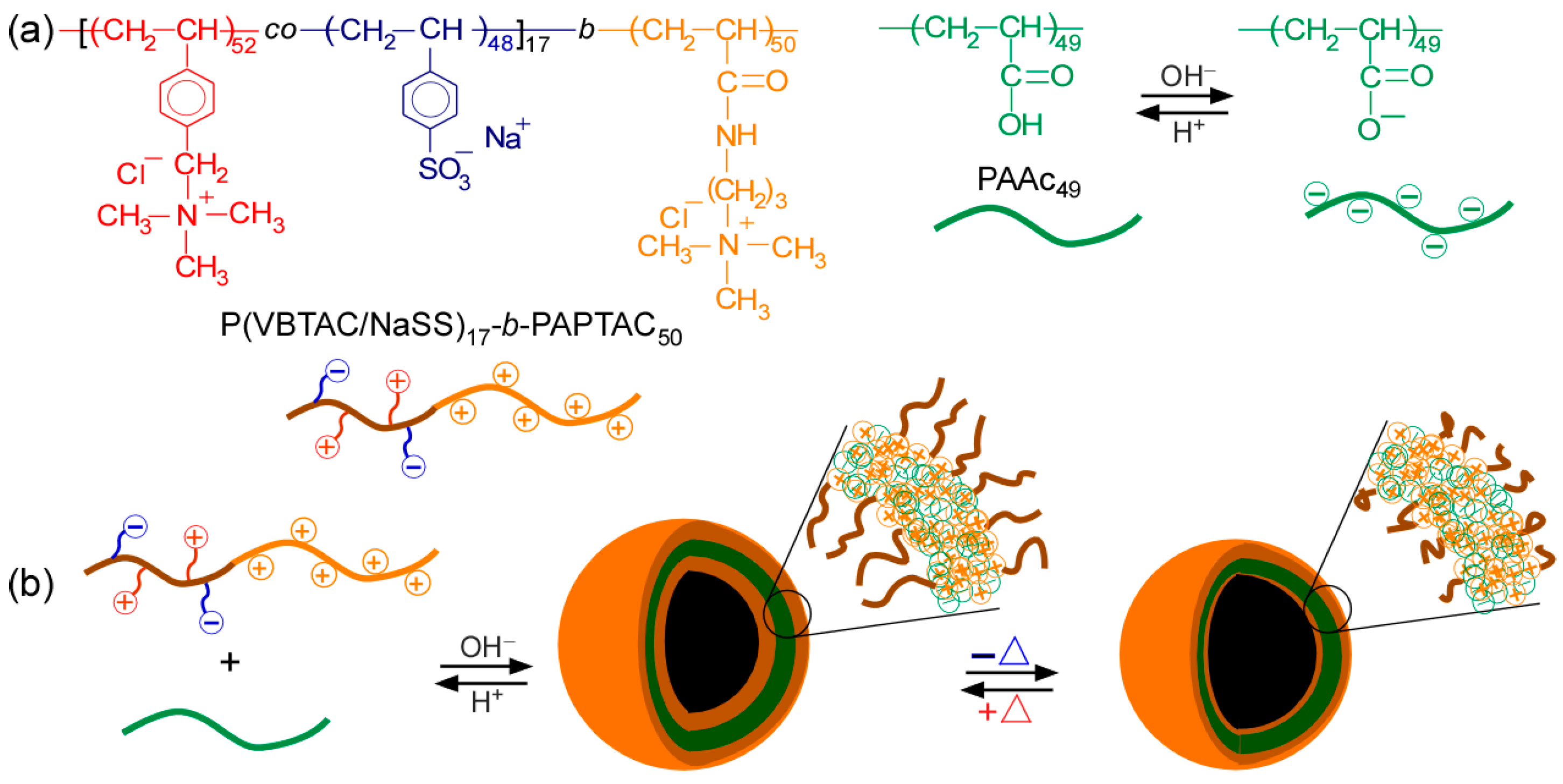
:A diblock copolymer (P(VBTAC/NaSS)17-b-PAPTAC50; P(VS)17A50) composed of amphoteric random copolymer, poly(vinylbenzyl trimethylammonium chloride-co-sodium p-styrensunfonate) (P(VBTAC/NaSS); P(VS)) and cationic poly(3-(acrylamidopropyl) trimethylammonium chloride) (PAPTAC; A) block, and poly(acrylic acid) (PAAc49) were prepared via a reversible addition−fragmentation chain transfer radical polymerization. Scrips V, S, and A represent VBTAC, NaSS, and PAPTAC blocks, respectively. Water-soluble polyion complex (PIC) vesicles were formed by mixing P(VS)17A50 and PAAc49 in water under basic conditions through electrostatic interactions between the cationic PAPTAC block and PAAc49 with the deprotonated pendant carboxylate anions. The PIC vesicle collapsed under an acidic medium because the pendant carboxylate anions in PAAc49 were protonated to delete the anionic charges. The PIC vesicle comprises an ionic PAPTAC/PAAc membrane coated with amphoteric random copolymer P(VS)17 shells. The PIC vesicle showed upper critical solution temperature (UCST) behavior in aqueous solutions because of the P(VS)17 shells. The pH- and thermo-responsive behavior of the PIC vesicle were studied using 1H NMR, static and dynamic light scattering, and percent transmittance measurements. When the ratio of the oppositely charged polymers in PAPTAC/PAAc was equal, the size and light scattering intensity of the PIC vesicle reached maximum values. The hydrophilic guest molecules can be encapsulated into the PIC vesicle at the base medium and released under acidic conditions. It is expected that the PIC vesicles will be applied as a smart drug delivery system.
1. Introduction
Owing to electrostatic interactions between cationic and anionic units, oppositely charged polyelectrolytes in water form a polyion complex (PIC) or polyelectrolytes complex [1,2]. Furthermore, PIC micelles with a PIC core and hydrophilic noncharged shells can be prepared without using an organic solvent in water [3,4]. Smart PIC aggregates have single- or multi-responsive properties against external stimuli, such as pH, temperature, and light irradiation. They show substantial changes in their properties with a slight change in the surrounding environments [5]. The PIC aggregates have attracted huge interest from many researchers because of their advantages. The PIC aggregates are expected to meet some drug delivery system (DDS) assignments, such as the targeted and controlled release of the drug. The pH of the medium is an important factor in the controlled release of a DDS. The microenvironment of cancer tumors is acidic, whereas it is nearly neutral for normal cells [6]. The change in the pH of the environment results in an ideal pH-responsive DDS carrier, which can be applied in cancer treatments. Therefore, many PIC aggregate systems, which target the release of drugs in response to the acidity of the surroundings, have been reported [7,8,9,10,11,12]. Water-soluble pH-responsive PIC vesicles covered with biocompatible poly(2-(methacryloyloxy)ethyl phosphorylcholine) (PMPC) shells have also been reported [3]. The PIC vesicle was coassembled via electrostatic interactions between PMAPTAC and ionized PAaH under a basic medium by mixing oppositely charged diblock copolymers consisting of PMPC and cationic poly(3-(methacrylamidopropyl) trimethylammonium chloride) (PMAPTAC), or anionic poly(sodium 6-acrylamidohexanoate) (PAaH) blocks. The hydrophilic guest molecule, Texas red-labeled dextran, was encapsulated into the inner water phase of the PIC vesicle. Under the acidic medium, PAaH was protonated to become noncharged resulting in a dissociation of the PIC vesicle, where the guest molecule was released. Therefore, the PIC vesicle is expected to effectively target the drug in the cytoplasm.
Similarly, the environment’s temperature is a widely used parameter in the controlled release of drugs [13,14,15,16,17]. Matsuoka et al. [13] reported on thermo-responsive PIC aggregates. Diblock copolymers composed of poly(sulfopropyl dimethylammonium propylacrylamide) (PSPP) and cationic PMAPTAC or anionic poly(sodium p-styrenesulfonate) (PNaSS) were mixed in water to form PIC micelles with a PMAPTAC/PNaSS core and PSPP shells. The PIC micelles showed an upper critical solution temperature (UCST) caused by the PSPP shells. They formed large aggregates of micelles below the phase transition temperature of the PSPP shells owing to the PSPP chain shrinkage. The UCST behavior of the PIC aggregates can be controlled using the polymer concentration and the degree of polymerization (DP) of the PSPP chains. Furthermore, some other PIC systems have been fabricated with the target to release a drug in response to environments containing glucose to deal with diabetes [18,19] or to use light irradiation to control the release of the drug [20,21].
These single stimulus-responsive PIC aggregates have been studied and applied. Besides stimuli response, PIC aggregates have shown other outstanding advantages [22,23,24,25,26,27]. For instance, dual pH- and thermo-responsive PIC micelles were prepared from a co-assembly of oppositely charged diblock copolymers, poly(N-methyl-2-vinyl pyridinium iodide)-block-poly(ethylene oxide) (P2MVP-b-PEO) and poly(acrylic acid)-block-poly(isopropyl acrylamide) (PAAc-b-PNIPAM) in water [27]. Under a basic medium at 25 °C, the PIC micelles were formed owing to the electrostatic interaction between the PAAc and P2MPV blocks with the PAAc/P2MVP core and hydrophilic mixed PNIPAM/PEG shells. However, under an acidic medium, the size of the aggregates increased, which can be attributed to the aggregation formed by hydrogen bonding between PAAc and PNIPAM/PEO. At high concentrations, the core–shell structure of PIC micelles switches to core–shell–corona (onion type) with a PNIPAM core, covered with a mixed P2MVP/PAAc shell, and a PEO outer corona at 60 °C.
Polyampholytes or amphoteric random copolymers are polymers that consist of cationic and anionic units in their structure [28,29,30]. Amphoteric random copolymers have no charge, which is comparable to the balance between oppositely charged groups since they contain both pendant cationic and anionic groups [31,32]. Therefore, to avoid complex precipitation, the polyampholytes can be used as a hydrophilic nonionic block. Furthermore, some amphoteric polymers show thermo-responsive and protein antifouling properties. Yusa et al. [28] reported that amphoteric diblock copolymers from poly(2-acrylamido-2-methylpropanesulfonic acid sodium salt) (PAMPS) and poly(3-(acrylamido)propyl trimethylammonium chloride) (PAPTAC) synthesized via a reversible addition−fragmentation chain transfer (RAFT) radical polymerization. The polymer exhibited a lower critical solution temperature (LCST) behavior due to the hydrogen-bonding interactions of the pendant amide groups and water molecules. The thermo-responsive behavior of the polymers can be controlled by adjusting the NaCl concentration. Meanwhile, P(APMPS/APTAC), a random copolymer prepared by RAFT with the same content as APMPS and APTAC, showed an ability in protein antifouling [33]. The PIC micelle consists of amphoteric random copolymer P(AMPS/APTAC) shells that show no interaction with proteins. Furthermore, a UCST-type thermo-responsive amphoteric random copolymer (P(VBTAC/NaSS)) composed of the pendant quaternary ammonium group in vinylbenzyl trimethylammonium chloride (VBTAC) and pendant sulfonate in NaSS with the same content of each monomer, was prepared via RAFT polymerization [34]. P(VBTAC/NaSS) showed the UCST behavior caused by electrostatic interactions between PVBTAC and PNaSS. The UCST behavior was affected by DP and concentrations of P(VBTAC/NaSS).
Based on prior studies, we designed pH- and thermos-responsive PIC micelles. The interaction of the PIC vesicle with the hydrophobic surface may be controlled by temperature if the PIC vesicle surface hydrophobicity is changed by heating. For example, if the PIC vesicle surface changes to be hydrophobic with cooling, the interactions increase between the PIC vesicle and cell. Furthermore, if the PIC vesicle is dissociated in response to pH, the encapsulated guest molecules are released by pH change. Particularly, cancer tissue is more acidic than healthy tissue, so designing a pH-responsive PIC vesicle is useful for cancer treatment. In this study, we have prepared a diblock copolymer (P(VS)17A50) consisting of polyampholyte (P(VBTAC/NaSS)17; P(VS)17) block and a cationic PAPTAC50 (A50) block (Scheme 1). Furthermore, the PIC aggregates were prepared by mixing P(VS)17A50 and polyacrylic acid (PAAc49) in a basic medium because of electrostatic interactions between the cationic PAPTAC block and deprotonated anionic PAAc49. The PIC aggregate had a vesicle structure, which confirmed that the PIC aggregate could encapsulate a hydrophilic guest molecule into the hollow core. PAAc indicated pH-responsive behavior caused by protonation and the pendant carboxyl groups [35,36,37]. The PAAc pendant carboxyl groups are protonated and neutralized under acidic conditions, leading to the dissociation of the PIC vesicles. The PIC vesicle was formed from the PAPTAC/PAAc membrane, covered with the amphoteric random copolymer P(VS)17 shells. In this study, to synthesize P(VS)17, equimolar amounts of styrene-type cationic VBTAC and anionic NaSS were randomly copolymerized via RAFT. Then, the cationic APTAC monomer was polymerized to synthesize P(SV)17A50. Furthermore, PAAc49 was also synthesized via RAFT. Polymers were characterized with NMR and gel-permeation chromatography (GPC) measurements. The pH- and thermo-responsive properties of the PIC vesicle were studied with percent transmittance, dynamic and static light scattering, zeta-potential, and NMR measurements. Additionally, since the P(SV)17 polyampholyte shells exhibited the UCST behavior, controlling the interaction with proteins and cells in response to temperature may be possible. Furthermore, the P(SV)17 polyampholyte can suppress protein adsorption. Therefore, the PIC vesicles are expected to modify the surfaces of medical devices.
2. Materials and Methods
2.1. Materials
Vinylbenzyl trimethylammonium chloride (VBTAC, 99%) methoxy poly(ethylene glycol) labeled pyrene (PEG-Py, Mn = 2000 g/mol) was purchased from Sigma-Aldrich (St. Louis, MO, USA), sodium p-styrenesulfonate (NaSS, 98%) from Tokyo Chemical Industry (Tokyo, Japan), and 4,4-azobis(4-cyanopentanoic acid) (V-501, 98%) and 2,2-azobisisobutyronitrile (AIBN, 98%) from Wako Pure Chemical (Osaka, Japan) were used without further purification. Acrylic acid (AAc, 98%) from Wako Pure Chemical (Osaka, Japan) was distilled under reduced pressure. 4-Cyanopentanoic acid dithiobenzoate (CPD) was synthesized using the previously reported method [38]. The water was purified using an ion-exchange column system.
2.2. Synthesis of P(VS)17 Random Copolymer
First, amphoteric random copolymer (P(VS)17) was prepared via RAFT polymerization in accordance with the previous report [34]. VTBAC (3.18 g, 15.0 mmol), NaSS (3.09 g, 15.0 mmol), CPD (419 mg, 1.50 mmol), and V-501 (210 mg, 0.749 mmol) were dissolved in 30 mL of mixed solvent of 1.2 M NaCl aqueous solution and MeOH (9/1, v/v) with the feed molar ratio of [VBTAC]/[NaSS]/[CPD]/[V-501] = 10/10/1/0.5. The mixture was then, degassed by purging with Ar gas for 30 min. Polymerization occurred at 70 °C for 5 h. After polymerization, 1H NMR spectra were used to calculate the polymerization conversion of VBTAC and NaSS (conversion = 84.5%). The reaction mixture was dialyzed against 1.2-M NaCl aqueous solution for two days and pure water for one day and collected using a freeze-drying method (4.69 g, 67.9%). The theoretical DP(theo)) and number-average molecular weight (Mn(theo)) were 17 and 3.83 × 10³ g/mol. The VBTAC and NaSS contents were determined using 1H NMR spectra of P(VS)17 and found to be 51.9 and 48.1 mol%, respectively.
2.3. P(VBTAC/NaSS)17-b-PAPTAC50
The diblock copolymer was prepared via RAFT polymerization using P(VS)17 as a macro-CTA. P(VS)17 (539 mg, 0.141 mmol), APTAC (1.46 g, 7.06 mmol), and V-501 (15.7 mg, 0.06 mmol) with a feed molar ratio of [P(VS)17]/[APTAC]/[AIBN] = 1/50/0.4 were dissolved in water (7.0 mL). The mixture was degassed by purging with Ar gas for 30 min. Polymerization occurred at 70 °C for 24 h. The polymerization conversion was calculated from 1H NMR integral intensity ratio of the vinyl protons and phenyl protons to 5.6 and 6.4–7.6 ppm, respectively, (conversion = 98.4%). Then, the reaction mixture was dialyzed against pure water for 5 days and collected using a freeze-drying method (1.55 g, 77.5%). From 1H NMR measurement, the DP(NMR)) of the APTAC block was estimated to be 50. The number-average molecular weight (Mn(GPC)) and polydispersity index (Mw/Mn) of P(VS)17A50 were estimated to be 5.70 × 10⁴ g/mol and 1.04, respectively, based on the GPC measurement.
2.4. Synthesis of PAAc49
RAFT polymerization was used to prepare poly(acrylic acid) (PAAc49). AAc (7.20 g, 100 mmol), CPD (280 mg, 0.99 mg), and AIBN (65.7 mg, 0.40 mmol) with a feed molar ratio of [AAc]/[CPD]/[AIBN] = 100/1/0.4 were dissolved in methanol (100 mL). The mixture was degassed by purging with Ar gas for 30 min. Polymerization occurred at 60 °C for 47 h. From the 1H NMR integral intensity ratio, the polymerization conversion of the vinyl proton signal at 5.8 ppm and terminal phenyl group protons at 7.3–7.9 ppm (conversion = 36.3%) was estimated. Then, the reaction mixture was dialyzed against pure water for one week and collected using a freeze-drying method (0.97 g, 12.9%). From 1H NMR measurement the DP(NMR) of PAAc49 was calculated to be 49. From GPC measurement, the Mn(GPC) and Mw/Mn of P(VS)17A50 were 1.46 × 10⁴ g/mol and 1.07, respectively.
2.5. Preparation of PIC Vesicles
P(VS)17A50 and PAAc49 were individually dissolved in water at Cp = 1.0 g/L as stock solutions to prepare the PIC vesicles. The PIC vesicles were fabricated by mixing the cationic P(VS)17A50 solution with the anionic PAAc49 solution while stirring for 5 min at room temperature. The pH of the mixed solution was adjusted using 0.1-M NaOH and 0.1-M HCl aqueous solutions, then left for one day to obtain an equilibrium state. The mole fraction of positively charged (f+ = [APTAC]/([APTAC] + [PAAc]) in the PIC vesicle solution corresponds to the mixing ratio of the opposite polymers
2.6. Encapsulation of PEG-Py
PEG-Py (0.01 g/L) aqueous solution was prepared. Then, P(VS)17A50 (0.1 g/L) and PAAc49 (0.1 g/L) were separately dissolved in the PEG-Py aqueous solution. Subsequently, the P(VS)17A50 aqueous solution was added to the PAAc49 aqueous solution with f+ = 0.5 and stirred for 5 min at room temperature. The mixed solution was adjusted to pH 10 using NaOH solution and then dialyzed for 20 days against an aqueous solution at pH 10 using a Harvard Apparatus (Holliston, MA, USA) polycarbonate membrane with 10-nm pores. The solvent was replaced every 12 h to remove the unbound PEG-Py molecules. A PEG-Py aqueous solution (0.01 g/L) without a PIC vesicle was dialyzed with the same procedure. Fluorescence spectroscopy measurements for PEG-Py in the presence and absence of the PIC vesicles were performed to detect PEG-Py content in the solution after dialysis. The loading capacity (LC) and loading efficiency (LE) were calculated according to the following Equations [39]:
2.7. Measurements
1H NMR measurements were performed to determine the complex characteristics of the polymer and polyion complex using a JEOL (Tokyo, Japan) JNM-ECZ 400 MHz NMR. The sample solutions were prepared in D2O and adjusted with NaOD or DCl solutions to desired pH. The standard pulse program, stebpgp1s19, used a stimulated echo, bipolar gradient pulse, and one spoil gradient with 3-9-19 pulse sequence (WATERGATE) solvent suppression of the water signal. The GPC measurements were used to determine the number-average molecular weight (Mn(GPC)) and polydispersity index (Mw/Mn). For cationic polymer, P(VS)17A50, a mixture of Na2SO4 (0.3 M) and CH3COOH (0.5 M) aqueous solution was used as a cationic eluent. P(VS)17A50 signal was detected using Jasco (Tokyo, Japan) RI-2031 Plus refractive index (RI) detector equipped with a PU-2080 Plus column operating at 40 °C. Using a calibrated curve of standard poly(2-vinylpyridine), the values of Mn(GPC) and Mw/Mn for P(VS)17A50 were estimated. For anionic homopolymer PAAc49, GPC spectra were obtained from Tosoh RI-8020 RI working at 40 °C with a Shodex (Tokyo, Japan) GF-7M column, Tosoh (Yamaguchi, Japan) DP-8020 pump and a mixture of phosphate buffer (50 mM) at pH 9 and acetonitrile (9/1, v/v) as an anionic eluent. Using a calibration made from standard PNaSS, the values of Mn and Mw/Mn for polymers were estimated. Fourier transform infrared (FTIR) spectra were obtained using a Jasco FT/IR-4200 spectrometer. The PIC vesicle structure was confirmed via transmission electron microscopy (TEM) observation using a JEOL (Tokyo, Japan) JEM-2100. The PIC vesicle aqueous solutions with f+ = 0.5 and Cp = 0.1 g/L at pH 10 were prepared. Then, a drop of the sample solution was put on a copper grid coated with thin films of Formvar. The oversupply sample solution was filtered using filter paper. The next step was to stain with sodium phosphotungstate and dry under a vacuum for one night. The hydrodynamic radius (Rh), light scattering intensity (LSI), and zeta-potential of polymers and PIC vesicles were calculated using a Malvern Nano ZS with He–Ne laser (4 mW at 632.8 nm) from Malvern (Kobe, Japan). Before taking the measurements, the sample solutions were filtered using a 0.45-μm pore size membrane filter. Furthermore, to estimate the weight-average molecular weight (Mw), a z-average radius of gyration (Rg), and the second viral coefficient (A2) values of the polymers and PIC, SLS measurements were performed using the Otsuka Electronics Photal (Osaka, Japan) SLS-6500 with a He–Ne laser (4 mW at 632.8 nm) as a light source working at 25 °C. An Otsuka Electronics Photal DRM-3000 (Osaka, Japan) differential refract meter was used to calculate dn/dCp values at 633 nm. Percentage transmittance (%T) was measured on a Jasco (Tokyo, Japan) V-730 UV-vis spectrophotometer equipped with a temperature control system. Fluorescence spectra for PEG-Py were measured using a Hitachi (Tokyo, Japan) F-2500 fluorescence spectrometer. The excitation and emission slit widths were kept constantly at 20 and 2.5 nm, respectively.
3. Results
3.1. Preparation of P(VS)17A50 and PAAc49
All polymers in this study were prepared via normal RAFT polymerization. An amphoteric random copolymer comprising cationic VBVTAC and anionic NaSS was prepared via RAFT radical polymerization. According to 1H NMR, the total conversion (p) of VBTAC and NaSS was estimated to be 84.5%. The theoretical DP(theo) and theoretical number-average molecular weight (Mn(theo)) of P(VS)17 were calculated using p and the following equation:
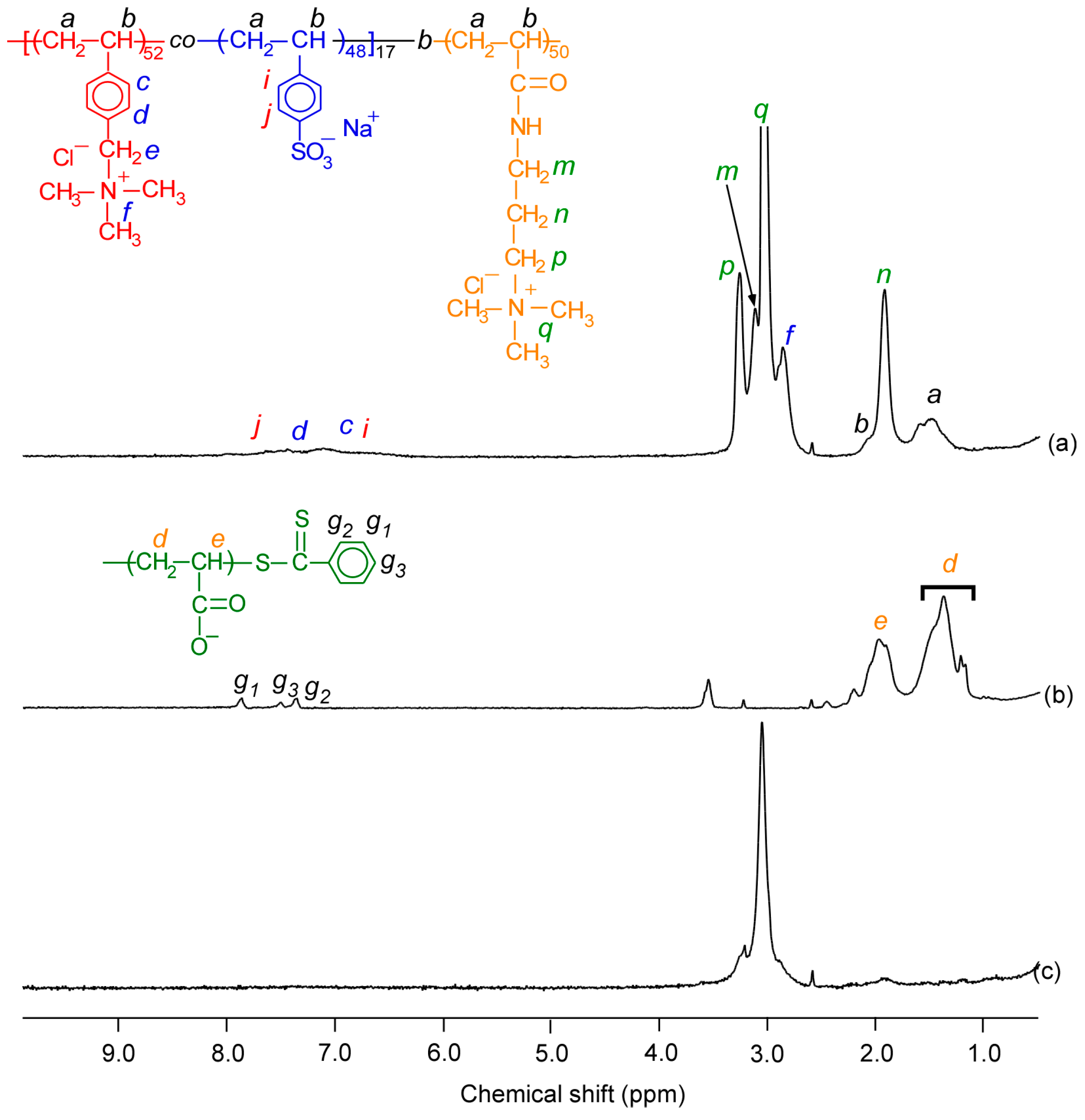
where, [M]0 and [CTA]0 are the initial concentrations of the monomer and CTA, respectively, and Mm and MCTA are the molecular weights of the monomer and CTA, respectively. The DP(theo) and Mn(theo) for P(VS)17 were calculated to be 17 and 3.83 × 10³ g/mol, respectively. The composition of P(VS)17 was estimated from 1H NMR in D2O containing 1.2 M NaCl at 70 °C (Figure S1). The main chain proton signals were observed at 1.5–2.1 ppm. The pendent methyl protons of the VTBAC unit were observed at 2.9 ppm. The pendant phenyl protons of the VBTAC and NaSS units are presented as 6.4–7.6 ppm. The VBTAC content in the P(VS)17 block was calculated to be 51.9 mol% based on the integral intensities between the pendant phenyl and VBTAC methyl protons. The DP(NMR) for the PAPTAC block in P(VS)17A50 was 50, as estimated from the integral intensity of the methylene proton from the PAPTAC unit at 1.6 ppm compared with that for the phenyl protons of VBTAC and NaSS at 6.4–7.6 ppm (Figure 1a). PAAc49 has a DP(NMR) of 49, estimated from the ratio of integral intensities between main chain protons at 1.5–2.3 ppm and terminal phenyl protons at 7.3–7.9 ppm, derived from the CTA fragment (Figure 1b). The Mn(NMR) and Mn(theo) values for P(VS)17A50 were close. The Mw/Mn values estimated from GPC for P(VS)17A50 and PAAc49 (Figure S2) were 1.04 and 1.07, respectively, indicating that the polymers showed well-controlled structures. The DP, Mn, and Mw/Mn values for all polymers are summarized in Table 1. P(VS)17 could not be measured using GPC because it cannot dissolve in the eluent. FTIR spectrum was measured for P(VS)17A50. The pendant carbonyl signal can be seen at 1700 cm−1 (Figure S3).
3.2. Preparation and Characterization of PIC Vesicles
The PIC vesicle was prepared by mixing P(VS)17A50 and PAAc49 with f+ = 0.5 at Cp = 2.0 g/L in D2O at pH 10 for the 1H NMR measurement (Figure 1c). For DLS measurements, the PIC vesicle was prepared at 0.1 g/L; however, the Cp was too low for NMR measurements so the PIC vesicle was prepared at Cp = 2.0 g/L. After mixing, the signals from the PAPTAC50 block and PAAc49 disappeared. The VBTAC unit’s methylene proton signal remained at 2.9 ppm, showing that the PAPTAC block and PAAc49 interacted to create the vesicle structure’s membrane and P(VS)17 produced shells.
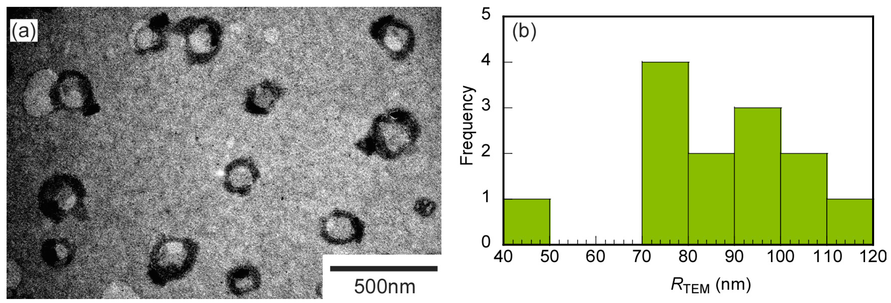
TEM was conducted to confirm the PIC aggregate shape. The TEM image was obtained for the stoichiometrically charge neutralized mixture of P(VS)17A50 and PAAc49 at pH 10 (Figure 2). The particles had a spherical shape with a contrasting white center and black edge, indicating a vesicle structure in the PIC aggregates. The average radius obtained from the TEM observations was 86.2 ± 17.3 nm, close to the hydrodynamic radius (Rh) estimated from the dynamic light scattering measurements (Rh = 86.6 nm). During TEM observation, the sample shrank due to the high vacuum conditions. The PIC vesicles collapsed as they shrank and stuck to the TEM grid membrane. At that time, the PIC vesicles expanded. Therefore, the size obtained from the TEM observation did not decrease much compared to the Rh value.
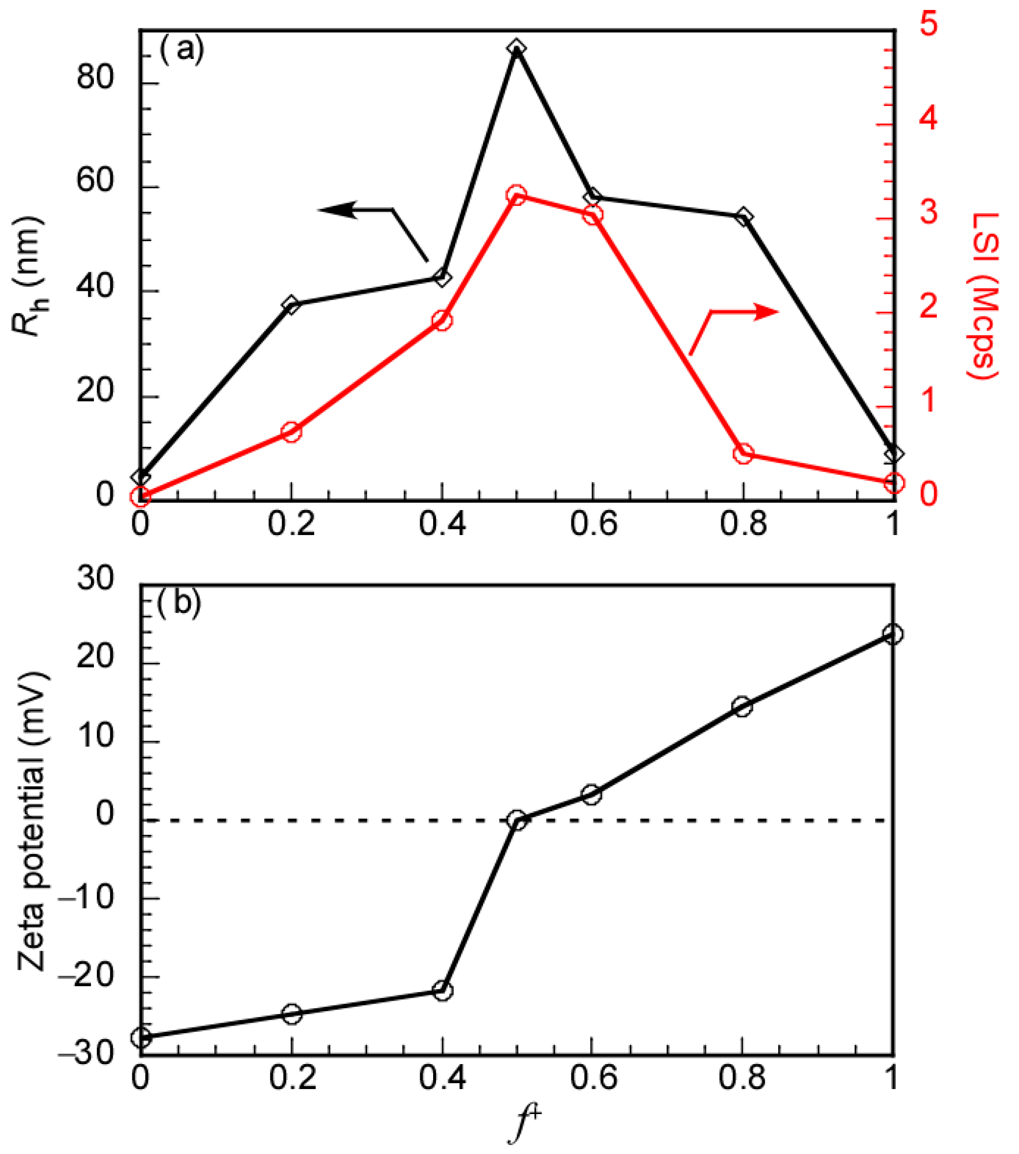
The mixing ratio of the oppositely charged polymers determines the development of the PIC vesicle [40,41]. The plots of Rh, LSI, and zeta-potential with f+ at pH 10 were prepared (Figure 3). The Cp values in the PIC vesicle solutions were kept at 0.1 g/L. However, samples at f+ = 0 and 1 attributed to anionic PAAc49 and cationic P(VS)17A50, respectively, were prepared at Cp = 2.0 g/L because Rh and zeta-potential values could not be obtained because of very low LSI at Cp = 0.1 g/L. The Rh and LSI values of the PIC vesicle reached maximum values at f+ = 0.5, and then the zeta-potential reached 0 mV, suggesting the neutralization of the oppositely charged PAPTAC50 block and PAAc49. Meanwhile, the zeta-potential values of PAAc49 and the PAPTAC50 block at f+ = 0 and 1 were −27.7 and 23.8 mV, respectively, indicating negative and positive charges.
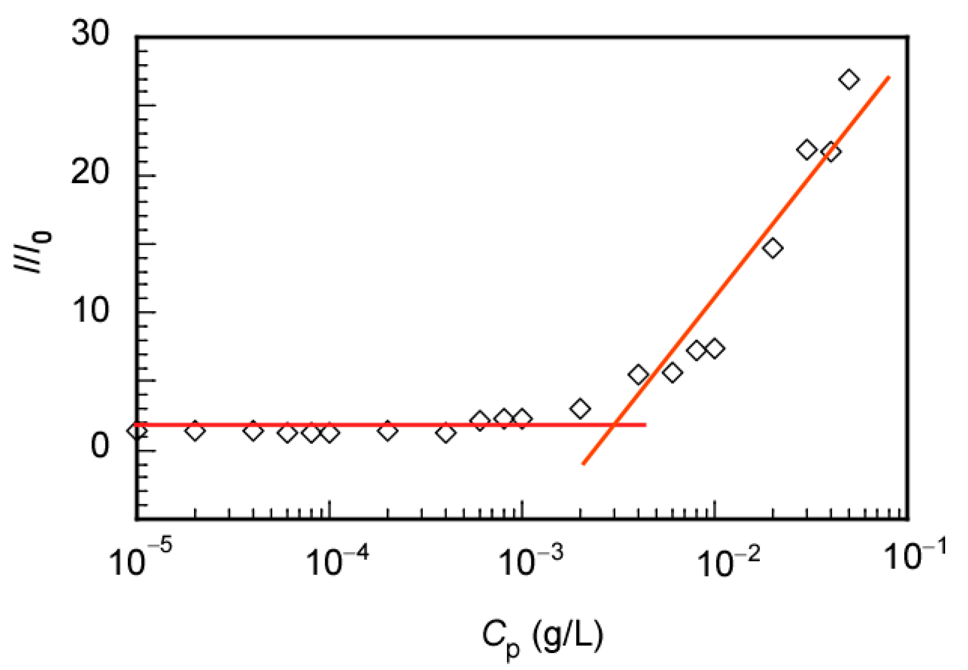
The critical micelle concentration (CMC) of the PIC vesicle was determined to certify the thermodynamic equilibrium properties. The LSI of the PIC vesicle aqueous solutions was measured as a function of Cp. The PIC vesicle aqueous solution was prepared by mixing P(VS)17A50 and PAAc49 in water at pH 10 with f+ = 0.5 at Cp = 0.1 g/L; the solution was diluted to the desired Cp using water at pH 10. The CMC for the PIC vesicle in water was determined from the relationship between the LSI ratio (I/I0) and Cp (Figure 4). I and I0 correspond to the LSIs of the PIC vesicle aqueous solution and water at pH 10, respectively. The crossing point of the linear portions in the low and high Cp regions was defined as the CMC value, which was 2.47 × 10⁻³ g/L. The defined CMC value implies that the PIC vesicle is in a dynamic equilibrium state. The PIC vesicle is stable above the CMC. The PIC vesicle, however, can disintegrate below the CMC into a small assembly of P(VS)17A50/PAAc49 because of strong electrostatic interactions between them [33,42].
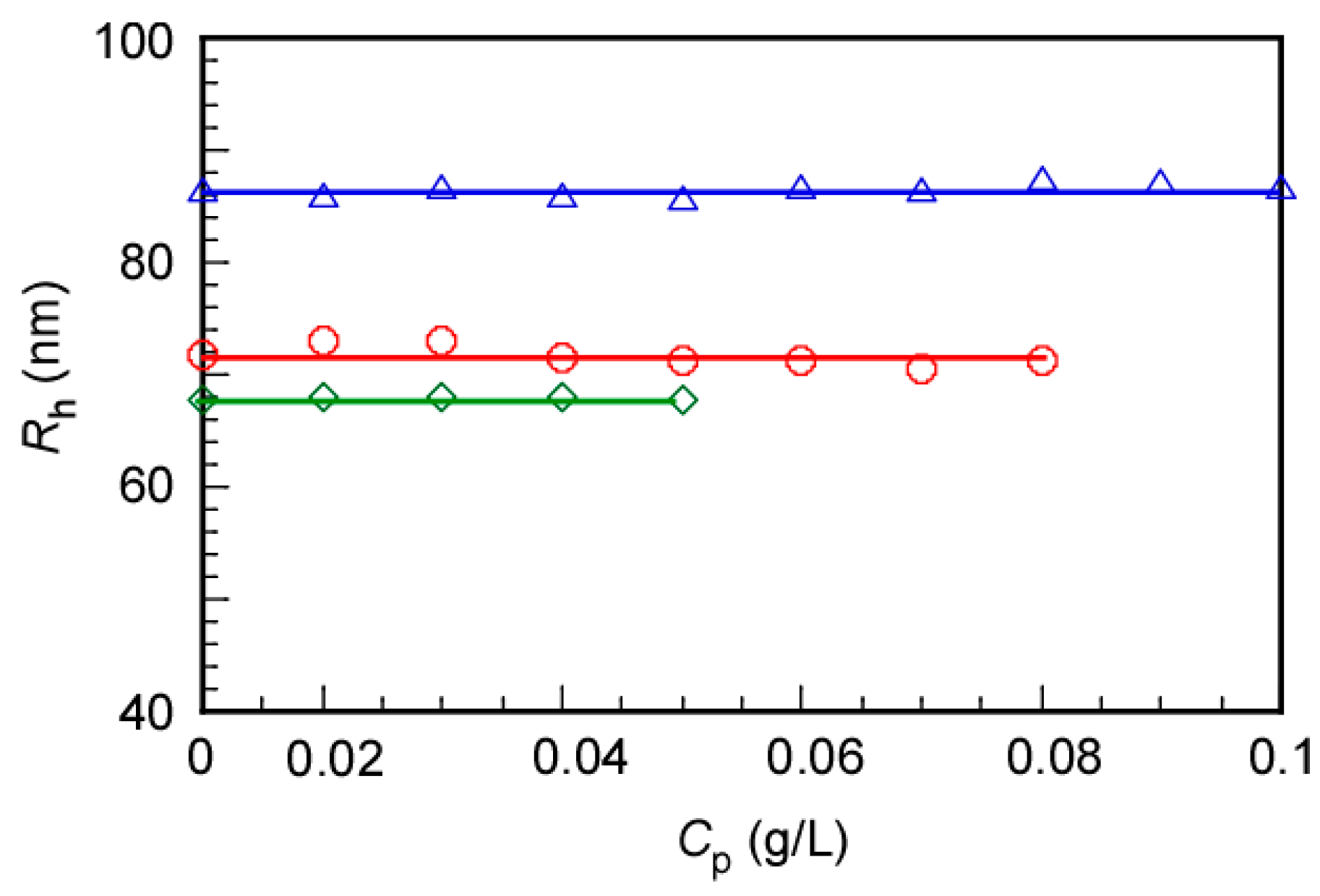
We studied the concentration dependence of the PIC vesicle because the SLS should be measured at different Cp values to perform the SLS measurements. The P(VS)17A50 and PAAc49 aqueous solutions were separately prepared at Cp = 0.1, 0.08, and 0.05 g/L. Then, P(VS)17A50 and PAAc49 Cp were mixed to form the PIC vesicle. The pH value of the aqueous solution was adjusted to 10 and left for one day to obtain an equilibrium state. Subsequently, the PIC vesicle aqueous solutions were diluted by water at pH 10 to adjust the target Cp. The Rh and LSI for the PIC vesicle were plotted as a function of the Cp after mixing (Figure 5). The Rh values for the PIC vesicles at Cp = 0.1, 0.08, and 0.05 g/L before mixing were 86.6, 71.2, and 67.8 nm, respectively. This indicated that the size of the PIC vesicles increased with an increasing Cp before mixing since the aggregation number (Nagg) of the PIC vesicle depends on the Cp. When the Cp increases, the Nagg increases to increase the size of the PIC vesicle [42]. Furthermore, the increase in the Nagg was confirmed by of the increase in the LSI value from 1.55 to 3.25 Mcps at Cp = 0.05 and 0.1 g/L, respectively (Figure S4). When the PIC vesicle was diluted with water at pH 10, the Rh values remained nearly constant, independent of the Cp. These findings suggest that the size of the PIC vesicle can be controlled by adjusting the Cp before mixing, and after the PIC vesicleformation they were stable against dilution.
SLS measurements at 25 °C were used to determine further properties of the polymers and PIC vesicle, such as evident weight-average molecular weight (Mw(SLS)), and radius of gyration (Rg) (Figure S5). The RI increment (dn/dCp) values for all samples were also separately estimated. Nagg is the number of polymer chains required to form a PIC vesicle. The PIC vesicle was prepared by mixing P(VS)17A50 and PAAc49 at Cp = 0.1 g/L with f+ = 0.5 at pH 10 then the solution was diluted to 0.025 g/L by water at pH 10. Table 2 indicates the value of Mw(SLS), Nagg, Rg, and Rh for all samples. The values of Mw(SLS) for P(VS)17A50 and PAAc49 were 1.55 × 10⁴ g/mol and 0.33 × 10⁴ g/mol, respectively, which were close to the Mn(NMR) values estimated from the 1H NMR measurements. P(VS)17A50 and PAAc49 had Mn(NMR) values of 1.42 × 10⁴ and 0.35 × 10⁴ g/mol, respectively. The Mw(SLS) for the PIC vesicle was 1.06 × 10⁸ g/mol. The value of Nagg for the PIC vesicle was estimated by dividing the Mw(SLS) of the PIC vesicle by that of the unimers and was found to be 5640. The Rg/Rh value for the PIC vesicle was calculated as 1.17, indicating a spherical shape [3,43,44]. The following equation can be used to calculate the density (d) of the polymers and PIC vesicles:
The d values for P(VS)17A50, PAAc49, and the PIC vesicle were calculated to be 0.033, 0.028, and 0.064 g/cm3, respectively. The d value for the PIC vesicle was larger than those for P(VS)17A50 and PAAc49, suggesting that the polymer chains in the PIC vesicle are more densely packed than those of the unimers.
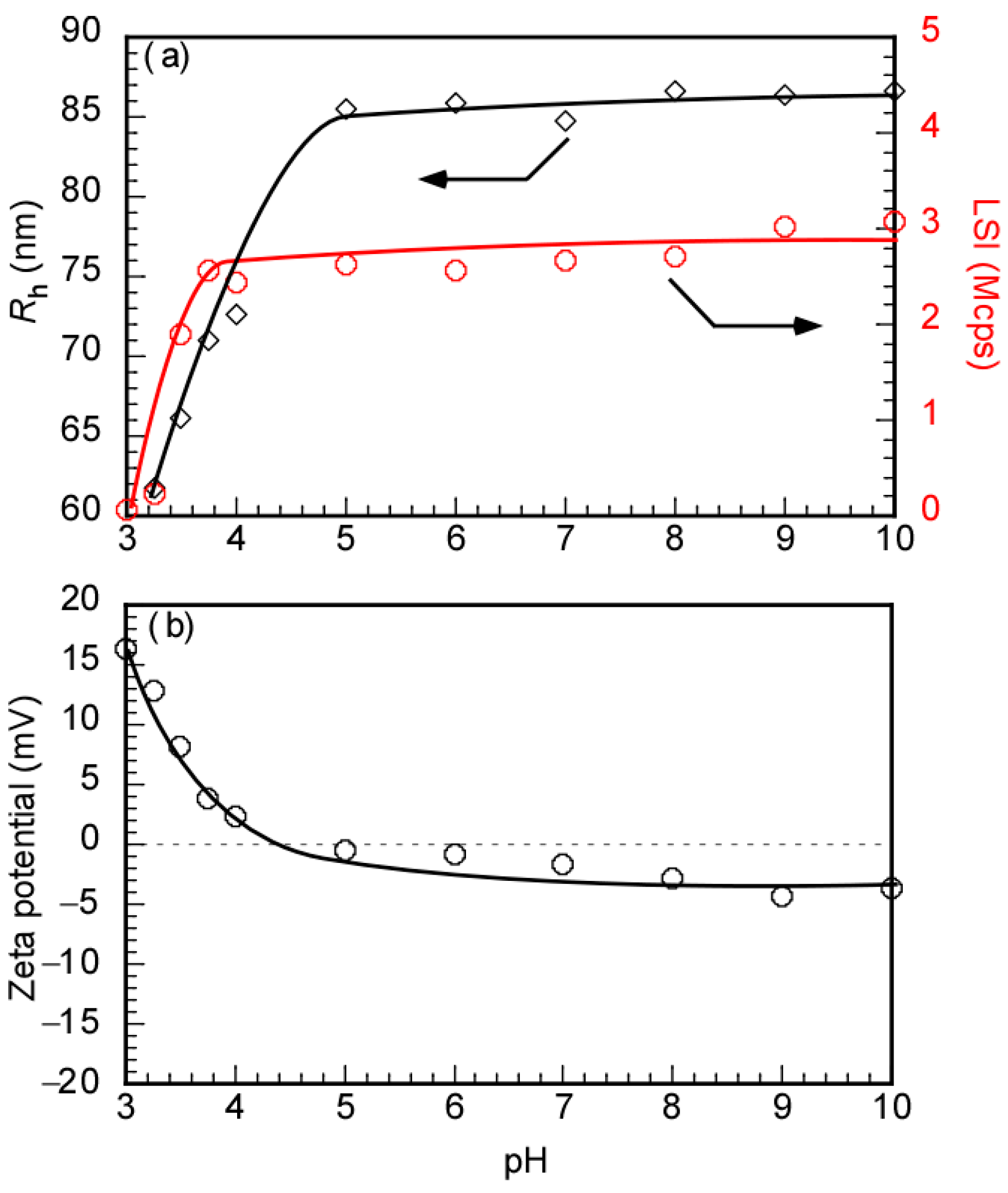
The pendant carboxylic groups in PAAc49 are deprotonated under basic conditions. Therefore, the pH of the solution is an important factor in the PIC vesicle formation. To understand the effect of the pH value on the PIC vesicle, the Rh and LSI were measured as a function of the pH values. Initially, the PIC vesicle aqueous solution was prepared in water at Cp = 0.1 g/L with f+ = 0.5, then adjusted to the appropriate pH using NaOH and HCl aqueous solutions. The Rh, LSI, and zeta-potential values were plotted against the pH. By increasing pH from 3 to 5, the Rh of the PIC vesicle increased from 60.9 to 85.5 nm, then remained at ca. 86 nm above pH 5 (Figure 6a). The LSI value of the PIC vesicle showed the same tendency as that of the Rh. The acid dissociation constant (pKa) of PAAc is about 4.5 [45,46,47]. At pH above 4.5 of the PAAc49 aqueous solutions, the pendant carboxyl groups were deprotonated to generate anionic charges. The anionic PAAc49 with pendant carboxylate anions can interact with the cationic PAPTAC block in P(VS)17A50 to form the PIC vesicle through electrostatic interactions. This was supported by the zeta-potential value of the PIC vesicle aqueous solutions. At pH < 5, the zeta-potential of the PIC vesicle showed a positive value which can be attributed to a charge of the cationic PAPTAC block in P(VS)17A50 with protonated PAAc49 (Figure 6b). After increasing the pH value, at 5 ≤ pH the zeta-potential reached nearly 0 mV. It implied that the PIC vesicle formation was affected by the pH value, which means that the PIC vesicle can be formed above pH 5 and collapse below pH 5. The pH influence on the association behavior of the PIC vesicle is attributed to the pH-responsive property of PAAc49. The conformation of PAAc in water was reported, and a reversible transition from the coil to the globule dependence of the pH was caused by deprotonation of the carboxylic group. At low pH, PAAc was a compact globular shape. When the media of the solution became baser, PAAc expanded into an open coil shape [47,48]. Furthermore, the Rh and LSI values for P(VS)17A50 were constant at ca. 6 nm and 70 kcps, respectively, at all pH values ranging from 3 to 10 (Figure S6), suggesting that P(VS)17A50 was not affected by pH. The small Rh and LSI values indicated that the polymer was in the unimer state. The zeta-potential values of P(VS)17A50 and PAAc49 were measured as a function of pH (Figure S7). The zeta-potential for P(VS)17A50 stayed constant at ca. 17 mV, indicating that the polymer always gives a positive charge independent of the solution’s pH. However, the zeta-potential of PAAc49 decreased from pH 3 to 5, then remained constant from pH 6. The pKa for PAAc49 was 4.5; therefore, from pH 5, the PAAc49 deprotonated and indicated a negative charge.
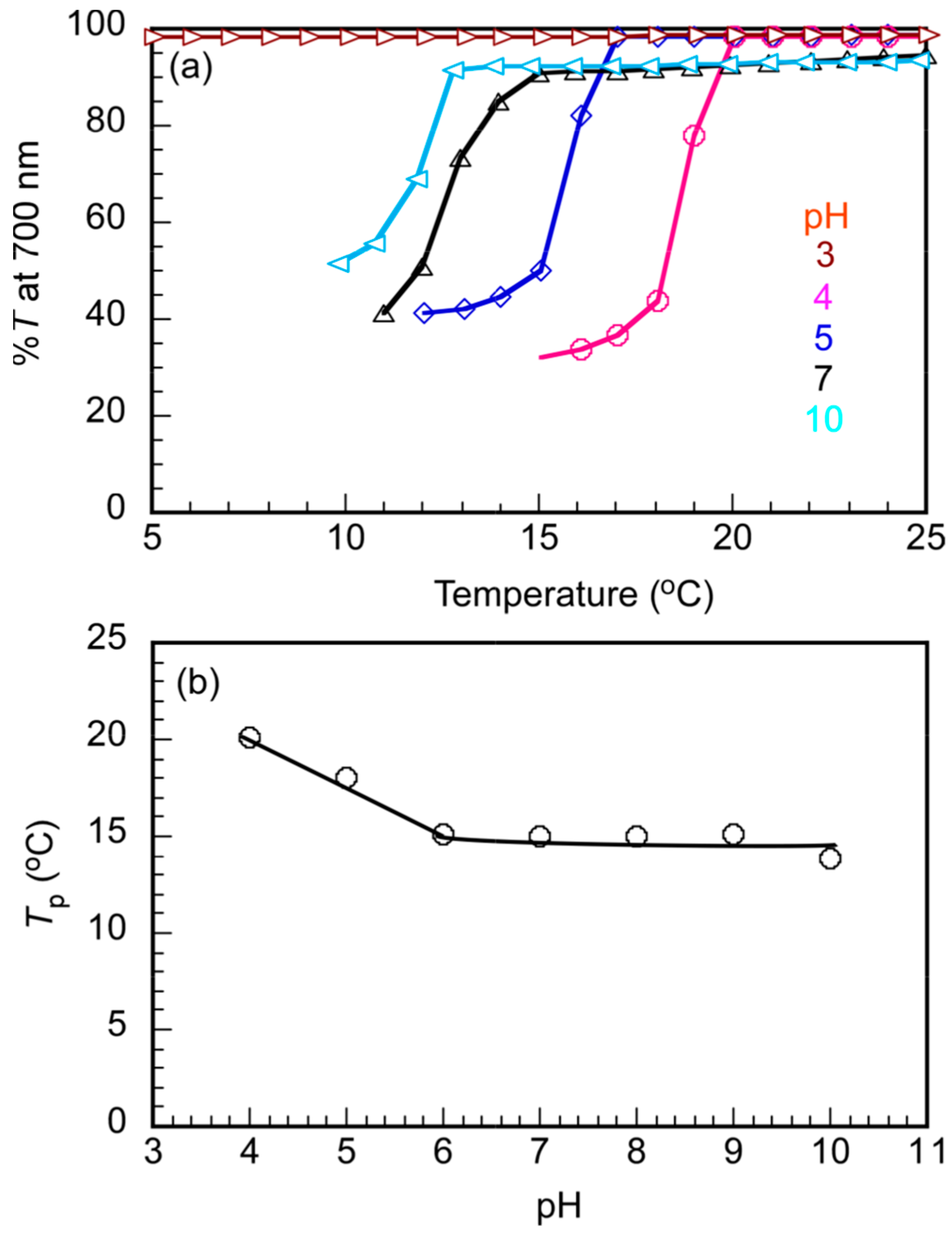
The 1H NMR spectra for the PIC vesicle suggested the A50/PAAc49 PIC membrane was covered with P(VS)17 amphoteric random copolymer shells. P(VS)17 showed the UCST behavior in aqueous solutions (Figure S8). Therefore, the PIC vesicle was expected to exhibit the UCST behavior owing to the P(VS)17 shells. The percentage transmittance (%T) of the PIC vesicle was plotted against temperature upon the cooling process indicating the UCST behavior of the PIC vesicle. The temperature dependences investigated the effect of pH on the UCST behavior on %T at different pH conditions ranging from pH 3 to 10 (Figure 7). The PIC vesicle was disrupted below pH 5 owing to protonation of the pendant carboxyl groups in PAAc49 (Figure 6). However, the aqueous solutions showed the UCST behavior excepted at pH 3. The phase transition temperature (Tp) of the PIC vesicle decreased from pH 4 to 6, then remained at 15 °C from pH 6. The UCST behavior of the PIC vesicle was affected by pH.
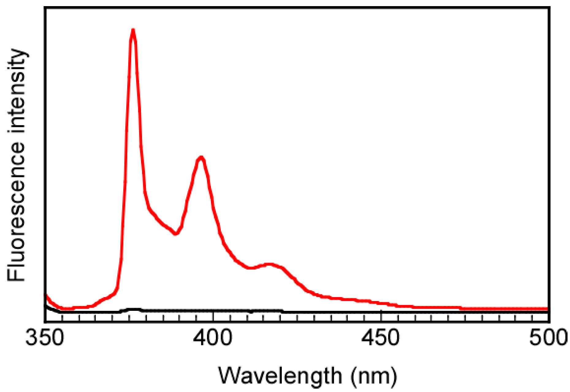
We have studied incorporating a hydrophilic noncharged guest molecule, PEG-Py, into the PIC vesicles. The hydrophilic drugs can be encapsulated inside the PIC vesicle thanks to the inner water phase. Herein, P(VS)17A50 and PAAc49 solutions containing PEG-Py were mixed to form the PIC vesicle. The PEG-Py molecules which could not be incorporated into the PIC vesicle were removed by dialysis against pure water at pH 10. In comparison, a blank experiment was conducted, and the PEG-Py aqueous solution in the absence of the PIC vesicle was dialyzed with the same procedure. After dialysis for 20 days, fluorescence spectroscopy for PEG-Py in the presence and absence of the PIC aggregate was measured (Figure 8). The maximum emission wavelengths at 376 and 397 nm for PEG-Py were observed in the PIC vesicle solution, suggesting that the PIC vesicles can encapsulate PEG-Py. Hydrophobic pyrene molecules associate to produce excimer emission with a peak at 480 nm. PEG-Py can dissolve in water without interpolymer association in this study because no excimer emission was observed from PEG-Py fluorescence. In contrast, no signal of PEG-Py was detected from the reference solution in the absence of the PIC vesicle owing to the removal of the small PEG-Py molecules after dialysis membrane with a pore size of 10 nm. These findings suggested the PIC aggregate might be a vesicle structure because it can encapsulate hydrophilic PEG-Py. From the calibration curve of PEG-Py (Figure S9) and the fluorescence intensity, the weight of PEG-Py encapsulated into a PIC vesicle was estimated to be 0.0012 g. The LE and LC were calculated to be 12% and 1.2%, respectively, from Equations (1) and (2).
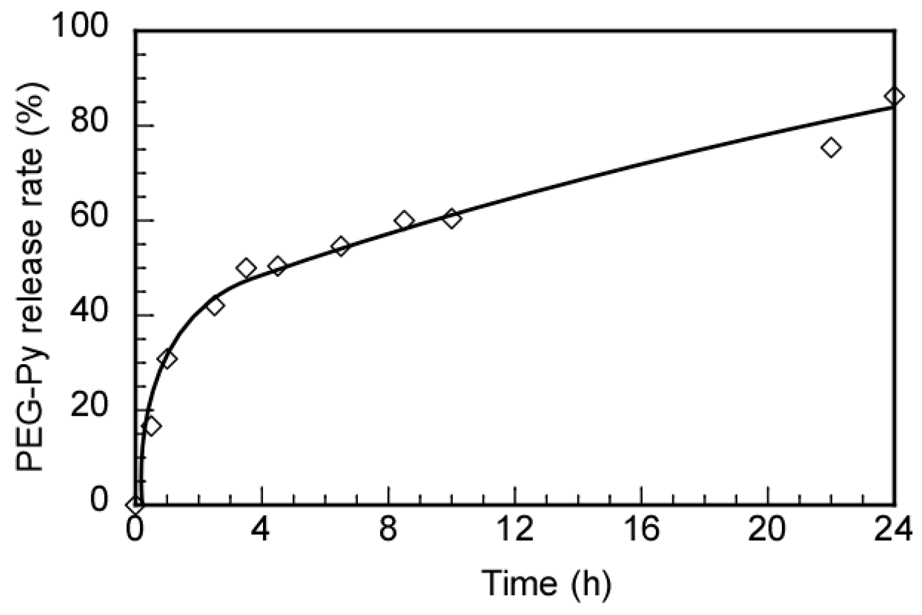
Using an HCl aqueous solution, the PIC vesicle encapsulating PEG-Py at pH 10 was adjusted to pH 3. A dialysis bag with a 10-nm pore size was used to dialyze the solution with an aqueous solution at pH 3. The solution sample inside the dialysis bag was taken out at varying times to measure the fluorescence intensities to calculate the release rate of the encapsulated PEG-Py from the PIC vesicle (Figure 9). After 24 h, the release amount of PEG-Py at pH 3 was 86.2%. Noncharged hydrophilic guest molecules can be encapsulated into the PIC vesicle at pH 10 and released at pH 3 owing to the PIC vesicle’s disruption. The release rate of the encapsulated guest molecule may be changed above and below the UCST of the P(VS)17 shell.
4. Conclusions
P(VS)17A50, a cationic diblock copolymer, and an anionic homopolymer, PAAc49, were successfully synthesized via a RAFT polymerization with a well-controlled structure. The pH- and thermo-responsive P(VS)17A50/PAAc49 PIC vesicle was prepared by mixing P(VS)17A50 and PAAc49 in basic aqueous solutions at room temperature due to electrostatic interactions. The Rh and LSI values of the PIC vesicle showed maximum values at f+ = 0.5, whereas the zeta-potential was near 0 mV. At low pH, the PIC vesicle disintegrated. The spherical shape with a vesicle structure was confirmed by TEM. The Rg/Rh ratio obtained from SLS and DLS measurements showed a value close to 1, indicating the spherical shape of the PIC vesicle. The thermo-responsive P(VS)17 shells caused UCST behavior in the PIC vesicle. The thermo-responsive behavior of the PIC vesicle was affected by the pH of the solution. The PIC vesicle can encapsulate water-soluble nonionic guest molecules at a high pH and release them under acidic conditions, implying that it could be used as a DDS material. The interaction of the PIC vesicle with the cell can be controlled by temperature because the surface hydrophobicity increases with decreasing temperature. Therefore, it is expected that the PIC vesicle can be taken in the cell at a low temperature below the UCS of the P(VS)17 shells. It is known that the pH around the cancer tissue is low. The encapsulated hydrophilic anticancer drugs can be released under acidic conditions from the P(VS)17A50/PAAc49 PIC vesicle.
Supplementary Materials
The following supporting information can be downloaded at: https://0-www-mdpi-com.brum.beds.ac.uk/article/10.3390/polym14091659/s1, Figure S1. 1H NMR spectrum for P(VBTAC/NaSS)17 in D2O containing 1.2-M NaCl at 70 °C; Figure S2. Gel-permeation chromatography (GPC) elution curves for (a) P(VS)17A50 and (b) PAAc49 monitored with refractive index (RI) detector working at 40 °C; Figure S3. FTIR spectrum for P(VS)17A50; Figure S4. Light scattering intensity (LSI) for the P(VS)17A50/PAAc49 PIC vesicles with f+ = 0.5 in water at pH 10 as a function of polymer concentration after mixing P(VS)17A50 and PAAc49. At 25 °C the PIC aggregates solution at 0.1 (triangle), 0.08 (circle), and 0.05 (diamond) g/L were diluted with pH 10 aqueous solution; Figure S5. Typical examples of Zimm plots for (a) P(VS)17, (b) PAAc49, and (c) the P(VS)17A50/PAAc49 PIC vesicle with f+ = 0.5 in aqueous solutions at pH 10; Figure S6. Hydrodynamic radius (Rh, diamond) and light scattering intensity (LSI, circle) for P(VS)17A50 at Cp = 2.0 g/L in water as a function of pH at 25 °C; Figure S7. Zeta-potential for P(VS)17A50 (circle) and PAAc49 (triangle) in water at Cp = 2.0 g/L as a function of pH at 25 °C; Figure S8. Percent transmittance (%T) at 700 nm for the 0.1-M NaCl aqueous P(VS)17 solution at Cp = 1.0 g/L as a function of temperature upon the heating and cooling processes; Figure S9. (a) Fluorescence spectra for PEG-Py in water at pH 10 excited at 334 nm at various PEG-Py concentrations ([PEG-Py]) and (b) a calibration curve of fluorescence intensity at 376 nm using [PEG-Py] in water at pH 10.
Author Contributions
Conceptualization, T.T.P. and S.-i.Y.; methodology, T.T.P., T.D.P. and S.-i.Y.; software, T.T.P.; validation, T.T.P. and S.-i.Y.; formal analysis, T.T.P. and S.-i.Y.; investigation, T.T.P.; writing—original draft preparation, T.T.P. and S.-i.Y.; writing—review and editing, T.T.P., T.D.P. and S.-i.Y.; visualization, T.T.P. and S.-i.Y.; supervision, S.-i.Y.; project administration, S.-i.Y.; funding acquisition, S.-i.Y. All authors have read and agreed to the published version of the manuscript.
Funding
This research was partially supported by KAKENHI grants (21H02005, 21K19931, 21H05027, 21H05535) from the Japan Society for the Promotion of Science (JSPS), JSPS Bilateral Joint Research Projects (JPJSBP120203509), the Cooperative Research Program of “Network Joint Research Center for Materials and Devices (20214044)”, the International Collaborative Research Program of Institute for Chemical Research, Kyoto University (2022-121), and MEXT Promotion of Distinctive Joint Research Center Program (JPMXP 0621467946).
Data Availability Statement
The data used to support the findings of this study are available from the corresponding authors upon request.
Acknowledgments
The authors would like to express their thanks to Thi Ngoc Anh Doan, Shota Fujii, Kazuo Sakurai for their suggestions.
Conflicts of Interest
The authors declare no conflict of interest.
References
- Lankalapalli, S.; Kolapalli, V.R.M. Polyelectrolyte complexes: A review of their applicability in drug delivery technology. Indian J. Pharm. Sci. 2009, 71, 481–487. [Google Scholar] [CrossRef] [PubMed] [Green Version]
- Insua, I.; Wilkinson, A.; Fernandez-Trillo, F. Polyion complex (PIC) particles: Preparation and biomedical applications. Eur. Polym. J. 2016, 81, 198–215. [Google Scholar] [CrossRef] [PubMed] [Green Version]
- Ohara, Y.; Nakai, K.; Ahmed, S.; Matsumura, K.; Ishihara, K.; Yusa, S. pH-responsive polyion complex vesicle with polyphosphobetaine shells. Langmuir 2019, 35, 1249–1256. [Google Scholar] [CrossRef] [PubMed]
- Slyusarenko, N.V.; Vasilyeva, N.Y.; Kazachenko, A.S.; Gerasimova, M.A.; Romanchenko, A.S.; Slyusareva, E.A. Synthesis and properties of interpolymer complexes based on chitosan and sulfated arabinogalactan. Polym. Sci. B 2020, 62, 272–278. [Google Scholar] [CrossRef]
- Bhat, A.H.; Khan, I.; Bhawani, S.A.; Abdul Rahim, M.K.; Gazal, U. Stimuli-Responsive Polymer Composites for Fabric Applications. Smart Polym. Nanocomposites; Woodhead Publishing: Sawston, UK, 2021; Chapter Fifteen. [Google Scholar]
- Justus, C.R.; Dong, L.; Yang, L.V. Acidic tumor microenvironment and pH-sensing G protein-coupled receptors. Front. Physiol. 2013, 4, 354. [Google Scholar] [CrossRef] [PubMed] [Green Version]
- Ohno, S.; Ishihara, K.; Yusa, S. Formation of polyion complex (PIC) micelles and vesicles with anionic pH-responsive unimer micelles and cationic diblock copolymers in water. Langmuir 2016, 32, 3945–3953. [Google Scholar] [CrossRef]
- Chen, J.; Ding, J.; Zhang, Y.; Xiao, C.; Zhuang, X.; Chen, X. Polyion complex micelles with gradient pH-sensitivity for adjustable intracellular drug delivery. Polym. Chem. 2015, 6, 397–405. [Google Scholar] [CrossRef]
- Oishi, M.; Sasaki, S.; Nagasaki, Y.; Kataoka, K. pH-responsive oligodeoxynucleotide (ODN)-poly(ethylene glycol) conjugate through acid-labile β-thiopropionate linkage: Preparation and polyion complex micelle formation. Biomacromolecules 2003, 4, 1426–1432. [Google Scholar] [CrossRef]
- Luo, Y.L.; Yuan, J.F.; Liu, X.J.; Xie, H.; Gao, Q.Y. Self-assembled polyion complex micelles based on PVP-b-PAMPS and PVP-b-PDMAEMA for drug delivery. J. Bioact. Compat. Polym. 2010, 25, 292–304. [Google Scholar] [CrossRef]
- Tian, S.; Liu, G.; Wang, X.; Wu, T.; Yang, J.; Ye, X.; Zhang, G.; Hu, J.; Liu, S. pH-regulated reversible transition between polyion complexes (PIC) and hydrogen-bonding complexes (HBC) with tunable aggregation-induced emission. ACS Appl. Mater. Interfaces 2016, 8, 3693–3702. [Google Scholar] [CrossRef]
- Oishi, M.; Nagatsugi, F.; Sasaki, S.; Nagasaki, Y.; Kataoka, K. Smart polyion complex micelles for targeted intracellular delivery of PEGylated antisense oligonucleotides containing acid-labile linkages. Chembiochem 2005, 6, 718–725. [Google Scholar] [CrossRef]
- Kim, D.; Matsuoka, H.; Saruwatari, Y. Formation of sulfobetaine-containing entirely ionic PIC (polyion complex) micelles and their temperature responsivity. Langmuir 2020, 36, 10130–10137. [Google Scholar] [CrossRef]
- De Santis, S.; Ladogana, R.D.; Diociaiuti, M.; Masci, G. Pegylated and thermosensitive polyion complex micelles by self-assembly of two oppositely and permanently charged diblock copolymers. Macromolecules 2010, 43, 1992–2001. [Google Scholar] [CrossRef]
- Li, G.; Guo, L.; Meng, Y.; Zhang, T. Self-assembled nanoparticles from thermo-sensitive polyion complex micelles for controlled drug release. Chem. Eng. J. 2011, 174, 199–205. [Google Scholar] [CrossRef]
- Pua, M.L.; Yoshitomi, T.; Chonpathompikunlert, P.; Hirayama, A.; Nagasaki, Y. Redox-active injectable gel using thermo-responsive nanoscale polyion complex flower micelle for noninvasive treatment of local inflammation. J. Control. Release 2013, 172, 914–920. [Google Scholar] [CrossRef]
- Kim, D.; Matsuoka, H.; Yusa, S.; Saruwatari, Y. Collapse behavior of polyion complex (PIC) micelles upon salt addition and reforming behavior by dialysis and its temperature responsivity. Langmuir 2020, 36, 15485–15492. [Google Scholar] [CrossRef]
- Dong, S.; Jiang, Y.; Qin, G.; Liu, L.; Zhao, H. Methionine-based pH and oxidation dual-responsive block copolymer: Synthesis and fabrication of protein nanogels. Biomacromolecules 2020, 21, 4063–4075. [Google Scholar] [CrossRef]
- Ma, R.; Yang, H.; Li, Z.; Liu, G.; Sun, X.; Liu, X.; An, Y.; Shi, L. Phenylboronic acid-based complex micelles with enhanced glucose-responsiveness at physiological pH by complexation with glycopolymer. Biomacromolecules 2012, 13, 3409–3417. [Google Scholar] [CrossRef]
- Jin, Q.; Cai, T.; Wang, Y.; Wang, H.; Ji, J. Light-responsive polyion complex micelles with switchable surface charge for efficient protein delivery. ACS Macro Lett. 2014, 3, 679–683. [Google Scholar] [CrossRef]
- Zhou, W.; Ding, P.; Zhang, C.; Wu, B.; Guo, X.; Stuart, M.A.C.; Zhuo, X.; Wang, J. Regulated polyion complex vesicles for efficient photothermal therapy. Adv. Funct. Mater. 2022, 2022, 2108729. [Google Scholar] [CrossRef]
- Chiang, W.H.; Hsu, Y.H.; Chen, Y.W.; Chern, C.S.; Chiu, H.C. Thermoresponsive interpolymeric complex assemblies from co-association of linear PAAc homopolymers with PNIPAAm segments containing PAAc-based graft copolymer. Macromol. Chem. Phys. 2011, 212, 1869–1878. [Google Scholar] [CrossRef]
- Zhang, D.; Liu, J.; Xie, H.; Zhu, A.; Xu, Y.; Zeng, B.; Luo, W.; Dai, L. Polyion complex micelles formed by azobenzene-based polymer with multi-responsive properties. J. Appl. Polym. Sci. 2021, 138, 50580. [Google Scholar] [CrossRef]
- Ishii, S.; Kaneko, J.; Nagasaki, Y. Dual stimuli-responsive redox-active injectable gel by polyion complex based flower micelles for biomedical applications. Macromolecules 2015, 48, 3088–3094. [Google Scholar] [CrossRef]
- Liu, Y.; Li, C.; Wang, H.Y.; Zhang, X.Z.; Zhuo, R.X. Synthesis of thermo- and pH-sensitive polyion complex micelles for fluorescent imaging. Chem. A Eur. J. 2012, 18, 2297–2304. [Google Scholar] [CrossRef]
- Ren, J.; Zhang, Y.; Zhang, J.; Gao, H.; Liu, G.; Ma, R.; An, Y.; Kong, D.; Shi, L. pH/sugar dual responsive core-cross-linked PIC micelles for enhanced intracellular protein delivery. Biomacromolecules 2013, 14, 3434–3443. [Google Scholar] [CrossRef]
- Voets, I.K.; Moll, P.; Aqli, A.; Jerome, C.; Detrembleur, C.; Waard, P.; Keizet, A.; Stuart, M. Temperature responsive complex coacervate core micelles with a PEO and PNIPAAm corona. J. Phys. Chem. B 2008, 35, 10833–10840. [Google Scholar] [CrossRef]
- Kawata, Y.; Kozuka, S.; Yusa, S. Thermo-responsive behavior of amphoteric diblock copolymers bearing sulfonate and quaternary amino pendant groups. Langmuir 2019, 35, 1458–1464. [Google Scholar] [CrossRef]
- Kazachenko, A.S.; Akman, F.; Malyar, Y.N.; Issaoui, N.; Vasilieva, N.Y.; Karacharov, A.A. Synthesis optimization, DFT and physicochemical study of chitosan sulfates. J. Mol. Struct. 2021, 1245, 131083. [Google Scholar] [CrossRef]
- Kazachenko, A.; Akman, F.; Medimagh, M.; Issaoui, N.; Vasilieva, N.; Malyar, Y.N.; Sudakova, I.G.; Karacharov, A.; Miroshnikova, A.; Marzook Al-Dossary, O.M. Sulfation of Diethylaminoethyl-cellulose: QTAIM topological analysis and experimental and DFT studies of the properties. ACS Omega 2021, 6, 22603–22615. [Google Scholar] [CrossRef]
- Rabiee, A.; Ershad-Langroudi, A.; Jamshidi, H. Polyacrylamide-based polyampholytes and their applications. Rev. Chem. Eng. 2014, 30, 501–519. [Google Scholar] [CrossRef]
- North, S.M.; Armes, S.P. Synthesis of polyampholytic diblock copolymers via RAFT aqueous solution polymerization. Polym. Chem. 2021, 12, 4846–4855. [Google Scholar] [CrossRef]
- Nakahata, R.; Yusa, S. Preparation of water-soluble polyion complex (PIC) micelles covered with amphoteric random copolymer shells with pendant sulfonate and quaternary amino groups. Polymers 2018, 10, 205. [Google Scholar] [CrossRef] [Green Version]
- Sharker, K.K.; Ohara, Y.; Shigeta, Y.; Ozoe, S.; Yusa, S. Upper critical solution temperature (UCST) behavior of polystyrene-based polyampholytes in aqueous solution. Polymers 2019, 11, 265. [Google Scholar] [CrossRef] [Green Version]
- Seki, K.; Tirrell, D.A. pH-Dependent complexation of poly(acrylic acid) derivatives with phospholipid vesicle membranes. Macromolecules 1984, 17, 1692–1698. [Google Scholar] [CrossRef]
- Li, R.; Zhang, C.; Wang, C.; Cheng, Y.; Hu, D. Study on the mechanism of the reversible color change of polyacrylic acid modified gold nanoparticles responding to pH. Materials 2021, 14, 3679. [Google Scholar] [CrossRef]
- Fukumoto, H.; Ishihara, K.; Yusa, S. Thermo-responsive behavior of mixed aqueous solution of hydrophilic polymer with pendant phosphorylcholine group and poly(acrylic acid). Polymers 2021, 13, 148. [Google Scholar] [CrossRef]
- Mitsukami, Y.; Donovan, M.S.; Lowe, A.B.; Mccormick, C.L. Water-soluble polymers. 81. Direct synthesis of hydrophilic styrenic-based homopolymers and block copolymers in aqueous solution via RAFT. Macromolecules 2001, 34, 2248–2256. [Google Scholar] [CrossRef]
- Kozuka, S.; Kuroda, K.; Ishihara, K.; Yusa, S. Interpolymer association of amphiphilic diblock copolymers bearing pendant siloxane and phosphorylcholine groups. J. Polym. Sci. Part A Polym. Chem. 2019, 57, 1500–1507. [Google Scholar] [CrossRef]
- Nakamura, N.; Mochida, Y.; Toh, K.; Fukushima, S.; Anraku, Y.; Cabral, H. Effect of mixing ratio of oppositely charged block copolymers on polyion complex micelles for in vivo application. Polymers 2020, 13, 5. [Google Scholar] [CrossRef]
- Anraku, Y.; Kishimura, A.; Oba, M.; Yamasaki, Y.; Kataoka, K. Spontaneous formation of nanosized unilamellar polyion complex vesicles with tunable size and properties. J. Am. Chem. Soc. 2010, 132, 1631–1636. [Google Scholar] [CrossRef]
- Pramod, P.S.; Takamura, K.; Chaphekar, S.; Balasubramanian, N.; Jayakannan, M. Dextran vesicular carriers for dual encapsulation of hydrophilic and hydrophobic molecules and delivery into cells. Biomacromolecules 2012, 13, 3627–3640. [Google Scholar] [CrossRef]
- Burchard, W. Static and dynamic light scattering from branched polymers and biopolymers. Springer: Berlin/Heidelberg, Germany, 1983. [Google Scholar]
- Geismann, C.; Tomicki, F.; Ulbricht, M. Block copolymer photo-grafted poly(ethylene terephthalate) capillary pore membranes distinctly switchable by two different stimuli. Sep. Sci. Technol. 2009, 44, 3312–3329. [Google Scholar] [CrossRef]
- Michaels, A.S.; Morelos, O. Polyelectrolyte adsorption by kaolinite. Ind. Eng. Chem 1995, 47, 1801–1809. [Google Scholar] [CrossRef]
- Paek, K.; Yang, H.; Lee, J.; Park, J.; Kim, B.J. Efficient colorimetric pH sensor based on responsive polymer-quantum dot integrated graphene oxide. ACS Nano 2014, 8, 2848–2856. [Google Scholar] [CrossRef]
- Katchalsky, A.; Eisenberg, H. Molecular weight of polyacrylic and polymethacrylic acid. J. Polym. Sci. 1951, 6, 145–154. [Google Scholar] [CrossRef]
- Lim, P.F.C.; Chee, L.Y.; Chen, S.B. Study of interaction between cetyltrimethylammonium bromide and poly(acrylic acid) by rheological measurements. J. Phys. Chem. B 2003, 107, 6491–6496. [Google Scholar] [CrossRef]
Scheme 1.
(a) Chemical structures of P(VBTAC/NaSS)17-b-PAPTAC50 ((P(VS)17A50) and PAAc49, and (b) conceptual formation of the PIC vesicle consist of P(VS)17A50 and PAAc49.
Scheme 1.
(a) Chemical structures of P(VBTAC/NaSS)17-b-PAPTAC50 ((P(VS)17A50) and PAAc49, and (b) conceptual formation of the PIC vesicle consist of P(VS)17A50 and PAAc49.

Figure 1.
1H NMR with water suppression spectra for P(VS)17A50 (a), PAAc49 (b), and the P(VS)17A50/PAAc49 PIC vesicle with f+ = 0.5 (c) at Cp = 2.0 g/L in D2O at pH 10 at 25 °C.
Figure 1.
1H NMR with water suppression spectra for P(VS)17A50 (a), PAAc49 (b), and the P(VS)17A50/PAAc49 PIC vesicle with f+ = 0.5 (c) at Cp = 2.0 g/L in D2O at pH 10 at 25 °C.

Figure 2.
(a) TEM image and (b) histogram of particle size distribution for the P(VS)17A50/PAAc49 PIC vesicle with f+ = 0.5 at Cp = 0.1 g/L in water at pH 10.
Figure 2.
(a) TEM image and (b) histogram of particle size distribution for the P(VS)17A50/PAAc49 PIC vesicle with f+ = 0.5 at Cp = 0.1 g/L in water at pH 10.

Figure 3.
(a) Hydrodynamic radius (Rh, diamond) and light scattering intensity (LSI, circle) and (b) zeta-potential for the P(VS)17A50/PAAc49 PIC vesicle at Cp = 0.1 g/L in water at pH 10 as a function of f+ at 25 °C.
Figure 3.
(a) Hydrodynamic radius (Rh, diamond) and light scattering intensity (LSI, circle) and (b) zeta-potential for the P(VS)17A50/PAAc49 PIC vesicle at Cp = 0.1 g/L in water at pH 10 as a function of f+ at 25 °C.

Figure 4.
Light scattering intensity ratio (I/I0) for the P(VS)17A50/PAAc49 PIC vesicle in water pH 10 as a function of polymer concentration (Cp). I and I0 are the light scattering intensities of the PIC vesicle solution and solvent, respectively.
Figure 4.
Light scattering intensity ratio (I/I0) for the P(VS)17A50/PAAc49 PIC vesicle in water pH 10 as a function of polymer concentration (Cp). I and I0 are the light scattering intensities of the PIC vesicle solution and solvent, respectively.

Figure 5.
Hydrodynamic radius (Rh) light scattering intensity (LSI) for the P(VS)17A50/PAAc49 PIC vesicle with f+ = 0.5 in water at pH 10 as a function of polymer concentration after mixing P(VS)17A50 and PAAc49. The PIC vesicle solution at 0.1 (triangle), 0.08 (circle), and 0.05 (diamond) g/L were diluted with pH 10 aqueous solution at 25 °C.

Figure 6.
(a) Hydrodynamic radius (Rh, diamond) and LSI (circle), and (b) zeta-potential for the P(VS)17A50/PAAc49 PIC vesicle with f+ = 0.5 at Cp = 0.1 g/L in water as a function of pH at 25 °C.
Figure 6.
(a) Hydrodynamic radius (Rh, diamond) and LSI (circle), and (b) zeta-potential for the P(VS)17A50/PAAc49 PIC vesicle with f+ = 0.5 at Cp = 0.1 g/L in water as a function of pH at 25 °C.

Figure 7.
(a) Percent transmittance (%T) at 700 nm for the P(VS)17A50/PAAc49 PIC vesicle aqueous solution with f+ = 0.5 as a function of temperature upon the cooling process at different pH at Cp = 0.1 g/L, and (b) pH dependence of the phase transition temperature (Tp) for the PIC vesicle aqueous solution with f+ = 0.5 at Cp = 0.1 g/L.

Figure 8.
Fluorescence spectra for PEG-Py after dialysis against water at pH 10 for 10 days excited at 334 nm in the presence (—) and absence (—) of the PIC vesicle.
Figure 8.
Fluorescence spectra for PEG-Py after dialysis against water at pH 10 for 10 days excited at 334 nm in the presence (—) and absence (—) of the PIC vesicle.

Figure 9.
Release profiles of the encapsulated PEG-Py from the PIC vesicle.


Table 1.
Degree of polymerization (DP), number-average molecular weight (Mn), and polydispersity index (Mw/Mn).
Table 1.
Degree of polymerization (DP), number-average molecular weight (Mn), and polydispersity index (Mw/Mn).
| Sample | DP (theo) | DP (NMR) | Mn(theo) 1 × 10⁻⁴ (g/mol) | Mn(NMR) 2 × 10⁻⁴ (g/mol) | Mn(GPC) 3 × 10⁻⁴ (g/mol) | Mw/Mn |
|---|---|---|---|---|---|---|
| P(VS)17 | 17 | - | 0.38 | - | - | - |
| P(VS)17A50 | 49 | 50 | 1.40 | 1.41 | 5.70 | 1.04 |
| PAAc49 | 36 | 49 | 0.26 | 0.34 | 1.40 | 1.07 |
1 Calculated from Equation (4) 2 Obtained from 1H NMR 3 Obtained from GPC.
Table 2.
Dynamic and static light scattering data for P(VS)17A50, PAAc49, and the P(VS)17A50/PAAc49 PIC vesicle.
Table 2.
Dynamic and static light scattering data for P(VS)17A50, PAAc49, and the P(VS)17A50/PAAc49 PIC vesicle.
| Sample | Mw(SLS) × 10−4 (g/mol) | Nagg | Rg (nm) | Rh (nm) | Rg/Rh | dn/dCp | d1 (g/cm3) |
|---|---|---|---|---|---|---|---|
| P(VS)17A50 | 1.55 | 1 | - | 5.7 | - | 0.1323 | 0.033 |
| PAAc49 | 0.33 | 1 | - | 5.3 | - | 0.2278 | 0.028 |
| PIC vesicle | 10,600 | 5640 | 101 | 86.6 | 1.17 | 0.0736 | 0.064 |
1 Calculated from Equation (5).
Publisher’s Note: MDPI stays neutral with regard to jurisdictional claims in published maps and institutional affiliations. |
© 2022 by the authors. Licensee MDPI, Basel, Switzerland. This article is an open access article distributed under the terms and conditions of the Creative Commons Attribution (CC BY) license (https://creativecommons.org/licenses/by/4.0/).
Share and Cite
MDPI and ACS Style
Pham, T.T.; Pham, T.D.; Yusa, S.-i. pH- and Thermo-Responsive Water-Soluble Smart Polyion Complex (PIC) Vesicle with Polyampholyte Shells. Polymers 2022, 14, 1659. https://0-doi-org.brum.beds.ac.uk/10.3390/polym14091659
AMA Style
Pham TT, Pham TD, Yusa S-i. pH- and Thermo-Responsive Water-Soluble Smart Polyion Complex (PIC) Vesicle with Polyampholyte Shells. Polymers. 2022; 14(9):1659. https://0-doi-org.brum.beds.ac.uk/10.3390/polym14091659
Chicago/Turabian StylePham, Thu Thao, Tien Duc Pham, and Shin-ichi Yusa. 2022. "pH- and Thermo-Responsive Water-Soluble Smart Polyion Complex (PIC) Vesicle with Polyampholyte Shells" Polymers 14, no. 9: 1659. https://0-doi-org.brum.beds.ac.uk/10.3390/polym14091659
Note that from the first issue of 2016, this journal uses article numbers instead of page numbers. See further details here.