
Mechanical Properties and Ion Release from Fibre-Reinforced Glass Ionomer Cement
 , , , and
Abstract
:1. Introduction
2. Materials and Methods
2.1. Examined Materials and Preparation of Samples
2.2. Microhardness
2.3. Flexural Strength
2.4. Compressive and Diametral Tensile Strengths
2.5. Fracture Toughness
2.6. Fluoride and Calcium Release
2.7. Scanning Electron Microscopy
2.8. Statistical Analysis
3. Results
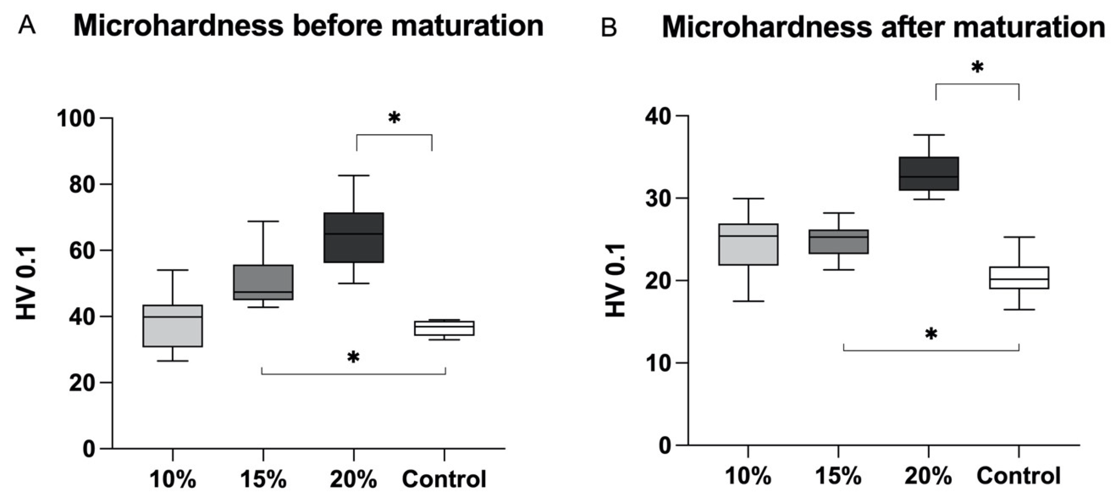
3.1. Microhardness
3.2. Flexural Strength, Compressive Strength, Diametral Tensile Strength and Fracture Toughness
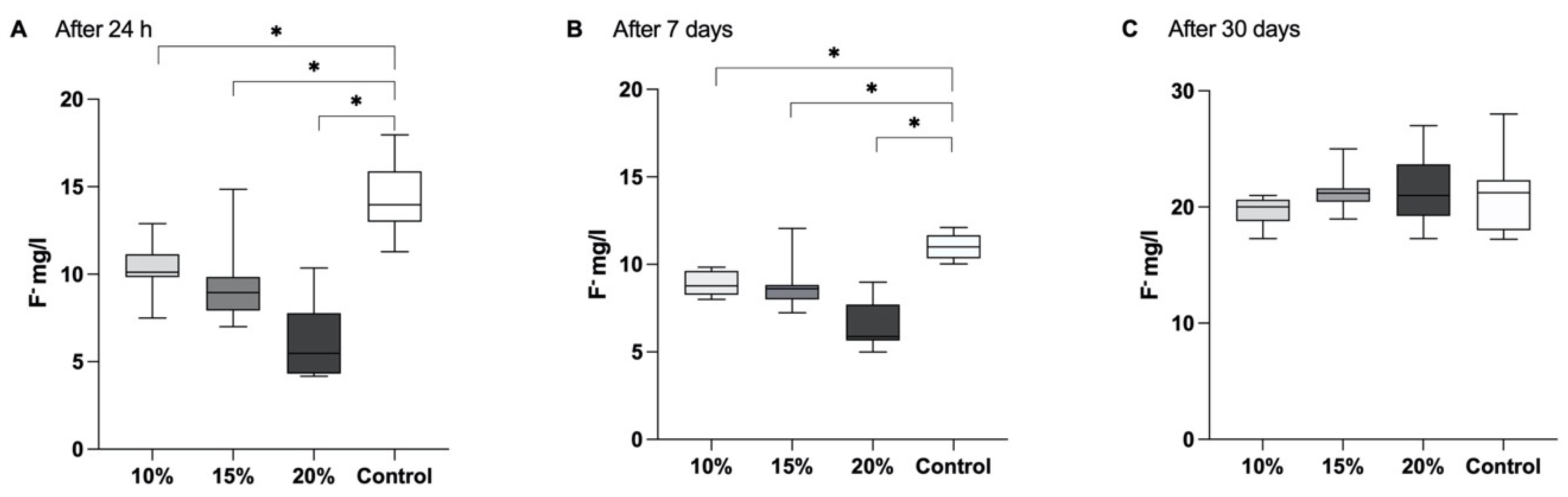
3.3. Fluoride Release
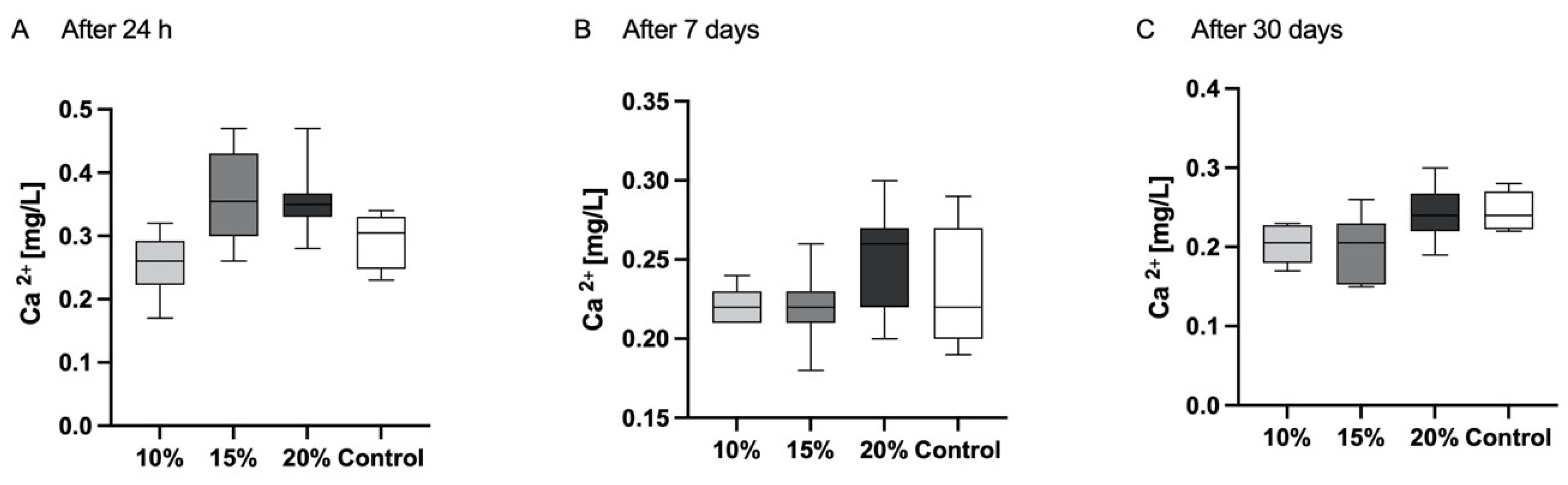
3.4. Calcium Release
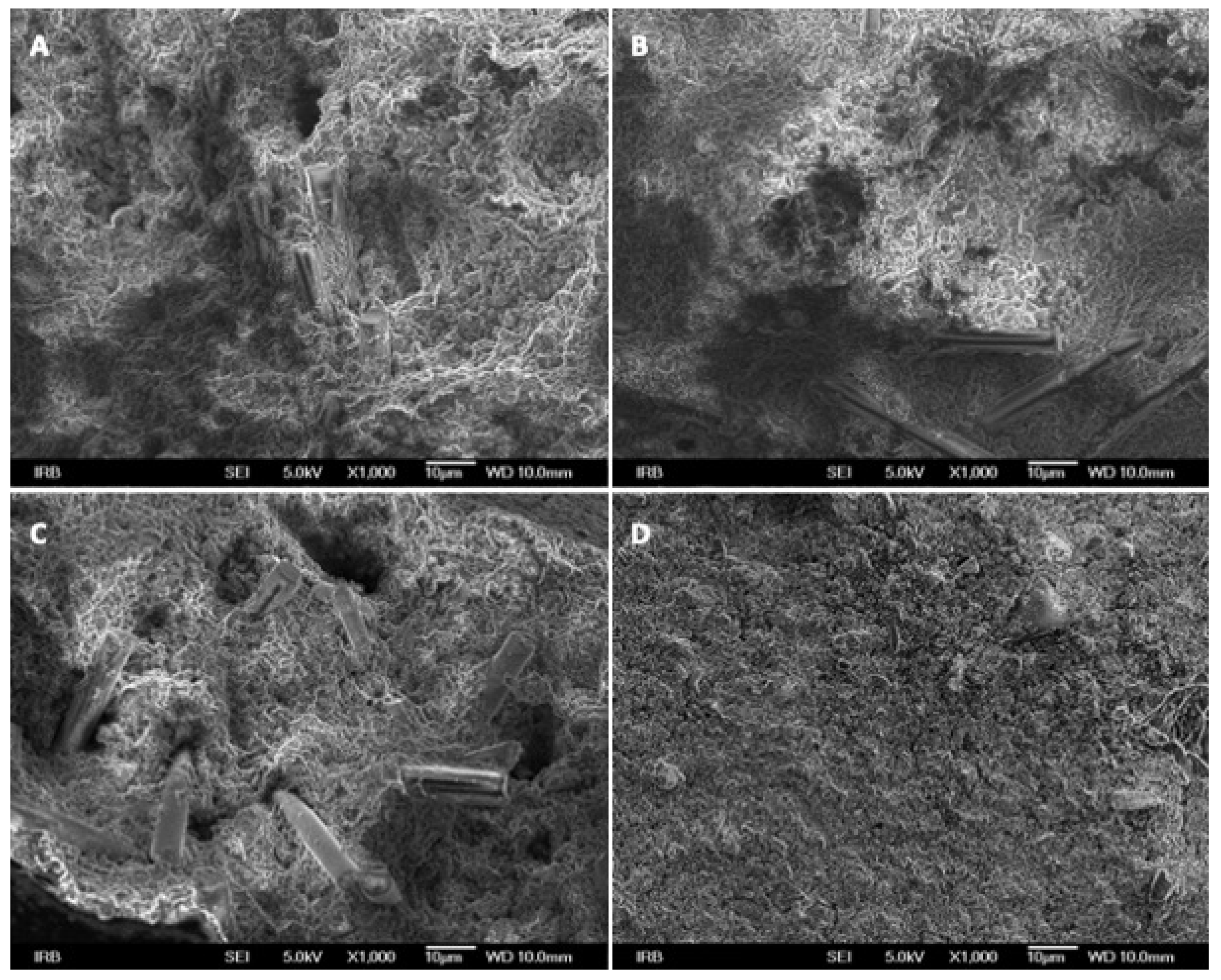
3.5. SEM
4. Discussion
5. Conclusions
Author Contributions
Funding
Institutional Review Board Statement
Data Availability Statement
Conflicts of Interest
References
- Spagnuolo, G. Bioactive Dental Materials: The Current Status. Materials 2022, 15, 2016. [Google Scholar] [CrossRef]
- Heintze, S.D.; Loguercio, A.D.; Hanzen, T.A.; Reis, A.; Rousson, V. Clinical efficacy of resin-based direct posterior restorations and glass-ionomer restorations—An updated meta-analysis of clinical outcome parameters. Dent. Mater. 2022, 38, e109–e135. [Google Scholar] [CrossRef]
- Opdam, N.J.; van de Sande, F.H.; Bronkhorst, E.; Cenci, M.S.; Bottenberg, P.; Pallesen, U.; Gaengler, P.; Lindberg, A.; Huysmans, M.C.; van Dijken, J.W. Longevity of posterior composite restorations: A systematic review and meta-analysis. J. Dent. Res. 2014, 93, 943–949. [Google Scholar] [CrossRef] [PubMed]
- Francois, P.; Fouquet, V.; Attal, J.P.; Dursun, E. Commercially Available Fluoride-Releasing Restorative Materials: A Review and a Proposal for Classification. Materials 2020, 13, 2313. [Google Scholar] [CrossRef]
- Bueno, L.S.; Silva, R.M.; Magalhães, A.P.R.; Navarro, M.F.L.; Pascotto, R.C.; Buzalaf, M.A.R.; Nicholson, J.W.; Sidhu, S.K.; Borges, A.F.S. Positive correlation between fluoride release and acid erosion of restorative glass-ionomer cements. Dent. Mater. 2019, 35, 135–143. [Google Scholar] [CrossRef]
- Naganuma, Y.; Takahashi, M.; Takada, Y.; Hoshi, K.; Kitaoka, A.; Takahashi, A.; Sasaki, K. Usefulness of conventional glass ionomer cements in an environment of insufficient moisture exclusion. J. Oral. Sci. 2022, 64, 242–246. [Google Scholar] [CrossRef] [PubMed]
- Burke, F.J. Dental Materials: What Goes Where? Class V Restorations. Dent. Update 2015, 42, 829–839. [Google Scholar] [CrossRef]
- Tanaka, C.B.; Ershad, F.; Ellakwa, A.; Kruzic, J.J. Fiber reinforcement of a resin modified glass ionomer cement. Dent. Mater. 2020, 36, 1516–1523. [Google Scholar] [CrossRef] [PubMed]
- Ilie, N.; Hickel, R. Resin composite restorative materials. Aust. Dent. J. 2011, 56 (Suppl. S1), 59–66. [Google Scholar] [CrossRef] [PubMed]
- Baig, M.S.; Fleming, G.J. Conventional glass-ionomer materials: A review of the developments in glass powder, polyacid liquid and the strategies of reinforcement. J. Dent. 2015, 43, 897–912. [Google Scholar] [CrossRef]
- Nicholson, J.W.; Sidhu, S.K.; Czarnecka, B. Enhancing the Mechanical Properties of Glass-Ionomer Dental Cements: A Review. Materials 2020, 13, 2510. [Google Scholar] [CrossRef]
- Alshabib, A.; Jurado, C.A.; Tsujimoto, A. Short fiber-reinforced resin-based composites (SFRCs); Current status and future perspectives. Dent. Mater. J. 2022, 41, 647–654. [Google Scholar] [CrossRef]
- Rajak, D.K.; Pagar, D.D.; Menezes, P.L.; Linul, E. Fiber-Reinforced Polymer Composites: Manufacturing, Properties, and Applications. Polymers 2019, 11, 1667. [Google Scholar] [CrossRef]
- Nicholson, J.W.; Sidhu, S.K.; Czarnecka, B. Fluoride exchange by glass-ionomer dental cements and its clinical effects: A review. Biomater. Investig. Dent. 2023, 10, 2244982. [Google Scholar] [CrossRef]
- Xie, D.; Brantley, W.A.; Culbertson, B.M.; Wang, G. Mechanical properties and microstructures of glass-ionomer cements. Dent. Mater. 2000, 16, 129–138. [Google Scholar] [CrossRef] [PubMed]
- Kukreja, R.; Singla, S.; Bhadoria, N.; Pawar, P.; Gupta, K.; Khandelwal, D.; Dewani, N. An In Vitro Study to Compare the Release of Fluoride from Glass Ionomer Cement (Fuji IX) and Zirconomer. Int. J. Clin. Pediatr. Dent. 2022, 15, 35–37. [Google Scholar] [CrossRef] [PubMed]
- ISO 4049:2009; Dentistry—Polymer-Based Filling, Restorative and Luting Materials. International Organization for Standardization: Geneva, Switzerland, 2000.
- Garoushi, S.; Vallittu, P.; Lassila, L. Hollow glass fibers in reinforcing glass ionomer cements. Dent. Mater. 2017, 33, e86–e93. [Google Scholar] [CrossRef] [PubMed]
- ISO 9917-1:2007; Dentistry Water-Based cements Part 1: Powder/Liquid Acid-Base Cements. International Organization for Standardization: Geneva, Switzerland, 2007.
- Garoushi, S.; He, J.; Obradovic, J.; Fardim, P.; Vallittu, P.K.; Lassila, L. Incorporation of cellulose fiber in glass ionomer cement. Eur. J. Oral Sci. 2020, 128, 81–88. [Google Scholar] [CrossRef]
- Garoushi, S.; Vallittu, P.K.; Lassila, L. Reinforcing effect of discontinuous microglass fibers on resin-modified glass ionomer cement. Dent. Mater. J. 2018, 37, 484–492. [Google Scholar] [CrossRef]
- ISO 19448:2018; Dentistry Analysis of Fluoride Concentration in Aqueous Solutions by Use of Fluoride Ion-Selective Electrode. International Organization for Standardization: Geneva, Switzerland, 2018.
- de Lima Navarro, M.F.; Pascotto, R.C.; Borges, A.F.S.; Soares, C.J.; Raggio, D.P.; Rios, D.; Bresciani, E.; Molina, G.F.; Ngo, H.C.; Miletić, I.; et al. Consensus on glass-ionomer cement thresholds for restorative indications. J. Dent. 2021, 107, 103609. [Google Scholar] [CrossRef]
- Garoushi, S.K.; He, J.; Vallittu, P.K.; Lassila, L.V.J. Effect of discontinuous glass fibers on mechanical properties of glass ionomer cement. Acta Biomater. Odontol. Scand. 2018, 4, 72–80. [Google Scholar] [CrossRef] [PubMed]
- Dowling, A.H.; Stamboulis, A.; Fleming, G.J. The influence of montmorillonite clay reinforcement on the performance of a glass ionomer restorative. J. Dent. 2006, 34, 802–810. [Google Scholar] [CrossRef] [PubMed]
- Cökeliler, D.; Erkut, S.; Zemek, J.; Biederman, H.; Mutlu, M. Modification of glass fibers to improve reinforcement: A plasma polymerization technique. Dent. Mater. 2007, 23, 335–342. [Google Scholar] [CrossRef]
- Tezvergil, A.; Lassila, L.V.; Vallittu, P.K. The shear bond strength of bidirectional and random-oriented fibre-reinforced composite to tooth structure. J. Dent. 2005, 33, 509–516. [Google Scholar] [CrossRef] [PubMed]
- Song, F.V.; Yang, B.; Tommaso, D.D.; Donnan, R.S.; Chass, G.A.; Yada, R.Y.; Farrar, D.H.; Tian, K.V. Resolving nanoscopic structuring and interfacial THz dynamics in setting cements. Mater. Adv. 2022, 3, 4982–4990. [Google Scholar] [CrossRef]
- Souza, J.C.M.; Fernandes, V.; Correia, A.; Miller, P.; Carvalho, O.; Silva, F.; Özcan, M.; Henriques, B. Surface modification of glass fiber-reinforced composite posts to enhance their bond strength to resin-matrix cements: An integrative review. Clin. Oral. Investig. 2022, 26, 95–107. [Google Scholar] [CrossRef]
- Hammouda, I.M. Reinforcement of conventional glass-ionomer restorative material with short glass fibers. J. Mech. Behav. Biomed. Mater. 2009, 2, 73–81. [Google Scholar] [CrossRef]
- Imataki, R.; Shinonaga, Y.; Nishimura, T.; Abe, Y.; Arita, K. Mechanical and Functional Properties of a Novel Apatite-Ionomer Cement for Prevention and Remineralization of Dental Caries. Materials 2019, 12, 3998. [Google Scholar] [CrossRef]
- Feng, J.; Cheng, L.; Zhou, X.; Xu, H.H.K.; Weir, M.D.; Li, Q.; Hannig, M.; Rupf, S. Effects of water aging on the mechanical and anti-biofilm properties of glass-ionomer cement containing dimethylaminododecyl methacrylate. Dent. Mater. 2019, 35, 434–443. [Google Scholar] [CrossRef]
- Mitra, S.B. Adhesion to dentin and physical properties of a light-cured glass-ionomer liner/base. J. Dent. Res. 1991, 70, 72–74. [Google Scholar] [CrossRef]
- Dowling, A.H.; Fleming, G.J.; McGinley, E.L.; Addison, O. Improving the standard of the standard for glass ionomers: An alternative to the compressive fracture strength test for consideration? J. Dent. 2012, 40, 189–201. [Google Scholar] [CrossRef]
- Heintze, S.D.; Ilie, N.; Hickel, R.; Reis, A.; Loguercio, A.; Rousson, V. Laboratory mechanical parameters of composite resins and their relation to fractures and wear in clinical trials—A systematic review. Dent. Mater. 2017, 33, e101–e114. [Google Scholar] [CrossRef]
- Attik, N.; Colon, P.; Gauthier, R.; Chevalier, C.; Grosgogeat, B.; Abouelleil, H. Comparison of physical and biological properties of a flowable fiber reinforced and bulk filling composites. Dent. Mater. 2022, 38, e19–e30. [Google Scholar] [CrossRef]
- Molnár, J.; Fráter, M.; Sáry, T.; Braunitzer, G.; Vallittu, P.K.; Lassila, L.; Garoushi, S. Fatigue performance of endodontically treated molars restored with different dentin replacement materials. Dent. Mater. 2022, 38, e83–e93. [Google Scholar] [CrossRef]
- Kumari, P.D.; Khijmatgar, S.; Chowdhury, A.; Lynch, E.; Chowdhury, C.R. Factors influencing fluoride release in atraumatic restorative treatment (ART) materials: A review. J. Oral. Biol. Craniofacial Res. 2019, 9, 315–320. [Google Scholar] [CrossRef]
- Moshaverinia, M.; Borzabadi-Farahani, A.; Sameni, A.; Moshaverinia, A.; Ansari, S. Effects of incorporation of nano-fluorapatite particles on microhardness, fluoride releasing properties, and biocompatibility of a conventional glass ionomer cement (GIC). Dent. Mater. J. 2016, 35, 817–821. [Google Scholar] [CrossRef]
- Tiwari, S.; Kenchappa, M.; Bhayya, D.; Gupta, S.; Saxena, S.; Satyarth, S.; Singh, A.; Gupta, M. Antibacterial Activity and Fluoride Release of Glass-Ionomer Cement, Compomer and Zirconia Reinforced Glass-Ionomer Cement. J. Clin. Diagn. Res. 2016, 10, Zc90–Zc93. [Google Scholar] [CrossRef] [PubMed]
- Jingarwar, M.M.; Pathak, A.; Bajwa, N.K.; Sidhu, H.S. Quantitative assessment of fluoride release and recharge ability of different restorative materials in different media: An in vitro study. J. Clin. Diagn. Res. 2014, 8, Zc31–Zc34. [Google Scholar] [CrossRef] [PubMed]
- Sauro, S.; Spagnuolo, G.; Del Giudice, C.; Neto, D.M.A.; Fechine, P.B.A.; Chen, X.; Rengo, S.; Chen, X.; Feitosa, V.P. Chemical, structural and cytotoxicity characterisation of experimental fluoride-doped calcium phosphates as promising remineralising materials for dental applications. Dent. Mater. 2023, 39, 391–401. [Google Scholar] [CrossRef] [PubMed]
- Zalizniak, I.; Palamara, J.E.; Wong, R.H.; Cochrane, N.J.; Burrow, M.F.; Reynolds, E.C. Ion release and physical properties of CPP-ACP modified GIC in acid solutions. J. Dent. 2013, 41, 449–454. [Google Scholar] [CrossRef] [PubMed]
- Silva, K.G.; Pedrini, D.; Delbem, A.C.; Cannon, M. Effect of pH variations in a cycling model on the properties of restorative materials. Oper. Dent. 2007, 32, 328–335. [Google Scholar] [CrossRef] [PubMed]
- Perera, D.; Yu, S.C.H.; Zeng, H.; Meyers, I.A.; Walsh, L.J. Acid Resistance of Glass Ionomer Cement Restorative Materials. Bioengineering 2020, 7, 150. [Google Scholar] [CrossRef] [PubMed]
- Nica, I.; Stoleriu, S.; Iovan, A.; Tărăboanță, I.; Pancu, G.; Tofan, N.; Brânzan, R.; Andrian, S. Conventional and Resin-Modified Glass Ionomer Cement Surface Characteristics after Acidic Challenges. Biomedicines 2022, 10, 1755. [Google Scholar] [CrossRef] [PubMed]
- Martins, R.A.; Marti, L.M.; Mendes, A.C.B.; Fragelli, C.; Cilense, M.; Zuanon, A.C.C. Brushing Effect on the Properties of Glass Ionomer Cement Modified by Hydroxyapatite Nanoparticles or by Bioactive Glasses. Int. J. Dent. 2022, 2022, 1641041. [Google Scholar] [CrossRef]
- Karatas, O.; Gul, P.; Akgul, N.; Celik, N.; Gundogdu, M.; Duymus, Z.Y.; Seven, N. Effect of staining and bleaching on the microhardness, surface roughness and color of different composite resins. Dent. Med. Probl. 2021, 58, 369–376. [Google Scholar] [CrossRef]
- Altaie, S.F. Tribological, microhardness and color stability properties of a heat-cured acrylic resin denture base after reinforcement with different types of nanofiller particles. Dent. Med. Probl. 2023, 60, 295–302. [Google Scholar] [CrossRef]


| Material | FS (MPa) | CS (MPa) | DTS (MPa) | FT (MPam1/2) |
|---|---|---|---|---|
| Control | 52 (9) A | 191 (12) AB | 21 (3) A | 0.8 (0.1) A |
| 10% | 60 (6) AB | 174 (8) B | 23 (2) AB | 1.4 (0.3) B |
| 15% | 62 (10) AB | 173 (9) B | 25 (3) AB | 1.4 (0.2) B |
| 20% | 78 (12) BC | 172 (18) B | 30 (4) BC | 1.7 (0.1) C |
Disclaimer/Publisher’s Note: The statements, opinions and data contained in all publications are solely those of the individual author(s) and contributor(s) and not of MDPI and/or the editor(s). MDPI and/or the editor(s) disclaim responsibility for any injury to people or property resulting from any ideas, methods, instructions or products referred to in the content. |
© 2024 by the authors. Licensee MDPI, Basel, Switzerland. This article is an open access article distributed under the terms and conditions of the Creative Commons Attribution (CC BY) license (https://creativecommons.org/licenses/by/4.0/).
Share and Cite
Ivica, A.; Šalinović, I.; Jukić Krmek, S.; Garoushi, S.; Lassila, L.; Säilynoja, E.; Miletić, I. Mechanical Properties and Ion Release from Fibre-Reinforced Glass Ionomer Cement. Polymers 2024, 16, 607. https://0-doi-org.brum.beds.ac.uk/10.3390/polym16050607
Ivica A, Šalinović I, Jukić Krmek S, Garoushi S, Lassila L, Säilynoja E, Miletić I. Mechanical Properties and Ion Release from Fibre-Reinforced Glass Ionomer Cement. Polymers. 2024; 16(5):607. https://0-doi-org.brum.beds.ac.uk/10.3390/polym16050607
Chicago/Turabian StyleIvica, Anja, Ivan Šalinović, Silvana Jukić Krmek, Sufyan Garoushi, Lippo Lassila, Eija Säilynoja, and Ivana Miletić. 2024. "Mechanical Properties and Ion Release from Fibre-Reinforced Glass Ionomer Cement" Polymers 16, no. 5: 607. https://0-doi-org.brum.beds.ac.uk/10.3390/polym16050607