Article

Phytophthora Antagonism of Endophytic Bacteria Isolated from Roots of Black Pepper (Piper nigrum L.)

1
Institute of Biotechnology & Environment, Tay Nguyen University, 567 Le Duan, Buon Ma Thuot city 630000, Vietnam
2
Department of Chemistry, Tamkang University, New Taipei City 25137, Taiwan
3
Department of Science and Technology, Tay Nguyen University, 567 Le Duan, Buon Ma Thuot city 630000, Vietnam
*
Authors to whom correspondence should be addressed.
Submission received: 13 January 2020 / Revised: 7 February 2020 / Accepted: 13 February 2020 / Published: 17 February 2020
(This article belongs to the Special Issue Integrating Pest Management into Agricultural Production Systems)

Abstract

:
In this study, 90 root samples were collected from 30 black pepper farms in three provinces in the Central Highlands of Vietnam. A total of 352 endophytic bacteria were isolated and their morphology described. An in vitro assay on the antifungal activity of these isolates was then conducted and 47 isolates were found to have antagonistic activity on Phytophthora fungi. The antifungal activity of the 47 isolates was evaluated in vivo by shoot assay. Among these 47 isolates, 6 were selected for further investigation. The six isolates were classified and identified by sequencing the 16S RNA gene and phylogeny. The results showed that all six endophytic bacteria belong to the following species of Bacillus genus: B. siamensis, B. amyloliquefaciens, B. velezenis, and B. methylotrophiycus. Enzymatic activity related to the antifungal activity of the six potent isolates was determined; it showed that they possessed high chitinase and protease activities. These isolates were applied for black pepper seedlings in greenhouse. The results showed three promising isolates: B. siamensis EB.CP6, B. velezensis EB.KN12, and B. methylotrophycus EB.KN13. Black pepper seedlings treated with the promising bacteria had the lowest rate of root disease (8.45–11.21%) and lower fatal rate (11.11–15.55%) compared to the control group (24.81% and 24.44%). In addition, the three promising isolates strongly affected the growth of the black pepper seedlings in greenhouse. The plant height, length of roots, and fresh biomass of the seedlings in the treated plots were higher than those in the control plots. Thus, the endophytic bacterial isolates have the potential to act as biocontrol agent for the sustainable production of black pepper.

1. Introduction

Endophytic bacteria belong to a bacterial group living in the tissues of plants, including roots, shoots, leaves, and even fruits. Endophytic bacteria interact with the host in growth promotion, nutrition uptake, pathogenic fungal antagonism, and nematode resistance [1,2,3,4,5,6]. For fungal antagonism, endophytic bacteria can release chitinases, beta glucanases, proteases, chemical compounds, and antibiotics, improving the environmental competitiveness [7,8,9]. In addition, endophytic bacteria have beneficial bioactivities such as nitrogen fixing, phosphorous solubility, and IAA biosynthesis to stimulate the growth of crops, reduce fertilizer application, and increase drought tolerance [10,11,12]. Therefore, studies on the application of endophytic bacteria for sustainable agricultural production have received significantly more attention worldwide.
Phytophthora is a pathogenic fungus causing diseases and serious damage to the growth and productivity of agricultural crops. Phytophthora is the most common fungus associated with root rot disease of agricultural crops, such as tomato, potato, onion, coffee, rubber, pepper, and black pepper [13,14,15]. Root rot disease caused by Phytophthora is one of the sources of serious production reduction in black pepper in Vietnam, India, Malaysia, and other countries [16,17]. Using chemical fungicides to manage Phytophthora in black pepper production is the most common application. But this method causes toxic pollution, harmful to the farmers and ecological system. In order to decrease the usage of toxic chemical fungicides for black pepper production, various options are recommended, such as the development of resistant varieties, agricultural practices, and the use of biocontrol agents [18,19,20].
Among these solutions, biocontrol has attracted much more attention with good reason. Aravind et al. (2009) isolated 71 endophytic strains; three strains with 70% Phytophthora antagonism in greenhouse were selected [17]. Jasim et al. [21] isolated endophytic bacteria from black pepper, which belong to Klebsiella, Enterobacter genera; they may promote the growth of black pepper and inhibit Phytopthora fungus. Munjal et al. [22] pointed out that Bacillus megaterium produced some antibiotics that strongly inhibit Phytophthora capsici, Pythium myriotylum, and Rhizotocnia solani. Nguyen et al. [12] isolated 106 endophytic isolates and found that Bacillus amyloliquefaciens EB.EK2 produces seven potent biochemical compounds for Phytophthora resistance. Trinh et al. [16] found a promising antifugal rhizobacteria Bacillus velezensis RB. DS29. This strain inhibited 98.75% Phytopthora growth in an in vivo bioassay because of the impacts of various enzymes and biochemical compounds. A few global reports on endophytic bacteria have emerged, but no report on this field in Vietnam has been presented. Therefore, for further investigation in this study, endophytic bacteria were isolated from the roots of black pepper and screened for their biochemical characteristics. Promising strains that might have strong antagonism against Phytophthora fungus were selected. They will be used for further investigation of biochemical compounds for Phytophthora antagonism and used as biocontrol agents for sustainable production of black pepper.

2. Materials and Methods

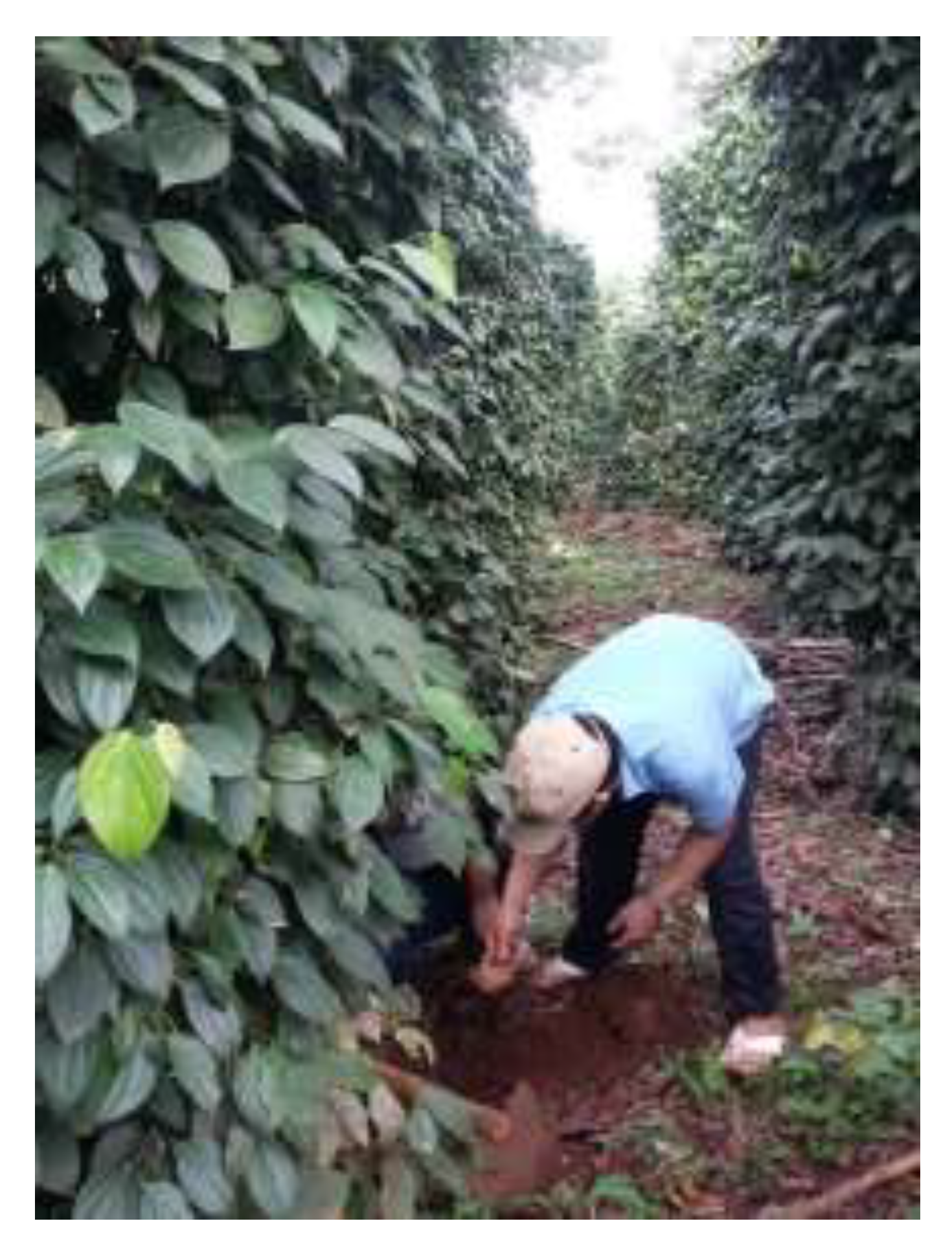
2.1. Method for Sampling

Ninety black pepper root samples were collected from 30 farms in Gia Lai, Dak Lak, and Dak Nong provinces in the Central Highlands, the largest area of black pepper production in Vietnam. The young root samples were collected from healthy 4–5 years old pepper plants of three local varieties: Vinh Linh, Trau, and Loc Ninh. Each farm selected three plants for sampling. The root samples were collected 1 m from the trunk at 0–30 cm depths (Figure 1). The root samples were then transported in sterilized polyethylene bags in ice pack to the laboratory. If the samples could not be processed immediately, they were kept at 4 ℃ for no longer than 18 to 24 h.

2.2. Isolation of Endophytic Bacteria

Endophytic bacteria were isolated from the internal tissues of the roots by the method of Aravind et al. (2009) [17]. In brief, the root samples were washed with water and cut into 1–2 cm pieces. The surface of the roots was sterilized with 2% sodium hypochlorite for 10 min and then 70% ethanol for 1 min. After that, the samples were washed six times with sterilized distilled water. Finally, the samples were checked for the efficacy of the disinfection procedure by inoculation of the last wash solution on TSA medium. One gram of the root tissue samples was grinded in 5 mL phosphate buffer (PBS) and centrifuged (600 rpm) at 5 ℃ for 1 min. One mL of the supernatant was diluted up to 105 and, 100 μL solution was then plated onto TSA medium; the plates were incubated at 30 °C for 48 h. Each individual colony of each sample was separated and grown on TSA medium, and then stored at −32 ℃ in 20% glycerol for further investigation. The morphology of the bacterial colonies and cells was characterized according to Bergey Manual [23]. The isolates having different morphology and biological activity were kept for further investigation. The characters and number of the isolates are named by districts collected as CP: Cu Prong; CS: Cu Se; DC: Duc Co District (Gia Lai Province); EH: Ea Hleo; KN: Krong Nang; CK: Cu Kuin district (Dak Lak Province); DS: Dak Song; DM: Dak Min, and DL: Dak RLap District (Dak Nong Province). EB prefix of the names of bacteria is endophytic bacteria.

2.3. In Vitro Phytophthora Antagonism by the Endophytic Bacteria

The pathogen Phytophthora is a fungal strain collected from the Institute of Biotechnology and Environment at Tay Nguyen University at Vietnam. It was grown on PDA medium at 30 °C.
All bacterial isolates were evaluated for their antifungal activity against Phytophthora on PDA plates by the method described by Tran et al. [24]. In brief, a mycelial plug of growing Phytophthora was placed in the center of the PDA medium and endophytic bacteria were streaked 2 cm on three sides of it. The plates were then incubated at 28 °C for 5 d or until the leading edge of Phytophthora in the control group reached the edge of the plate. The tests were conducted in triplicates (Figure 2).
The radial growth of fungal mycelium was measured and the percentage of growth inhibition calculated as follows:
Rate of mycelium growth inhibition (%) = [(D1 – D2)]/D1] × 100, where D1 = diameter of the fungus mycelium grown on the control disk (cm) and D2 = diameter of the fungus mycelium grown on the treated bacteria disk (cm).

2.4. In Vivo Phytophthora Antagonism

In vivo antagonism tests of the endophytic bacteria were conducted following the Dinus method [25]. Black pepper shoots (about 8 cm in length with at least one node) were excised from healthy black pepper vines (Vinh Linh local variety) and washed thoroughly with tap water before surface sterilization with 0.1% sodium hypochlorite for 10 min. The shoots were then washed five times with sterile distilled water. After that, the shoots were dried on sterilized paper. The shoots were put into the endophytic bacteria suspension (107 CFU/mL) for 60 min, and then placed on sterilized papers to remove excess moisture. Each experiment was conducted in triplicates. The treated shoots were incubated with Phytophthora fungus and kept in a plastic tray at 30 °C for 3 d in the dark. The moisture filter paper was kept at the bottom of the tray to provide high humidity. Three replications were maintained with three shoots in each bacterial treatment. The length of the dark lesions that developed along the inoculated spots on the shoots was determined after the 96-h experiment. In the control group, the shoots were inoculated with Phytophthora but not treated with the endophytic bacteria:
Lesion inhibition (%) = [(D1 – D2)]/D1] × 100
where D1 = length of the dark lesion inoculated with fungus Phytophthora (cm) and D2 = length of the dark lesion inoculated with the fungus treated with endophytic bacteria (cm).

2.5. Bioassay in the Greenhouse

Black pepper seedlings, a Vinh Linh local variety with seven leaves, were used for Phytophthora antagonism testing in greenhouse. Six potent endophytic bacteria were cultivated in LB (composition L−1: 10 g tryptone, 5 g yeast extract, and 10 g NaCl) for 72 h at 25 °C with a shaking speed of 150 rpm. The bacteria culture was adjusted to 107 CFU/mL by optical density (UV Vis spectrophotometer, Jasco V630, Japan).
Phytophthora fungus was grown on potato dextrose medium for 3 d at 28 °C and a shaking speed of 150 rpm; spore density was adjusted to 107 spores/mL. The evaluation of the Phytophthora antagonism by the endophytic bacteria was conducted in greenhouse. The six potent endophytic bacteria and two control groups were used for this experiment. Each seedling was irrigated with 10 mL bacterial suspension (107 CFU/mL), and irrigated with 10 mL (107 spore/mL) one month later. The bioassay had a total of eight formulas in triplicates (24 plots) and twenty seedlings per plot. The experiment was designed as a random completed block design (RCBD). The greenhouse conditions were: 75–80% humidity, temperature of 25–30 °C, and light intensity of 2000–3000 lx. All the seedlings in the experiment in greenhouse were taken care of, following technical guidance by the Vietnam agriculture department.
All plots except for Control groups 1 and 2 were treated with 10 mL of endophytic bacteria suspension (107 CFU/mL)/plant. After a month, the seedlings were treated with 10 mL of Phytophthora spore suspension (107 spores/mL). The experiment was conducted for three months under greenhouse conditions. The growth data on the black pepper seedlings, rate of Phytophthora infection and rate of fatality were observed; mean values were calculated from six plants.

2.6. Chitinase Activity of the Endophytic Bacteria

The endophytic bacteria were grown in LB medium supplemented with 0.1% colloidal chitin for 5 d at 30 °C and a shaking speed of 150 rpm. Cells were separated by centrifugation at 6000 × g and 4 °C for 5 min. The supernatant was dialyzed overnight at 4 °C using 20 mM sodium phosphate buffer (pH 6.0). The dialyzed protein solution was used to measure the chitinase activity. The chitinase activity assay was conducted in a 600 μL reaction mixture containing 0.1% colloidal chitin as the substrate and an appropriate volume of crude enzymes in 20 mM sodium phosphate buffer (pH 6.0) in triplicates. The reaction mixture was incubated at 37 °C for 15 min. Chitinase activity was determined via the method described by Imoto [26].

2.7. Protease Activity of the Endophytic Bacteria

The bacteria were grown in LB medium for 5 d, at 30 °C with shaking speed of 150 rpm. The supernatant was prepared as per the procedures described above for chitinase activity. The reaction mixture, containing 5 mL of 1% casein and 1 mL of crude enzymes, was kept at 35.5 °C for 10 min in triplicates. The reaction was terminated by the addition of 10 mL of 5% TCA (trichloroacetic acid). After filtration, 3 mL Folin Ciocalteau reagent was added to the solution. It was kept for 10 min at room temperature before being measured at 660nm by UV Vis (Jasco V630, Japan), following Ansons methods [27].

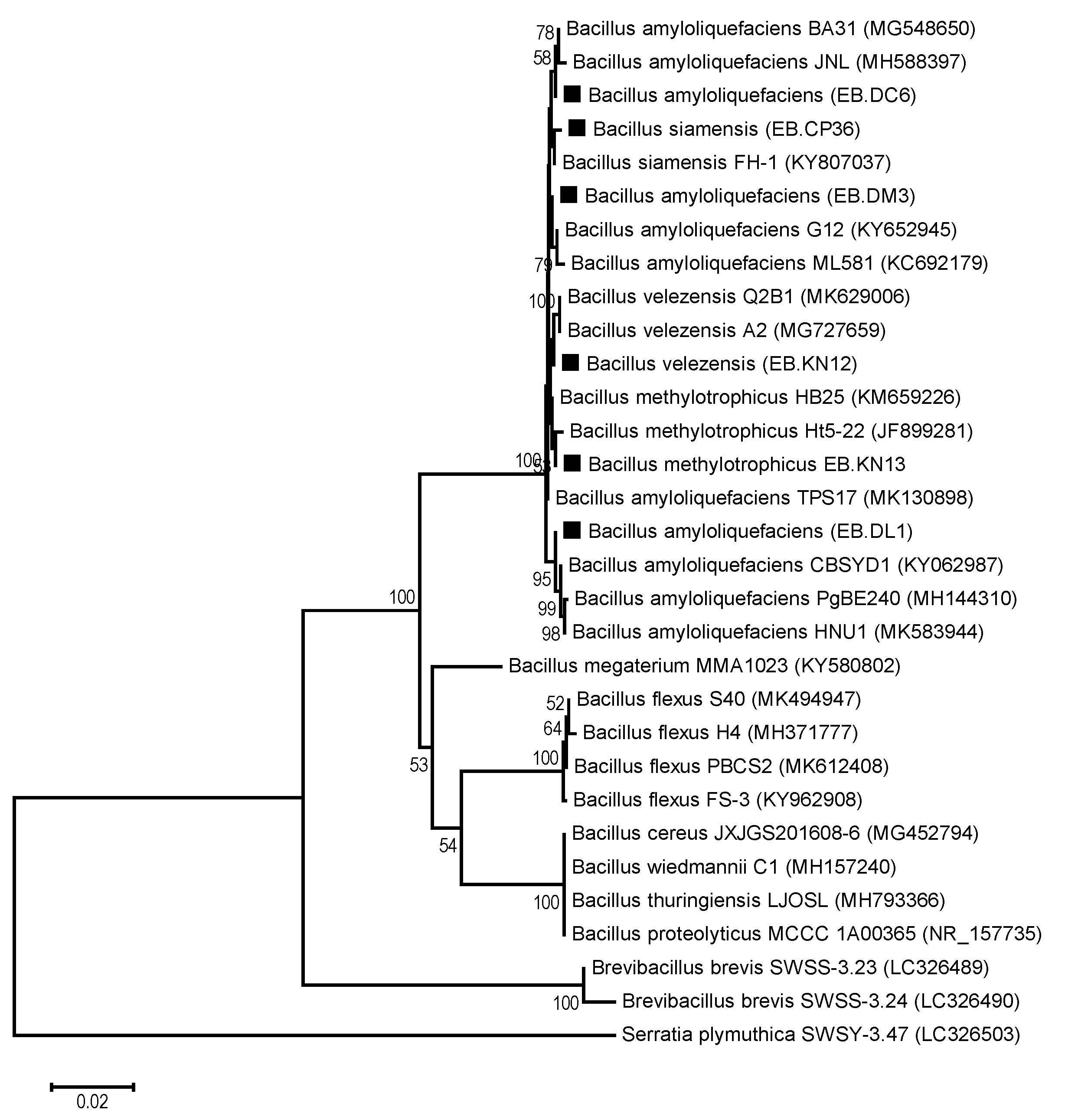
2.8. PCR Amplification, Sequencing, and Phylogenetic Analysis of the 16S rRNA Gene

Genomic DNA from an overnight culture of each strain was extracted and used as a template for amplification by PCR. A nearly full-length segment of 16S rRNA gene nucleotides was amplified in a 100 μL reaction tube using universal primers 27f (5′-AGAGTTTGATCMTGGCTCAG-3′) and 1492r (5′-TACGGYTACCTTGTTACGACTT-3′). The 16S rRNA gene was amplified by iCycler thermal cycler (Bio-Rad, Hercules, California, USA) with the following schedule: 94 °C for 5 min, repeated in 30 cycles of denaturation at 95 °C for 30 s, annealing at 55 °C for 30 s, and extension at 72 °C for 2 min. The amplified products were then separated by electrophoresis on agarose gel (1.5% w/v). The target bands in the agarose gel were cut out and purified using a QIA quick PCR purification (Promega Co., USA). Sequencing reactions were carried out in a CEQ8000 Genetic Analysis System (Beckman Coulter Inc., USA) using a CEQ Dye Terminator Cycle Sequencing Kit (Beckman Coulter Inc., USA).
The sequences (1300 to 1440 bps) of the 16S rRNA genes were compared to known sequences in the DDBJ/Genbank/EMBL databases using BLAST (https://blast.ncbi.nlm.nih.gov/Blast.cgi) for determining the taxonomic positions of the endophytic bacteria isolates. A phylogenetic tree was built using MEGA version 6.0 software after multiple alignments of data by CLUSTAL W [28,29].

2.9. Data Analysis

Statistical analysis was performed by one-way analysis of variance (ANOVA), followed by Duncan’s multiple range tests in triplicates using SAS 9.1 software. α ≤ 0.05 was considered to be significant.

3. Results and Discussion

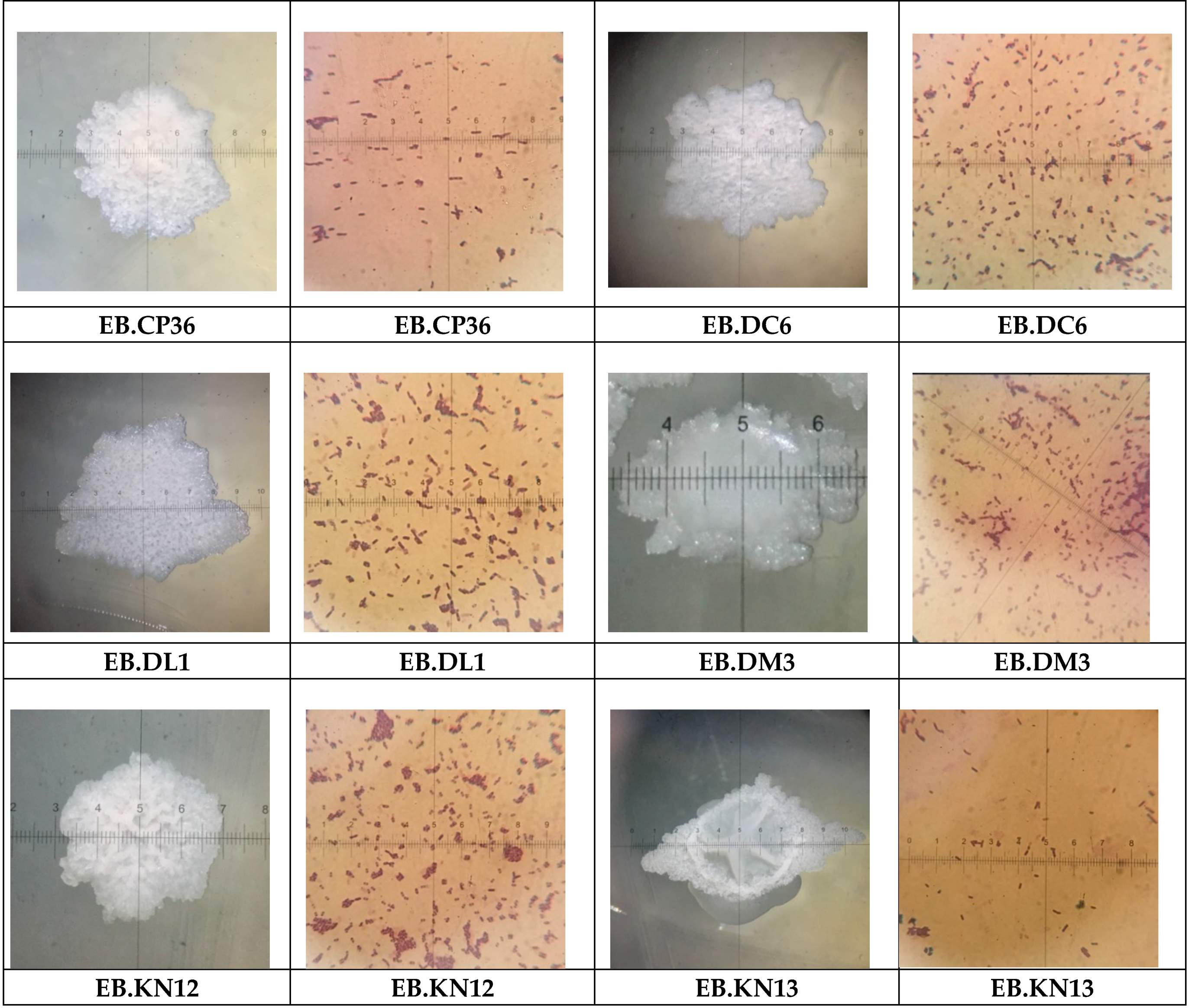
3.1. Isolation and Morphology of Endophytic Bacteria

From 90 collected root samples, 352 endophytic bacterial isolates were isolated. Most of them have quite different morphologies in the colonies, such as color, size, shape, etc., and different Phytophthora antagonism activity. The isolates of same morphology, bioactivity, and same sampling locations were removed from the collection. Some isolates of same morphology, but quite different activity, were kept separately for further study. The endophytic bacterial community in the root of black pepper plants in the Central Highland exhibited rich diversity. The results showed 106 endophytic bacteria isolated from the root collected in Gia Lai Province; 186 isolates from Dak Lak province and 60 isolates from Dak Nong province. By preliminary identification, they belong to Bacillus, Pseudomonas, Klebsiella, Enterobacter, Serratia, and Micrococcus, with most species belonging to the genus Bacillus (55.2%). The diversity of the endophytic bacteria in this study was higher than those in other reports. Toh et al. [14] screened 129 endophytic bacteria from the roots of black pepper and selected three isolates (Entrobacter cancerogenus, E. cloacae, and E. asburiae) against Phytophthora. Aravind et al. [17] reported that 74 endophytic bacteria were isolated, characterized, and evaluated against Phytophthora capsici; six genera belong to Pseudomonas spp. (20 strains), Serratia sp. (1 strain), Bacillus spp. (22 strains), Arthrobacter spp. (15 strains), Micrococcus spp. (7 strains), Curtobacterium sp. (1 strain), while eight unidentified strains were isolated from internal tissues of root and stem. Among them, three isolates (Pseudomonas aeruginosa, P. putida, and Bacillus megaterium) were identified as effective antagonistic endophytes for biological control of Phytophthora root rot in black pepper. This diversity may be caused by different varieties of local black pepper, diversity of geography in the Central Highlands, such as elevation, different soil, different weather and agricultural cultivation practice. The survey showed at least three local varieties, such as Vinh Linh, Trau, and Loc Ninh in mainly two kinds of soil: ferrasols and grey soil. It is reported that diversity of endophytic bacteria depends on the host, such as genus, species, and varieties by interaction and adaptation between plant hosts and the bacteria [30]. The diversity of endophytic bacteria community in this study was the same diversity as reported by Trinh et al. [16] who indicated that there were approximately 500 rhizobacteria isolated from the root of black pepper collected at five provinces of the Central Highlands, Vietnam. Nguyen et al. [12] isolated endophytic bacteria from the roots of black pepper cultivated in the Central Highlands that were screened for plant growth-promoting activity. It reported that 106 endophytic bacteria strains were isolated. It is clear that the Central Highlands, Vietnam with the diversity of the ecological systems is a main source of rich diversity of endophytic microorganism community.

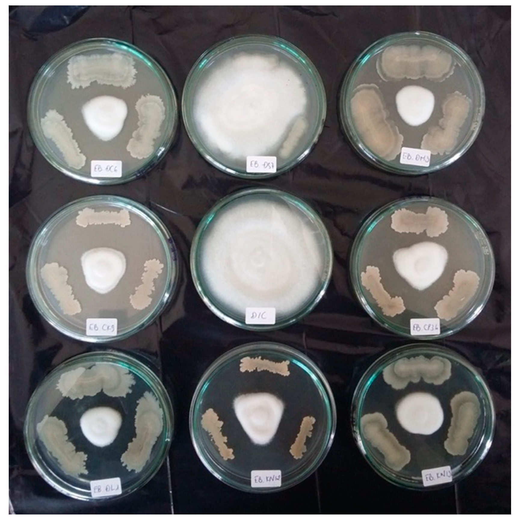
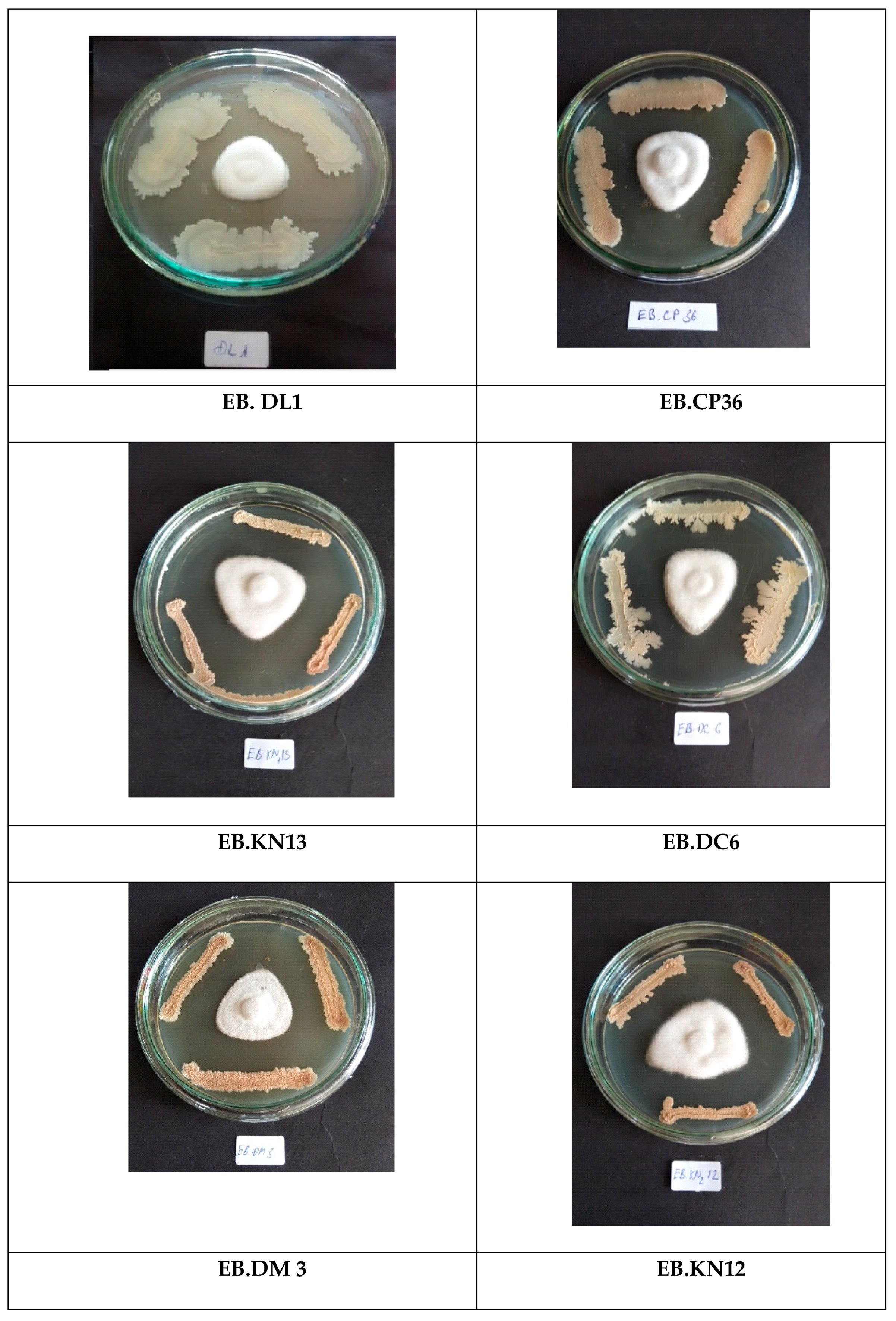
3.2. Screening Phytophthora Antagonism Activity of the Endophytic Bacteria

All 352 isolates were evaluated for Phythophtora antagonism activity in vitro. The obtained results showed that 170 isolates had less than 30% Phytophthora mycelium growth inhibition, whereas 136 and 47 isolates had 30–50% and more than 50% Phytophthora mycelium growth inhibition, respectively (Figure 2).
The results shown in Table 1 indicate that there were 47 isolates having higher than 50% Phytophthora growth inhibition on disk. From these results, six potent isolates with more than 60% fungal growth inhibition were selected for further study by EB.CP36, EB.DC6, EB.DL1, EB.DM3, EB.KN12, and EB.KN13 (Figure 3). These endophytic bacteria isolates show higher antifungal activity than those reported by Toh et al. [14]. Toh reported that among 19 endophytic isolates of black pepper, two isolates showed the highest antagonism against Phytopthora capsici, with the percentage of inhibition up to 47.63% and 43.33%, respectively [14].
The morphology of the six selected endophytic bacteria is shown in Figure 4. They are Gram positive rod-shaped bacteria and the colonies have convex irregular shape, slotted surface, with translucent, opaque, and brilliant color. The morphology of the bacteria had the characteristics of Bacillus genus. For further investigation, these bacteria were classified and identified by sequencing of 16S rRNA gene. The phylogenic analysis (Figure 5) indicated that all six bacteria belonged to Bacillus genus. They were associated to the species B. siamensis, B. amyloliquefaciens, B. velezensis, and B. methylotrophicus. All data of the gene fragments were accessed on GenBank as shown in Table 2.

3.3. In Vivo Phytophthora Antagonism by the Endophytic Bacteria

After evaluation of Phytophthora antagonism in vitro, the activity of six bacteria were determined in vivo by cut shoot bioassay. The results shown in Table 3 indicate that among the six bacterial isolates, five show 90–96% lesion inhibition: EB.CP36, EB.DL1, EB.DM3, EB.KN12, and EB.KN13. It is clear that these isolates might protect the shoots from Phytophthora infection. The activity of these isolates was higher than those reported by Dinu (2007). Dinu (2007) reported that among the 19 isolates screened, there were 5 endophytic bacterial isolates having 60–70% lesion inhibition [25]. Aravind (2008) isolated 74 endophytic bacterial isolates from black pepper with 22 Bacillus strains. Three out of the selected endophytic bacterial isolates had 70% Phytophthora capsici disease suppression in greenhouse: Bacillus megaterium, Pseudomonas putida, and Pseudomonas aeruginosa [17]. In particular, EB.DC6 isolates seemed to have no activity in vivo by shoot assay, although the Phytophthora growth inhibition in vitro was 65.84%. It means that EB.DC6 may not protect black pepper shoot from Phytophthora infection. For more discussion on mechanism of the fungal antagonism, some previous works reported that bacteria may inhibit the pathogenic fungi in combination of antibiotics, chemical compounds, enzymes, and nutrient competitions [2,3].
It is known that chitinases, proteases, and beta glucanases play very important roles in the inhibition of pathogenic fungi [14]. Therefore, chitinase and protease activity of the potent isolates was evaluated. As shown in Table 3, all isolates had chitinase and protease activity. Higher chitinase activity was found in EB.CP36 (0.507 U/mL), EB.KN13 (0.502 U/mL), EB.DM3 (0.406 U/mL), and EB.DL1 (0.343 U/mL). In regard to the protease activity, the highest was found in EB.DC6 (7.880 U/mL) and then EB.CP36 (7.372 U/mL), EB.DL1 (4.218 U/mL), and EB.KN13 (3.965 U/mL). Generally, chitinase activity of the endophytic bacterial isolates was of 0.154–0.507 U/mL and protease activity from 1.33–7.37 U/mL, the same as those from the rhizobacteria isolated from roots of black pepper [12,16]. In this study, the results show a positive correlation between chitinase activity and the Phytophthora antagonism activity (r = 0.36, n = 18), but a negative correlation between protease activity and the Phytophthora antagonism activity (r = −0.23, n = 18). This result appears to be same as results of recent works by Trinh (2019), who also indicated that proteases, chitinases, and beta glucanses produced by rhizobacteria of black pepper showed no effect on Phytophthora antagonism [16].
In most cases, pathogenic fungal inhibition of bacteria depends on toxic chemical compounds [12,16] and volatile organic compounds [18,22]. Trinh (2019) reported that rhizobacteria Bacillus velezensis RB.DS 29 isolated from root of black pepper can produce many potential fungal inhibition compounds such as: pregn-4-ene-3, 20-dione, 17-hydroxy-6-methyl-, bis (O-methyloxime)]; disulfide, methyl 1-(methylthio) propyl; propanoic acid, 2-methyl-, decyl ester; 1-propanone, 1-(2-benzofuranyl)-3-[(4-methoxyphenyl) amino]; and propanethioic acid, S-pentyl ester, metronidazole-oh and sulfadiazine. Munjal [22] also reported that endophytic bacteria of black pepper Bacillus megaterium BP17 produced some pyrazine derivatives which play a very important role in the inhibition of pathogenic fungi.

3.4. Evaluation of Phytophthora Antagonism in Greenhouse by Endophytic Bacteria

The efficacy of 6 endophytic bacteria on Phytophthora antagonism and the growth of black pepper seedlings in greenhouse was further investigated. The results (Table 4) show that the seedlings treated with EB.CP36, EB.KN12, and EB.KN13 isolates had low rate of root disease 8.45–14.21% compared to 24.81% in Control group 2. These results led to the fatal rate (%) in these plots being 11.11–15.55% lower than those in Control 2 (24.44%) in three months, similar to the report by Trinh (2019) treating with B. velezensis [16]. The results (Table 4) show that EB.DC6 isolate seems to have low efficacy on the Phytophthora antagonism, the same as in vivo by shoot assay (Table 3). It indicates that three bacteria, EB.CP36, EB.KN12, and EB.KN13 were affected most by Phytophthora antagonism activity in vivo under greenhouse condition. Three endophytic bacteria, B. siamensis, B. amyloliquefaciens, and B. velezensis are known as potential biocontrol agents in general [12,16,17,31,32,33]. B. velezensis FZB42 was able to form biofilm for increasing biocontrol. This strain can also synthesize antifungal compounds, such as fengycin, bacillomycin D, difficidin, bacilysin, and amylocyclicin [34]. B. velezensis has been used in the biocontrol of both wheat, powdery mildew by Blumeria granminis and Wilt Disease Fusarium oxysporium [35].
Recently, Cheng et al. (2019) reported that B. methylotrophicus showed efficacy on the management of Maize stalk rot [36]. But there have not been any reports on biocontrol of B. siamensis and B. methylotrophicus in black pepper.
EB.CP36, EB.KN12, and EB.KN13 isolates have not only Phytophthora antagonism activity but also stimulated growth of the seedlings in greenhouse (Table 4). Growth data on the seedlings treated by these isolates, such as number of leaves, plant height, biomass, and length of roots, were significantly higher than the Control 1 and 2 groups, and the other three bacteria observed (p < 0.05). Endophytic bacteria have been known as biocontrol agents and for plant growth promotion because of nitrogen fixing, soluble phosphorous, and IAA biosynthesis activities [12,14,16,17,25]. The three endophytic bacteria: EB.CP36, EB.KN12, and EB.KN13 are promising biocontrol agents; therefore, further investigation and application should be performed.

4. Conclusions

It is concluded that three selected endophytic bacteria of the screened bacteria show high Phytophthora antagonism activity in vivo in greenhouse. These isolates belong to the B. siamensis, B. velezensis, and B. methylotrophicus species, which have both pathogenic fungal antagonism and plant growth promotion activities. The three selected endophytic bacteria are promising endophytic bacteria to apply for sustainable production of black pepper. They are also important resources for further investigation.

Author Contributions

Conceptualization, A.D.N. and S.-L.W.; methodology, A.D.N. and V.A.N.; software, V.B.N., C.T.D. and T.N.T.; validation, T.D.T., S.-L.W. and A.D.N.; formal analysis, V.A.N., C.T.D. and V.B.N.; investigation, V.A.N., A.D.N. and D.M.T.; resources, A.D.N., S.-L.W. and T.D.T.; data curation, A.D.N.; writing—original draft preparation A.D.N. and S.-L.W.; writing—review and editing, A.D.N. and S.-L.W. All authors have read and agreed to the published version of the manuscript.

Funding

This work is supported by a grant of the ministry of Education and Training, Vietnam with titled: “Application of biotechnology for sustainable production of black pepper (Piper nigrum L.) in the Central Highland” (code B 2017–2019), and a grant from the Ministry of Science and Technology, Taiwan (MOST 106-2320-B-032-001-MY3).

Conflicts of Interest

The authors declare no conflict of interest.

References

  1. Tran, T.P.H.; Wang, S.L.; Nguyen, V.B.; Tran, M.D.; Nguyen, A.D. Study of novel endophytic bacteria for biocontrol of black pepper root-knot nematodes in the Central Highlands of Vietnam. Agronomy 2019, 9, 714. [Google Scholar] [CrossRef] [Green Version]
  2. Trivedi, P.C.; Malhotra, A. Bacteria in the Management of Plant Parasitic Nematodes, in Bacteria in Agrobiology: Disease Management; Maheshwari, D.K., Ed.; Springer: Berlin, Germany, 2013; pp. 349–377. [Google Scholar]
  3. Hardoim, P.; Nissinen, R.; Van Elsas, J.D. Ecology of Bacterial endophytes in sustainable agriculture. In Bacteria in Agrobiology: Plant probiotics; Springer: Berlin, Germany, 2012; pp. 97–126. [Google Scholar]
  4. Bouizgarne, B. Bacteria for plant growth promotion and disease management. In Bacteria in Agrobiology: Disease Management; Maheshwari, D.K., Ed.; Springer: Berlin, Germany, 2013; pp. 15–47. [Google Scholar]
  5. Ko, W.H.; Tsou, Y.J.; Lin, M.J.; Chern, L.L. Activity and characterization of secondary metabolites produced by a new microorganism for control of plant diseases. New Biotechnol. 2010, 27, 397–402. [Google Scholar] [CrossRef]
  6. Nguyen, V.B.; Wang, S.L.; Nguyen, T.H.; Nguyen, T.H.; Trinh, T.H.T.; Nong, T.T.; Nguyen, T.U.; Nguyen, V.N.; Nguyen, A.D. Reclamation of rhizobacteria newly isolated from black pepper plant roots as potential biocontrol agents of root-knot nematodes. Res. Chem. Intermed. 2019, 45, 5293–5307. [Google Scholar] [CrossRef]
  7. Banerjee, D.; Pandey, A.; Jana, M.; Strobel, G. Muscolor albus MOW12 an endophyte of Piper nigrum L. (Piperaceae) collected from North East India produces volatile antimicrobials. Indian J. Microbiol. 2014, 54, 27–32. [Google Scholar] [CrossRef] [Green Version]
  8. Fernando, W.G.D.; Nakkeenran, S.; Zhang, Y. Biosynthesis of antibiotics by PGPR and its relation in biocontrol of plant diseases. In Biocontrol and Biofertilization; Springer: Dordrecht, The Netherlands, 2005; pp. 67–109. [Google Scholar]
  9. Liang, T.W.; Chen, W.T.; Lin, Z.H.; Kuo, Y.H.; Nguyen, A.D.; Wang, S.L. An amphiprotic novel chitosanase from Bacillus mycoides and its application in the production of chitooligomers with their antioxidant and anti-inflammatory evaluation. Int. J. Mol. Sci. 2016, 17, 1302. [Google Scholar] [CrossRef] [PubMed] [Green Version]
  10. Wang, C.L.; Su, J.W.; Liang, T.W.; Nguyen, A.D.; Wang, S.L. Production, purification and characterization of a chitosanase from Bacillus cereus. Res. Chem. Intermed. 2014, 40, 2237–2248. [Google Scholar] [CrossRef]
  11. Dagesta, S.G.; Deepa, C.K.; Pandey, A. Potential plant growth promoting activity of Serratia nematodiphila NII-0928 on black pepper (Piper nigrum L.). World J. Microbiol. Biotechnol. 2011, 27, 259–265. [Google Scholar]
  12. Nguyen, A.D.; Wang, S.L.; Trinh, T.H.T.; Tran, T.N.; Nguyen, V.B.; Doan, C.T.; Huynh, V.Q.; Vo, T.P.K. Plant growth promotion and fungal antagonism of endophytic bacteria for the sustainable production of black pepper (Piper nigrum L.). Res. Chem. Intermed. 2019, 45, 5325–5339. [Google Scholar] [CrossRef]
  13. Bhattacharyya, P.N.; Jha, D.K. Plant growth-promoting rhizobacteria (PGPR): Emergence in agriculture. World J. Microbiol. Biotechnol. 2012, 28, 1327–1350. [Google Scholar] [CrossRef]
  14. Toh, S.C.; Samuel, L.; Awang, A.S.A.H. Screening for antifungal- producing bacteria from Piper nigrum plant against Phytopthora capsici. Int. Food Res. J. 2016, 23, 2616–2622. [Google Scholar]
  15. Anith, K.N.; Radhakrishnan, N.V.; Manomohandas, T.P. Screening of antagonistic bacteria for biological control of nursery wilt of black pepper (Piper nigrum). Microbiol. Res. 2003, 158, 91–97. [Google Scholar] [CrossRef] [PubMed]
  16. Trinh, T.H.T.; Wang, S.L.; Nguyen, V.B.; Tran, M.D.; Doan, C.T.; Vo, T.P.K.; Huynh, V.Q.; Nguyen, A.D. A potent antifungal rhizobacteria Bacillus velezensis RB.DS29 isolated from black pepper (Piper nigrum L.). Res. Chem. Intermed. 2019, 45, 5309–5323. [Google Scholar] [CrossRef]
  17. Aravind, R.; Kumar, A.; Eapen, S.J.; Ramana, K.V. Endophytic bacterial flora in root and stem tissues of black pepper (Piper nigrum L.) genotype: Isolation, identification and evaluation against Phytophthora capsici. Lett. Appl. Microbiol. 2009, 48, 58–64. [Google Scholar] [CrossRef] [PubMed]
  18. Sheoran, N.; Nadakkakath, A.V.; Munjal, V.; Kundu, A.; Subaharan, K.; Venugopal, V.; Rajamma, S.; Eapen, S.J.; Kumar, A. Genetic analysis of plant endophytic Pseudomonas putida BP25 and chemo-profiling of its antimicrobial volatile organic compounds. Microbiol. Res. 2015, 173, 66–78. [Google Scholar] [CrossRef]
  19. Partelli, F.L. Nutrient of black pepper: A Brasilian experience. J. Spice Aromat. Crops 2009, 18, 73–83. [Google Scholar]
  20. Dinesh, R.; Srinivasan, V.; Hamza, S.; Parthasarathy, V.A.; Aipe, K.C. Physico-chemical, biochemical and microbial properties of the rhizospheric soils of tree species used as supports for black pepper cultivation in the humid tropics. Geoderma 2010, 158, 252–258. [Google Scholar] [CrossRef]
  21. Jasim, B.; Jimtha, C.J.; Jyothis, M.; Radhakrishnan, E.K. Plant growth promoting potential of endophytic bacteria isolated from Piper nigrum. Plant Growth Regul. 2013, 71, 1–11. [Google Scholar] [CrossRef]
  22. Munjal, V.; Nadakkakath, V.A.; Sheoran, N.; Kundu, A.; Venugopal, V.; Subaharan, K.; Rajamma, S.; Eapen, S.J.; Kumar, A. Genotyping and identification of broad spectrum antimicrobial volatiles in black pepper root endophytic biocontrol agent, Bacillus megaterium BP17. Biol. Control 2016, 92, 66–76. [Google Scholar] [CrossRef]
  23. Krieg, N.R. Bergey’s Manual of Systematic Bacteriology; Springer: New York, NY, USA, 2010. [Google Scholar]
  24. Tran, M.D.; Sugimoto, H.; Nguyen, A.D.; Watanabe, T.; Suzuki, K. Identification and characterization of chitinolytic bacteria isolated from a freshwater lake. Biosci. Biotechnol. Biochem. 2018, 82, 343–355. [Google Scholar] [CrossRef] [Green Version]
  25. Dinu, A.; Kumar, A.; Aravind, R.; Eapen, S.J. Novel in planta assay for selection of antagonistic bacteria against Phytophthora capsici on black pepper (Piper nigrum L). J. Spices Aromat. Crops 2007, 16, 1–7. [Google Scholar]
  26. Imoto, T.; Yagishita, K. A simple activity measurement of lysozyme. Agric. Biol. Chem. 1971, 35, 1154–1156. [Google Scholar] [CrossRef]
  27. Anson, M.L. The estimation of pepsin, trypsin, papain and cathepsin with hemoglobin. J. Gen. Physiol. 1938, 22, 79–89. [Google Scholar] [CrossRef] [PubMed]
  28. Tamura, K.; Stecher, G.; Peterson, D.; Fillipski, A.; Kumar, S. Mega 6: Molecular evoluation genetic analysis version 6.0. Mol. Biol. Evol. 2013, 30, 2725–2729. [Google Scholar] [CrossRef] [PubMed] [Green Version]
  29. Larkin, M.A.; Blackshields, G.; Brown, N.P. Clustal W and clustal X version 2.0. Bioinformatics 2007, 23, 2947–2958. [Google Scholar] [CrossRef] [PubMed] [Green Version]
  30. Ulrich, K.; Ulrich, A.; Ewald, D. Diversity of endophytic bacterial communities in poplar grown under field conditions. FEMS Microbiol. Ecol. 2008, 63, 169–180. [Google Scholar] [CrossRef] [Green Version]
  31. Gu, Q.; Yang, Y.; Yuan, Q.; Shi, G.; Wu, L.; Lou, Z.; Huo, R.; Wu, H.; Borriss, R.; Gao, X. Bacillomycin D produced by Bacillus amyloliquefaciens is involved in the antagonistic interaction with the plant-pathogenic fungus Fusarium graminearum. Appl. Environ. Microbiol. 2017, 83, e01075. [Google Scholar] [CrossRef] [Green Version]
  32. Fan, B.; Blom, J.; Klenk, H.P.; Borris, R. Bacillus amyloliquefaciens, Bacillus velezensis, and Bacillus siamensis form an operational group B. amyloliquefaciens; within the B. subtilis species complex. Front. Microbiol. 2017, 8, 22. [Google Scholar] [CrossRef] [Green Version]
  33. Palazzini, J.M.; Dunlap, C.A.; Bowman, M.J.; Chulze, S.N. Bacillus velezensis RC 218 as a biocontrol agent to reduce Fusarium head blight and deoxynivalenol accumulation: Genome sequencing and secondary metabolite cluster profiles. Microbiol. Res. 2016, 192, 30–36. [Google Scholar] [CrossRef]
  34. Krober, M.M.; Verwaaijen, B.; Wibberg, D.; Winkler, A.; Pühler, A.; Schlüter, A. Comparative transcriptome analysis of the biocontrol strain Bacillus amyloliquefaciens FZB42 as response to biofilm formation analyzed by RNA sequencing. J. Biotechnol. 2016, 231, 212–223. [Google Scholar] [CrossRef]
  35. Cai, X.C.; Liu, C.H.; Wang, B.T.; Xue, Y.R. Genomic and metabolic traits endow Bacillus velezensis CC09 with a potential biocontrol agent in control of wheat powdery mildew disease. Microbiol. Res. 2017, 196, 89–94. [Google Scholar] [CrossRef]
  36. Cheng, X.; Ji, X.; Li, J.; Qi, W.; Qiao, K. Characterization of antagonistic Bacillus methylotrophicus isolated from rhizosphere and its biocontrol effects on maize stalk rot. Phytopathology 2019, 109, 571–581. [Google Scholar] [CrossRef] [PubMed] [Green Version]
Figure 1. Sampling roots of black pepper. Young root samples collected from healthy black pepper, 4–5 years old, 1 m from the trunk in 0–30 cm depths.
Agronomy 10 00286 g001
Figure 2. In vitro tests of Phytophthora growth inhibition by the endophytic bacteria. Phytophthora and endophytic bacterial isolates were grown on PDA medium. Phytophthora was placed in the center of medium and endophytic bacteria were streaked 2 cm on three side of it. The plates were incubated at 30 °C for 5 d. Control plate in the center of the picture and the antagonism was evaluated by comparing control and treatment groups.
Agronomy 10 00286 g002
Figure 3. In vitro antagonism of the selected endophytic bacteria against Phytophthora. Phytophthora and endophytic bacteria were grown on PDA medium. Phytophthora placed in the center of the medium and endophytic bacteria streaked 2 cm on three side of it. The plates were incubated at 30 °C for 5 d to evaluate the activity. Characters boxes are names of isolates.
Agronomy 10 00286 g003
Figure 4. Colony and cell morphology of the six selected endophytic bacteria grown on TSA medium. Pictures were taken by stereo microscope (Model: Olympus SZ61, Japan), bar 1 mm. Gram-stained bacterial cells, taken by optical microscope (Model: Olympus, CH 30RF200, Japan), bar 100 mm. Characters in the boxes are names of the isolates.
Agronomy 10 00286 g004
Figure 5. Phylogenic analysis of the endophytic bacteria. The phylogenetic tree was built as per Kimura’s method and created using Mega software version 6.0 after multiple alignments of the data by Clustal W. The numbers at the branches are bootstrap confidence percentages (%).
Agronomy 10 00286 g005
Table 1. Phytophthora antagonism activity of endophytic bacteria.
No.IsolatesPhytophthora Mycelium Growth Inhibition (%)
1EB.CP1760.83 ± 0.42 e
2EB.CP2454.58 ± 0.42 ijk
3EB.CP2656.25 ± 0.72 i
4EB.CP2752.50 ± 0.72 klm
5EB.CP3158.83 ± 0.65 fgh
6EB.CP3555.00 ± 0.72 ijk
7EB.CP3663.42 ± 0.80 d
8EB.CP3750.00 ± 0.72 op
9EB.CS0560.42 ± 0.55 ef
10EB.CS0852.29 ± 0.55 mno
11EB.CS2051.25 ± 0.36 nop
12EB.DC256.25 ± 0.36 i
13EB.DC358.96 ± 0.21 fgh
14EB.DC665.84 ± 0.75 c
15EB.DS0251.88 ± 0.36 no
16EB.DS0352.33 ± 0.40 mno
17EB.DS0755.00 ± 0.36 ijk
18EB.DL168.34 ± 0.91 b
19EB.DL260.00 ± 0.36 efg
20EB.DM371.17 ± 1.09 a
21EB.DM450.83 ± 0.55 op
22EB.DM2252.50 ± 0.36 klm
23EB.DM2355.83 ± 0.55 i
24EB.DM2653.96 ± 0.26 jkl
25EB.DM3455.96 ± 0.46 i
26EB.DM3158.96 ± 0.21 fgh
27EB.DM4150.83 ± 0.55 op
28EB.KN1.453.96 ± 0.26 jkl
29EB.KN1.555.96 ± 0.46 i
30EB.KN1.758.42 ± 0.49 gh
31EB.KN1.1363.63 ± 0.38 d
32EB.KN1.851.46 ± 0.18 nop
33EB.KN2.0151.08 ± 0.23 nop
34EB.KN2.0358.17 ± 0.47 h
35EB.KN2.0458.42 ± 0.49 gh
36EB.KN2.0555.21 ± 0.25 ijk
37EB.KN2.1266.46 ± 1.10 c
38EB.KN2.1455.13 ± 0.31 ijk
39EB.CK0160.96 ± 0.18 e
40EB.CK0751.13 ± 0.38 nop
41EB.CK0950.13 ± 0.83 p
42EB.CK1559.08 ± 0.18 fgh
43EB.CK1755.29 ± 0.18 ij
44EB.CK2251.79 ± 0.08 no
45EB.EH0558.13 ± 0.36 h
46EB.EH0961.17 ± 0.29 e
47EB.EH1152.46 ± 0.40 klm
48Control0
Endophytic isolates were evaluated for Phytopthora antagonism on PDA medium at 30 ℃ for 5 d in triplicates as shown in Figure 2. The values of Phytophthora mycelium growth inhibition (%) were the mean of triplicates and standard deviations. Superscripts a, ab, b, bc, c, cd, d, e, efg, fgh, gh, h, i, ijk, ij, jkl, klm, mno, no, op, nop, and p mean comparison with LSD 0.05 (least significant difference at α < 0.05). Different letters in the same column indicate significant differences (5%) between treatments, according to Duncan’s multiple range test using SAS 9.1 software.
Table 2. Classification, identification, and GenBank code of the potent endophytic bacterial isolates.
NoIsolatesScientific nameDDBJ/EMBL/GenBank Code
1EB.CP36Bacillus siamensisLC506615
2EB.DC6Bacillus amyloliquefaciensLC506616
3EB.DL1Bacillus amyloliquefaciensLC506617
4EB.DM3Bacillus amyloliquefaciensLC506618
5EB.KN12Bacillus velezensisLC506619
6EB.KN13Bacillus methylotrophicusLC506620
Table 3. Relationship between enzymatic activity and Phytophthora antagonism in vitro and in vivo.
IsolatesChitinase * Activity
(U/mL)
Protease Activity **
(U/mL)
Phytophthora Mycelium Growth Inhibition ***
(%)
Lesion Inhibition ****
(%)
EB.CP360.507 ± 0.009 a7.372 ± 0.60 a62.71 ± 0.62 d90.49 ± 2.26 b
EB.DC60.125 ± 0.001 d7.880 ± 0.03 a65.84 ± 1.30 bc12.00 ± 1.57 c
EB.DL10.343 ± 0.020 c4.218 ± 0.26 b68.34 ± 1.57 b93.57 ± 1.04 ab
EB.DM30.406 ± 0.010 b1.335 ± 0.55 c71.17 ± 1.88 a96.29 ± 0.24 a
EB.KN120.154 ± 0.000 d1.491 + 0.01 c66.46 ± 1.91 b96.38 ± 1.63 a
EB.KN130.502 ± 0.080 a3.965 ± 0.08 b63.63 ± 0.66 cd94.02 ± 1.97 ab
* Chitinase activity was measured with 0.1% colloidal chitin as a substrate by method described Imoto. ** Protease activity of six potent endophytic bacteria was determined with 1% casein as a substrate by Anson method. *** In vitro tests of the endophytic bacteria’s antagonistic activity to Phytophthora were conducted on PDA medium. **** In vivo tests were conducted on black pepper shoots as described above. All values in Table 3 are means of triplicates, and standard deviations was calculated. Superscripts a, ab, b, bc, c, cd, and d means were compared with LSD = 0.05. (least significant difference at alpha 0.05). Different letters in the same column indicate significant differences (5%) between treatments, according to Duncan’s multiple range test using SAS 9.1 software.
Table 4. Effect of the endophytic bacteria on the growth and Phytophthora resistance of black pepper seedlings in the greenhouse.
TreatmentsLeaf Number/PlantPlant Height (cm)Diameter of Shoot (mm)Length of Root (cm)Fresh Biomass (g)Rate of Root Disease (%)Fatal Rate of Plant (%)
EB.CP369.50 ± 0.39 ab60.73 ± 2.69 a3.34 ± 0.10 ab16.09 ± 0.55 a11.26 ± 0.21 b8.45 ± 1.55 d15.55 ± 3.85 bc
EB.DC69.26 ± 0.24 b53.40 ± 1.18 c3.45 ± 0.14 ab13.15 ± 0.63 d11.01 ± 0.12 b24.03 ± 0.19 a28.89 ± 7.69 a
EB.DL19.71 ± 0.28 ab46.68 ± 2.29 d3.48 ± 0.04 a14.33 ± 0.19 c9.40 ± 0.12 c17.33 ± 0.60 b20.00 ± 0.00 abc
EB.DM39.84 ± 0.33 ab55.00 ± 1.22 bc3.47 ± 0.16 a13.00 ± 0.40 d9.54 ± 0.23 c14.71 ± 2.77 c17.78 ± 7.70 bc
EB.KN1210.28 ± 0.57 a58.04 ± 2.00 ab3.41 ± 0.13 ab15.37 ± 0.66 b11.25 ± 0.40 b11.21 ± 1.65 cd11.11 ± 3.84 c
EB.KN1310.37 ± 0.24 a59.49 ± 2.19 a3.38 ± 0.26 ab15.70 ± 0.38 ab11.97 ± 0.07 a14.21 ± 1.15 c13.33 ± 6.66 c
Control 19.73 ± 0.19 ab48.01 ± 1.46 d3.17 ± 0.14 c12.93 ± 0.33 d8.71 ± 0.26 d9.38 ± 2.41 d8.89 ± 3.84 c
Control 26.90 ± 0.9 c43.47 ± 0.77 e3.17 ± 0.17 c12.07 ± 0.22 e7.98 ± 0.13 e24.81 ± 1.75 a24.44 ± 7.69 ab
EB.CP36, EB.DC6, EB.DL1, EB.DM3, EB.KN12, and EB.KN13 are potential strains for treatment of the black pepper seedlings in greenhouse in triplicates; Control 1 (without endophytic bacteria and without Phytophthora); Control 2 (with Phytophthora, without endophytic bacteria). Data means of triplicates and standard deviations. Superscripts a, ab, b, bc, c, cd, and d denote comparison with LSD 0.05 (least significant difference at alpha 0.05). Different letters in the same column indicate significant differences (5%) between treatments, according to Duncan’s multiple range test using SAS 9.1 software.

